Next Article in Journal
Psychological Profile of Laryngectomized Patients
Previous Article in Journal
Conservative Surgery of Breast Cancer in Women; Psychological Benefits
 
 
Journal of Mind and Medical Sciences is published by MDPI from Volume 12 Issue 1 (2025). Previous articles were published by another publisher in Open Access under a CC-BY (or CC-BY-NC-ND) licence, and they are hosted by MDPI on mdpi.com as a courtesy and upon agreement with Valparaiso University (ValpoScholar).
Review

Pain in Photodynamic Therapy

by
Mircea Tampa
1,
Maria Isabela Sârbu
1,*,
Mădălina-Irina Mitran
2,
Cristina-Iulia Mitran
2,
Adrian Dumitru
3,
Vasile Benea
2 and
Simona-Roxana Georgescu
1
1
Department of Dermatology and Venereology, Carol Davila University, Dionisie Lupu Street No. 37, Sect. 2, 020022 Bucharest, Romania
2
Department of Dermatology and Venereology, Victor Babes Hospital, 030303 Bucharest, Romania
3
Department of Histopathology, Carol Davila University, 050474 Bucharest, Romania
*
Author to whom correspondence should be addressed.
J. Mind Med. Sci. 2016, 3(1), 19-30; https://doi.org/10.22543/2392-7674.1038
Submission received: 15 January 2016 / Revised: 25 February 2016 / Accepted: 25 March 2016 / Published: 30 March 2016

Abstract

:
Photodynamic therapy is a modern treatment with applications in several medical specialties, which has been intensely studied in the last years. The main indications in dermatology are actinic keratosis, superficial basal cell carcinoma and Bowen's disease- common skin disorders in which photodynamic therapy proved its efficacy. At present, the use of photodynamic therapy for the treatment of other skin disorders is profoundly researched. Pain is the most common and redoubtable adverse effect of photodynamic therapy and it is the most important factor affecting the patient's adherence to treatment. The aim of this article is to look over the most recent medical studies regarding pain in PDT, with emphasis on the factors affecting the occurrence of pain and the most recent strategies for controlling photodynamic therapy- related pain.

Introduction

Photodynamic therapy (PDT) is a modern treatment which has been used in several medical specialties in the last years, especially for the treatment of various cancers and infections, but also inflammatory disorders and photo- rejuvenation. It involves the selective uptake of a photosensitizing agent by the targeted cell or tissue, light activation and cell destruction in the presence of oxygen [1,2].
PDT was first described by Oscar Raab, a medical student from Munich who was studying the effect of acridine orange on the cultures of Paramecium caudatum. The student noticed that the cultures were destroyed when they were exposed to both acridine and light but not when they were exposed to acridine alone. Based on this discovery, his professor dr. Von Tappeiner, further researched the phenomenon and later called it photodynamic action. The therapy was extensively studied in the last hundred years in an attempt to discover better sensitizers, light sources or techniques. The discovery of photosensitizers like hematoporphyrin (Meyer Betz, 1913), hematoporphyrin derivative (Schwartz, 1955) and 5-aminolevulinic acid (Kennedy and Pottier, 1990) are key moments in the history of PDT [3,4,5].
PDT requires the concomitant presence of a photosensitizer, light with an adequate wavelength and oxygen [2]. Most photosensitizers have a tetrapyrrole structure. They can be classified as porphyrins and non-porphyrins and are administered systemically or topically. A photosensitizer must be selectively taken by the targeted tissue, absorb light of an appropriate wavelength and destroy specific cells or tissues. The most commonly used photosensitizers in dermatology are 5-aminolevulinic acid (ALA) and its ester, methyl aminolevulinate (MAL) [3,6].
Several light sources can be used in PDT, including non-coherent light sources, light- emitting diodes and lasers. Natural light has also been used with some good results. The wavelength is also important. Tissue penetration is best between 600 and 1200 nm. However, oxygen cannot be generated by wavelengths longer than 800 nm. Porphyrins, the most commonly used sensitizers, maximally absorb light of 400-410 nm the Soret band, but also have minor absorption peaks at 630 nm [2,3,7].

Discussion

  • Mechanism of action
The topically or systemically administered photosensitizer is activated by light of an adequate wavelength. The sensitizer absorbs the light and is transformed from its ground state into the excited singlet state, which is very unstable. Therefore, the drug can either emit fluorescence and go back to the ground state, or it can undergo electron spin conversion to its triplet state which is more stable. In the presence of oxygen, this molecule either reacts with a substrate and forms radicals (type I reactions) or the energy is transferred to oxygen and forms reactive oxygen species (type II reaction). As a result, the lipids, proteins and nucleic acids of targeted cells are altered and apoptosis occurs. The blood vessels are also affected and ischemia contributes to the death of cells [8,9,10].
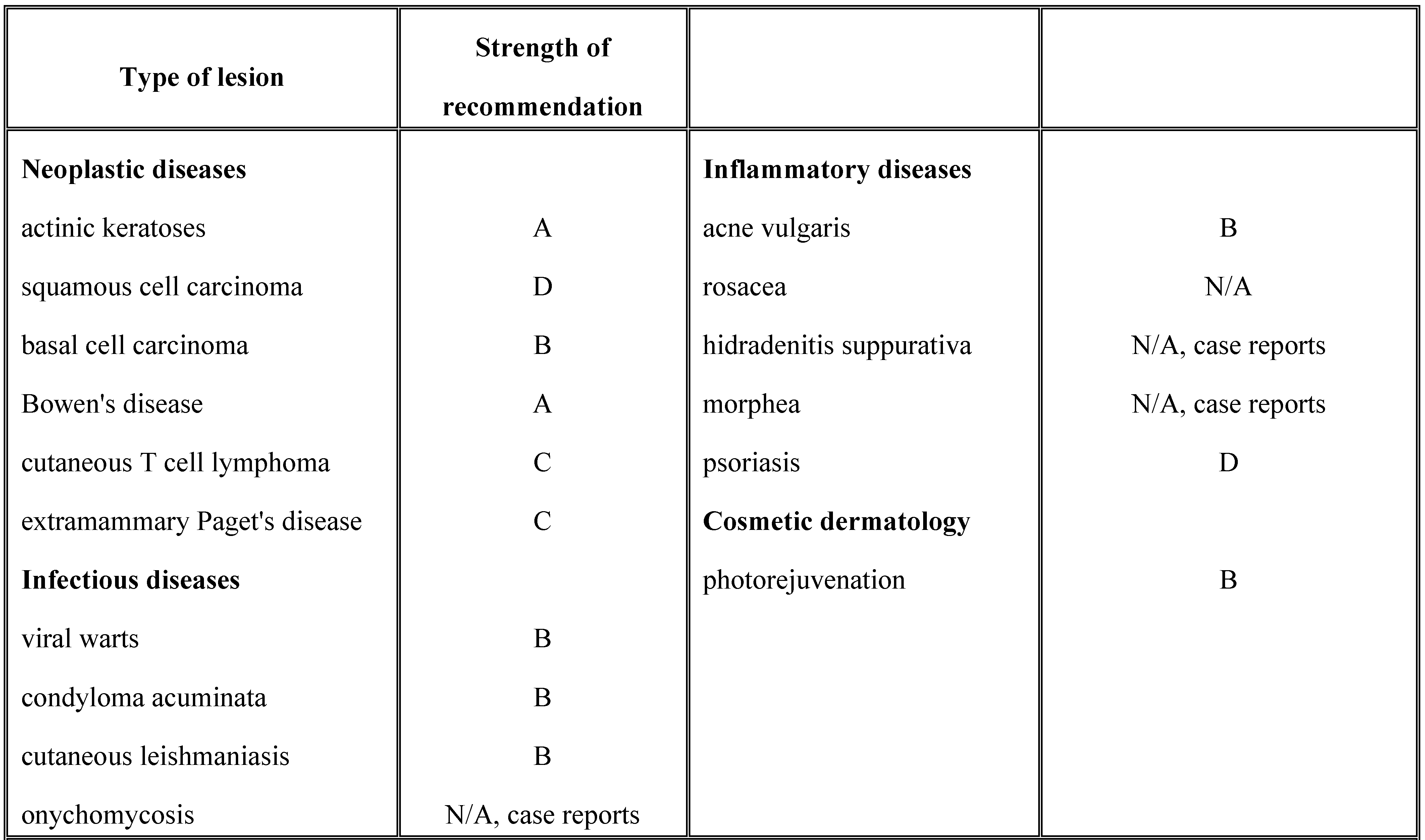
  • Applications of photodynamic therapy in dermatology. PDT has several applications in dermatology, including neoplastic diseases, inflammatory diseases, microbial diseases, photoaging and rejuvenation (Table 1). While there is clear evidence on the effectiveness of PDT for actinic keratosis, BCC, especially the superficial type and Bowen's disease, the data regarding the use of PDT for other dermatological disorders is still scarce. However, studies show promising results [11,12,13,14,15]. Contraindications to PDT are porphyria, systemic lupus erythematosus, non- responsive tumors, allergy to the photosensitizing agent and photosensitive dermatoses [13].
Table 1. Applications of PDT in dermatology. A—good evidence; B—fair evidence; C—poor evidence; D—fair evidence to support the rejection of the procedure; N/A not available.
Jmms 03 00003 i001

Adverse reactions

Acute adverse reactions are photosensitivity, pain and inflammation. Photosensitivity depends on the administration route. Therefore, it is greater after systemic administration and it is localized after topical administration. Pain is the most common and redoubtable adverse effect. Inflammation manifesting as edema, induration, purpura and blistering can occur and can sometimes be serious, leading to tissue necrosis [1,11]. Chronic adverse reactions are rare and include scarring, post inflammatory hypopigmentation and hyperpigmentation [1,11].
  • Pain in photodynamic therapy
Pain is the most common side-effect associated with PDT and it is the most important factor that affects the patient's adherence to treatment. Most patients undergoing PDT experience some degree of pain, which can be mild, manifesting as stinging or burning, or more severe. It usually occurs during exposure, being most intense after a few minutes of irradiation, and decreases towards the end of the session. Few patients however also experience post-procedural pain. Therefore, some patients interrupt treatment sessions while others do not return for further treatments [11,16,17].
Most studies assessing pain during PDT use the visual analogue scale (VAS) for pain. The VAS for pain is a measurement instrument which tries to measure the amount of pain felt by the patient across a continuum ranging from none (marked as 0) to unbearable pain (marked as 10) (Figure 1). The assessment of pain is subjective, the results depending on psychosocial factors and personal characteristics of the patient. According to some studies 20% of patients rate pain over six on the VAS [18,19,20].
A study performed by Sandberg et al. in 2006 investigated pain related to ALA-PDT in 91 patients treated for actinic keratosis. VAS was used for pain assessment. The authors found a mean value of VAS of 4.6. 21% of patients experienced severe pain (VAS 7-10) while 31% had little or no pain (VAS 0-3) [16].
The mechanism of pain in PDT is still unknown. Aδ and C fibers are the major pain- conducting nerve fiber systems. In PDT, pain is mainly mediated through the unmyelinated afferent C-fibers. C-fibers innervate polymodal receptors which respond to thermal, mechanical and chemical pain. The P substance and other neurotransmitters are also involved in PDT related pain. Hyperthermia, reactive oxygen species and inflammation have been discussed as possible triggers for pain in PDT. Nerve stimulation by ALA through Aδ and C fibers might also play a role [16].
  • Factors contributing to pain in PDT for dermatological disorders
Lesion type: actinic keratosis seem to be more painful than BCC and Bowen's disease. The treatment of acne lesions with PDT is also painful. A study performed by Schleyer et al. on 12 patients with psoriasis treated with ALA-PDT showed unsatisfactory results, irradiation being interrupted several times due to pain and severe burning sensation. PDT for viral warts is also painful [11,19,21,22].
Location: lesion located in well innervated areas such as the head, hands and perineum are more painful.
Extent of lesions: larger lesions or more extensive treated areas are associated with more pain than small lesions [16,17].
Type of photosensitizer: MAL and ALA are the most widely used photosensitizing agents in dermatology. Studies show that ALA-PDT is associated with more pain than MAL-PDT. A study performed by Gaal et al. in 2011, which included 87 patients with 182 lesions (AK, BCC and Bowen's disease) found that 21 of the 24 treatments associated with intolerable pain that required treatment discontinuation were done with ALA-PDT [23]. Wiegell et al. compared pain related to ALA-PDT with pain related to MAL- PDT in 20 volunteers with tape-stripped normal skin. Patients were randomized to receive either ALA-PDT or MAL-PDT. The authors found that ALA-PDT was associated with more pain than MAL-PDT [24]. Kasche et. al. performed a study on 69 patients with AK who received either ALA- PDT or MAL-PDT. The authors found that treatment had to be interrupted in 54% of patients receiving ALA-PDT and 14% of patients receiving MAL-PDT [25].
One explanation might be that MAL is more lipophilic than ALA and penetrates tissues better. It also determines better accumulation of protoporphyrin IX in abnormal cells [19,22]. Another explanation could be that ALA and γ- aminobutyric acid (GABA) have similar structures and are carried into cells by the same carrier systems, namely GABA-transporters. Therefore ALA might be transported into nerve endings by GABA receptors. GABA is the primary inhibitory neurotransmitter in the central nervous system and abnormal levels can determine neurological conditions and pain. The uptake of MAL on the other hand is cell dependent, MAL being transported by non-polar amino-acid transporters [19,24,26,27].
Total dose of light and light intensity. Total dose of light, measured in joules/cm2 must be differentiated from the intensity of light, measured in watts/cm2. Higher total dose of light and higher light intensity both seem to be associated with higher levels of pain. Zeitouni et al. performed a retrospective review of pain control by a two-step irradiance schedule during ALA-PDT, in an existing dermatology data base. Their study included 14 patients who had initially received an irradiance of 30 or 50 mW/cm2 for 20 J/cm2 followed by 150 mW/cm2 for 200-300 J/cm2. The author obtained a median VAS score of 1 and conlcuded that the two step irradiance protocole is effective in minimising pain [28]. The same group of researchers later performed a study on 21 patients with 25 superficial BCCs who were treated with MAL-PDT. Patients were randomized to receive either laser with irradiance at 40 or 50 mW/cm2 or LED with irradiance at 35 mW/cm2. After that all patients received irradiance at 70 mW/cm2 for a total of 75 J/cm2. Pain was measured using the VAS. The authors report that pain was minimal in the LED cohort (35/70 mW/cm2), was mild in the 40/70 mW/cm2 laser cohort and was higher in the 50/70 mW/cm2 laser cohort [29]. Radakovic et. al. performed a study on 27 patients with at least 3 AKs on the face or scalp who were treated with ALA and irradiated with 70, 100 or 140 j/cm2. The authors report no statistically significant difference between the three light doses regarding the degree of PDT- induced pain [30].
Wavelength. Morton et al. compared ALA- PDT with red light and green light in the treatment of Bowen's disease. The study included 61 patients. The authors concluded that PDT with green light is less effective than PDT with red light in the treatment of Bowen's disease. However, the severity of the experienced pain was similar between the two cohorts, with red light being more painful than green light [22,31].
Source of light. Several sources of light have been tried in order to increase efficacy and decrease pain. Babilas et al. performed a study on 25 patients with AKs who received MAL-PDT and irradiation with LED on one side of the face and variable pulsed light (VPL) on the other side of the face. The authors showed that pain was significantly lower on the side treated with VPL [32]. Kessels et al. compared PDT with LED with PDT with pulsed dye laser (PDL) in 61 patients with AK, the patients receiving LED-PDL on one side of the face and PDL-PDT on the other side. Pain was significantly lower in the PDL-PDT group. 78.7% of patients treated with PDL-PDT and 32.8 of patients in the LED group declared that they would undergo the treatment again [33]. Some studies showed that daylight PDT is associated with less pain than conventional PDT. Wiegell et al. showed in a study performed on 29 patients with AK, published in 2008, that LED- PDT and daylight PDT have similar efficacy, LED-PDT being more painful than daylight-PDT [34]. Braathen also conducted a study on 18 patients with AKs who were treated with daylight - PDT. Only one patients reported pain, scored as 5 on the VAS [35].
Number of sessions. The second session of PD is more painful than the first one.
Skin phototype. Phototypes and 2 are associated with more pain [19].
  • Pain management
Since pain is the most important side effect of PDT, several strategies for controlling pain have been researched. Until now no ideal method for pain management was found, probably because the precise mechanism of pain was not discovered [19].

Topical anesthetics

Topical anesthetics have failed to prove their efficacy in controlling the pain associated with PDT. EMLA, an eutectic mixture of lidocaine 2,5% and prilocaine 2.5%, is the most widely used topical anesthetic. Langan et al performed a randomized, double blind study on 14 patients with AK treated with two sessions of ALA, who received EMLA on one session and placebo (Aqueous cream) on the other session. The authors report no difference between EMLA and placebo [36].
Tetracaine gel was also evaluated as a potential local anesthetic in PDT. Holmes et al. performed a prospective, double blind, placebo controlled study on 42 patients with non- melanoma skin cancer who were randomized to receive either tetracaine gel or placebo gel. The pain in the tetracaine group was slightly lower than pain in the placebo group (VAS 4.0 vs. VAS 4.5). However, the difference was not statistically significant between the two groups [37].
Sandberg et al. tested the efficacy of capsaicin cream as a pain-reducing agent in 6 patients with AKs who applied he cream 3-5 times a day for a week before treatment. The authors obtained no significant pain relief. However, all patients presented local adverse reactions [16].
Morphine gel also showed no efficacy in controlling PDT-related pain in a double-blind, placebo controlled study performed on 28 patients (27 AKs and 1 BCC) who were randomized to receive either morphine 0.3% gel or placebo cream. The authors reported identical maximum pain scores (5.5) and concluded that opioid receptors might not be involved in the mechanism of pain determined by PDT [38]. Topical lidocaine cream associated with pretreatment with urea 40% to enhance lidocaine penetration also failed to prove its efficacy in reducing PDT -related pain [39].

Locally injected anesthetics

Locally injected anesthetics and nerve block showed better results in reducing pain than topical anesthetics. Therefore, locally injected lidocaine, mepivacaine, ropovacaine, prilocaine and epinephrine all showed some good results in the management of PDT-related pain. Paoli et al. performed a study on 16 patients with AKs and used nerve block with mepivacaine and adrenaline on one side of the face and then performed PDT on both sides of the face. The authors report that the pain was significantly reduced on the anaesthetized side compared to the non-anesthetized side [40]. Borelli et al. assessed the effect of subcutaneous infiltration anesthesia (SIA) on pain in PDT in 16 patients who received SIA on one side of the face and oral analgesics only for the other side of the face and concluded that SIA decreases pain in PDT significantly more than oral analgesia [41].

Conscious sedation

Conscious sedation with inhaled 50% nitrous oxide/ oxygen has been recently reported in association with PDT and according to the authors, it could be an alternative for classical anesthetics [42,43].
Transcutaneous electrical nerve stimulation (TENS) Halldin et al. performed a study on 14 patients with AKs who had previously experienced severe PDT-related pain. The electrodes for TENS were placed on the shoulders. Pain was assessed using the VAS. The authors observed a mean VAS of 6.2, as compared to a mean VAS of 8.1 at baseline treatments and concluded that TENS could be an efficient pain-relieving technique [44].

Other methods

Several other techniques were researched for decreasing PDT-related pain. Cooling of the treatment site seems to beneficial and is part of the treatment protocol in some centers. Cold air and cold water have both been used. Reducing irradiance, interruption of sessions and use of thermal water could also reduce pain in PDT [19,22,45].

Conclusion

PDT is a modern treatment with many applications in dermatology, but also other medical specialties. Pain is commonly experienced by patients undergoing PDT and is often associated with poor adherence to treatment. Several factors contribute to the occurrence of pain and many pain-relieving techniques have been studied in the attempt to alleviate it. However, since the exact mechanism is still unknown, most of those pain- relieving strategies were unsuccessful. Further studies are therefore required.

Acknowledgement

This work was possible with the financial support of Young Researchers Grant from the Carol Davila University of Medicine and Pharmacy, no. 33884/11.11.2014.

References

  1. Yokoyama, Y.; Shigeto, T.; Miura, R.; Kobayashi, A.; Mizunuma, M.; Yamauchi, A.; Futagami, M.; Mizunuma, H. A Strategy Using Photodynamic Therapy and Clofibric Acid to Treat Peritoneal Dissemination of Ovarian Cancer. Asian Pac. J. Cancer Prev. 2016, 17, 775–779. [Google Scholar]
  2. Agostinis, P.; Berg, K.; Cengel, K.A.; Foster, T.H.; Girotti, A.W.; Gollnick, S.O.; Hahn, S.M.; Hamblin, M.R.; Juzeniene, A.; Kessel, D.; et al. Photodynamic therapy of cancer: An update. CA Cancer J. Clin. 2011, 61, 250–281. [Google Scholar] [PubMed]
  3. Pervaiz, S.; Olivo, M. Art and science of photodynamic therapy. Clin. Exp. Pharmacol. Physiol. 2006, 33, 551–556. [Google Scholar] [CrossRef] [PubMed]
  4. Tong, H.; Wang, Y.; Li, H.; Jin, Q.; Ji, J. Dual pH-responsive 5-aminolevulinic acid pseudopolyrotaxane prodrug micelles for enhanced photodynamic therapy. Chem. Commun. 2016, 52, 3966–3969. [Google Scholar]
  5. Dougherty, T.J.; Gomer, C.J.; Henderson, B.W.; Jori, G.; Kessel, D.; Korbelik, M.; Moan, J.; Peng, Q. Photodynamic therapy. J. Natl. Cancer Inst. 1998, 90, 889–905. [Google Scholar]
  6. O’Connor, A.E.; Gallagher, W.M.; Byrne, A.T. Porphyrin and nonporphyrin photosensitizers in oncology: Preclinical and clinical advances in photodynamic therapy. Photochem. Photobiol. 2009, 85, 1053–1074. [Google Scholar]
  7. Katz, S.I.; Gilchrest, B.A.; Paller, A.S.; Leffell, D.J. Fitzpatrick’s Dermatology in General Medicine, 8th ed.; McGrawHill, 2012; ISBN 978-0071669047. [Google Scholar]
  8. Triesscheijn, M.; Baas, P.; Schellens, J.H.; Stewart, F.A. Photodynamic therapy in oncology. Oncologist 2006, 11, 1034–1044. [Google Scholar]
  9. Bacellar, I.O.; Tsubone, T.M.; Pavani, C.; Baptista, M.S. Photodynamic Efficiency: From Molecular Photochemistry to Cell Death. Int. J. Mol. Sci. 2015, 16, 20523–20559. [Google Scholar] [CrossRef]
  10. Matei, C.; Tampa, M.; Poteca, T.; Panea-Paunica, G.; Georgescu, S.R.; Ion, R.M.; Popescu, S.M.; Giurcaneanu, C. Photodynamic therapy in the treatment of basal cell carcinoma. J. Med. Life 2013, 6, 50–54. [Google Scholar]
  11. Morton, C.A.; McKenna, K.E.; Rhodes, L.E. Guidelines for topical photodynamic therapy: Update. Br. J. Dermatol. 2008, 159, 1245–1266. [Google Scholar] [CrossRef]
  12. Kim, M.; Jung, H.Y.; Park, H.J. Topical PDT in the Treatment of Benign Skin Diseases: Principles and New Applications. Int. J. Mol. Sci. 2015, 16, 23259–23278. [Google Scholar] [CrossRef] [PubMed]
  13. Wan, M.T.; Lin, J.Y. Current evidence and applications of photodynamic therapy in dermatology. Clin. Cosmet. Investig. Dermatol. 2014, 7, 145–163. [Google Scholar]
  14. Kharkwal, G.B.; Sharma, S.K.; Huang, Y.-Y.; Dai, T.; Hamblin, M.R. Photodynamic Therapy for Infections: Clinical Applications. Lasers Surg. Med. 2011, 43, 755–767. [Google Scholar] [PubMed]
  15. Dai, T.; Huang, Y.Y.; Hamblin, M.R. Photodynamic therapy for localized infections– state of the art. Photodiagnosis Photodyn. Ther. 2009, 6, 170–188. [Google Scholar] [PubMed]
  16. Sandberg, C.; Stenquist, B.; Rosdahl, I.; Ros, A.M.; Synnerstad, I.; Karlsson, M.; Gudmundson, F.; Ericson, M.B.; Larkö, O.; Wennberg, A.M. Important factors for pain during photodynamic therapy for actinic keratosis. Acta Derm.-Venereol. 2006, 86, 404–408. [Google Scholar]
  17. Halldin, C.B.; Gillstedt, M.; Paoli, J.; Wennberg, A.M.; Gonzalez, H. Predictors of pain associated with photodynamic therapy: A retrospective study of 658 treatments. Acta Derm.-Venereol. 2011, 91, 545–551. [Google Scholar]
  18. Crichton, N. Visual analogue scale (VAS). J. Clin. Nurs. 2001, 10, 706-6. [Google Scholar]
  19. Chaves, Y.N.; Torezan, L.A.; Niwa, A.B.; Sanches, J.A.; Festa, N.C. Pain in photodynamic therapy: Mechanism of action and management strategies. An. Bras. De. Dermatol. 2012, 87, 521–529. [Google Scholar]
  20. Figure one extracted from: http://www.trialdatasolutions.com/tds/howto/v as. jsp, last accessed 31.01.2016.
  21. Schleyer, V.; Radakovic-Fijan, S.; Karrer, S.; Zwingers, T.; Tanew, A.; Landthaler, M.; Szeimies, R.M. Disappointing results and low tolerability of photodynamic therapy with topical 5-aminolaevulinic acid in psoriasis. A randomized, double-blind phase I/II study. J. Eur. Acad. Dermatol. Venereol. 2006, 20, 823–828. [Google Scholar]
  22. Warren, C.B.; Karai, L.J.; Vidimos, A.; Maytin, E.V. Pain associated with aminolevulinic acid- photodynamic therapy of skin disease. J. Am. Acad. Dermatol. 2009, 61, 1033–1043. [Google Scholar]
  23. Gaál, M.; Otrosinka, S.; Baltás, E.; Ócsai, H.; Oláh, J.; Kemény, L.; Gyulai, R. Photodynamic therapy of non-melanoma skin cancer with methyl aminolaevulinate is associated with less pain than with aminolaevulinic acid. Acta Derm.-Venereol. 2012, 92, 173–175. [Google Scholar] [CrossRef] [PubMed]
  24. Wiegell, S.R.; Stender, I.M.; Na, R.; Wulf, H.C. Pain associated with photodynamic therapy using 5-aminolevulinic acid or 5-aminolevulinic acid methylester on tape-stripped normal skin. Arch. Dermatol. 2003, 139, 1173–1177. [Google Scholar]
  25. Kasche, A.; Luderschmidt, S.; Ring, J.; Hein, R. Photodynamic therapy induces less pain in patients treated with methyl aminolevulinate compared to aminolevulinic acid. J. Drugs Dermatol. 2006, 5, 353–356. [Google Scholar] [PubMed]
  26. Rud, E.; Gederaas, O.; Høgset, A.; Berg, K. 5-Aminolevulinic Acid, but not 5-Aminolevulinic acid esters, is transported into adenocarcinoma cells by system BETA transporters. Photochem. Photobiol. 2000, 71, 640–647. [Google Scholar]
  27. Baglo, Y.; Gabrielsen, M.; Sylte, I.; Gederaas, O.A. Homology Modeling of Human γ-Butyric Acid Transporters and the Binding of Pro-Drugs 5-Aminolevulinic Acid and Methyl Aminolevulinic Acid Used in Photodynamic Therapy. PLoS ONE. 2013, 8, e65200. [Google Scholar]
  28. Zeitouni, N.C.; Paquette, A.D.; Housel, J.P.; Shi, Y.; Wilding, G.E.; Foster, T.H.; Henderson, B.W. A Retrospective Review of Pain Control by a Two-Step Irradiance Schedule During Topical ALA-Photodynamic Therapy of Non- melanoma Skin Cancer. Lasers Surg. Med. 2013, 45, 89–94. [Google Scholar]
  29. Zeitouni, N.C.; Sunar, U.; Rohrbach, D.J.; Paquette, A.D.; Bellnier, D.A.; Shi, Y.; Henderson, B.W. A Prospective Study of Pain Control by a Two- Step Irradiance Schedule During Topical Photodynamic Therapy of Non-melanoma Skin Cancer. Dermatol. Surg. 2014, 40, 1390–1394. [Google Scholar] [PubMed]
  30. Radakovic-Fijan, S.; Blecha-Thalhammer, U.; Kittler, H.; Hönigsmann, H.; Tanew, A. Efficacy of 3 different light doses in the treatment of actinic keratosis with 5-aminolevulinic acid photodynamic therapy: A randomized, observer-blinded, intrapatient, comparison study. J. Am. Acad. Dermatol. 2005, 53, 823–827. [Google Scholar]
  31. Morton, C.A.; Whitehurst, C.; Moore, J.V.; MacKie, R.M. Comparison of red and green light in the treatment of Bowen’s disease by photodynamic therapy. Br. J. Dermatol. 2000, 143, 767–772. [Google Scholar] [CrossRef]
  32. Babilas, P.; Knobler, R.; Hummel, S.; Gottschaller, C.; Maisch, T.; Koller, M.; Landthaler, M.; Szeimies, R.M. Variable pulsed light is less painful than light-emitting diodes for topical photodynamic therapy of actinic keratosis: A prospective randomized controlled trial. Br. J. Dermatol. 2007, 157, 111–117. [Google Scholar]
  33. Kessels, J.P.; Nelemans, P.J.; Mosterd, K.; Kelleners-Smeets, N.W.; Krekels, G.A.; Ostertag, J.U. Laser-mediated Photodynamic Therapy: An Alternative Treatment for Actinic Keratosis? Acta Derm. Venereol. 2016, 96, 351–354. [Google Scholar] [CrossRef] [PubMed]
  34. Wiegell, S.R.; Hædersdal, M.; Philipsen, P.A.; Eriksen, P.; Enk, C.D.; Wulf, H.C. Continuous activation of PpIX by daylight is as effective as and less painful than conventional photodynamic therapy for actinic keratoses; a randomized, controlled, single-blinded study. Br. J. Dermatol. 2008, 158, 740–746. [Google Scholar]
  35. Braathen, L.R. Daylight photodynamic therapy in private practice in Switzerland: Gain without pain. Acta Derm.-Venereol. 2012, 92, 653–654. [Google Scholar] [CrossRef] [PubMed]
  36. Langan, S.M.; Collins, P. Randomized, double-blind, placebo-controlled prospective study of the efficacy of topical anaesthesia with a eutetic mixture of lignocaine 2.5% and prilocaine 2.5% for topical 5-aminolaevulinic acid–photodynamic therapy for extensive scalp actinic keratoses. Br. J. Dermatol. 2006, 154, 146–149. [Google Scholar] [PubMed]
  37. Holmes, M.V.; Dawe, R.S.; Ferguson, J.; Ibbotson, S.H. A randomized, double-blind, placebo-controlled study of the efficacy of tetracaine gel (Ametop®) for pain relief during topical photodynamic therapy. Br. J. Dermatol. 2004, 150, 337–340. [Google Scholar]
  38. Skiveren, J.; Haedersdal, M.; Philipsen, P.A.; Wiegell, S.R.; Wulf, H.C. Morphine gel 0.3% does not relieve pain during topical photodynamic therapy: A randomized, double- blind, placebo-controlled study. Acta Derm.-Venereol. 2006, 86, 409–411. [Google Scholar]
  39. Touma, D.; Yaar, M.; Whitehead, S.; Konnikov, N.; Gilchrest, B.A. A trial of short incubation, broad-area photodynamic therapy for facial actinic keratoses and diffuse photodamage. Arch. Dermatol. 2004, 140, 33–40. [Google Scholar]
  40. Paoli, J.; Halldin, C.; Ericson, M.B.; Wennberg, A.M. Nerve blocks provide effective pain relief during topical photodynamic therapy for extensive facial actinic keratoses. Clin. Exp. Dermatol. 2008, 33, 559–564. [Google Scholar]
  41. Borelli, C.; Herzinger, T.; Merk, K.; Berking, C.; Kunte, C.; Plewig, G.; Degitz, K. Effect of subcutaneous infiltration anesthesia on pain in photodynamic therapy: A controlled open pilot trial. Dermatol. Surg. 2007, 33, 314–318. [Google Scholar]
  42. Cabete, J.; Campos, S.; Lestre, S. Conscious sedation with inhaled 50% nitrous oxide/oxygen premix in photodynamic therapy sessions for vulvar lichen sclerosus treatment. An. Bras. Dermatol. 2015, 90, 120–122. [Google Scholar]
  43. Gholam, P.; Fink, C.; Uhlmann, L.; Enk, A. Pain reduction in patients after applying a nitrous oxide/oxygen mixture (Livopan) during photodynamic therapy: Study protocol for an observational study (Livopan study). BMJ Open 2015, 5, e006412. [Google Scholar] [CrossRef] [PubMed]
  44. Halldin, C.B.; Paoli, J.; Sandberg, C.; Ericson, M.B.; Wennberg, A.M. Transcutaneous electrical nerve stimulation for pain relief during photodynamic therapy of actinic keratoses. Acta Derm.-Venereol. 2008, 88, 311–312. [Google Scholar] [CrossRef] [PubMed]
  45. Wiegell, S.R.; Haedersdal, M.; Wulf, H.C. Cold water and pauses in illumination reduces pain during photodynamic therapy: A randomized clinical study. Acta Derm.-Venereol. 2009, 89, 145–149. [Google Scholar] [PubMed]

