Next Article in Journal
Evaluation of Short-Term Complications in Laparoscopic Peritoneal Dialysis Catheter Placement—A Single Tertiary Center Experience
Previous Article in Journal
Challenges in Diagnosing Nasopharyngeal Tumors
 
 
Journal of Mind and Medical Sciences is published by MDPI from Volume 12 Issue 1 (2025). Previous articles were published by another publisher in Open Access under a CC-BY (or CC-BY-NC-ND) licence, and they are hosted by MDPI on mdpi.com as a courtesy and upon agreement with Valparaiso University (ValpoScholar).
Article

Biological Effect of Modern Bioactive Materials Used in Direct and Indirect Capping; In Vitro Study

by
Irina Maria Gheorghiu
1,†,
Alexandru Andrei Iliescu
2,*,
George Alexandru Denis Popescu
3,
Stana Paunica
4,† and
Anca Silvia Dumitriu
4
1
Department of Restorative Odontotherapy, Carol Davila University of Medicine, Faculty of Stomatology, Bucharest, Romania
2
Department of Oral Rehabilitation, Craiova University of Dental Medicine, Craiova, Romania
3
Doctoral School, Carol Davila University of Medicine, Bucharest, Romania
4
Department of Periodontology, Faculty of Stomatology, Carol Davila University of Medicine, Bucharest, Romania
*
Author to whom correspondence should be addressed.
These authors contributed equally to this work.
J. Mind Med. Sci. 2024, 11(2), 444-451; https://doi.org/10.22543/2392-7674.1544
Submission received: 12 March 2024 / Revised: 12 April 2024 / Accepted: 5 July 2024 / Published: 31 October 2024

Abstract

:
Objective. In this study, the biological effect of MTA Repair HP (Mineral Trioxide Aggregate Repair High Plasticity) and Biodentine have been tested on a stabilized fibroblast cell line NCTC clone 929. Materials and Methods. We assessed quantitative and qualitative parameters related to cytotoxic effect of the investigated products. The experimental period was 96 hours. Statistical analysis was performed with Kruskal-Wallis and Wilcoxon tests. Results. The detached cells test showed no statistically significant difference on cell culture for Biodentine and MTA Repair HP, while for the cellular density assay we found the same biological effect on the tested fibroblasts in the first 24 and 48 h, but a significant different cellular response for the investigated pulp capping materials for the next 48 h of the experiment. Conclusions. The results demonstrated that the materials presented a very low level of cytotoxicity. Biodentine showed in all parameters better biological effects than MTA Repair HP, expressed by lower and limited cellular damage and a higher cell density.

Introduction

Nowadays, when the biological, minimally invasive and conservative concept dominates the entire approach in dentistry, treatments for preserving dental pulp vitality are even more current. In order to achieve this objective, different cavity preparation techniques are used (for example selective caries removal strategies), as well as modern bioactive materials which induce tissues regeneration, especially for cases where the pulp exposure occurred, in order to avoid pulpectomy [1,2].
If the “gold-standard” of pulp capping was calcium hydroxide, an interactive material that possesses restorative properties, today pulp regenerative dentistry has bioactive materials as its fundamental element. Whether it is about indirect pulp capping in a deep dental caries situation, or direct capping in the case of a pulp chamber accidental opening, the first approach is to use materials that exert a biological effect, they promote specific cellular or tissue responses, which support and stimulates the activity of the pulp-dentin complex, an essential biological entity of the tooth [3].
The concept of biomaterial refers to a material that is active from the host’s local response point of view, thus inducing a certain biological activity, necessarily having properties of bioadhesion, tissue inducer, biodegradation and bioresorbable [4]. The field of bioactive dental restorative materials is extremely advanced today, and in operative dentistry and endodontics the term bioactive usually refers to the ability of a material to stimulate dentin-pulp complex and induce the formation of hydroxyapatite crystals on their surface [5].
As a result of the evolution of a dental caries or of a restorative treatment to preserve pulp vitality (perceived as an aggression of a mechanical or chemical nature), a cascade of molecular mechanisms is triggered [6]. That lead to tertiary dentin production, either secreted by odontoblasts (reaction dentin), or by other cells that replace them (repair dentin), or by cells differentiated from dental pulp stem cells (DPSC dental pulp stem cells), activated by growth factors.
The latter, like the other types of odontogenic stem cells, can transform into cells specific to dental tissues, like odontoblasts, as well into other types of cells, like nerve or smooth muscle cells, opening a large window to regenerative medicine. In this context, the battle for new bioactive dental material which can possess properties capable of inducing cell proliferation and tissue healing is becoming almost a necessity [7]. The first and most well-known material used for indirect and direct capping (introduced in 1920) was calcium hydroxide, which remains today the most used material in this field, given its successful clinical results over time [8]. At the present time, in the framework of treatments to preserve the vitality of the dental pulp, modern materials are mainly bioactive silicate cements [9].
If in the case of some indirect capping are essential avoiding bacterial contamination and hermetic sealing restoration to prevent microleakage for a successful approach, for direct pulp capping the therapeutic result is conditioned by a series of biological factors related to the preoperative pulp status, or the general status of the patient, along with the bioactive dental materials that are being increasingly used with a widening application option [10].
The first bioactive material introduced was MTA (mineral trioxide aggregate), which revolutionized regenerative endodontic therapy since 1995, when it was launched [11]. The main properties of this tricalcium and dicalcium silicate cement are biocompatibility, bioactivity (conductive and inductive hard tissue properties) and antimicrobial activity.
The MTA clinical indications refer to vital and non-vital pulp therapy: vital pulp capping, pulpotomy, internal resorption, perforation repair, apexification, apexogenesis endodontic sealers [12]. Being a hydrophilic cement, the setting is favored by moisture, from this it is clinically widely and successfully used in root perforations and furcation defects [13]. Limitations of MTA are related to a prolonged setting time, technically difficult manipulation and cost.
In 2009 a new bioactive tricalcium silicate cement was released, Biodentine, known as “dentin substitute”, which has improved mechanical, chemical and biological properties over MTA, regarding bioactivity, biocompatibility, adhesion, setting time, discoloration potential of tooth structures, color stability. Clinical indications for Biodentine are similar to MTA, including deep and/or large coronal and radicular carious lesions (sandwich technique), dentine long term replacement and temporary enamel replacement [14,15].
The bioactive silicate cements stimulate dentin-pulp complex, inducing pulp cell lines proliferation, and stimulate reparative dentine formation, while no signs of inflammation occur. This mechanism is mediated by the calcium hydroxide ions released which offer the conditions for apatite formation [16,17]. These materials are also known as “bioceramics” [18].
The goal of the in-vitro present study is to determine the biological effect on fibroblasts of the modern bioactive materials used for indirect and direct pulp capping, considering that they are placed in indirect or even direct contact with the pulp tissue. We assessed parameters related to cytotoxic effect of the most used bioactive silicate cements in a stabilize cell line.
Cell culture tests are the first line in toxicity testing of medical devices, both from financial and moral and ethical considerations. They offer valuable information about the effects on cell proliferation, viability, cellular metabolism, changes of cell permeability, enzymatic activity, cellular morphological lesions and alterations, cell death, as a direct expression of the toxicity of the investigated product [19]. Clinic direct extrapolation of the results is not possible, due to the fact that cell cultures toxicity studies exclude the action of other cellular and tissues factors and mechanisms specific to a living organism, as such they must be corroborated with subsequent in-vitro and in-vivo studies.

Materials and Methods

Materials

In this study, the biological effect of MTA Repair HP (Mineral Trioxide Aggregate Repair High Plasticity) and Biodentine have been tested on a stabilized fibroblast cell line NCTC clone 929(L-929) from ATCC (American Type Culture Collection). This is a one of the first stabilized clone line cells and it is widely used for various cytotoxicity tests. The origin is mouse connective tissue, with no contaminants and a limited in-vitro life span. The cell line was maintained by serial passages at the rate of dispersion of 1:2 and over 96 hours of follow-up, the number of detached cells from the substrate was determined, as well as the gradual changes in the cell concentration (density) in the confluent layer. Also, different parameters/modifications expressing the possible effects on the cellular morphology and the appearance of the cellular layer were visualized under the optical microscope.
The purpose of this study is the comparative analysis of the investigated substances regarding the in-vitro biological effects (cytotoxicity) on a fibroblast cell line, quantified and validated by the parameters mentioned above. A control group was also investigated.
The main characteristics of the tested products are summarized in Table 1 and their biological properties are listed in Table 2.

Methods

In this study we determined both quantitative (numerical data) and qualitative (microscopically assessments of cell damage) in-vitro biological effects of the tested materials. The entire experimental period was 96 hours, the parameters being determined at every 24 hours. The tested products were abbreviated MTA HP and BD, respectively in this study. They have been investigated in cured phase, using indirect contact method, according to ISO 10993-5 [20]. MTA HP and BD were prepared according to the manufacturer’s instructions. For MTA HP: 1 powder capsule was manually mixed with 1 liquid capsule on a sterile pad till the optimal consistency was achieved (putty consistency), also high plasticity. BD was prepared by adding to the powder capsule the content of the liquid capsule and mechanically mix for 30 seconds. The consistency is similar to the investigated MTA HP product. We prepared and insert the materials into sterile molds with sterile dental cement spatulas and carriers, so the test won’t be contaminated and false data obtained/induced. In this experiment we investigate the cytotoxic effect of these materials after setting (15 minutes for MTA HP, respectively 12 minutes for BD), although the biological effect on cell substrate is equally important during the first initial contact with the host and cellular effects can appear during the setting period. In order to obtain a large surface of sample in contact with cell substrate we use a dish form molder, of 4 mm diameter and 2 mm height. After the setting time, the materials were placed in 1 ml cell culture medium Eagle’s Minimum Essential Medium (EMEM) (ATCC) without fetal calf serum, and incubated for 72 hours at 37O Celsius in humified atmosphere, in order to obtain medium extracts of the investigated materials. For the entire experiment a number of 12 samples of each material have been used, for more appropriate statistically significance behaviour of the two tested materials.
The fibroblasts were incubated in cell culture microplates, at a density of 7.5 x10⁴celule/cm2, in EMEM (ATCC), which contains Earle’s Balanced Salt Solution, non-essential amino acids, L-glutamine, sodium pyruvate and sodium bicarbonate, supplemented with 10% fetal bovine serum (SFB) and 1% antibiotic mixture (penicillin, streptomycin and neomycin), in a humified 95% air, 5% CO2 atmosphere, at 37o Celsius and an optimal 7.2-7.4 pH was maintained. After 24 hours the Eagle medium was sucked in and 0,1 ml of the undiluted extracts medium of each material type (which contained products released from the investigated products) and fetal bovine serum were added. The cell cultures were kept in standard incubation in thermostat for 96 hours at 37 °C. A control group for every tested lot was prepared, in which fibroblasts were incubated in cellular medium.
In order to monitor the cytotoxic effect, we sequentially assed two parameters: the number of detached cells and the cellular density. The cellular layer was detached with trypsin, resumed in a known environment volume (0.1 mL of 0.4% trypan blue staining solution to 0.1 mL of cell suspension), incubate at room temperature for 3 minutes and immediately the cells have been counted in the hemocytometer. In trypan blue exclusion assay, only damaged cells take up the dye and the proportion of damaged cells can be counted. Evaluation of cellular density in the presence of the investigated pulp capping materials was made also by counting in hemocytometer.
The cell morphology was evaluated by optical microscopy and it was addressed to possible changes in the general cellular appearance, vacuolization, damage to the cell membranes, the appearance of some alterations of the nucleus and nucleoli, as well as the number and characteristics of the detached cells from the substrate.

Statistical Analysis
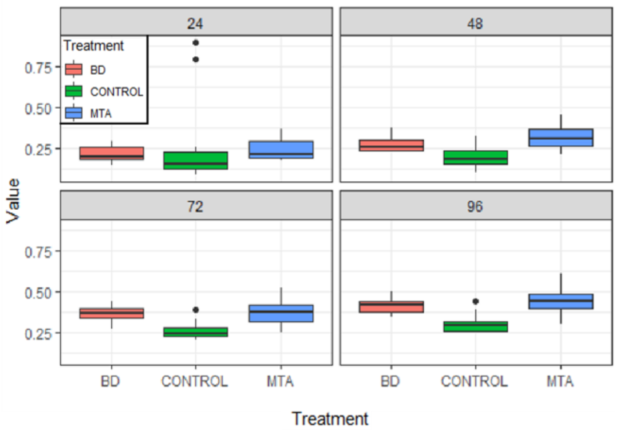
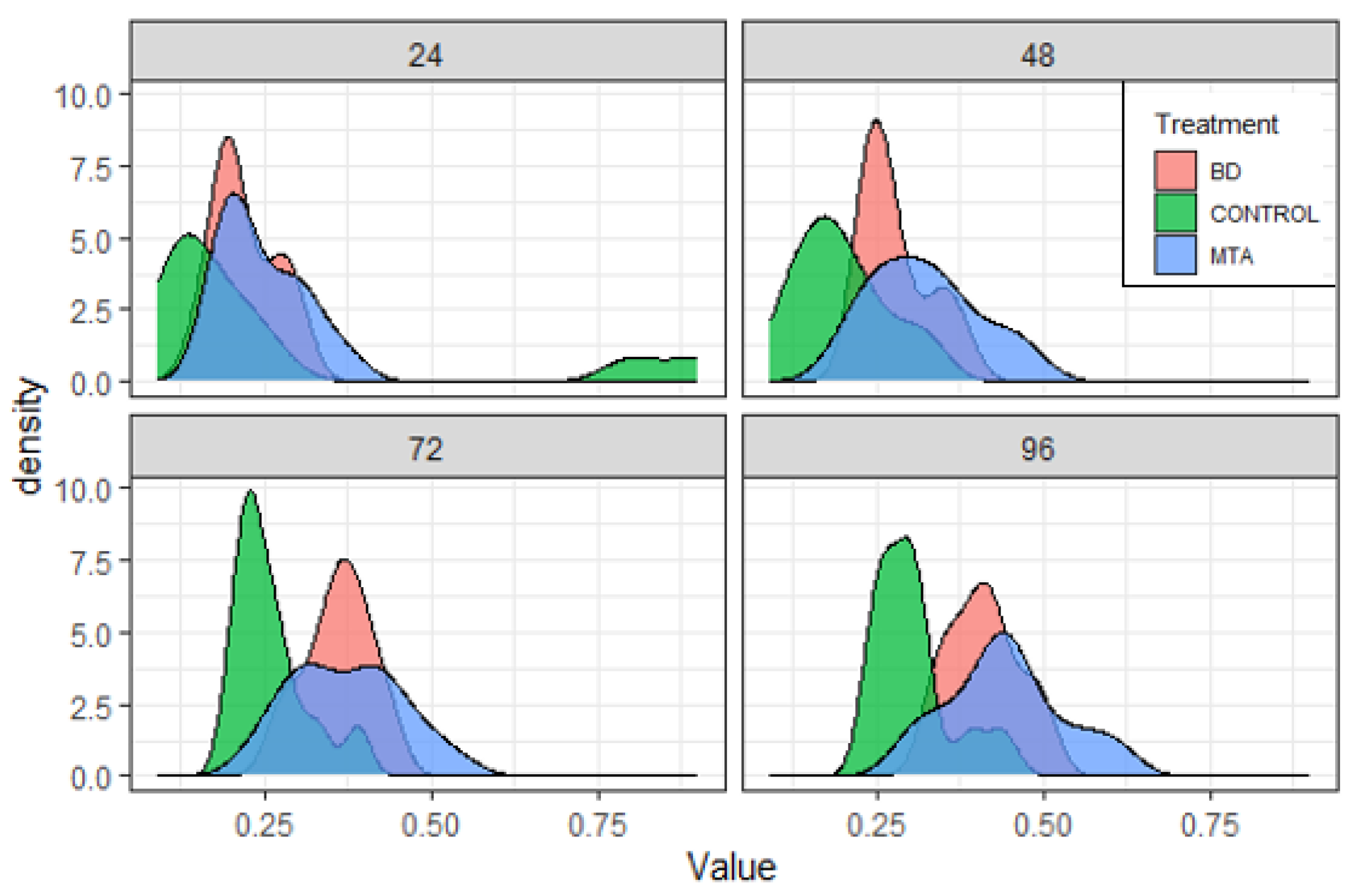
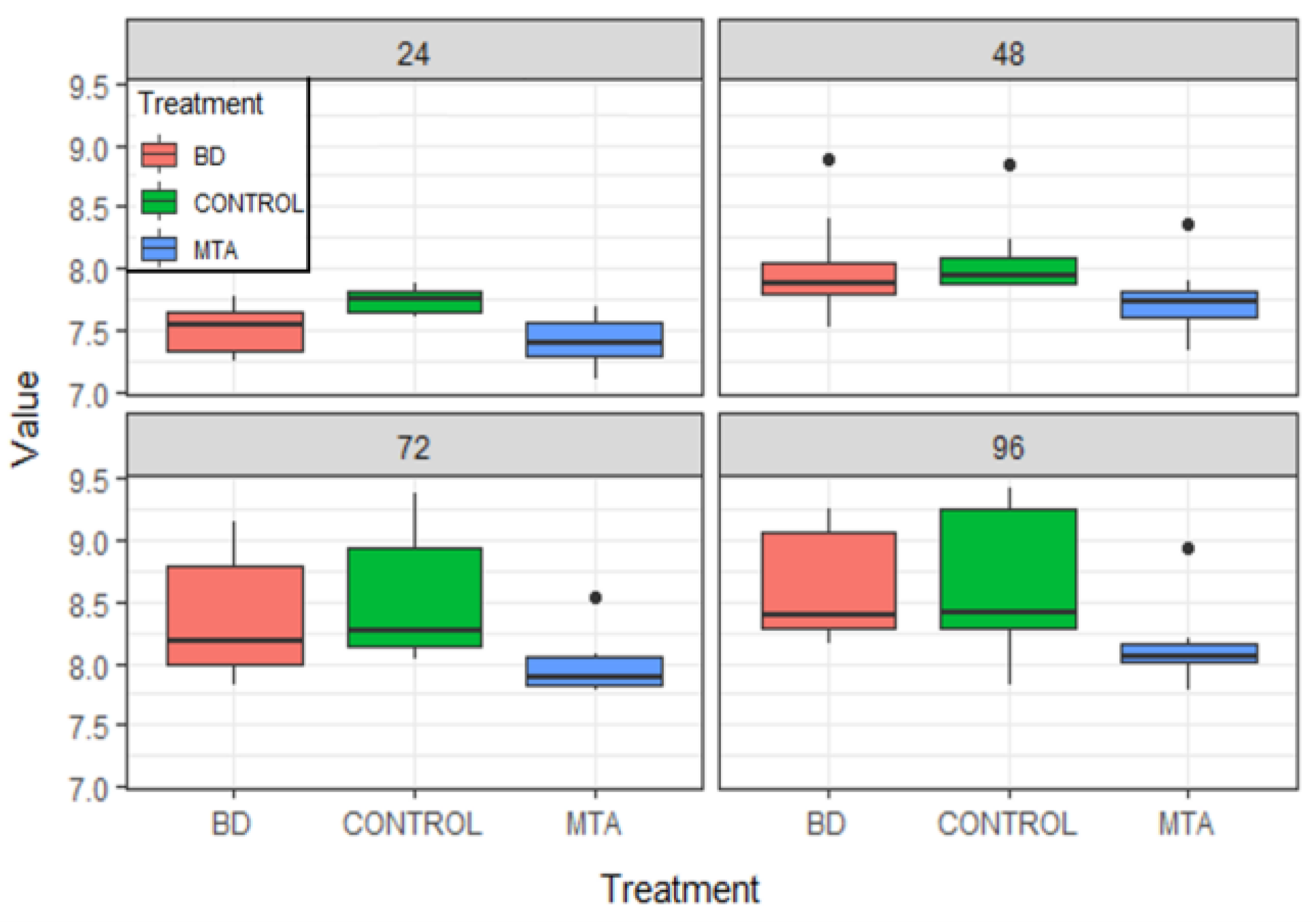
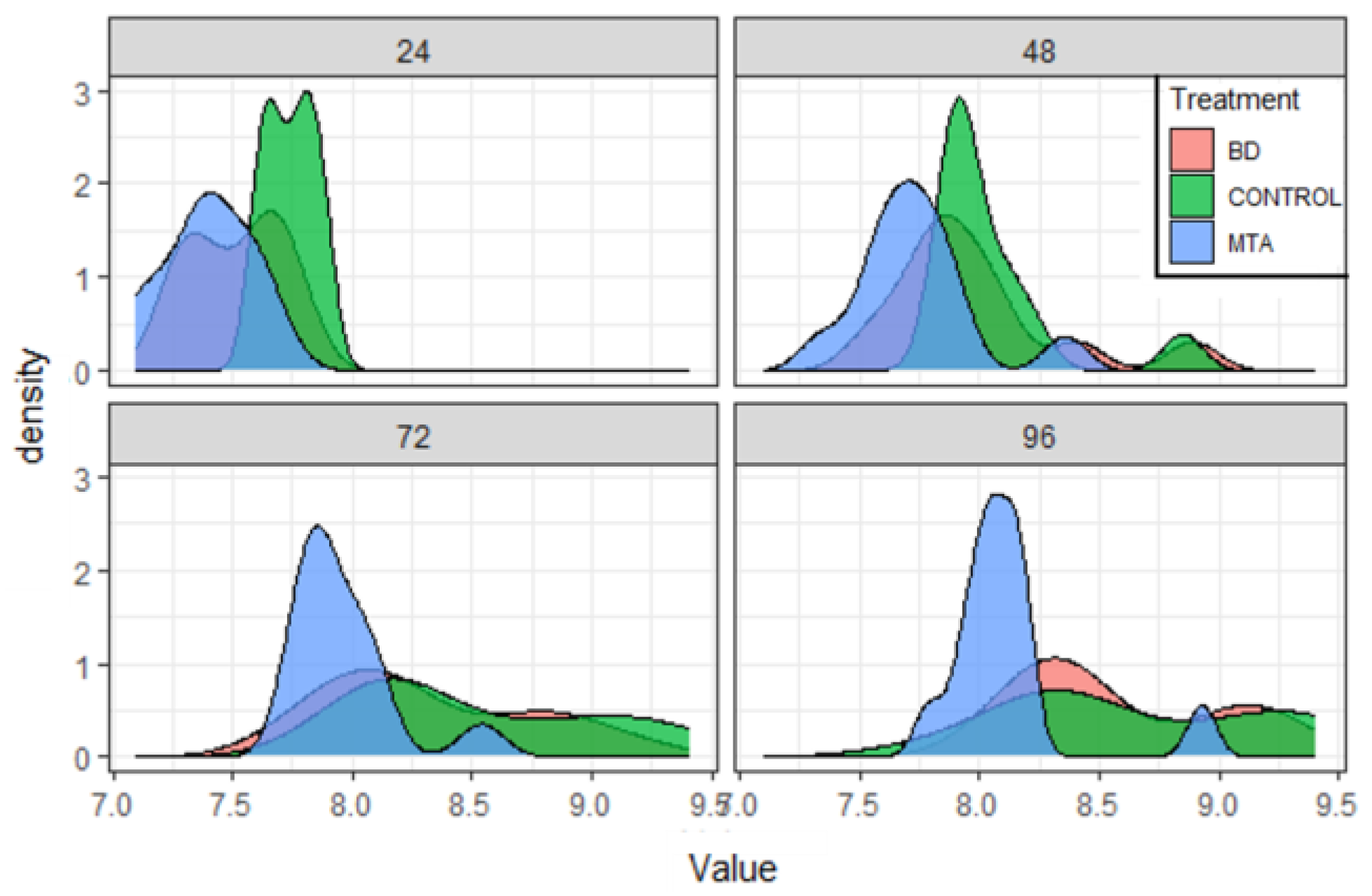
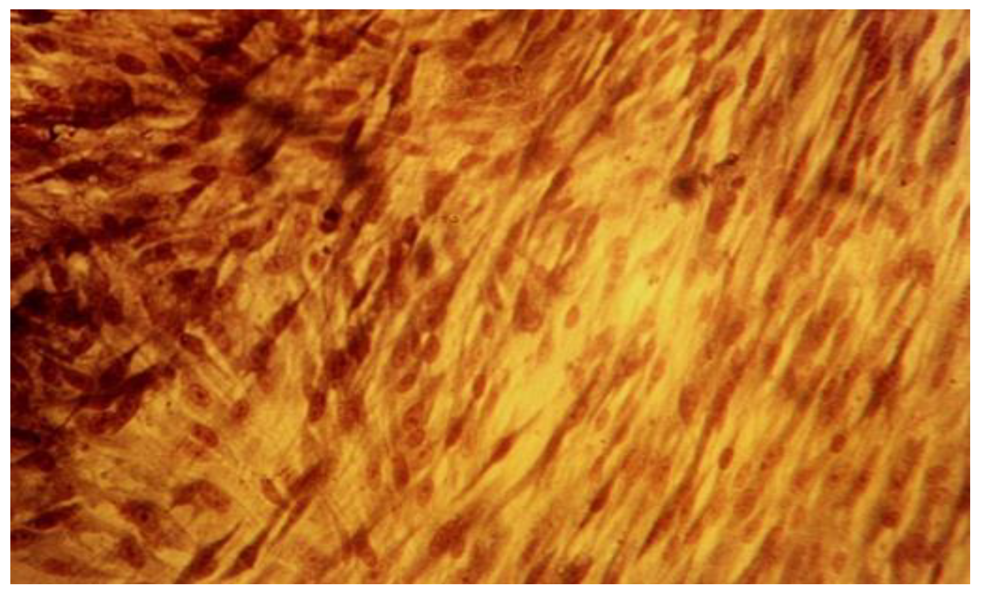
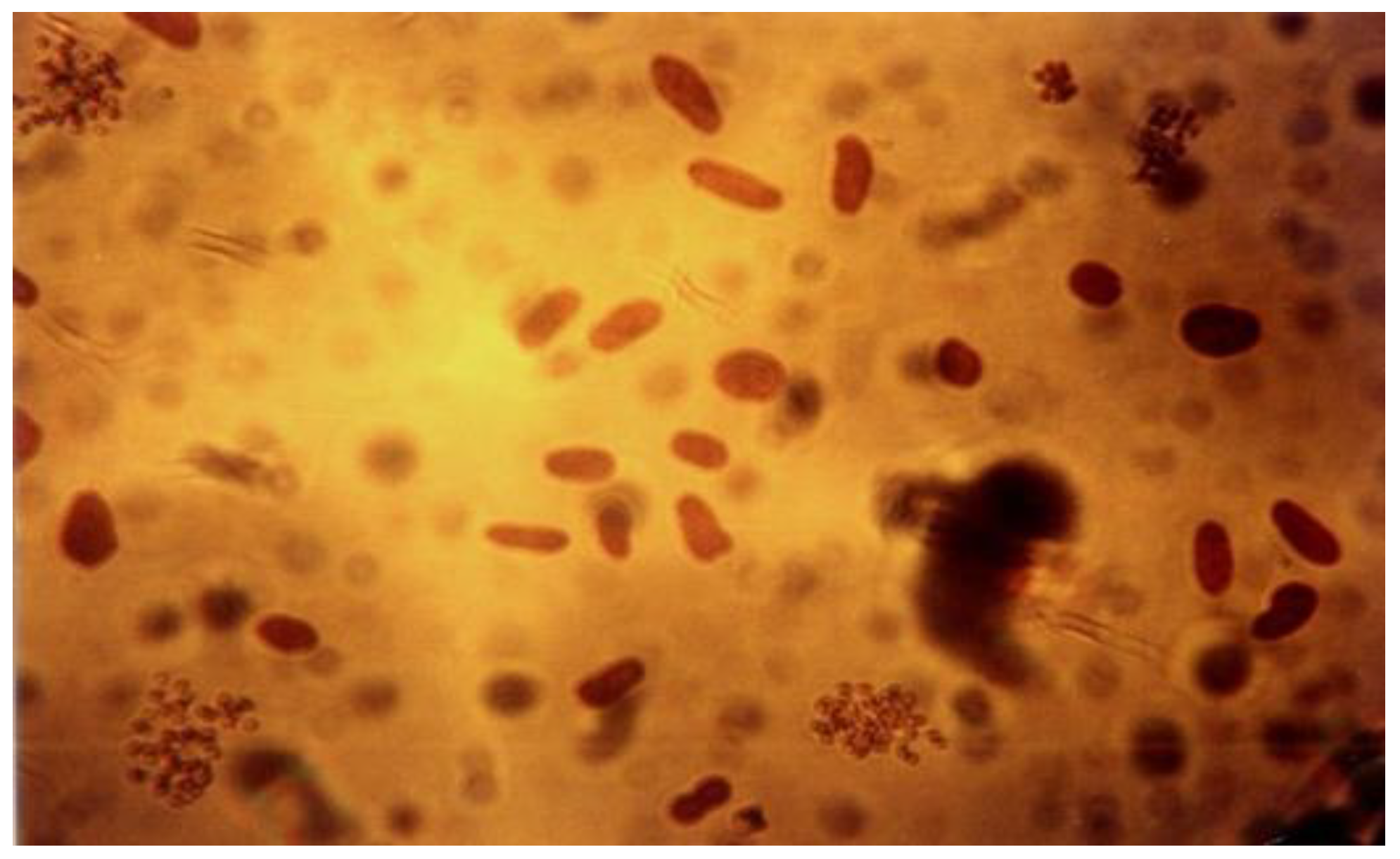
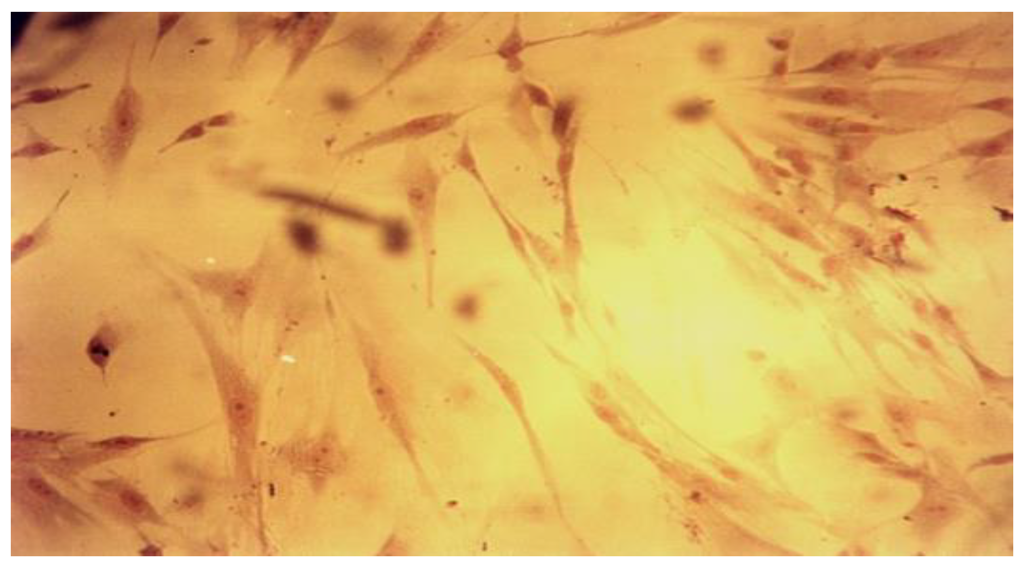
Statistical analysis was performed using two nonparametric tests: Kruskal-Wallis test in order to identify if there are differences between the control and the investigated products during the experiment. Further, the comparisons of groups with statistically significant differences were investigated with a Wilcoxon test with continuity correction in order to precisely identify the differences. The level of significance was set at p < 0.05.

Results

The results for cytotoxicity assessment by the mean of number of detached cells from the monolayer, for BD, MTA HP and control group, at 24, 48, 72 and 96 hours, are presented in Figure 1 and Figure 2.
The evolution of cells density in the presence of tested substances and control at every 24 hours point, during the 4 days experiment, are presented below in Figure 3 and Figure 4.
The qualitative expression of cytotoxicity was carried out microscopically and assessed by the cell cultures morphology analysis, reveals different degrees of cellular degeneration in contact with the investigated substances. We could observe different stages of membrane disintegration, nucleus changes and appearance of pathological pseudopodia.
Another specific aspect noted in this study is related to the appearance of the rounded cells detached form the monolayer, these being death or dying cells, as loss of adherence usually initiates apoptosis. The presence of dead cells is a result of pathological progression of cells alterations in contact with tested materials used in the study. They were floating in the suspension and their number increased during the entire experiment in the investigated lots as an indicator of the substances cytologic effect (Figure 5, Figure 6, Figure 7 and Figure 8).

Discussions

In this study the biological effects of bioactive MTA HP and BD have been investigated in a widespread used stabilized cellular line (L929). Cellular death (detached cells) and also evolution of cellular density of fibroblasts have been assessed in monolayer.
We choose to evaluate the direct effect on fibroblasts because the tested substances are used in close or even direct contact to pulp cells (fibroblasts and dental pulp stem cells), with which the proposed cell line is demonstrated to have similarities in characteristics and behavior [21].
Cell line tests for in-vitro cytotoxicity for medical devices can be done by direct or indirect contact tests. In-vivo, MTA HP and BD interact with dental pulp both in semisolid phase (when they are applied on the tooth structures) and in solid phase, after the setting time, for an extended period of time. As a result, their effect is different, depending on the elements that have been released from their structure. In the present study, given their limited solubility, as we mention before, we choose the indirect contact method, and the samples were tested in cured state, using the extracts medium of each material type.
Cell death in culture is a normal biological process, part of the cell growing and development. The passive death, known as necrosis, is usually a result of extracellular stresses that induce cell lysis, like nutrient depletion, hypoxia, waste byproduct accumulation, effect of specific chemicals to which the cells are in contact. The other types of cell death are programed (genetically control death known as apoptosis) or by autophagy.
Dead cell counting assays are a popular method to assess cell viability, using different staining methods which penetrate only damaged membrane cells (dead or dying cells). Trypan blue dye which we use is excluded from unaffected cells (the ones that remain colorless).
The quantitative results of cytotoxicity by evaluating the detached cells parameter, showed a continuous increasing of the detached cells number during the entire experiment both for BD and MTA HP. The aspect is similar to fibroblasts behaviour in control groups in the first 24 hours, but the death cells counting indicated lower values in the second phase of the study, then the continuous increasing aspect was maintained. For the first 24 hours no statistically significant difference between BD, MTA HP and control has been found (BD-control: p = 0.390; MTA HP-control: p = 0.280; BD-MTA HP: p = 0.390). At 48 hours for both capping material the significantly differences have been identified, compared to control (BD-control: p = 0.012; MTA HP-control: p = 0.004), but they presented similar effect on cellular culture (p = 0.247). The results at 72 hours confirmed the same effect of tested products, with no statistically significant difference between them (p = 0.794), but we found significant difference regarding BD-control behaviour (p = 0.002) and MTA HP – control (p = 0.002). At the end of the experiment, at 96 hours, the same results were found: differences in cell death of fibroblasts p = 0.001 for BD and p = 0.001 for MTA HP, compared to control, but no statistically significant difference was found between both materials regarding cellular death at this point (p = 0.340). The results indicated a higher number of dead cells for MTA HP versus BD in all tests, but not a significantly difference regarding detached cells assay between the two investigated products. Also, there were statistically significant difference between control and both investigated products, with the first 24 hours exception (when similar cell response was observed and no difference has been found between control and tested cements). This assay results of cell mortality indicated a slightly different cytotoxic effect depending on the type of tested cellular extract, more pronounced for MTA HP, and with Kruskal-Wallis test and Wilcoxon rank sum test aid we documented that in the first 24 hours of experiment no statistically significant difference between all tested groups was confirmed, but it was identified and maintained for the entire remaining period of the study.
The cell density is a continuously changing parameter, as representing a parameter of cell health [22,23]. Evaluation of cellular density in the presence of the investigated pulp capping materials is a way to monitor proliferation and viability in cultured cell lines as biological cell indicators for MTA HP and BD.
In our study, both as a result of a normal cell proliferation process and the tested materials effect, the cell density generally increased. The evolution of cell density was higher for BD than MTA HP in all tested lots, presenting a lower rate in the first 24 hours, when MTA determined a decreased cellular density, then a continuous increasing level, till the end of the experiment. For the control, the density in monolayer was continuously progressing for each lot, at different rates, during the entire study.
In the first 24 hours a statistically significant difference was found between both the MTA HP and BD groups and control groups, but not between the two investigated products (p = 0.00021). At 48 hours the results indicate a statistically significant difference between MTA HP and control (p = 0.0044), but not between BD and MTA HP (p = 0.0652) and BD-control (p = 0.2874). At 72 hours there is no statistically significant difference between BD and control (p = 0.3405). Still, at this point, statistically significant difference has been identified between MTA HP and control (p = 0.0011) and between the two pulp capping materials (p = 0.0204) and the same results were found at the end of the experiment, at 96 hours (BD-control: p = 0.686; MTA HP -control: p = 0.004; MTA HP-BD: p = 0.001). Based on cellular density parameter, using Kruskal-Wallis test, we can conclude that there are statistically significant differences between the fibroblasts reaction in the tested lots for MTA HP compared to control, at every 24 hours mark, during the entire experiment, but for BD and control group the differences could be identified only in the first 24 hours, then they exhibit the same biological behaviour. Regarding comparative statistically analysis of BD and MTA HP effects on fibroblasts culture, there were no differences in the first 24 and 48 hours, but a significant different cellular behaviour appeared for the last half period of time of the experiment. Pairwise comparisons using Wilcoxon test with continuity correction all statistically significant differences have been precisely identified.
The continuous increase of cellular density is an expression of the biocompatibility and inductive properties of the bioactive cements. There is a large number of studies which confirm these aspects, although we found a slightly cell growth inhibition in tested lots compared to control lots, especially for MTA HP [24,25].
The qualitative assessment of biological effect by microscopic examination of cellular appearance is consistent with values for the cellular dead rates and cellular density. Different changes in general cellular morphology, whose progressive evolution during the experiment, conducted to death cell, and detachment from substrate, have been observed. The observations indicate more obvious and visible aspects for MTA HP than BD and they have been presented descriptively. Nevertheless, the microscopic evaluations are in general more difficult to be precisely identified and must be always referred to the entire experiment and numerical data obtained. For MTA HP, more visible cell membrane integrity damages were observed, in association with a higher number of floating death cells. Loss of membrane integrity is a clear indicator for an unfavorable prognosis for cell survival [26]. For BD the morphological degenerative cell alterations were more discrete, and the presence of round detached cells less important. The rounded aspect is related to first sign of cellular injury, swelling, later followed by different nuclei and nucleoli alterations. No clear-cut delimitation between the healthy and affected cells were observed.
According to qualitative morphological grading of cytotoxicity ISO-10993-5 [20], we can conclude that the cell injuries can be considered slight (not more than 20 % of the cells are affected).
MTA’ cytotoxicity is an expression of the calcium hydroxide released during the setting period, as the subsequent high alkalinity is cytotoxic [27]. When it is cured, MTA shows almost none or slightly cytotoxicity, although some studies discovered that calcium ions are released form the material for 28 days after setting [28]. On the other side, MTA is capable of partially releasing its soluble fraction to an aqueous environment over a long period of time, showing material solubility, with consecutive cellular toxicity [29]. The MTA HP product we have tested is a new bioactive cement with improved physical parameters than the conventional MTA cement and better biological capacities due to higher calcium ions released rate which induce the differentiation of pulp cells and thus the healing effect on mineralization [30]. MTA HP releases calcium most intensively in the first month after application, compared to conventional MTA, which showed the highest release of calcium after 1 week [31]. The high-plasticity of this material is also a key factor in better properties exerted by MTA Repair HP as pulp-capping product, allowing a better handling and insertion, along with higher push-out bond strength values [32].
Different recent studies have demonstrated very good biocompatibility and cytocompatibility of MTA HP in human dental pulp cells, and an intense bioactive response with which the findings of this present study are in line/agreement [33,34].
Biodentine exhibits minor microscopically cell damage and permitted an important proliferation, identified as an increased cell density evolution during the entire experiment. Its outstanding bioactivity is attributed to TGF-β1 (transforming growth factor beta 1) activation in pulp cells, responsible for tertiary dentinogenesis and the secretion of the dentin matrix [35,36]. At the same time, Biodentine cytotoxic effect is proven to be insignificant in a large number of studies, being recommended as the silicate-based cement of choice for vital pulp therapy [37,38]. The excellent cytocompatibility of Biodentine is related to its improved composition, with no calcium aluminate and calcium sulfate and other heavy metals. Also, physical properties (easy manipulation, shorter setting time, better sealing with no microleakage and subsequent bacterial infiltration) allow a higher success rate in pulp capping treatments [39].
Cell toxicity assays are important tools for assessing biological response in order to predict the future in-vivo behaviour of the tested materials. The results of this in-vitro test are consistent with other studies on MTA HP and Biodentine regarding cytocompatibility. The best parameters were obtained for Biodentine, both for numerical data of cellular death and cell density and microscopic examination. We must also emphasize that at all time intervals, both investigated cements presented a very low level of cytotoxicity.

Conclusions

With the limitations of a cell culture in-vitro test, the present study which investigated fibroblasts behavior in the presence of MTA Repair HP(Angelus) and Biodentine demonstrated that cells are sensitive to both substances. The products induced different cellular degenerative alterations, which finally led to cell death. The quantitative results showed, however, that they present low levels of cytotoxicity and the biological risk is not a very high one. Biodentine showed in all parameters better biological effects than MTA Repair HP, expressed by lower and limited cellular damage and a higher cell density. The detached cells assay indicated that there is not a statistically significant difference regarding the fibroblast’s response in the presence of the two investigated products. The cellular density parameter showed no differences in the first 24 and 48 hours, but a significant different cellular behaviour appeared for the tested products for the second 48 hours of the study. Regarding the comparison with control, in cell death test we found statistically significant difference between control and both investigated products, with the first 24 hours exception (when similar response was identified), and for cellular density parameter we found statistically significant differences between the fibroblasts reaction in the tested lots for MTA compared to control, at every 24 hours mark, during the entire experiment, but for BD and control the differences could be identified only in the first 24 hours, then they exhibit the same biological behaviour.
In-vivo there is a large number of factors that intervene, in association with other immunological mechanisms and reactions, so the clinical response of human pulp to these products are a very good one for indirect and direct pulp capping and they have been demonstrated efficient clinical behavior.

Institutional Review Board Statement

Any aspect of the work covered in this manuscript has been conducted with the ethical approval of all relevant bodies and that such approvals are acknowledged within the manuscript. Informed consent was obtained from all subjects involved in the study.

Conflicts of Interest

There are no known conflicts of interest in the publication of this article. The manuscript was read and approved by all authors.

References

  1. Morotomi, T.; Washio, A.; Kitamura, C. Current and future options for dental pulp therapy. Jpn Dent Sci Rev. 2019, 55, 5–11. [Google Scholar] [CrossRef] [PubMed]
  2. European Society of Endodontology (ESE) developed by: Duncan, H.F.; Galler, K.M.; et al. European Society of Endodontology position statement: Management of deep caries and the exposed pulp. Int Endod J. 2019, 52, 923–934. [Google Scholar] [CrossRef]
  3. Ladino, L.G.; Bernal, A.; Calderón, D.; Cortés, D. Bioactive Materials in Restorative Dentistry: A Literature Review. SVOA Dentistry. 2021, 2, 74–81. [Google Scholar]
  4. Bioactive Restorative Materials. Int Dent J. 2023, 73, 9–10. [CrossRef] [PubMed]
  5. Walsh, R.M.; He, J.; Schweitzer, J.; Opperman, L.A.; Woodmansey, K.F. Bioactive endodontic materials for everyday use: A review. Gen Dent. 2018, 66, 48–51. [Google Scholar]
  6. Iliescu, A.A.; Gheorghiu, I.M.; Tănase, M.; Iliescu, A.; Mitran, L.; Mitran, M.; Perlea, P. Reactionary Versus Reparative Dentine in Deep Caries. ARS Medica Tomitana. 2019, 25, 15–21. [Google Scholar] [CrossRef]
  7. Li, Y.; Liu, C.; Han, G. Research progress of odontogenic extracellular vesicles in regeneration of dental pulp. Oral Dis. 2023, 29, 2565–2577. [Google Scholar] [CrossRef]
  8. Willershausen, B.; Willershausen, I.; Ross, A.; Velikonja, S.; Kasaj, A.; Blettner, M. Retrospective study on direct pulp capping with calcium hydroxide. Quintessence Int. 2011, 42, 165–171. [Google Scholar]
  9. Al-Saudi, K.W. A paradigm shift from calcium hydroxide to bioceramics in direct pulp capping: A narrative review. J Conserv Dent Endod. 2024, 27, 2–10. [Google Scholar] [CrossRef]
  10. Islam, R.; Islam, M.R.R.; Tanaka, T.; Alam, M.K.; Ahmed, H.M.A.; Sano, H. Direct pulp capping procedures—Evidence and practice. Jpn Dent Sci Rev. 2023, 59, 48–61. [Google Scholar] [CrossRef]
  11. Torabinejad, M.; Hong, C.U.; McDonald, F.; Pitt Ford, T.R. Physical and chemical properties of a new root-end filling material. J Endod. 1995, 21, 349–353. [Google Scholar] [CrossRef] [PubMed]
  12. Göhring, K.S.; Lehnert, B.; Zehnder, M. Indications for use of MTA, a review. Part 1: Chemical, physical and biological properties of MTA. Schweiz Monatsschr Zahnmed. 2004, 114, 143–153. [Google Scholar] [PubMed]
  13. Pinheiro, L.S.; Kopper, P.M.P.; Quintana, R.M.; Scarparo, R.K.; Grecca, F.S. Does MTA provide a more favourable histological response than other materials in the repair of furcal perforations? A systematic review. Int Endod J. 2021, 54, 2195–2218. [Google Scholar] [CrossRef]
  14. Bachoo, I.K.; Seymour, D.; Brunton, P. A biocompatible and bioactive replacement for dentine: Is this a reality? The properties and uses of a novel calcium-based cement. Br Dent J. 2013, 214, E5. [Google Scholar] [CrossRef] [PubMed]
  15. Rajasekharan, S.; Martens, L.C.; Cauwels, R.G.E.C.; Anthonappa, R.P. Biodentine™ material characteristics and clinical applications: A 3 year literature review and update. Eur Arch Paediatr Dent. 2018, 19, 1–22. [Google Scholar] [CrossRef]
  16. Ford, T.R.; Torabinejad, M.; Abedi, H.R.; et al. Using mineral trioxide aggregate as a pulp-capping material. J Am Dent Assoc. 1996, 127, 1491–1494. [Google Scholar] [CrossRef]
  17. Pérard, M.; Le Clerc, J.; Watrin, T.; et al. Spheroid model study comparing the biocompatibility of Biodentine and MTA. J Mater Sci Mater Med. 2013, 24, 1527–1534. [Google Scholar] [CrossRef]
  18. Iliescu, A.; Perlea, P.; Tulus, G.; Gheorghiu, I.M.; Iliescu, M.G.; Manolea, H.O. Bioceramics in endodontics. In Bioceramics and Biocomposites: From Research to Clinical Practice; Antoniac, I., Ed.; Wiley: Hoboken, NJ, USA, 2019; pp. 241–290. [Google Scholar]
  19. Silano, V.; Paganuzzi-Stammati, A.; Zucco, F. Toxicity Tests with Mammalian Cell Cultures. 2005; https://api.semanticscholar.org/CorpusID:53128858. [Google Scholar]
  20. ISO 10993-5. Biological evaluation of medical devices. Part 5: Tests for in vitro cytotoxicity. 2009; http://www.iso.org/iso/catalogue_detail.htm?csnumber=36406.
  21. Cohen, B.I.; Pagnillo, M.K.; Musikant, B.L.; Deutsch, A.S. An in vitro study of the cytotoxicity of two root canal sealers. J Endod. 2000, 26, 228–229. [Google Scholar] [CrossRef]
  22. Gilbert, D.F.; Mofrad, S.A.; Friedrich, O.; Wiest, J. Proliferation characteristics of cells cultured under periodic versus static conditions. Cytotechnology. 2019, 71, 443–452. [Google Scholar] [CrossRef]
  23. Adan, A.; Kiraz, Y.; Baran, Y. Cell Proliferation and Cytotoxicity Assays. Curr Pharm Biotechnol. 2016, 17, 1213–1221. [Google Scholar] [CrossRef]
  24. Miranda, R.B.; Fidel, S.R.; Boller, M.A. L929 cell response to root perforation repair cements: An in vitro cytotoxicity assay. Braz Dent J. 2009, 20, 22–26. [Google Scholar] [CrossRef] [PubMed]
  25. Koh, E.T.; McDonald, F.; Pitt Ford, T.R.; Torabinejad, M. Cellular response to Mineral Trioxide Aggregate. J Endod. 1998, 24, 543–547. [Google Scholar] [CrossRef] [PubMed]
  26. Ammendolia, D.A.; Bement, W.M.; Brumell, J.H. Plasma membrane integrity: Implications for health and disease. BMC Biol. 2021, 19, 71. [Google Scholar] [CrossRef]
  27. Ha, W.N.; Nicholson, T.; Kahler, B.; Walsh, L.J. Mineral Trioxide Aggregate-A Review of Properties and Testing Methodologies. Materials (Basel). 2017, 10, 1261, Published 2017 Nov 2. [Google Scholar] [CrossRef]
  28. Gandolfi, M.G.; Siboni, F.; Primus, C.M.; Prati, C. Ion release, porosity, solubility, and bioactivity of MTA Plus tricalcium silicate. J Endod. 2014, 40, 1632–1637. [Google Scholar] [CrossRef]
  29. Silva, E.J.; Accorsi-Mendonça, T.; Pedrosa, A.C.; Granjeiro, J.M.; Zaia, A.A. Long-Term Cytotoxicity, pH and Dissolution Rate of AH Plus and MTA Fillapex. Braz Dent J. 2016, 27, 419–423. [Google Scholar] [CrossRef] [PubMed]
  30. Palczewska-Komsa, M.; Kaczor-Wiankowska, K.; Nowicka, A. New Bioactive Calcium Silicate Cement Mineral Trioxide Aggregate Repair High Plasticity (MTA HP)-A Systematic Review. Materials (Basel). 2021, 14, 4573. [Google Scholar] [CrossRef]
  31. Guimarães, B.M.; Prati, C.; Duarte, M.A.H.; Bramante, C.M.; Gandolfi, M.G. Physicochemical properties of calcium silicate-based formulations MTA Repair HP and MTA Vitalcem. J Appl Oral Sci. 2018, 26, e2017115, Published 2018 Apr 5. [Google Scholar] [CrossRef]
  32. Silva, E.J.; Carvalho, N.K.; Zanon, M.; Senna, P.M.; et al. Push-out bond strength of MTA HP, a new high-plasticity calcium silicate-based cement. Braz Oral Res. 2016, 30, S1806–83242016000100269. [Google Scholar] [CrossRef]
  33. Tomás-Catalá, C.J.; Collado-González, M.; García-Bernal, D.; et al. Comparative analysis of the biological effects of the endodontic bioactive cements MTA-Angelus, MTA Repair HP and NeoMTA Plus on human dental pulp stem cells. Int Endod J 2017, 50 Suppl 2, e63–e72. [Google Scholar] [CrossRef]
  34. Jiménez-Sánchez, M.D.C.; Segura-Egea, J.J.; Díaz-Cuenca, A. MTA HP Repair stimulates in vitro an homogeneous calcium phosphate phase coating deposition. J Clin Exp Dent. 2019, 11, e322–e326, Published 2019 Apr 1. [Google Scholar] [CrossRef] [PubMed]
  35. Laurent, P.; Camps, J.; About, I. Biodentine(TM) induces TGF-β1 release from human pulp cells and early dental pulp mineralization. Int Endod J. 2012, 45, 439–448. [Google Scholar] [CrossRef] [PubMed]
  36. Abuarqoub, D.; Aslam, N.; Zaza, R.; et al. The Immunomodulatory and Regenerative Effect of Biodentine™ on Human THP-1 Cells and Dental Pulp Stem Cells: In Vitro Study. Biomed Res Int. 2022, 2022, 2656784. [Google Scholar] [CrossRef]
  37. Birant, S.; Gokalp, M.; Duran, Y.; Koruyucu, M.; Akkoc, T.; Seymen, F. Cytotoxicity of NeoMTA Plus, ProRoot MTA and Biodentine on human dental pulp stem cells. J Dent Sci. 2021, 16, 971–979. [Google Scholar] [CrossRef] [PubMed]
  38. Jang, Y.E.; Lee, B.N.; Koh, J.T.; et al. Cytotoxicity and physical properties of tricalcium silicate-based endodontic materials. Restor Dent Endod. 2014, 39, 89–94. [Google Scholar] [CrossRef]
  39. Nowicka, A.; Lipski, M.; Parafiniuk, M.; et al. Response of human dental pulp capped with biodentine and mineral trioxide aggregate. J Endod. 2013, 39, 743–747. [Google Scholar] [CrossRef]
Figure 1. Boxplot diagram of the evolution of the detached cells from the monolayer induced by the investigated pulp capping materials in fibroblasts cell culture, compared to control, at 24, 48, 72 and 96 hours.
Jmms 11 00056 g001
Figure 2. The evolution of the detached cells from the monolayer induced by the investigated pulp capping materials in fibroblasts cell culture, compared to control group, at 24, 48, 72 and 96 hours.
Jmms 11 00056 g002
Figure 3. Boxplot diagram of the evolution of cells density in the presence of investigated pulp capping materials in fibroblasts cell culture, compared to control group, at 24, 48, 72 and 96 hours.
Jmms 11 00056 g003
Figure 4. The evolution of cells density in the presence of investigated pulp capping materials in fibroblasts cell culture, compared to control group, at 24, 48, 72 and 96 hours.
Jmms 11 00056 g004
Figure 5. Fibroblasts cell line L929 at 24-hour mark, MTA Repair HP (Angelus, Brazil) lot, 20x magnification. Degenerative cytoplasmic and membrane alterations.
Jmms 11 00056 g005
Figure 6. Fibroblasts cell line L929 at 96-hour mark, MTA Repair HP (Angelus, Brazil) lot, 40x magnification. Detached cells from monolayer, in suspension.
Jmms 11 00056 g006
Figure 7. Fibroblasts cell line L929 at 24-hour mark, Biodentine lot, 20x magnification. Cell membrane damage in association with nuclei alterations.
Jmms 11 00056 g007
Figure 8. Fibroblasts cell line L929 at 96-hour mark, Biodentine lot, 40x magnification. Numerous cellular morphological alterations and floating dead cells.
Jmms 11 00056 g008
Table 1. Main characteristics of MTA Repair HP and Biodentine.
ProductDental Material CategoryPresentation/CompositionPropertiesField of Applications
MTA Repair HPBioactive, reparative calcium silicate-based cementPowder/liquid capsulesPowder: Tricalcium Silicate, Dicalcium Silicate, Tricalcium Aluminate, Calcium Oxide, Calcium TungstateLiquid: Water and plasticizer
-
high plasticity, high alkalinity
-
setting time 15 minutes
-
bismuth – free
-
hydrophilic/low solubility
-
calcium ions release
-
antibacterial effect
-
small particles size
-
radiopaque
-
indirect and direct pulp capping
-
pulpotomy
-
root canal perforation
-
root resorption
-
apexification
-
retrograde endodontic surgery
BIODENTINEBioactive new calcium silicate-based cement, generally known as “dentine substitute”Capsules/single–dose containersPowder: Tricalcium Silicate, Dicalcium Silicate, Calcium carbonate and oxide filler, iron oxide, zirconium oxideLiquid: hydro-soluble polymer, calcium chloride
-
high plasticity, alkaline pH
-
setting time 12 minutes
-
mechanical/flexural properties similar to dentin
-
resistance to microleakage
-
color stability
-
great bond to dental tissues
-
radiopaque
-
indirect and direct pulp capping
-
dentin replacement, in deep/large coronal restorations (sandwich technique)
-
pulpotomy
-
root and furcation perforations
-
apexification
-
retrograde endodontic surgery
Table 2. Biological properties of MTA Repair HP and Biodentine.
Dental ProductBiological Properties
MTA Repair HP
-
biocompatibility
-
dentin remineralization
-
cementum neoformation
-
antibacterial effect
BIODENTINE
-
bioactivity (biomimetic remineralization)
-
biocompatibility
-
sealing ability
-
antibacterial action
-
anti-inflammatory properties
-
stability in the oral environment

