
Patterns of Pneumatization of the Posterior Nasal Roof


 ,
,
Abstract
:1. Introduction
2. Materials and Methods
3. Results
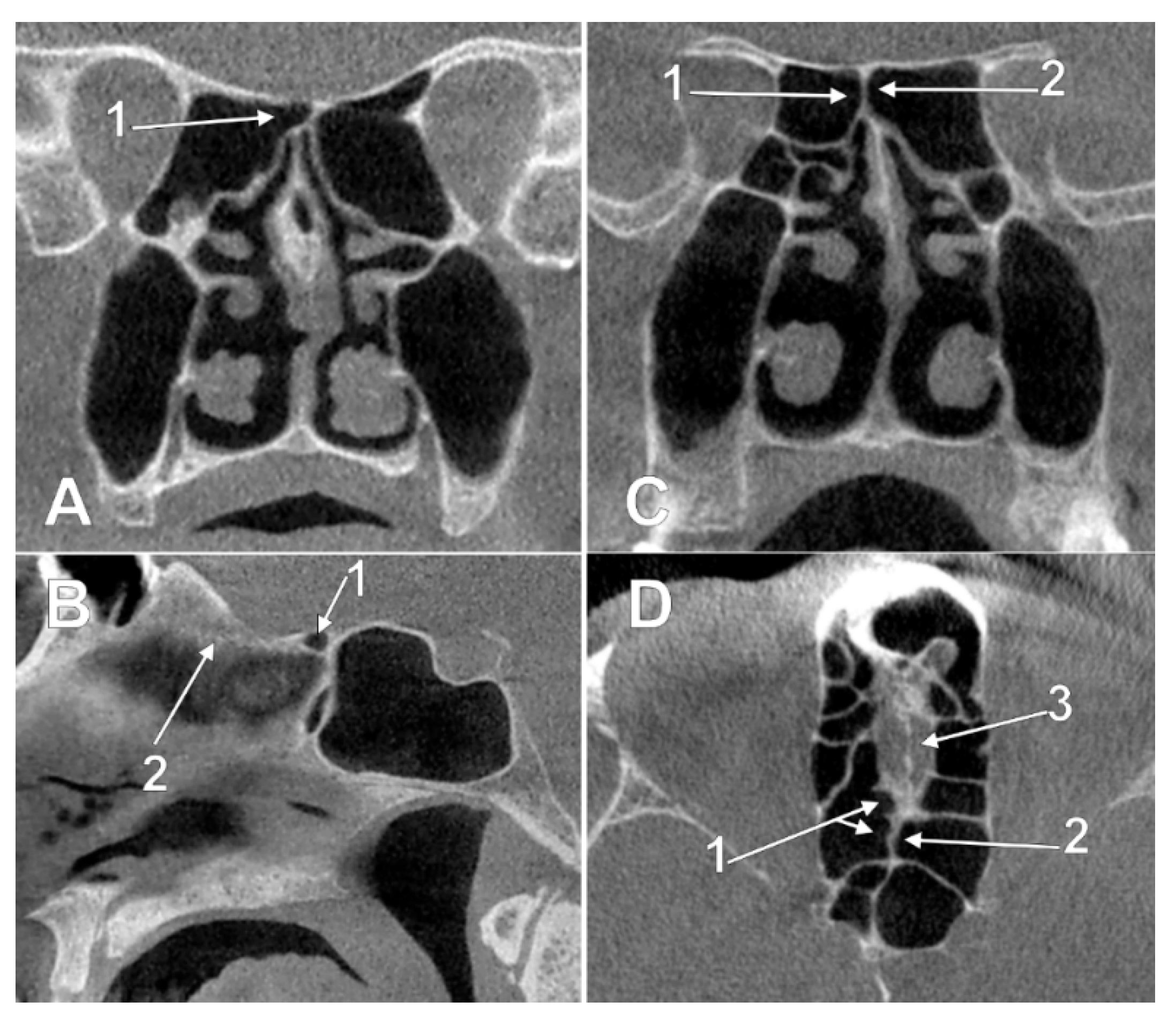
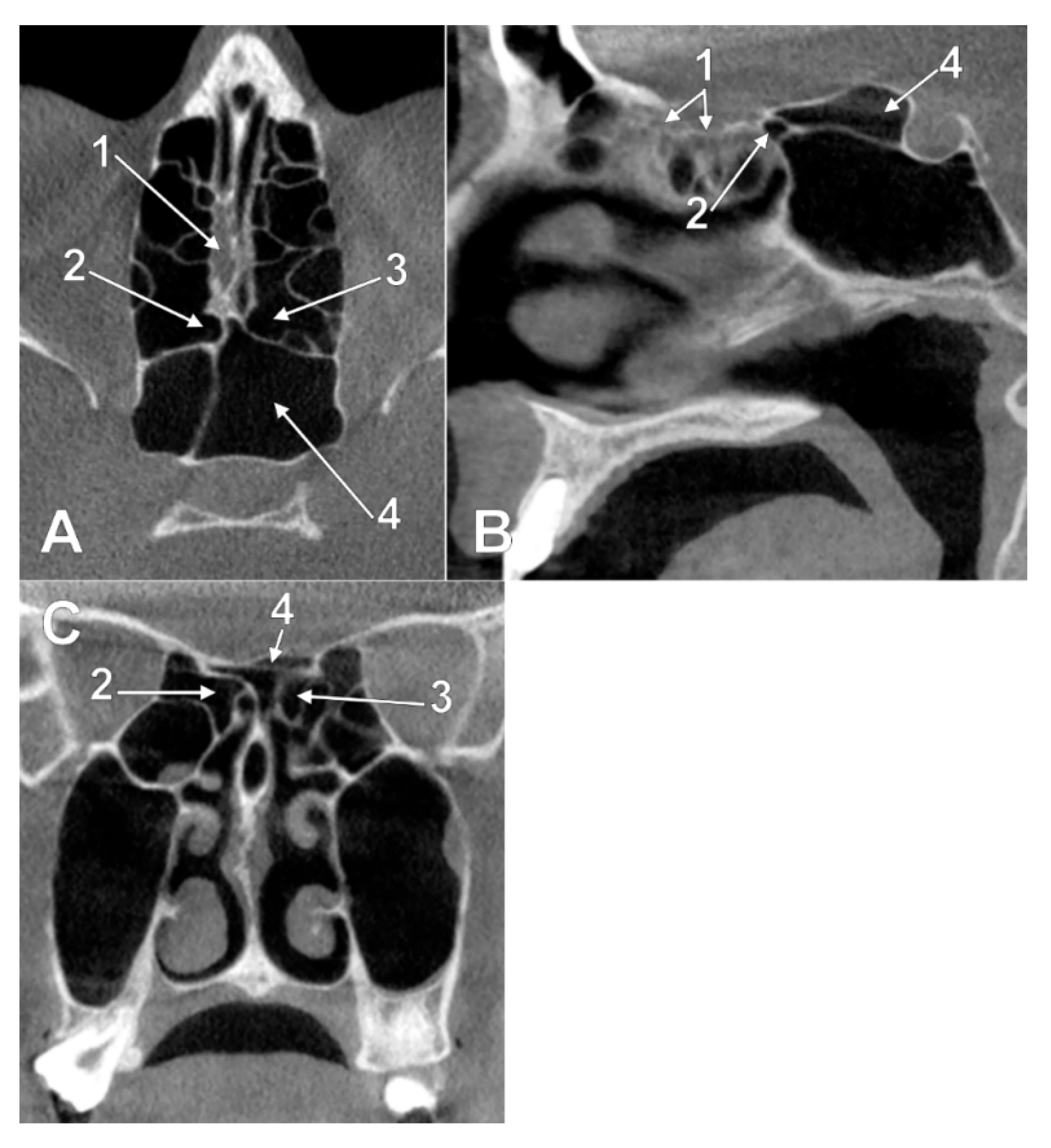
3.1. Ethmoidal Origin of Nasal Roof Pneumatizations
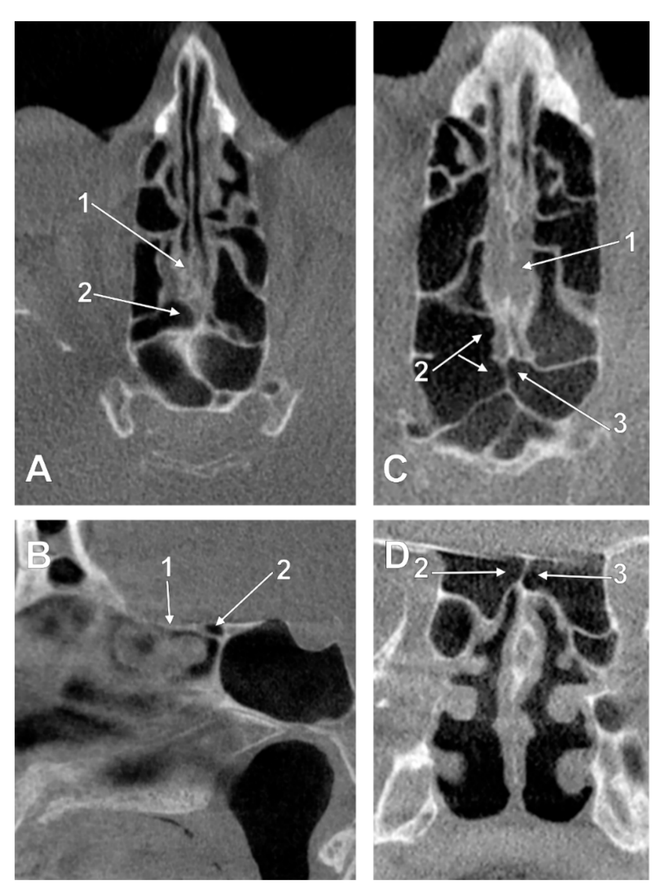
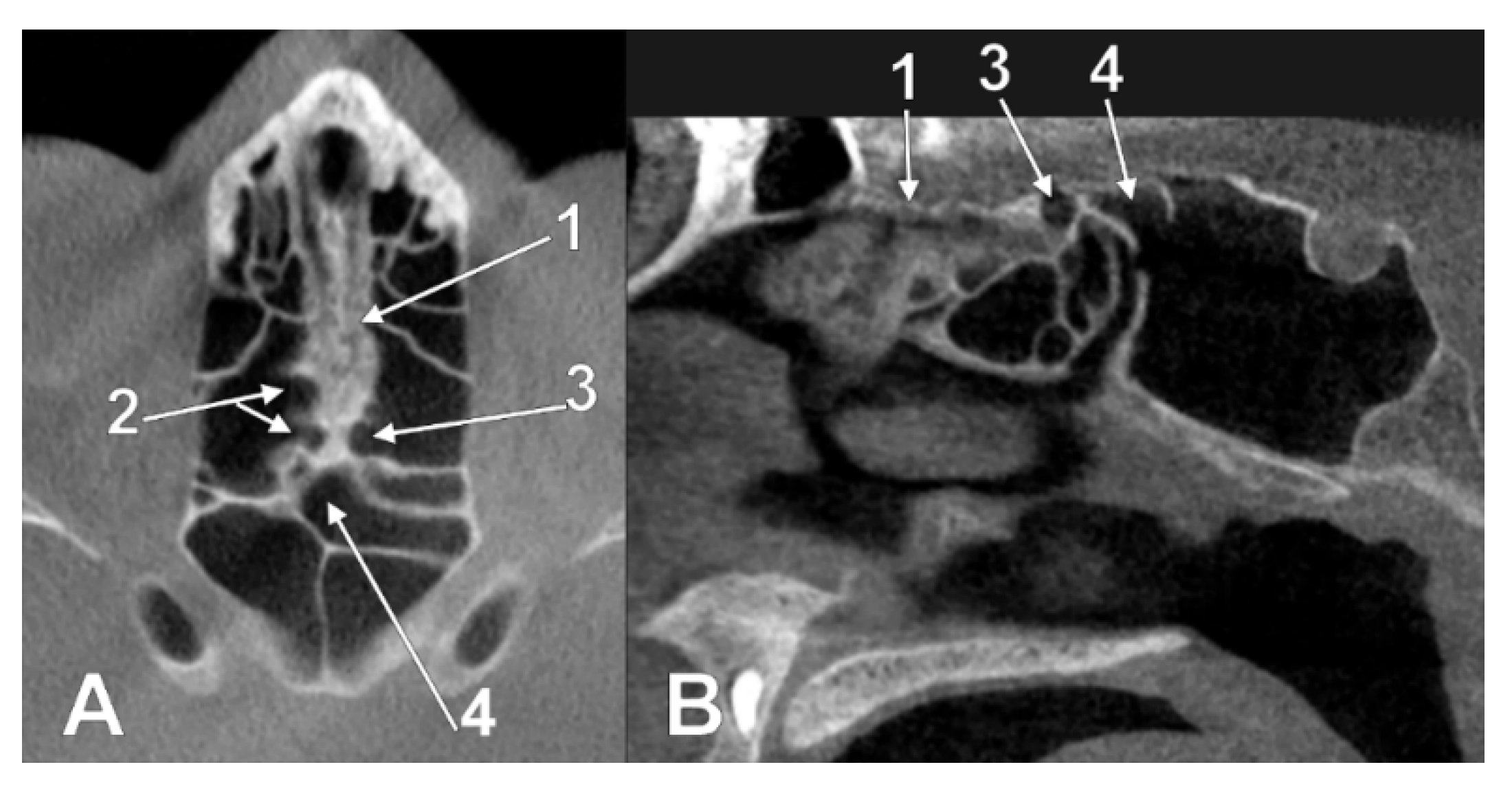
3.2. Sphenoidal Origin of Nasal Roof Pneumatization
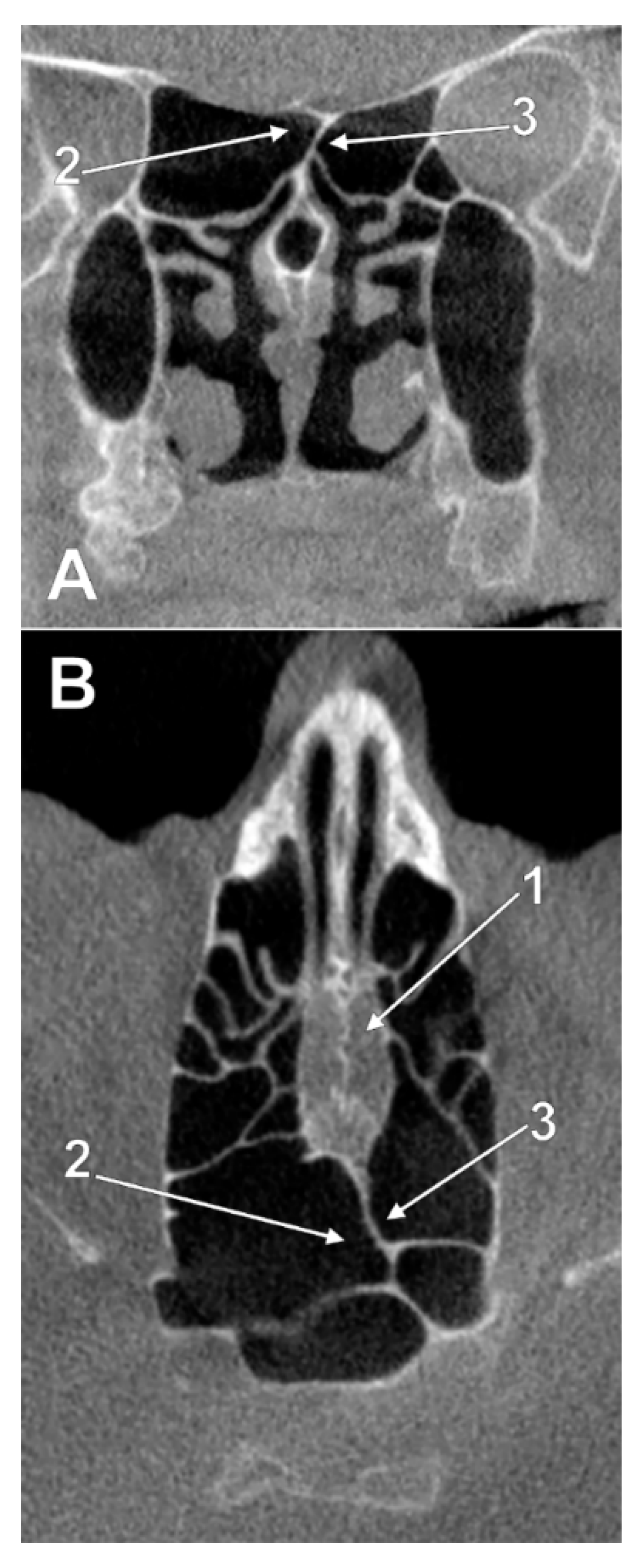
3.3. Sphenoethmoidal (Onodi Cell) Origin of Nasal Roof Pneumatizations
4. Discussion
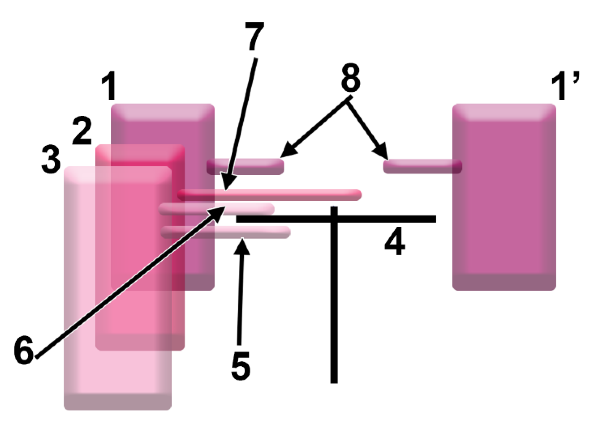
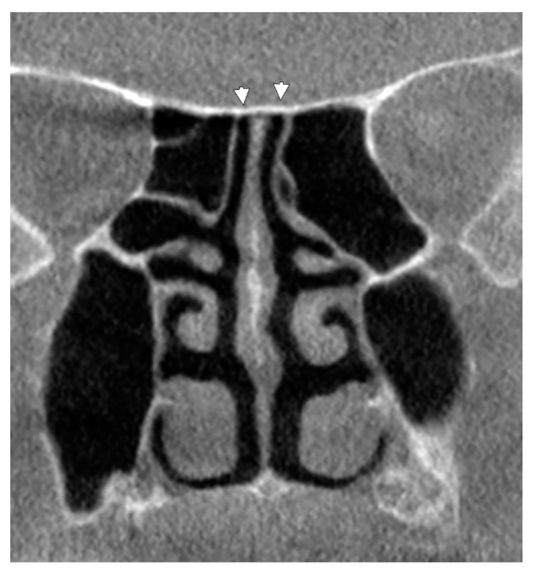
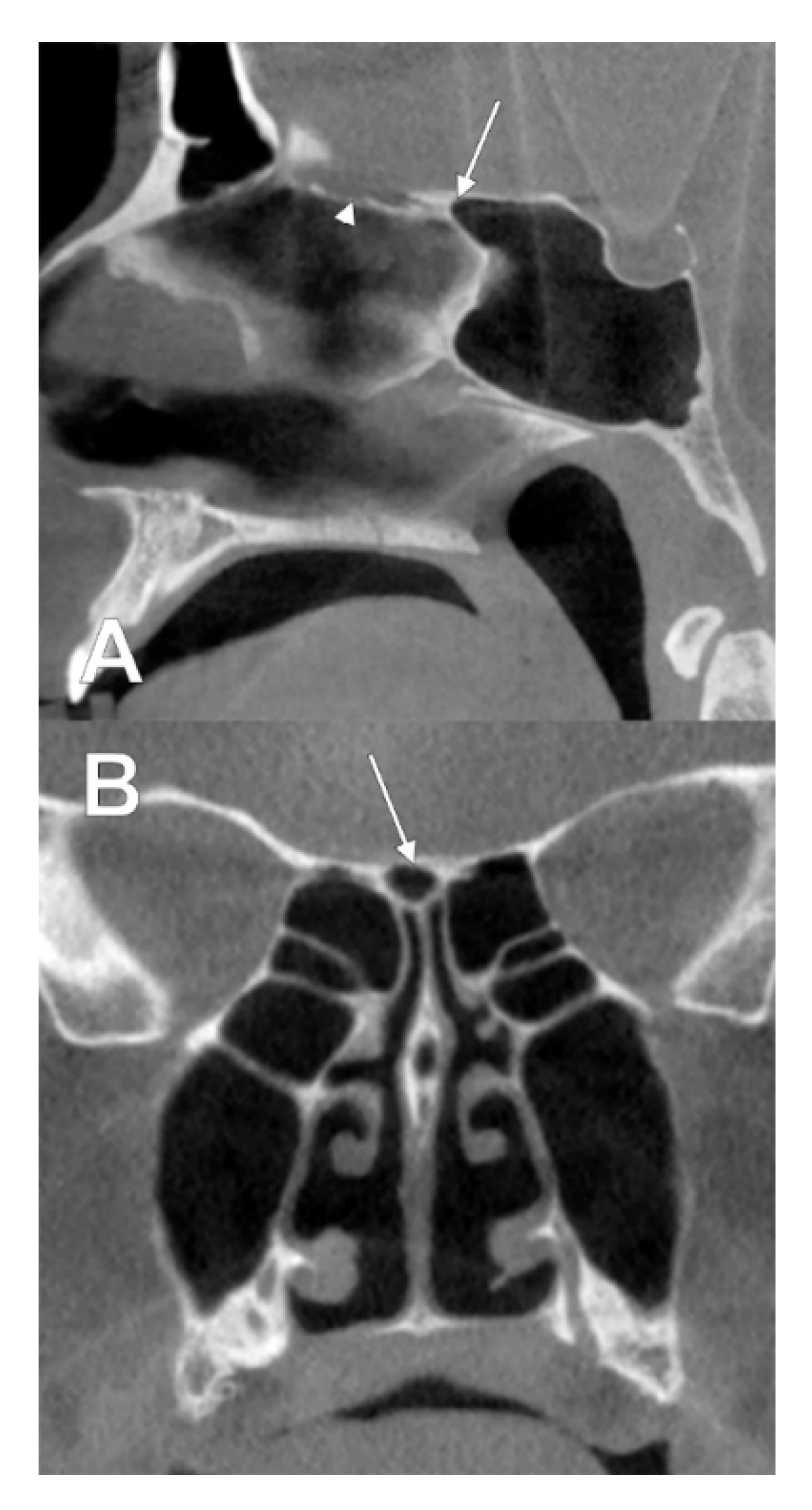
4.1. Gore’s Supraseptal Air Cell
4.2. Ethmoidal and Sphenoidal Pneumatizations
4.3. Clinical Anatomy
5. Conclusions
Author Contributions
Funding
Institutional Review Board Statement
Informed Consent Statement
Conflicts of Interest
References
- Vasvari, G.; Reisch, R.; Patonay, L. Surgical anatomy of the cribriform plate and adjacent areas. min-Minim. Invasive Neurosurg. 2005, 48, 25–33. [Google Scholar] [CrossRef] [PubMed]
- Palade, O.; Severin, F.; Toader, M.; Cobzeanu, M.; Toader, C. Combined approach for large tumors of the nose and paranasal sinuses-case report. Med.-Surg. J. 2016, 120, 380–383. [Google Scholar]
- Gray, H.; Standring, S.; Anand, N.; Birch, R.; Collins, P.; Crossman, A.; Gleeson, M.; Jawaheer, G.; Smith, A.L.; Spratt, J.D.; et al. Gray’s Anatomy: The Anatomical Basis of Clinical Practice, 41th ed.; Elsevier: London, UK, 2016. [Google Scholar]
- Teatini, G.; Simonetti, G.; Salvolini, U.; Masala, W.; Meloni, F.; Rovasio, S.; Dedola, G.L. Computed tomography of the ethmoid labyrinth and adjacent structures. Ann. Otol. Rhinol. Laryngol. 1987, 96, 239–250. [Google Scholar] [CrossRef] [PubMed]
- Hajiioannou, J.; Owens, D.; Whittet, H.B. Evaluation of anatomical variation of the crista galli using computed tomography. Clin. Anat. 2010, 23, 370–373. [Google Scholar] [CrossRef] [PubMed]
- Kaur, J.; Mogulla, S.; Malik, A.; Garg, S. Unusual Presentation of a Sphenoidal Sinus Neuroendocrine Tumor: A Case Report and Review of Literature. Cureus 2021, 13, e13689. [Google Scholar] [CrossRef] [PubMed]
- Gomez, J.; Pickup, S. Cribiform Plate Fractures; StatPearls: Treasure Island, FL, USA, 2021. [Google Scholar]
- Jaworek-Troc, J.; Zarzecki, M.; Bonczar, A.; Kaythampillai, L.N.; Rutowicz, B.; Mazur, M.; Urbaniak, J.; Przybycien, W.; Piatek-Koziej, K.; Kuniewicz, M.; et al. Sphenoid bone and its sinus—Anatomo-clinical review of the literature including application to FESS. Folia Med. Crac. 2019, 59, 45–59. [Google Scholar]
- Ali, Q.M.; Dietrich, B.; Becker, H. Patterns of skull base fracture: A three-dimensional computed tomographic study. Neuroradiology 1994, 36, 622–624. [Google Scholar] [CrossRef] [PubMed]
- Knizek, Z.; Michalek, R.; Vodicka, J.; Zdobinska, P. Cribriform Plate Injury after Nasal Swab Testing for COVID-19. JAMA Otolaryngol.—Head Neck Surg. 2021, 147, 915–917. [Google Scholar] [CrossRef]
- Radoi, P.M.; Rusu, M.C.; Dinca, D.; Toader, C. Combined rare anatomic variants: Persistent primitive olfactory artery and azygos pericallosal artery. Surg. Radiol. Anat. 2021, 43, 1305–1308. [Google Scholar] [CrossRef]
- Krmpotic-Nemanic, J.; Vinter, I.; Jalsovec, D.; Hat, J. Relation of the ethmoidal cells to the floor of the anterior cranial fossa. Ann. Anat. Anat. Anz. Off. Organ Anat. Ges. 2000, 182, 533–536. [Google Scholar] [CrossRef]
- Babu, A.C.; Nair, M.; Kuriakose, A.M. Olfactory fossa depth: CT analysis of 1200 patients. Indian J. Radiol. Imaging 2018, 28, 395–400. [Google Scholar] [CrossRef] [PubMed]
- Rusu, M.C.; Sandulescu, M.; Sava, C.J.; Dinca, D. Bifid and secondary superior nasal turbinates. Folia Morphol. 2019, 78, 199–203. [Google Scholar] [CrossRef] [PubMed]
- Sava, C.J.; Rusu, M.C.; Sandulescu, M.; Dinca, D. Vertical and sagittal combinations of concha bullosa media and paradoxical middle turbinate. Surg. Radiol. Anat. 2018, 40, 847–853. [Google Scholar] [CrossRef]
- Carstocea, L.; Rusu, M.C.; Matesica, D.S.; Sandulescu, M. Air spaces neighbouring the infraorbital canal. Morphologie 2020, 104, 44–50. [Google Scholar] [CrossRef] [PubMed]
- Çalışkan, A.; Sumer, A.P.; Bulut, E. Evaluation of anatomical variations of the nasal cavity and ethmoidal complex on cone-beam computed tomography. Oral Radiol. 2017, 33, 51–59. [Google Scholar] [CrossRef]
- Pedziwiatr, Z.F. Structural pattern of the ethmoidal labyrinth in adult man. II. Arch. Fur Klin. Und Exp. Ohren-Nasen-Und Kehlkopfheilkd. 1973, 204, 8–16. [Google Scholar] [CrossRef]
- Rusu, M.C.; Sandulescu, M.; Derjac-Arama, A.I. The pterygopalatine recess of the superior nasal meatus. Surg. Radiol. Anat. 2016, 38, 979–982. [Google Scholar] [CrossRef]
- Rusu, M.C.; Sava, C.J.; Ilie, A.C.; Sandulescu, M.; Dinca, D. Agger Nasi Cells versus Lacrimal Cells and Uncinate Bullae in Cone-Beam Computed Tomography. Ear Nose Throat J. 2019, 98, 334–339. [Google Scholar] [CrossRef] [Green Version]
- Kaplanoglu, H.; Kaplanoglu, V.; Dilli, A.; Toprak, U.; Hekimoglu, B. An analysis of the anatomic variations of the paranasal sinuses and ethmoid roof using computed tomography. Eurasian J. Med. 2013, 45, 115–125. [Google Scholar] [CrossRef]
- Marquez, S.; Tessema, B.; Clement, P.A.; Schaefer, S.D. Development of the ethmoid sinus and extramural migration: The anatomical basis of this paranasal sinus. Anat. Rec. 2008, 291, 1535–1553. [Google Scholar] [CrossRef]
- Basic, N.; Basic, V.; Jukic, T.; Basic, M.; Jelic, M.; Hat, J. Computed tomographic imaging to determine the frequency of anatomical variations in pneumatization of the ethmoid bone. Eur. Arch. Oto-Rhino-Laryngol. 1999, 256, 69–71. [Google Scholar]
- Meloni, F.; Mini, R.; Rovasio, S.; Stomeo, F.; Teatini, G.P. Anatomic variations of surgical importance in ethmoid labyrinth and sphenoid sinus. A study of radiological anatomy. Surg. Radiol. Anat. 1992, 14, 65–70. [Google Scholar] [CrossRef] [PubMed]
- Kantarci, M.; Karasen, R.M.; Alper, F.; Onbas, O.; Okur, A.; Karaman, A. Remarkable anatomic variations in paranasal sinus region and their clinical importance. Eur. J. Radiol. 2004, 50, 296–302. [Google Scholar] [CrossRef] [PubMed]
- von Arx, T.; Lozanoff, S.; Bornstein, M.M. Extraoral anatomy in CBCT—A literature review. Part 1: Nasoethmoidal region. Swiss Dent. J. 2019, 129, 804–815. [Google Scholar] [PubMed]
- Gore, M.R. The supraseptal ethmoid sinus cell: A previously unreported ethmoid sinus variant. Clin. Case Rep. 2019, 7, 1306–1308. [Google Scholar] [CrossRef] [Green Version]
- Van Alyea, O. Ethmoid labyrinth: Anatomic study, with consideration of the clinical significance of its structural characteristics. Arch. Otolaryngol. 1939, 29, 881–902. [Google Scholar] [CrossRef]
- Van Alyea, O.E. Sphenoid sinus: Anatomic study, with consideration of the clinical significance of the structural characteristics of the sphenoid sinus. Arch. Otolaryngol. 1941, 34, 225–253. [Google Scholar] [CrossRef]
- Wang, J.; Bidari, S.; Inoue, K.; Yang, H.; Rhoton, A., Jr. Extensions of the sphenoid sinus: A new classification. Neurosurgery 2010, 66, 797–816. [Google Scholar] [CrossRef] [Green Version]
- Jaworek-Troc, J.; Zarzecki, M.; Zamojska, I.; Iwanaga, J.; Przybycien, W.; Mazur, M.; Chrzan, R.; Walocha, J.A. The dimensions of the sphenoid sinuses: Evaluation before the functional endoscopic sinus surgery. Folia Morphol. 2021, 80, 275–282. [Google Scholar] [CrossRef]
- Jaworek-Troc, J.; Zarzecki, M.; Przybycien, W.; Lipski, M.; Curlej-Wadrzyk, A.; Iwanaga, J.; Walocha, J.; Mazurek, A.; Chrzan, R.; Urbanik, A. The course of the main septum in the sphenoid sinuses—Evaluation before the FESS. Folia Med. Crac. 2021, 61, 35–51. [Google Scholar]
- Jaworek-Troc, J.; Walocha, J.A.; Loukas, M.; Tubbs, R.S.; Iwanaga, J.; Zawilinski, J.; Brzegowy, K.; Zarzecki, J.J.; Curlej-Wadrzyk, A.; Kucharska, E.; et al. Extensive pneumatisation of the sphenoid bone: Anatomical investigation of the recesses of the sphenoid sinuses and their clinical importance. Folia Morphol. 2021, 80, 935–946. [Google Scholar] [CrossRef] [PubMed]
- Rusu, M.C.; Didilescu, A.C.; Jianu, A.M.; Paduraru, D. 3D CBCT anatomy of the pterygopalatine fossa. Surg. Radiol. Anat. 2013, 35, 143–159. [Google Scholar] [CrossRef] [PubMed]
- Rusu, M.C.; Hostiuc, S.; Motoc, A.G.M.; Mogoanta, C.A.; Sava, J.C.; Sandulescu, M. The sphenoethmoidal sinus and the modified anatomy of the related structures. Rom. J. Morphol. Embryol. 2020, 61, 143–148. [Google Scholar] [CrossRef] [PubMed]
- Jaworek-Troc, J.; Walocha, J.A.; Skrzat, J.; Iwanaga, J.; Tubbs, R.S.; Mazur, M.; Lipski, M.; Curlej-Wadrzyk, A.; Gladysz, T.; Chrzan, R.; et al. A computed tomography comprehensive evaluation of the ostium of the sphenoid sinus and its clinical significance. Folia Morphol. 2021. [Google Scholar] [CrossRef]
- Craiu, C.; Rusu, M.C.; Hostiuc, S.; Sandulescu, M.; Derjac-Arama, A.I. Anatomic variation in the pterygopalatine angle of the maxillary sinus and the maxillary bulla. Anat. Sci. Int. 2017, 92, 98–106. [Google Scholar] [CrossRef]
- Sandulescu, M.; Rusu, M.C.; Ciobanu, I.C.; Ilie, A.; Jianu, A.M. More actors, different play: Sphenoethmoid cell intimately related to the maxillary nerve canal and cavernous sinus apex. Rom. J. Morphol. Embryol. 2011, 52, 931–935. [Google Scholar]
- Tesfaye, S.; Hamba, N.; Gerbi, A.; Negeri, Z. Radio-anatomic variability in sphenoid sinus pneumatization with its relationship to adjacent anatomical structures and their impact upon reduction of complications following endonasal transsphenoidal surgeries. Transl. Res. Anat. 2021, 24, 100126. [Google Scholar] [CrossRef]
- Yanagisawa, E.; Weaver, E.M.; Ashikawa, R. The Onodi (sphenoethmoid) Cell. Ear Nose Throat J. 1998, 77, 578–580. [Google Scholar] [CrossRef]
- Kainz, J.; Stammberger, H. Danger areas of the posterior rhinobasis. An endoscopic and anatomical-surgical study. Acta Oto-Laryngol. 1992, 112, 852–861. [Google Scholar] [CrossRef]
- Yanagisawa, E. Endoscopic view of sphenoethmoidal recess and superior meatus. Ear Nose Throat J. 1993, 72, 331–332. [Google Scholar] [CrossRef]
- Castle-Kirszbaum, M.; Uren, B.; Goldschlager, T. Anatomic Variation for the Endoscopic Endonasal Transsphenoidal Approach. World Neurosurg. 2021, 156, 111–119. [Google Scholar] [CrossRef] [PubMed]
- Majmundar, N.; Kamal, N.H.; Reddy, R.K.; Eloy, J.A.; Liu, J.K. Limitations of the endoscopic endonasal transcribriform approach. J. Neurosurg. Sci. 2018, 62, 287–296. [Google Scholar] [CrossRef] [PubMed]
- Erdem, G.; Erdem, T.; Miman, M.C.; Ozturan, O. A radiological anatomic study of the cribriform plate compared with constant structures. Rhinology 2004, 42, 225–229. [Google Scholar] [PubMed]
- Keros, P. On the practical value of differences in the level of the lamina cribrosa of the ethmoid. Z. Fur Laryngol. Rhinol. Otol. Und Ihre Grenzgeb. 1962, 41, 809–813. [Google Scholar]
- Elwany, S.; Medanni, A.; Eid, M.; Aly, A.; El-Daly, A.; Ammar, S.R. Radiological observations on the olfactory fossa and ethmoid roof. J. Laryngol. Otol. 2010, 124, 1251–1256. [Google Scholar] [CrossRef]
- Mollanji, R.; Bozanovic-Sosic, R.; Zakharov, A.; Makarian, L.; Johnston, M.G. Blocking cerebrospinal fluid absorption through the cribriform plate increases resting intracranial pressure. Am. J. Physiol.-Regul. Integr. Comp. Physiol. 2002, 282, R1593–R1599. [Google Scholar] [CrossRef] [Green Version]
- Hudgins, P.A.; Browning, D.G.; Gallups, J.; Gussack, G.S.; Peterman, S.B.; Davis, P.C.; Silverstein, A.M.; Beckett, W.W.; Hoffman, J.C., Jr. Endoscopic paranasal sinus surgery: Radiographic evaluation of severe complications. Am. J. Neuroradiol. 1992, 13, 1161–1167. [Google Scholar]
- Okushi, T.; Mori, E.; Nakayama, T.; Asaka, D.; Matsuwaki, Y.; Ota, K.; Chiba, S.; Moriyama, H.; Otori, N. Impact of residual ethmoid cells on postoperative course after endoscopic sinus surgery for chronic rhinosinusitis. Auris Nasus Larynx 2012, 39, 484–489. [Google Scholar] [CrossRef]
- Shin, J.H.; Kim, S.W.; Hong, Y.K.; Jeun, S.S.; Kang, S.G.; Kim, S.W.; Cho, J.H.; Park, Y.J. The Onodi cell: An obstacle to sellar lesions with a transsphenoidal approach. Otolaryngol.—Head Neck Surg. Off. J. Am. Acad. Otolaryngol.-Head Neck Surg. 2011, 145, 1040–1042. [Google Scholar] [CrossRef]
- Żytkowski, A.; Tubbs, R.S.; Iwanaga, J.; Clarke, E.; Polguj, M.; Wysiadecki, G. Anatomical normality and variability: Historical perspective and methodological considerations. Transl. Res. Anat. 2021, 23, 100105. [Google Scholar] [CrossRef]


| Combination of Types | Count | On the Same Side | On Opposite Sides | Males | Females |
|---|---|---|---|---|---|
| IIa1 + IIIa1 | 1 | 1 | 1 | ||
| IIa1 + IIa2 | 2 | 2 | 2 | ||
| IIa1 + IIIa2 | 1 | 1 | 1 | ||
| IIa3 + IIIa2 | 1 | 1 | 1 | ||
| IIb1 + IVb2 | 1 | 1 | 1 | ||
| IIb3 + IIIb2 | 1 | 1 | 1 | ||
| IIIb1 + IVb1 | 3 | 1 | 2 | 2 | 1 |
Publisher’s Note: MDPI stays neutral with regard to jurisdictional claims in published maps and institutional affiliations. |
© 2022 by the authors. Licensee MDPI, Basel, Switzerland. This article is an open access article distributed under the terms and conditions of the Creative Commons Attribution (CC BY) license (https://creativecommons.org/licenses/by/4.0/).
Share and Cite
Mureșan, A.N.; Rusu, M.C.; Rădoi, P.M.; Toader, C. Patterns of Pneumatization of the Posterior Nasal Roof. Tomography 2022, 8, 316-328. https://doi.org/10.3390/tomography8010026
Mureșan AN, Rusu MC, Rădoi PM, Toader C. Patterns of Pneumatization of the Posterior Nasal Roof. Tomography. 2022; 8(1):316-328. https://doi.org/10.3390/tomography8010026
Chicago/Turabian StyleMureșan, Alexandru Nicolae, Mugurel Constantin Rusu, Petrinel Mugurel Rădoi, and Corneliu Toader. 2022. "Patterns of Pneumatization of the Posterior Nasal Roof" Tomography 8, no. 1: 316-328. https://doi.org/10.3390/tomography8010026
APA StyleMureșan, A. N., Rusu, M. C., Rădoi, P. M., & Toader, C. (2022). Patterns of Pneumatization of the Posterior Nasal Roof. Tomography, 8(1), 316-328. https://doi.org/10.3390/tomography8010026