Abstract
Biological hydroxyapatite (HA) contains the different minor ions which favour its bio-reactivity in vivo. In this study, the preparation of HA particles containing both silicate and carbonate ions under the presence of sodium silicate was investigated, and the physicochemical properties were evaluated according to the contents and states of silicate and carbonate ions. The increment in the silicate ion reduced the crystallinity and expanded the crystalline size along with a-axis. Solid-state 29Si–NMR spectra indicated the increase in the adsorption of oligomeric silicate species on the HA particle surfaces in addition to the substitution state of silicate ions, suggesting the occurrence of the surface coating of silicates on the surfaces. The possible states of carbonate and silicate ions at the HA surfaces will provide the bioactivity.
1. Introduction
The similarity in composition and structure of hydroxyapatite (HA, Ca10(PO4)6(OH)2) to the inorganic phase of bones and teeth and its biocompatibility allow it to be used for biomedical applications [1,2]. The low-crystalline HA is suitable for ion substitution in both calcium (Ca2+) and phosphate (PO43−) ions due to its labile crystal structure [3]. The biological HA usually contains the different minor ions such as Na+, K+, Mg2+, Sr2+, F−, Cl−, carbonate (CO32−), and silicate (SiO44−) ions [4,5,6]. The substitution or inclusion of these minor ions in HA changes its morphology, crystalline structure and physicochemical properties [7], leading to the enhanced bioactivity [8,9]. Among them, the important ions in the synthetic HA for biological applications are the SiO44− and CO32− ions since the mineral bone contains substantial amounts of the ions [10]. In biological bones, the contents of CO32− and SiO44− ions are approximately 4–8 wt% [8,11] and 0.4–0.5 wt% [12,13], respectively. The SiO44− ions (e.g., silicate ion) are indispensable in the early stages of bone and cartilage formation, since they promote the mineralization, contributing to proper osteointegration and helping to bond the implanted material with the biological bone [9,14]. Thus, the incorporation of silicate ions in the HA structure can significantly improve the biological reactivity of HA [15]. On the other hand, the PO43− ions of HA can be substituted by the CO32− ions, generating the B–type carbonated hydroxyapatite (CHA) [16,17]. The B–type CHA has greater solubility, subserving the Ca2+ and PO43− ion concentration, which favours the osteointegration [18,19]. Furthermore, CHA enhances the osteoclast resorption allowing new bone formation [20,21]. Therefore, the understanding of the inclusion of silicate and carbonate ions in HA is the basis for effective control of the bioactivity. Although diverse studies have been carried out on the substitution of carbonate and silicate ions in HA [22], the mechanism of substitution and/or inclusion in low-crystalline HA has not been completely clarified [23,24,25].
The aim of this study is to control the simultaneous incorporation of SiO44− and CO32− ions into HA to obtain low-crystalline HA particles (SiHA) under the presence of sodium silicate in order to increase the HA bioreactivity. The preparation conditions are similar to the physiological conditions used to obtain the particles that are close to the biological HA. The mechanism of substitution with SiO44− and CO32− ions in the HA particles was investigated using the Fourier transform infrared spectrometer, X-ray diffraction, field emission scanning electron microscope, and solid-state 29Si–NMR spectra recorded by dipolar decoupled magic-angle spinning.
2. Materials and Methods
The SiHA particles with the assumed formula of Ca10(PO4)6−x(SiO4)x(OH)2−x (x = 0~8.0, Ca/(P + Si) = 1.67) were synthesized. Dipotassium hydrogen phosphate (K2HPO4, FUJIFILM Wako Pure Chemical Co. (Osaka, Japan), purity: 99.0+ wt%), sodium silicate (Water glass, Na2O⋅2SiO2 with molar ratio (SiO2/Na2O): 2.06–2.31, FUJIFILM Wako Pure Chemical Co. (Osaka, Japan)), and calcium chloride dihydrate (CaCl2⋅2H2O, FUJIFILM Wako Pure Chemical Co. (Osaka, Japan), purity: 99.0–103.0 wt%) were used as the starting reagents. The theoretical silicon concentration in SiHA was set at 0, 0.8, 1.5, 4.0, 6.0 and 8.0 wt%. The detailed initial addition amounts were listed in ESM, Table S1. The required amount of K2HPO4 and Na2O⋅2SiO2 were dissolved in deionized water (0.04 L) at 60 °C. Then, the deionized water (0.02 L) containing CaCl2⋅2H2O (0.01 mol) was added to the phosphate and silicate ion solution with continuous stirring at 60 °C. Then, the pH level was adjusted to be 13, and the solution was stirred at 80 °C. After centrifugation, the solid product was washed with ultrapure water and dried at 65 °C for 24 h, and then heated at 250 °C for 3 h. The samples were named as HA, 0.8SiHA, 1.5SiHA, 4.0SiHA, 6.0SiHA and 8.0SiHA depending on the feed Si concentration.
The characterization was performed by using Fourier transform infrared (FT-IR) spectrometer, wavelength dispersive X-ray fluorescence (XRF) spectrometer, X-ray diffraction (XRD), field emission scanning electron microscope (FE-SEM), and solid-state 29Si–nuclear magnetic resonance recorded by dipolar decoupled magic-angle spinning (29Si–NMR DD-MAS). Based on the FT-IR spectra methodology, the quantification of CO32− ions (wt%) was performed by the relative absorbance area ratio of v1CO32− (1570–1330 cm−1) to v1v3PO43− (~900–1230 cm−1) [26]. According to Scherer’s equation (K = 0.89), crystalline size along the a–axis (D300) was calculated from the half width of the D300 diffraction patterns in the XRD. In the solid state 29Si–NMR spectra, the chemical shift was adjusted by referencing the peak position of tetramethylsilane, and was evaluated by the spectral separation technique based on the peaks of Q0 (–72 ± 2 ppm) due to isolated silicon unit, Q1 (–74 ± 2 ppm) due to one Si–O–Si and three Si–OH bonds [27] and Q2 (–90 ± 2 ppm) due to two Si–O–Si and two Si–OH bonds [28].
3. Results and Discussion
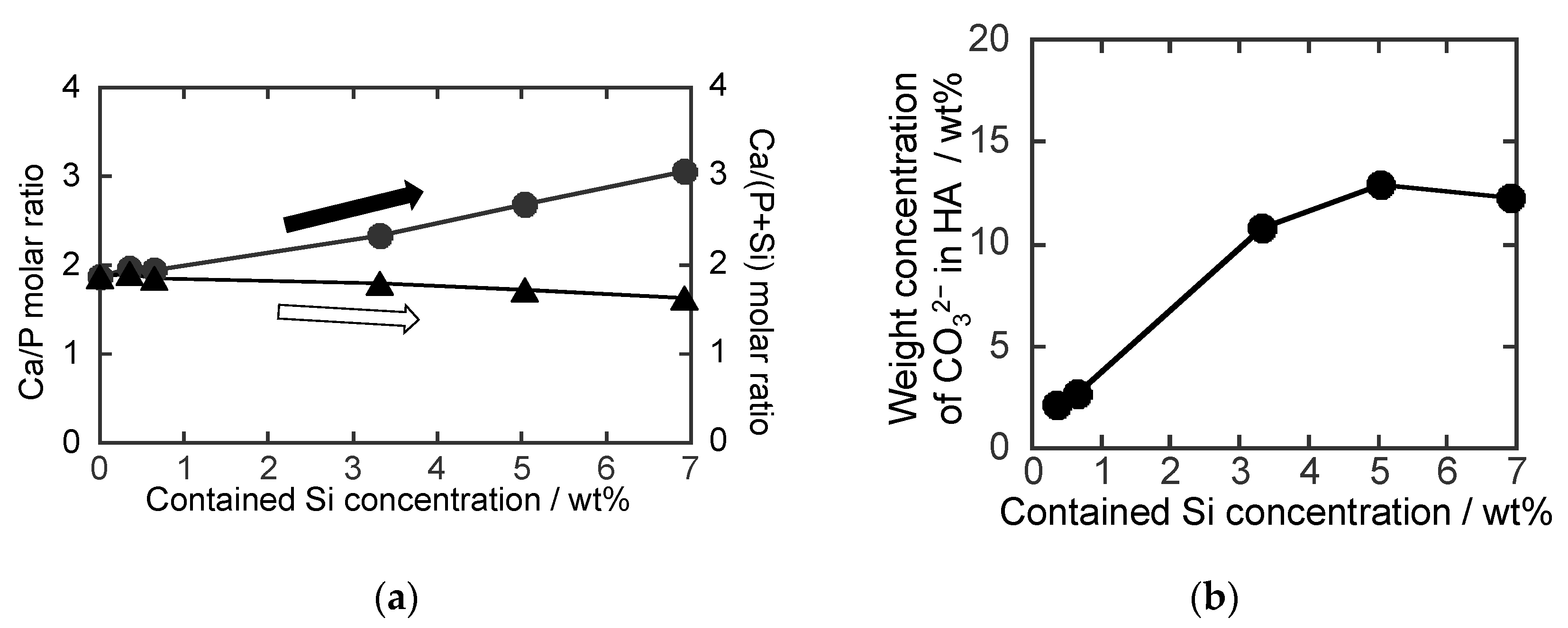
The Si concentrations obtained by XRF were 0.4, 0.7, 3.3. 5.0 and 6.9 wt% for 0.8SiHA, 1.5SiHA, 4.0SiHA, 6.0SiHA and 8.0SiHA. As shown in Figure 1a, the Ca/P molar ratio in the SiHA particles increased with the increase in feed Si concentration, suggesting the substitutions of PO43− with SiO44− ions in the HA. Due to the ion substitution, the amount of PO43− ions decreased, causing an increase in the Ca/P molar ratio [18]. On the other hand, the Ca/(Si + P) molar ratio only decreased slightly with the increase in the feeding of Si, indicating the inclusion of SiO44− ions in the HA.

Figure 1.
(a) Resultant molar ratios of the (●) Ca to P (Ca/P) and the (▲) Ca to (P + Si) (Ca/(P + Si)) and (b) carbonate weight concentration in the SiHA particles. Here, the contained Si concentrations were 0.35, 0.65, 3.32. 5.03 and 6.92 wt% for 0.8SiHA, 1.5SiHA, 4.0SiHA, 6.0SiHA and 8.0SiHA.
The FT–IR spectra of the SiHA particles (Figure S1) show the stretching vibration band of v1(PO43−) ions at 960 cm−1. The band assigned to Si-OH vibrational modes of SiO44− ions appears at 890 cm−1. The P–O stretching vibration bands of PO43− ions (1100, 1045 cm−1) and the Si–O–Si asymmetric bands of SiO44− ions (1040–1200 cm−1) are located very close, which makes interpretation difficult. The bands at around 1470, 1550 cm−1 correspond to the asymmetric stretching mode of v3CO32− ions. The band at ~3570 cm−1 is assigned to the OH− stretching mode [29,30,31,32]. It was observed that the increase in Si concentration caused an increase in the intensity of the Si–OH band and a decrease in the intensity of the v1(PO43−) band, corroborating the inclusion of SiO44− ions by the substitution of PO43− ions. Furthermore, increasing the Si concentration increased the intensity of the CO32− bands, indicating the simultaneous substitution of PO43− ions with SiO44− and CO32− ions in the HA to form SiHA. This can be described by the possible formula (e.g., 2PO43−→SiO44− + CO32−) [33]. The incorporation of SiO44− ions caused a decrease in the intensity of the OH− band until it almost disappeared in 8.0SiHA, implying the loss of OH− ions to compensate for the excess negative charges generated by the substitution of the PO43− ions with more negative ions (SiO44−). Using the Kröger–Vink notation [34], the charge compensation mechanism [35] can be expressed as follows:
Therefore, the increase in Si concentration effectively caused an increase in the content of CO32− ions, as shown in Figure 1b, indicating that the presence of SiO44− ions induced the inclusion of CO32− ions in HA. The incorporation of SiO44− and CO32− ions into HA increased the defects in the HA structure, resulting in more vacancies of OH− () due to more substitutions of PO43− ions. The simultaneous substitution of the PO43− ions in the HA with SiO44− and CO32− ions can be described as follows [13,36]
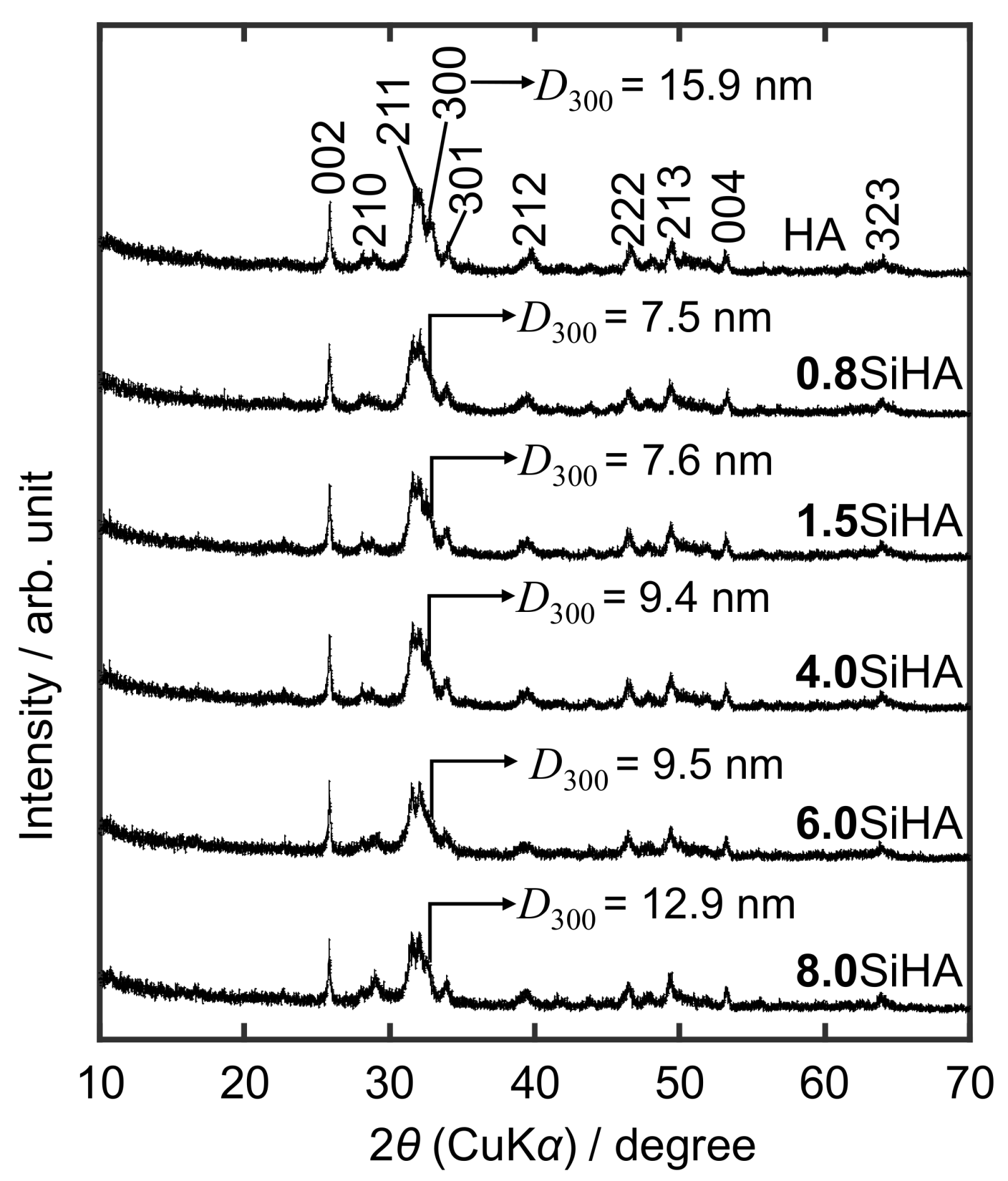
Figure 2 shows the XRD patterns of HA and SiHA particles at the different Si concentrations. All the patterns were ascribed to a HA (Ca10(PO4)6(OH)2, ICDD: 00-009-0432). It was observed that the increase in Si concentration resulted in a decrease in crystallinity, which was caused by the defects and the formation of due to the substitution of PO43− ions with SiO44− and CO32− ions in the HA structure. The calculated D300 crystalline sizes along with the a–axis of all the SiHA samples were smaller than the HA (Figure 2 (inset)) due to the incorporation of SiO44− and CO32− ions into the HA structure [37]. It was observed that the value of D300 first decreased (0.8SiHA) and then began to increase with increasing the Si concentration. Especially, 8.0SiHA had the highest value of D300, suggesting that the substitution of PO43− ions with SiO44− ions would occur predominantly, since the radius of the Si4+ (0.042 nm) is greater than that of P5+ (0.035 nm), and the bond length of the Si–O bond (0.16 nm) is greater than that of the P–O bond (0.15 nm) [20,38], which must result in a higher D300 value. These results are consistent with the crystal sizes observed in FE-SEM images (Figure S2). In the images, all the SiHA particles exhibit needle-like shapes with the smaller sizes than HA, and theaggregation forms were observed.

Figure 2.
XRD patterns of the HA and SiHA particles (Inset: D300 values).
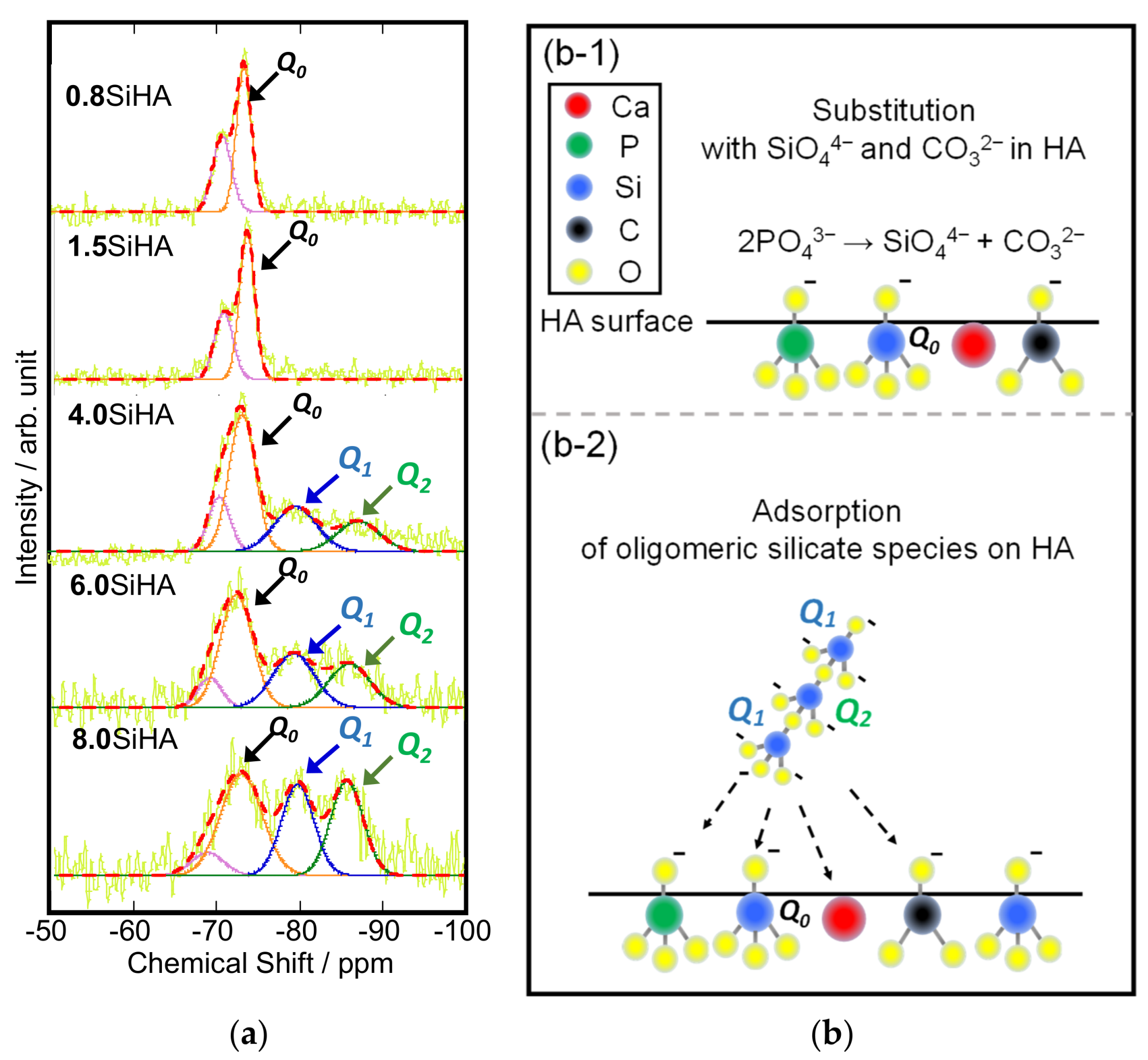
Figure 3a shows the deconvolution curves and fitting results of the spectra of the SiHA particles provided by their Q0~Q2 components. At the lower Si concentration (0.8SiHA and 1.5SiHA), only Q0 peak was observed. However, at the higher Si concentration (4.0SiHA, 6.0SiHA and 8.0SiHA), the Q1 and Q2 peaks appeared. The Q0 peak of the SiO4 tetrahedron corresponded to the stretching Si–O band in the FT–IR spectra of SiHA. The possible inclusion mechanism of the silicates ions into HA structure is shown in Figure 3b. It was suggested that the substitution with SiO44− ions in the HA structures was related to the inclusion of CO32− ions, which was proposed by the reaction mechanism based on the substitution of phosphate ions in Figure 3(b-1). Polymerized silicate chains (e.g., oligomeric silicates) due to Q1 and Q2 peaks appeared at the Si concentration of 3.32 wt%, suggesting that the unsubstituted SiO44− ions began to adsorb on the SiHA with the polymerization and condensation reactions. At the Si concentrations of 5.03 and 6.92 wt%, the intensities of Q1 and Q2 increased, suggesting that the unsubstituted SiO44− ions started to form oligomeric fragments and adsorbed on the SiHA. These results indicated that the oligomeric fragments are larger in the presence of more unsubstituted SiO44− ions as shown in Figure 3(b-2).

Figure 3.
(a) Deconvolution curves (red dotted lines) and fitting results (coloured lines) of solid-state 29Si–NMR DD-MAS spectra of the SiHA particles with the Qn (n = 0, 1, 2) derived from raw data (yellow lines). Schemes of the possible states of carbonate and silicate ions at the HA surfaces for the case in (b-1) 0.8SiHA and 1.5SiHA and (b-2) 4.0SiHA, 6.0SiHA and 8.0SiHA. The increment in the Si concentration increases the relative Q2 peak intensity, suggesting the absorption of oligomer derived from unsubstituted silicate ions on the HA surface.
4. Conclusions
The inclusion of both silicate and carbonate ions was successfully achieved in the presence of sodium silicate. The content of silicate ions increased with increasing Si feed. The silicate ions promoted the inclusion of carbonate ions in the HA. The substitution of phosphate ions with silicate and carbonate ions into the HA structure produced defects and hydroxyl vacancies, generating a loss in crystallinity and smaller crystalline sizes. The substitution of phosphate ions was mainly dominated with silicate ions in the structure, inducing the particle growth along with the a–axis. The results of solid-state 29Si–NMR DD-MAS demonstrated that at the lower Si feed, only ion substitution has occurred. At the higher Si content, the remaining silicate ions with the saturation of substitution formed the (poly)silicate species (i.e., oligomeric silicates). It was observed that these (poly)silicate species have a strong tendency to adsorb to the HA surfaces, modulating the crystal growth of the SiHA. Thus, the solid state 29Si-NMR spectra indicated that upon reaching saturation in the substitution of phosphate ions with silicate ions, the remaining silicate ions polymerized to form (poly)silicate species adsorbed on the surface of the HA, suggesting the appearance of the silicate surface layer on the SiHA surfaces. There is a possibility to control the silicate ion states, the inclusion amount of carbonate ions in SiHA and its crystalline size by the feed concentration of sodium silicate. The incorporation of silicate and carbonate ions into the HA structure is expected to enhance the bioactivity of the SiHA particles.
Supplementary Materials
The following supporting information can be downloaded at: https://www.mdpi.com/article/10.3390/biomimetics7020040/s1, Table S1: Added amounts of the reagents in the synthesis of the SiHA particles; Figure S1: FT–IR spectra of the HA and SiHA particles; Figure S2: FE−SEM images of the HA and SiHA particles.
Author Contributions
Conceptualization, T.G.P.G., K.S., S.Y., T.S. and M.T.; methodology, K.S., S.Y. and T.S.; software, K.S. and T.S.; validation, K.S., S.Y., Z.L., M.T. and T.G.P.G.; formal analysis, T.S., T.G.P.G. and Z.L.; investigation, T.G.P.G.; resources, M.T.; data curation, K.S. and T.S.; writing—original draft preparation, T.G.P.G.; writing—review and editing, Z.L. and M.T.; supervision, M.T.; project administration, M.T. All authors have read and agreed to the published version of the manuscript.
Funding
This research received no external funding.
Institutional Review Board Statement
Not applicable.
Informed Consent Statement
Not applicable.
Data Availability Statement
Data available on request from the authors.
Acknowledgments
The authors would like to thank Analysis and Instrumentation Center in Nagaoka University of Technology for providing their facilities.
Conflicts of Interest
The authors declare no conflict of interest.
References
- Palard, M.; Champion, E.; Foucaud, S. Synthesis of silicated hydroxyapatite Ca10(PO4)6−x(SiO4)x(OH)2−x. J. Solid State Chem. 2008, 181, 1950–1960. [Google Scholar] [CrossRef]
- Solonenko, A.P. Biomaterials based on mixtures of calcium phosphates and silicates: Investigation of possible production by precipitation from water solutions. Glas. Ceram. 2017, 73, 386–389. [Google Scholar] [CrossRef]
- Galindo, T.G.P.; Chai, Y.; Tagaya, M. Hydroxyapatite nanoparticle coating on polymer for constructing effective biointeractive interfaces. J. Nanomater. 2019, 2019, 6495239. [Google Scholar] [CrossRef] [Green Version]
- Posner, A.S.; Betts, F. Synthetic Amorphous Calcium Phosphate and Its Relation to Bone Mineral Structure. Acc. Chem. Res. 1975, 8, 273–281. [Google Scholar] [CrossRef]
- Patel, K.D.; Singh, R.K.; Lee, J.H.; Kim, H.W. Electrophoretic coatings of hydroxyapatite with various nanocrystal shapes. Mater. Lett. 2019, 234, 148–154. [Google Scholar] [CrossRef]
- Singh, R.K.; Kim, T.H.; Patel, K.D.; Kim, J.J.; Kim, H.W. Development of biocompatible apatite nanorod-based drug-delivery system with in situ fluorescence imaging capacity. J. Mater. Chem. B 2014, 2, 2039–2050. [Google Scholar] [CrossRef]
- Solonenko, A.P.; Golovanova, O.A. Silicate-substituted carbonated hydroxyapatite powders prepared by precipitation from aqueous solutions. Russ. J. Inorg. Chem. 2014, 59, 1228–1236. [Google Scholar] [CrossRef]
- Solonenko, A.P.; Blesman, A.I.; Polonyankin, D.A.; Bel’skaya, L.V. Effect of sodium silicate on the nature of crystallization products in calcium phosphate systems. Russ. J. Inorg. Chem. 2017, 62, 1286–1292. [Google Scholar] [CrossRef]
- Sindu, P.A.; Kolanthai, E.; Suganthi, R.V.; Arul, K.T.; Manikandan, E.; Catalani, L.H.; Kalkura, S.N. Green synthesis of Si-incorporated hydroxyapatite using sodium metasilicate as silicon precursor and in vitro antibiotic release studies. J. Photochem. Photobiol. B Biol. 2017, 175, 163–172. [Google Scholar] [CrossRef]
- Boanini, E.; Gazzano, M.; Bigi, A. Ionic substitutions in calcium phosphates synthesized at low temperature. Acta Biomater. 2010, 6, 1882–1894. [Google Scholar] [CrossRef]
- Tagaya, M.; Ikoma, T.; Takeguchi, M.; Hanagata, N.; Tanaka, J. Interfacial serum protein effect on biological apatite growth. J. Phys. Chem. C 2011, 115, 22523–22533. [Google Scholar] [CrossRef]
- Sprio, S.; Tampieri, A.; Landi, E.; Sandri, M.; Martorana, S.; Celotti, G.; Logroscino, G. Physico-chemical properties and solubility behaviour of multi-substituted hydroxyapatite powders containing silicon. Mater. Sci. Eng. C 2008, 28, 179–187. [Google Scholar] [CrossRef]
- Gibson, I.R.; Best, S.M.; Bonfield, W. Chemical characterization of silicon-substituted hydroxyapatite. J. Biomed. Mater. Res. 1999, 44, 422–428. [Google Scholar] [CrossRef]
- Hijón, N.; Cabañas, M.V.; Peña, J.; Vallet-Regí, M. Dip coated silicon-substituted hydroxyapatite films. Acta Biomater. 2006, 2, 567–574. [Google Scholar] [CrossRef]
- Xu, J.L.; Khor, K.A. Chemical analysis of silica doped hydroxyapatite biomaterials consolidated by a spark plasma sintering method. J. Inorg. Biochem. 2007, 101, 187–195. [Google Scholar] [CrossRef]
- Suetsugu, Y.; Takahashi, Y.; Okamura, F.P.; Tanaka, J. Structure Analysis of A-Type Carbonate Apatite by a Single-Crystal X-ray Diffraction Method. J. Solid State Chem. 2000, 155, 292–297. [Google Scholar] [CrossRef]
- Fleet, M.E.; Liu, X. Coupled substitution of type A and B carbonate in sodium-bearing apatite. Biomaterials 2007, 28, 916–926. [Google Scholar] [CrossRef]
- Bang, L.T.; Long, B.D.; Othman, R. Carbonate hydroxyapatite and silicon-substituted carbonate hydroxyapatite: Synthesis, mechanical properties, and solubility evaluations. Sci. World J. 2014, 2014, 969876. [Google Scholar] [CrossRef]
- Ibrahim, D.M.; Mostafa, A.A.; Korowash, S.I. Chemical characterization of some substituted hydroxyapatites. Chem. Cent. J. 2011, 5, 74. [Google Scholar] [CrossRef] [Green Version]
- Bang, L.T.; Ramesh, S.; Purbolaksono, J.; Ching, Y.C.; Long, B.D.; Chandran, H.; Othman, R. Effects of silicate and carbonate substitution on the properties of hydroxyapatite prepared by aqueous co-precipitation method. Mater. Des. 2015, 87, 788–796. [Google Scholar] [CrossRef]
- Reffitt, D.M.; Ogston, N.; Jugdaohsingh, R.; Cheung, H.F.J.; Evans, B.A.J.; Thompson, R.P.H.; Powell, J.J.; Hampson, G.N. Orthosilicic acid stimulates collagen type 1 synthesis and osteoblastic differentiation in human osteoblast-like cells in vitro. Bone 2003, 32, 127–135. [Google Scholar] [CrossRef]
- Ratnayake, J.T.B.; Mucalo, M.; Dias, G.J. Substituted hydroxyapatites for bone regeneration: A review of current trends. J. Biomed. Mater. Res. Part B Appl. Biomater. 2016, 105, 1285–1299. [Google Scholar] [CrossRef] [PubMed]
- Biedrzycka, A.; Skwarek, E.; Hanna, U.M. Hydroxyapatite with magnetic core: Synthesis methods, properties, adsorption and medical applications. Adv. Colloid Interface Sci. 2021, 291, 102401. [Google Scholar] [CrossRef] [PubMed]
- Skwarek, E.; Gładysz-Płaska, A.; Choromańska, J.B.; Broda, E. Adsorption of uranium ions on nano-hydroxyapatite and modified by Ca and Ag ions. Adsorption 2019, 25, 639–647. [Google Scholar] [CrossRef] [Green Version]
- Skwarek, E.; Gładysz–Płaska, A.; Bolbukh, Y. Adsorption of Uranyl Ions at the Nano-hydroxyapatite and Its Modification, Nanosca. Res. Lett. 2017, 12, 278. [Google Scholar]
- Grunenwald, A.; Keyser, C.; Sautereau, A.M.; Crubézy, E.; Ludes, B.; Drouet, C. Revisiting carbonate quantification in apatite (bio)minerals: A validated FTIR methodology. J. Archaeol. Sci. 2014, 49, 134–141. [Google Scholar] [CrossRef] [Green Version]
- Tagaya, M.; Abe, S.; Motozuka, S.; Shiba, K.; Takemura, T.; Hayashi, I.; Sakaguchi, Y. Surface-engineered mesoporous silica particles with luminescent, cytocompatible and targeting properties for cancer cell imaging. RSC Adv. 2017, 7, 13643–13652. [Google Scholar] [CrossRef] [Green Version]
- Yamada, S.; Tagaya, M.; Yamada, S.; Motozuka, S. Synthesis of nanostructured silica/hydroxyapatite hybrid particles containing amphiphilic triblock copolymer for effectively controlling hydration layer structures with cytocompatibility. J. Mater. Chem. B 2020, 8, 1524–1537. [Google Scholar] [CrossRef]
- Koutsopoulos, S. Synthesis and characterization of hydroxyapatite crystals: A review study on the analytical methods. J. Biomed. Mater. Res. 2002, 62, 600–612. [Google Scholar] [CrossRef]
- Aminian, A.; Solati-Hashjin, M.; Samadikuchaksaraei, A.; Bakhshi, F.; Gorjipour, F.; Farzadi, A.; Moztarzadeh, F.; Schmücker, M. Synthesis of silicon-substituted hydroxyapatite by a hydrothermal method with two different phosphorous sources. Ceram. Int. 2011, 37, 1219–1229. [Google Scholar] [CrossRef]
- Antonakos, A.; Liarokapis, E.; Leventouri, T. Micro-Raman and FTIR studies of synthetic and natural apatites. Biomaterials 2007, 28, 3043–3054. [Google Scholar] [CrossRef] [PubMed]
- Hayakawa, S.; Kanaya, T.; Tsuru, K.; Shirosaki, Y.; Osaka, A.; Fujii, E.; Kawabata, K.; Gasqueres, G.; Bonhomme, C.; Babonneau, F.; et al. Heterogeneous structure and in vitro degradation behavior of wet-chemically derived nanocrystalline silicon-containing hydroxyapatite particles. Acta Biomater. 2013, 9, 4856–4867. [Google Scholar] [CrossRef] [PubMed]
- Mostafa, N.Y.; Hassan, H.M.; Abd Elkader, O.H. Preparation and Characterization of Na+, SiO44−, and CO32−Co-Substituted Hydroxyapatite. J. Am. Ceram. Soc. 2011, 94, 1584–1590. [Google Scholar] [CrossRef]
- Kröger, F.A.; Vink, H.J. Relations between the Concentrations of Imperfections in Crystalline Solids. Solid State Phys. 1956, 3, 307–435. [Google Scholar]
- Targonska, S.; Wiglusz, R.J. Investigation of physicochemical properties of the structurally modified nanosized silicate-substituted hydroxyapatite co-doped with eu3+ and sr2+ ions. Nanomaterials 2021, 11, 27. [Google Scholar] [CrossRef]
- Gibson, I.R.; Bonfield, W. Novel synthesis and characterization of an AB-type carbonate-substituted hydroxyapatite. J. Biomed. Mater. Res. 2001, 59, 697–708. [Google Scholar] [CrossRef] [PubMed]
- Pietak, A.M.; Reid, J.W.; Stott, M.J.; Sayer, M. Silicon substitution in the calcium phosphate bioceramics. Biomaterials 2007, 28, 4023–4032. [Google Scholar] [CrossRef] [PubMed]
- Tang, X.L.; Xiao, X.F.; Liu, R.F. Structural characterization of silicon-substituted hydroxyapatite synthesized by a hydrothermal method. Mater. Lett. 2005, 59, 3841–3846. [Google Scholar] [CrossRef]
Publisher’s Note: MDPI stays neutral with regard to jurisdictional claims in published maps and institutional affiliations. |
© 2022 by the authors. Licensee MDPI, Basel, Switzerland. This article is an open access article distributed under the terms and conditions of the Creative Commons Attribution (CC BY) license (https://creativecommons.org/licenses/by/4.0/).