Article

The Development of Biomimetic Aligned Skeletal Muscles in a Fully 3D Printed Microfluidic Device

1
Ritsumeikan Global-Innovation Research Organization (R-GIRO), Ristumeikan University, Kyoto 525-8577, Japan
2
Department of Mechanical Engineering, Ritsumeikan University, Kyoto 525-8577, Japan
3
Department of Pharmaceutical Sciences, Ritsumeikan University, Kyoto 525-8577, Japan
*
Author to whom correspondence should be addressed.
Biomimetics 2022, 7(1), 2; https://doi.org/10.3390/biomimetics7010002

Submission received: 3 December 2021 / Revised: 17 December 2021 / Accepted: 20 December 2021 / Published: 21 December 2021

Abstract

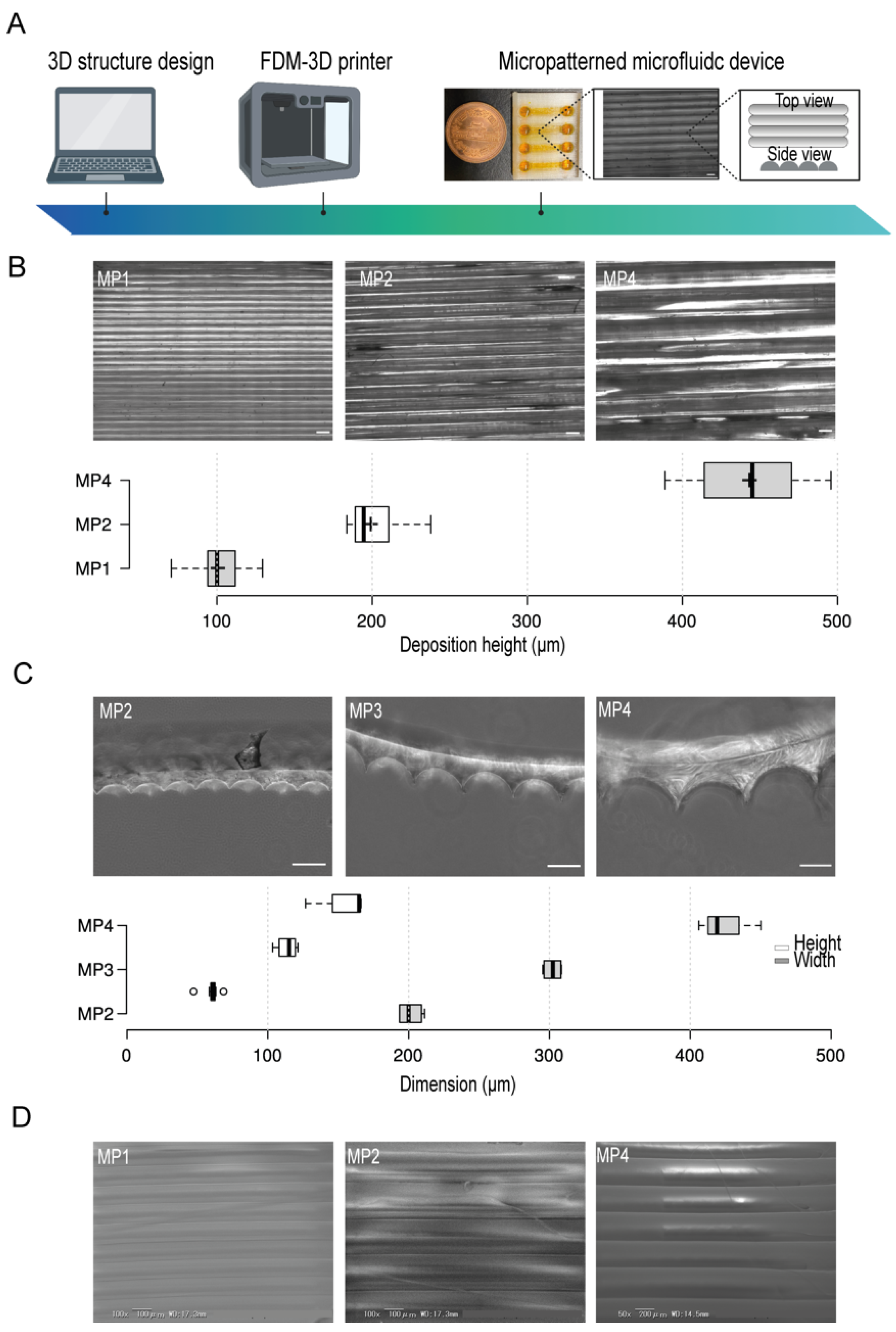
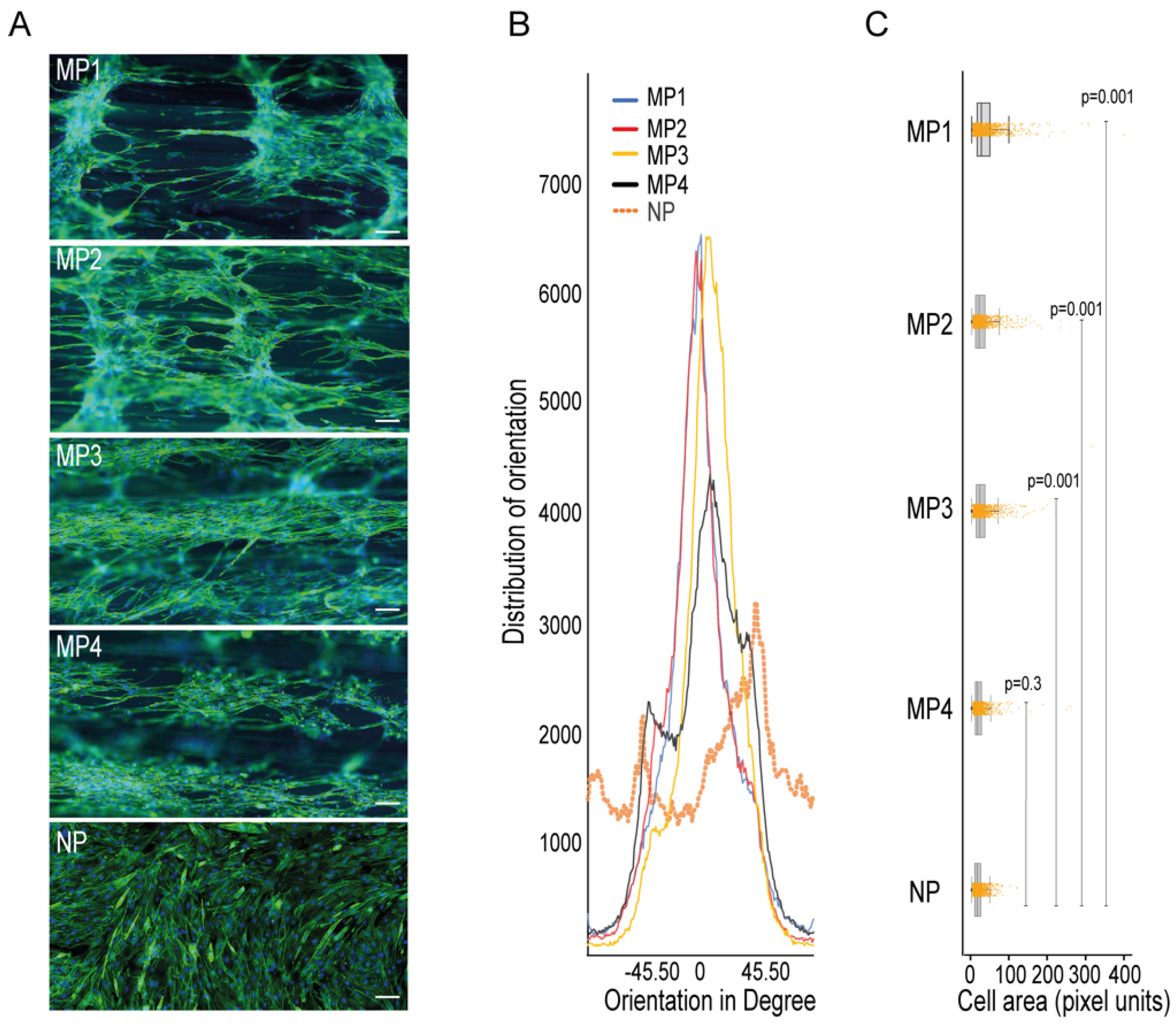
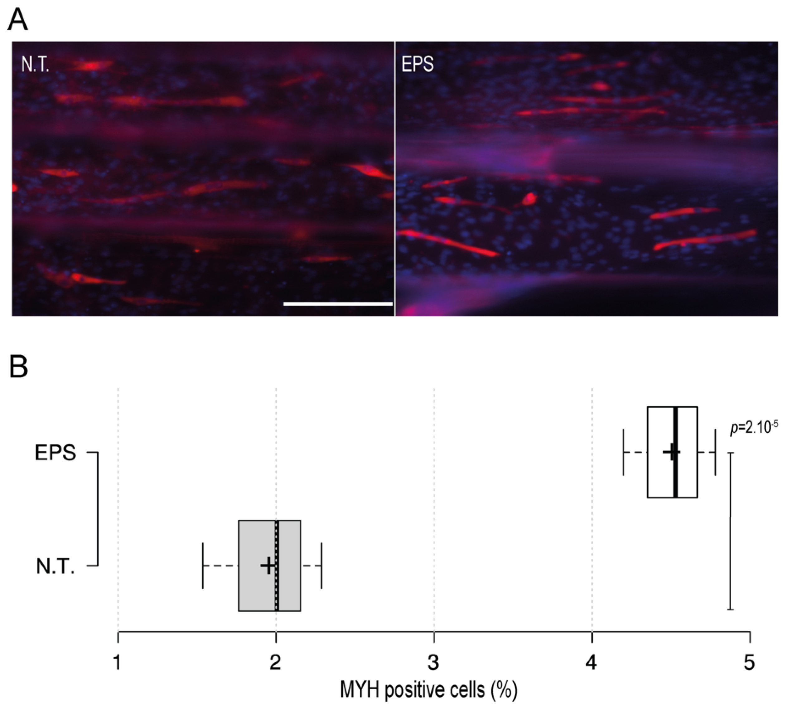
Human skeletal muscles are characterized by a unique aligned microstructure of myotubes which is important for their function as well as for their homeostasis. Thus, the recapitulation of the aligned microstructure of skeletal muscles is crucial for the construction of an advanced biomimetic model aimed at drug development applications. Here, we have developed a 3D printed micropatterned microfluid device (3D-PMMD) through the employment of a fused deposition modeling (FDM)-based 3D printer and clear filaments made of biocompatible polyethylene terephthalate glycol (PETG). We could fabricate micropatterns through the adjustment of the printing deposition heights of PETG filaments, leading to the generation of aligned half-cylinder-shaped micropatterns in a dimension range from 100 µm to 400 µm in width and from 60 µm to 150 µm in height, respectively. Moreover, we could grow and expand C2C12 mouse myoblast cells on 3D-PMMD where cells could differentiate into aligned bundles of myotubes with respect to the dimension of each micropattern. Furthermore, our platform was applicable with the electrical pulses stimulus (EPS) modality where we noticed an improvement in myotubes maturation under the EPS conditions, indicating the potential use of the 3D-PMMD for biological experiments as well as for myogenic drug development applications in the future.
Keywords: skeletal muscles; micropattern; fused deposition modeling (FDM)-3D printer skeletal muscles; micropattern; fused deposition modeling (FDM)-3D printer