Living Light 2018: Conference Report
Abstract
1. Introduction
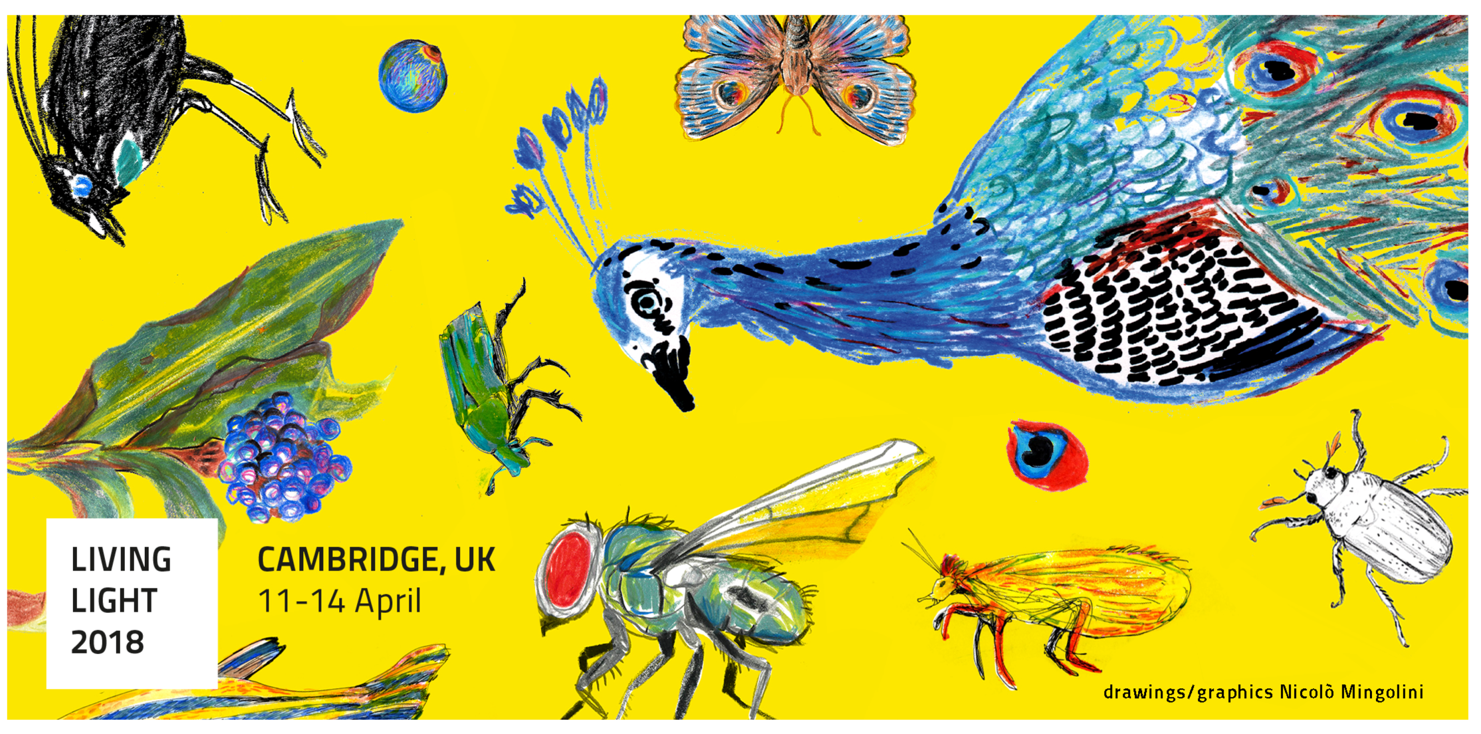
 Illustration of the conference themes by Nicolò Mingolini, Living Light’s artist in residence who curated the graphics of the conference materials. The artist’s impressions show some of the colorful organisms discussed at the meeting.
2. Sessions
2.1. Vision
2.2. Plant Photonics
2.3. Development and Pattern Formation in Nature
2.4. Fossilization of Structural Colors
2.5. Evolution and Ecology
2.6. Modelling of Light–Matter Interaction
2.7. Biomimetics and Bioinspiration
3. Poster Contributions
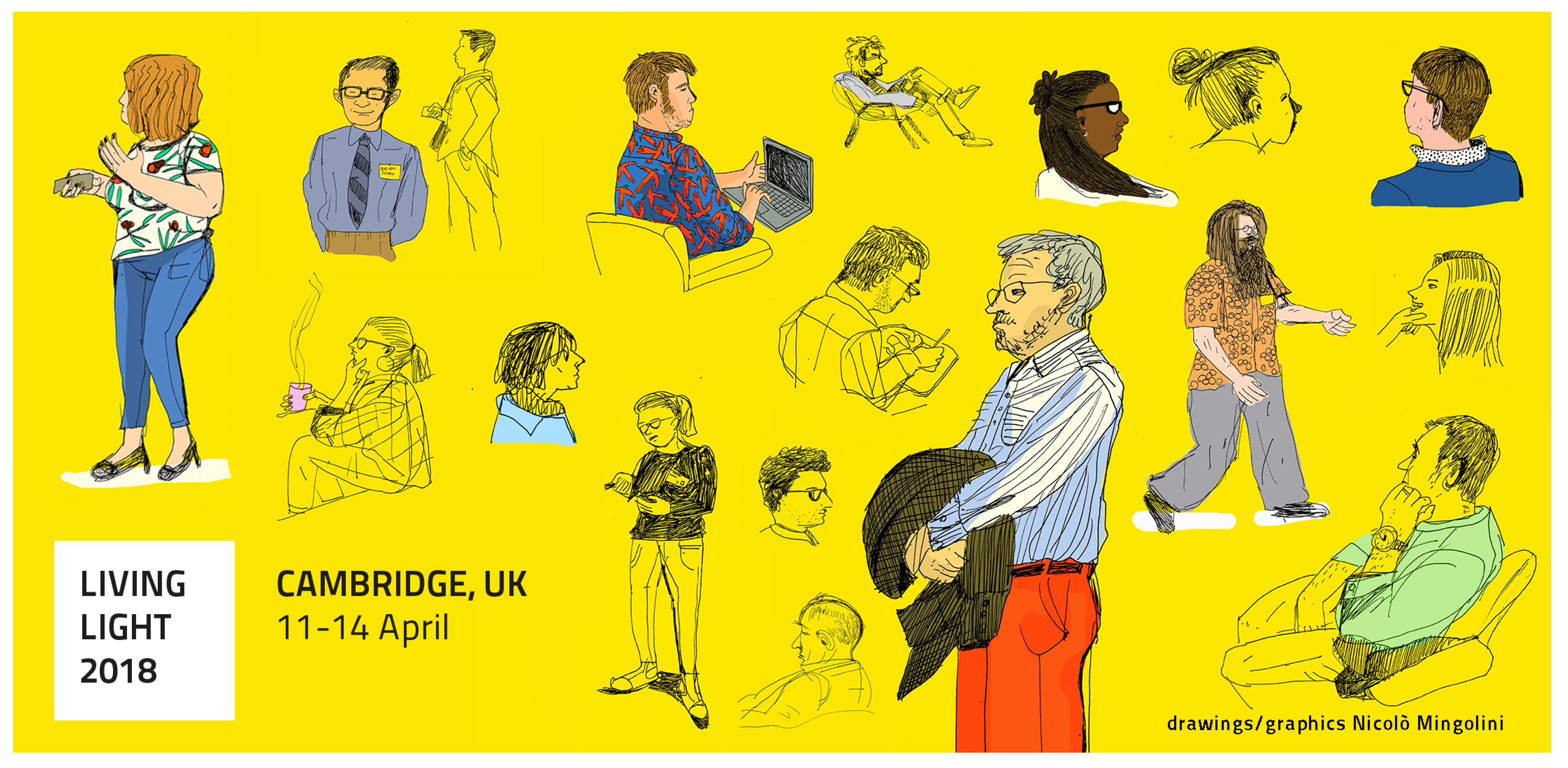
 Some of the conference attendees drawn through the eyes of Nicolò Mingolini, Living Light’s artist in residence.
4. Plenary Discussions
4.1. Living Light: Tools, Relevance, and Outlook
- E-archives: the panel debated whether the Living Light community would benefit from using e-print services such as arXiv, bioRxiv, vixRa, etc. as repository for unpublished work. Interestingly, the discussion of this topic was rather controversial and no agreement could be reached. While some researchers supported such platforms as an opportunity to shorten publication time and make their work citable early on, others believed that the use of repositories that are not peer-reviewed is dangerous as it can lead to the spread of inaccurate and potentially erroneous studies.
- Networking and new tools: the discussion on new tools highlighted that while most hardware (e.g., advanced electron microscopes) and software (e.g., code for numerical simulations) required for the study of biological photonics are already in place, the connection between researchers with different skill sets is still lagging behind. The community agreed that multidisciplinary conferences such as Living Light are crucial to develop new collaborations and broaden the network of scientists working in the field.
4.2. The Future of Biomimetics: Scaling Up
- Developmental studies as a gateway to biomimetics: there was general consensus that by studying the growth of photonics in living organisms one can gain information useful to mimic the processes in vitro in order to produce low-cost high-performance materials. However, the panel agreed that the complexity of living organisms makes this task extremely challenging and that replicating processes such as DNA synthesis is still far from the capabilities of the current available technologies.
- Bioinspiration vs. biomimetics: interestingly, there was no agreement on the use of these two terms and which one of the two processes should be pursued by the Living Light community. While some panelists believed that biomimetics should be the final aim, others were more prone to support bioinspiration as this allows for more freedom in terms of choosing the functions needed for new materials and combine strategies that are not necessarily observed in the same species.
- Sustainability: the panel highlighted the need for environmental awareness. Even though biomimetic technologies are inspired by natural structures, they are not necessarily based upon sustainable materials. Therefore the need arises to reflect on the environmental impact of one’s own research with the goal to not only mimic the natural architectures and processes but also to try and questions one’s choice of materials.
5. Conclusions
Acknowledgments
Conflicts of Interest
References
- Daly, I.M.; How, M.J.; Partridge, J.C.; Temple, S.E.; Marshall, N.J.; Cronin, T.W.; Roberts, N.W. Dynamic polarization vision in mantis shrimps. Nat. Commun. 2016, 7, 12140. [Google Scholar] [CrossRef] [PubMed]
- Thoen, H.H.; Chiou, T.H.; Marshall, N.J. Intracellular recordings of spectral sensitivities in stomatopods: A comparison across species. Integr. Comp. Biol. 2017, 57, 1117–1129. [Google Scholar] [CrossRef] [PubMed]
- Thoen, H.H.; Sayre, M.E.; Marshall, J.; Strausfeld, N.J. Representation of the stomatopod’s retinal midband in the optic lobes: Putative neural substrates for integrating chromatic, achromatic and polarization information. J. Comp. Neurol. 2018, 526, 1148–1165. [Google Scholar] [CrossRef] [PubMed]
- Michiels, N.K.; Seeburger, V.C.; Kalb, N.; Meadows, M.G.; Anthes, N.; Mailli, A.A.; Jack, C.B. Controlled iris radiance in a diurnal fish looking at prey. R. Soc. Open Sci. 2018, 5, 170838. [Google Scholar] [CrossRef] [PubMed]
- Wilkins, L.; Marshall, N.J.; Johnsen, S.; Osorio, D. Modelling colour constancy in fish: Implications for vision and signalling in water. J. Exp. Biol. 2016, 219, 1884–1892. [Google Scholar] [CrossRef] [PubMed]
- Lind, O.; Henze, M.J.; Kelber, A.; Osorio, D. Coevolution of coloration and colour vision? Phil. Trans. R. Soc. B 2017, 372, 20160338. [Google Scholar] [CrossRef] [PubMed]
- Kingston, A.C.; Wardill, T.J.; Hanlon, R.T.; Cronin, T.W. An unexpected diversity of photoreceptor classes in the longfin squid, Doryteuthis pealeii. PLoS ONE 2015, 10, e0135381. [Google Scholar] [CrossRef] [PubMed]
- Feller, K.D.; Cronin, T.W. Spectral absorption of visual pigments in stomatopod larval photoreceptors. J. Comp. Phys. A 2016, 202, 215–223. [Google Scholar] [CrossRef] [PubMed]
- Jacobs, M.; Lopez-Garcia, M.; Phrathep, O.P.; Lawson, T.; Oulton, R.; Whitney, H.M. Photonic multilayer structure of Begonia chloroplasts enhances photosynthetic efficiency. Nat. Plant. 2016, 2, 16162. [Google Scholar] [CrossRef] [PubMed]
- Lopez-Garcia, M.; Masters, N.; O’Brien, H.E.; Lennon, J.; Atkinson, G.; Cryan, M.J.; Oulton, R.; Whitney, H.M. Light-induced dynamic structural color by intracellular 3D photonic crystals in brown algae. Sci. Adv. 2018, 4, eaan8917. [Google Scholar] [CrossRef] [PubMed]
- Ignatov, M.S.; Ignatova, E.A.; Belousova, A.A.; Sigaeva, A.O. Additional observations on protonemata of Schistostega pennata (Bryophyta). Arctoa 2012, 21, 1–20. [Google Scholar] [CrossRef]
- Gebeshuber, I.C.; Lee, D.W. Nanostructures for Coloration (Organisms other than Animals); Springer: Dordrecht, The Netherlands, 2012; pp. 1790–1803. [Google Scholar]
- Vignolini, S.; Rudall, P.J.; Rowland, A.V.; Reed, A.; Moyroud, E.; Faden, R.B.; Baumberg, J.J.; Glover, B.J.; Steiner, U. Pointillist structural color in Pollia fruit. Proc. Natl. Acad. Sci. USA 2012, 109, 15712–15715. [Google Scholar] [CrossRef] [PubMed]
- Vignolini, S.; Gregory, T.; Kolle, M.; Lethbridge, A.; Moyroud, E.; Steiner, U.; Glover, B.J.; Vukusic, P.; Rudall, P.J. Structural colour from helicoidal cell-wall architecture in fruits of Margaritaria nobilis. J. R. Soc. Interface 2016, 13, 20160645. [Google Scholar] [CrossRef] [PubMed]
- Wilts, B.D.; Zubiri, B.A.; Klatt, M.A.; Butz, B.; Fischer, M.G.; Kelly, S.T.; Spiecker, E.; Steiner, U.; Schröder-Turk, G.E. Butterfly gyroid nanostructures as a time-frozen glimpse of intracellular membrane development. Sci. Adv. 2017, 3, e1603119. [Google Scholar] [CrossRef] [PubMed]
- Onelli, O.D.; van de Kamp, T.; Skepper, J.N.; Powell, J.; dos Santos Rolo, T.; Baumbach, T.; Vignolini, S. Development of structural colour in leaf beetles. Sci. Rep. 2017, 7, 1373. [Google Scholar] [CrossRef] [PubMed]
- Ghiradella, H. Structure of butterfly scales: Patterning in an insect cuticle. Microsc. Res. Tech. 1994, 27, 429–438. [Google Scholar] [CrossRef] [PubMed]
- Dinwiddie, A.; Null, R.; Pizzano, M.; Chuong, L.; Krup, A.L.; Tan, H.E.; Patel, N.H. Dynamics of F-actin prefigure the structure of butterfly wing scales. Dev. Biol. 2014, 392, 404–418. [Google Scholar] [CrossRef] [PubMed]
- Hollergschwandtner, E.; Schwaha, T.; Neumüller, J.; Kaindl, U.; Gruber, D.; Eckhard, M.; Stöger-Pollach, M.; Reipert, S. Novel mesostructured inclusions in the epidermal lining of Artemia franciscana ovisacs show optical activity. PeerJ 2017, 5, e3923. [Google Scholar] [CrossRef] [PubMed]
- Gur, D.; Nicolas, J.D.; Brumfeld, V.; Bar-Elli, O.; Oron, D.; Levkowitz, G. Development of high-order organization of guanine-based reflectors underlies the dual functionality of the zebrafish iris. bioRxiv 2018, 240028. [Google Scholar] [CrossRef]
- Johansen, V.E.; Catón, L.; Hamidjaja, R.; Oosterink, E.; Wilts, B.D.; Rasmussen, T.S.; Sherlock, M.M.; Ingham, C.J.; Vignolini, S. Genetic manipulation of structural color in bacterial colonies. Proc. Natl. Acad. Sci. USA 2018, 201716214. [Google Scholar] [CrossRef] [PubMed]
- Parnell, A.J.; Bradford, J.E.; Curran, E.V.; Washington, A.L.; Adams, G.; Brien, M.N.; Burg, S.L.; Morochz, C.; Fairclough, J.P.A.; Vukusic, P.; et al. Wing scale ultrastructure underlying convergent and divergent iridescent colours in mimetic Heliconius butterflies. J. R. Soc. Interface 2018, 15, 20170948. [Google Scholar] [CrossRef] [PubMed]
- Vignolini, S.; Moyroud, E.; Hingant, T.; Banks, H.; Rudall, P.J.; Steiner, U.; Glover, B.J. The flower of Hibiscus trionum is both visibly and measurably iridescent. New Phytol. 2015, 205, 97–101. [Google Scholar] [CrossRef] [PubMed]
- Totz, J.F.; Rode, J.; Tinsley, M.R.; Showalter, K.; Engel, H. Spiral wave chimera states in large populations of coupled chemical oscillators. Nat. Phys. 2018, 14, 282. [Google Scholar] [CrossRef]
- McNamara, M.; Field, D. Maturation Experiments Reveal Bias in the Fossil Record of Feathers. In Proceedings of the EGU General Assembly Conference Abstracts, Vienna, Austria, 17–22 April 2016; EGU General Assembly: Vienna, Austria, 2016; Volume 18, p. 16944. [Google Scholar]
- Zhang, Q.; Mey, W.; Ansorge, J.; Starkey, T.A.; McDonald, L.T.; McNamara, M.E.; Jarzembowski, E.A.; Wichard, W.; Kelly, R.; Ren, X.; et al. Fossil scales illuminate the early evolution of lepidopterans and structural colors. Sci. Adv. 2018, 4, e1700988. [Google Scholar] [CrossRef] [PubMed]
- McNamara, M.E.; Saranathan, V.; Locatelli, E.R.; Noh, H.; Briggs, D.E.; Orr, P.J.; Cao, H. Cryptic iridescence in a fossil weevil generated by single diamond photonic crystals. J. R. Soc. Interface 2014, 11, 20140736. [Google Scholar] [CrossRef] [PubMed]
- Stoddard, M.C.; Kupán, K.; Eyster, H.N.; Rojas-Abreu, W.; Cruz-López, M.; Serrano-Meneses, M.A.; Küpper, C. Camouflage and clutch survival in plovers and terns. Sci. Rep. 2016, 6, 32059. [Google Scholar] [CrossRef] [PubMed]
- Stoddard, M.C.; Hauber, M.E. Colour, vision and coevolution in avian brood parasitism. Philos. Trans. R. Soc. B 2017, 372, 20160339. [Google Scholar] [CrossRef] [PubMed]
- Finkbeiner, S.D.; Fishman, D.A.; Osorio, D.; Briscoe, A.D. Ultraviolet and yellow reflectance but not fluorescence is important for visual discrimination of conspecifics by Heliconius erato. J. Exp. Biol. 2017, 220, 1267–1276. [Google Scholar] [CrossRef] [PubMed]
- Willmott, K.R.; Willmott, J.C.R.; Elias, M.; Jiggins, C.D. Maintaining mimicry diversity: Optimal warning colour patterns differ among microhabitats in Amazonian clearwing butterflies. Proc. R. Soc. B 2017, 284, 20170744. [Google Scholar] [CrossRef] [PubMed]
- Goessling, J.W.; Frankenbach, S.; Ribeiro, L.; Serôdio, J.; Kühl, M. Modulation of the light field related to valve optical properties of raphid diatoms: Implications for niche differentiation in the microphytobenthos. Mar. Ecol. Prog. Ser. 2018, 588, 29–42. [Google Scholar] [CrossRef]
- Goessling, J.W.; Su, Y.; Cartaxana, P.; Maibohm, C.; Rickelt, L.F.; Trampe, E.C.; Walby, S.L.; Wangpraseurt, D.; Wu, X.; Ellegaard, M.; et al. Structure-based optics of centric diatom frustules: Modulation of the in vivo light field for efficient diatom photosynthesis. New Phytol. 2018. [Google Scholar] [CrossRef] [PubMed]
- Holt, A.L.; Vahidinia, S.; Gagnon, Y.L.; Morse, D.E.; Sweeney, A.M. Photosymbiotic giant clams are transformers of solar flux. J. R. Soc. Interface 2014, 11, 20140678. [Google Scholar] [CrossRef] [PubMed]
- Levenson, R.; DeMartini, D.G.; Morse, D.E. Molecular mechanism of reflectin’s tunable biophotonic control: Opportunities and limitations for new optoelectronics. APL Mater. 2017, 5, 104801. [Google Scholar] [CrossRef]
- Plyushcheva, M.V.; da Silva, K.P.; Aneli, N.B.; Vays, V.B.; Kondrashov, F.A.; Goñi, A.R. Two-color fluorescence in elytra of the scale-worm Lepidonotus squamatus (Polychaeta, Polynoidae): In vivo spectral characteristic. Mater. Today Proc. 2017, 4, 4998–5005. [Google Scholar] [CrossRef]
- Xiao, M.; Hu, Z.; Wang, Z.; Li, Y.; Tormo, A.D.; Le Thomas, N.; Wang, B.; Gianneschi, N.C.; Shawkey, M.D.; Dhinojwala, A. Bioinspired bright noniridescent photonic melanin supraballs. Sci. Adv. 2017, 3, e1701151. [Google Scholar] [CrossRef] [PubMed]
- Peteya, J.A.; Clarke, J.A.; Li, Q.; Gao, K.Q.; Shawkey, M.D. The plumage and colouration of an enantiornithine bird from the Early Cretaceous of China. Palaeontology 2017, 60, 55–71. [Google Scholar] [CrossRef]
- Hsiung, B.K.; Siddique, R.H.; Jiang, L.; Liu, Y.; Lu, Y.; Shawkey, M.D.; Blackledge, T.A. Tarantula-inspired noniridescent photonics with long-range order. Adv. Opt. Mater. 2017, 5. [Google Scholar] [CrossRef]
- Hsiung, B.K.; Siddique, R.H.; Stavenga, D.G.; Otto, J.C.; Allen, M.C.; Liu, Y.; Lu, Y.F.; Deheyn, D.D.; Shawkey, M.D.; Blackledge, T.A. Rainbow peacock spiders inspire miniature super-iridescent optics. Nat. Commun. 2017, 8, 2278. [Google Scholar] [CrossRef] [PubMed]
- Vukusic, P.; Hallam, B.; Noyes, J. Brilliant whiteness in ultrathin beetle scales. Science 2007, 315, 348. [Google Scholar] [CrossRef] [PubMed]
- Burresi, M.; Cortese, L.; Pattelli, L.; Kolle, M.; Vukusic, P.; Wiersma, D.S.; Steiner, U.; Vignolini, S. Bright-white beetle scales optimise multiple scattering of light. Sci. Rep. 2014, 4, 6075. [Google Scholar] [CrossRef] [PubMed]
- Greenewalt, C.H.; Brandt, W.; Friel, D.D. Iridescent colors of hummingbird feathers. J. Opt. Soc. Am. 1960, 50, 1005–1013. [Google Scholar] [CrossRef]
- Durrer, H. Colouration. In Biology of the Integument; Springer: Berlin/Heidelberg, Germany, 1986; pp. 239–247. [Google Scholar]
- Cai, J.; Townsend, J.; Dodson, T.; Heiney, P.; Sweeney, A. Eye patches: Protein assembly of index-gradient squid lenses. Science 2017, 357, 564–569. [Google Scholar] [CrossRef] [PubMed]
- Mendoza-Galván, A.; Muñoz-Pineda, E.; Ribeiro, S.J.; Santos, M.V.; Järrendahl, K.; Arwin, H. Mueller matrix spectroscopic ellipsometry study of chiral nanocrystalline cellulose films. J. Opt. 2018, 20, 024001. [Google Scholar] [CrossRef]
- Mouchet, S.R.; Lobet, M.; Kolaric, B.; Kaczmarek, A.M.; Van Deun, R.; Vukusic, P.; Deparis, O.; Van Hooijdonk, E. Controlled fluorescence in a beetle’s photonic structure and its sensitivity to environmentally induced changes. Proc. R. Soc. B 2016, 283, 20162334. [Google Scholar] [CrossRef] [PubMed]
- Ordinario, D.D.; Leung, E.M.; Phan, L.; Kautz, R.; Lee, W.K.; Naeim, M.; Kerr, J.P.; Aquino, M.J.; Sheehan, P.E.; Gorodetsky, A.A. Protochromic devices from a cephalopod structural protein. Adv. Opt. Mater. 2017, 5. [Google Scholar] [CrossRef]
- Aizenberg, J.; Kolle, M.; Vukusic, P.; Howe, R.D. Band-Gap Tunable Elastic Optical Multilayer Fibers. U.S. Patent App. 14/760,306, 17 December 2015. [Google Scholar]
- Schenk, F.; Wilts, B.D.; Stavenga, D.G. The Japanese jewel beetle: A painter’s challenge. Bioinspir. Biomim. 2013, 8, 045002. [Google Scholar] [CrossRef] [PubMed]
- Schenk, F. Biomimetics, color, and the arts. In Bioinspiration, Biomimetics, and Bioreplication 2015; Lakhtakia, A., Knez, M., Martín-Palma, R.J., Eds.; Proc. SPIE: San Diego, CA, USA, 2015; Volume 9429, p. 94290Z. [Google Scholar]
- Siddique, R.H.; Donie, Y.J.; Gomard, G.; Yalamanchili, S.; Merdzhanova, T.; Lemmer, U.; Hölscher, H. Bioinspired phase-separated disordered nanostructures for thin photovoltaic absorbers. Sci. Adv. 2017, 3, e1700232. [Google Scholar] [CrossRef] [PubMed]
- Syurik, J.; Siddique, R.H.; Dollmann, A.; Gomard, G.; Schneider, M.; Worgull, M.; Wiegand, G.; Hölscher, H. Bio-inspired, large scale, highly-scattering films for nanoparticle-alternative white surfaces. Sci. Rep. 2017, 7, 46637. [Google Scholar] [CrossRef] [PubMed]
- Hill, S.A.; Benito-Alifonso, D.; Morgan, D.J.; Davis, S.A.; Berry, M.; Galan, M.C. Three-minute synthesis of sp3 nanocrystalline carbon dots as non-toxic fluorescent platforms for intracellular delivery. Nanoscale 2016, 8, 18630–18634. [Google Scholar] [CrossRef] [PubMed]
- Wangpraseurt, D.; Holm, J.B.; Larkum, A.W.; Pernice, M.; Ralph, P.J.; Suggett, D.J.; Kühl, M. In vivo microscale measurements of light and photosynthesis during coral bleaching: Evidence for the optical feedback loop? Front. Microbiol. 2017, 8, 59. [Google Scholar] [CrossRef] [PubMed]
- Koren, K.; Jakobsen, S.L.; Kühl, M. In-vivo imaging of O2 dynamics on coral surfaces spray-painted with sensor nanoparticles. Sens. Actuator B Chem. 2016, 237, 1095–1101. [Google Scholar] [CrossRef]
- Zi, J.; Yu, X.; Li, Y.; Hu, X.; Xu, C.; Wang, X.; Liu, X.; Fu, R. Coloration strategies in peacock feathers. Proc. Natl. Acad. Sci. USA 2003, 100, 12576–12578. [Google Scholar] [CrossRef] [PubMed]
- Schmidt, O.G.; Eberl, K. Nanotechnology: Thin solid films roll up into nanotubes. Nature 2001, 410, 168. [Google Scholar] [CrossRef] [PubMed]
- Sharma, V.; Crne, M.; Park, J.O.; Srinivasarao, M. Structural origin of circularly polarized iridescence in jeweled beetles. Science 2009, 325, 449–451. [Google Scholar] [CrossRef] [PubMed]
- Gould, K.S.; Lee, D.W. Physical and ultrastructural basis of blue leaf iridescence in four Malaysian understory plants. Am. J. Bot. 1996, 45–50. [Google Scholar] [CrossRef]
© 2018 by the authors. Licensee MDPI, Basel, Switzerland. This article is an open access article distributed under the terms and conditions of the Creative Commons Attribution (CC BY) license (http://creativecommons.org/licenses/by/4.0/).
Share and Cite
Onelli, O.D.; Wilts, B.D.; Vignolini, S. Living Light 2018: Conference Report. Biomimetics 2018, 3, 11. https://doi.org/10.3390/biomimetics3020011
Onelli OD, Wilts BD, Vignolini S. Living Light 2018: Conference Report. Biomimetics. 2018; 3(2):11. https://doi.org/10.3390/biomimetics3020011
Chicago/Turabian StyleOnelli, Olimpia D., Bodo D. Wilts, and Silvia Vignolini. 2018. "Living Light 2018: Conference Report" Biomimetics 3, no. 2: 11. https://doi.org/10.3390/biomimetics3020011
APA StyleOnelli, O. D., Wilts, B. D., & Vignolini, S. (2018). Living Light 2018: Conference Report. Biomimetics, 3(2), 11. https://doi.org/10.3390/biomimetics3020011


