Correction: Almutary et al. Development of 3D-Bioprinted Colitis-Mimicking Model to Assess Epithelial Barrier Function Using Albumin Nano-Encapsulated Anti-Inflammatory Drugs. Biomimetics 2023, 8, 41

 , , ,
, , ,  , and
, and 
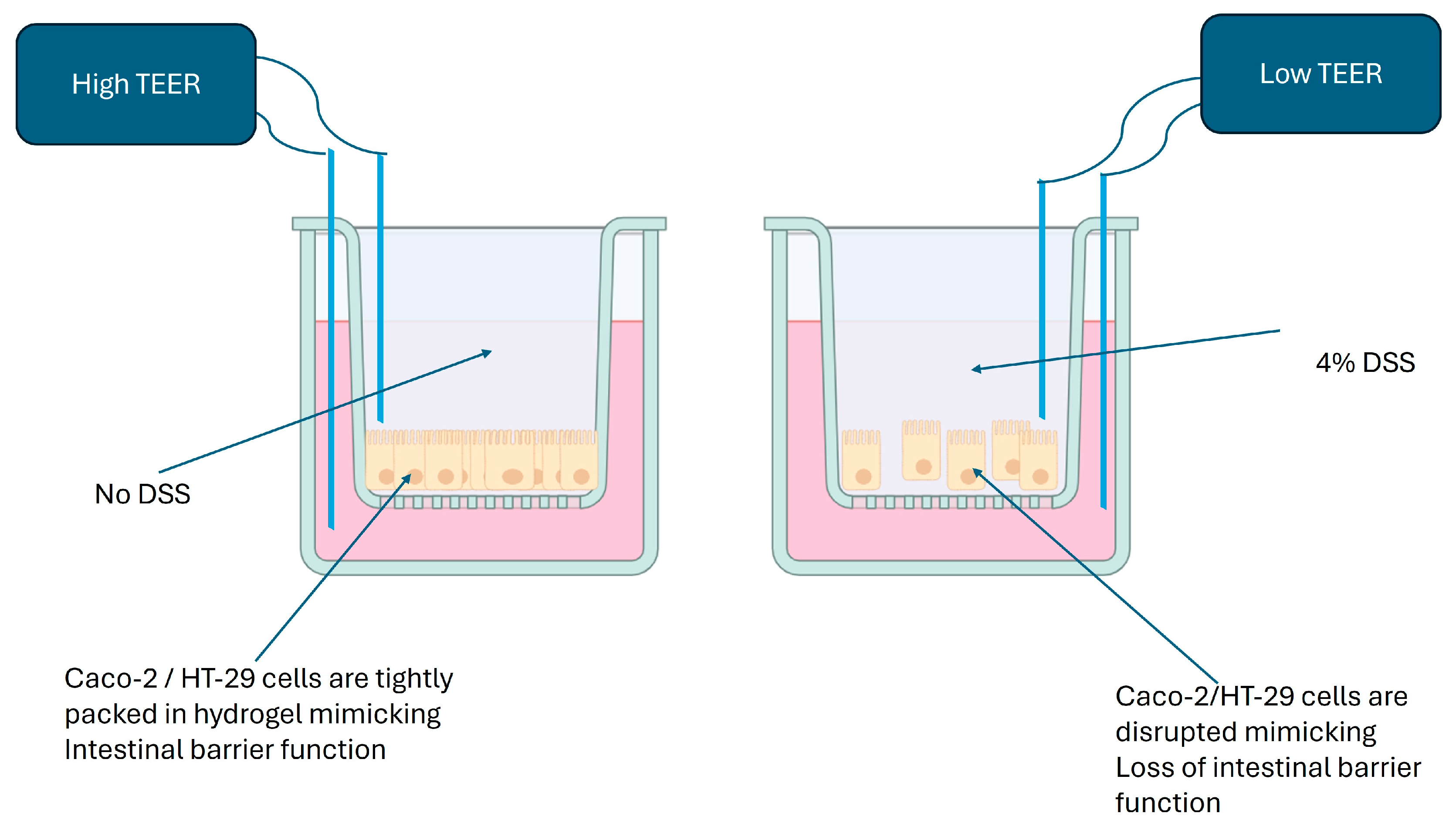
1. Error in Figure
2. Text Correction
3.2. Induction of Disease in a 3D-Printed Caco-2 and HT-29 Model
3. References
References
- Almutary, A.G.; Alnuqaydan, A.M.; Almatroodi, S.A.; Bakshi, H.A.; Chellappan, D.K.; Tambuwala, M.M. Development of 3D-Bioprinted Colitis-Mimicking Model to Assess Epithelial Barrier Function Using Albumin Nano-Encapsulated Anti-Inflammatory Drugs. Biomimetics 2023, 8, 41. [Google Scholar] [CrossRef] [PubMed]
- Araki, Y.; Sugihara, H.; Hattori, T. In vitro effects of dextran sulfate sodium on a Caco-2 cell line and plausible mechanisms for dextran sulfate sodium-induced colitis. Oncol. Rep. 2006, 16, 1357–1362. [Google Scholar] [CrossRef] [PubMed]
Disclaimer/Publisher’s Note: The statements, opinions and data contained in all publications are solely those of the individual author(s) and contributor(s) and not of MDPI and/or the editor(s). MDPI and/or the editor(s) disclaim responsibility for any injury to people or property resulting from any ideas, methods, instructions or products referred to in the content. |
© 2025 by the authors. Licensee MDPI, Basel, Switzerland. This article is an open access article distributed under the terms and conditions of the Creative Commons Attribution (CC BY) license (https://creativecommons.org/licenses/by/4.0/).
Share and Cite
Almutary, A.G.; Alnuqaydan, A.M.; Almatroodi, S.A.; Bakshi, H.A.; Chellappan, D.K.; Tambuwala, M.M. Correction: Almutary et al. Development of 3D-Bioprinted Colitis-Mimicking Model to Assess Epithelial Barrier Function Using Albumin Nano-Encapsulated Anti-Inflammatory Drugs. Biomimetics 2023, 8, 41. Biomimetics 2025, 10, 265. https://doi.org/10.3390/biomimetics10050265
Almutary AG, Alnuqaydan AM, Almatroodi SA, Bakshi HA, Chellappan DK, Tambuwala MM. Correction: Almutary et al. Development of 3D-Bioprinted Colitis-Mimicking Model to Assess Epithelial Barrier Function Using Albumin Nano-Encapsulated Anti-Inflammatory Drugs. Biomimetics 2023, 8, 41. Biomimetics. 2025; 10(5):265. https://doi.org/10.3390/biomimetics10050265
Chicago/Turabian StyleAlmutary, Abdulmajeed G., Abdullah M. Alnuqaydan, Saleh A. Almatroodi, Hamid A. Bakshi, Dinesh Kumar Chellappan, and Murtaza M. Tambuwala. 2025. "Correction: Almutary et al. Development of 3D-Bioprinted Colitis-Mimicking Model to Assess Epithelial Barrier Function Using Albumin Nano-Encapsulated Anti-Inflammatory Drugs. Biomimetics 2023, 8, 41" Biomimetics 10, no. 5: 265. https://doi.org/10.3390/biomimetics10050265
APA StyleAlmutary, A. G., Alnuqaydan, A. M., Almatroodi, S. A., Bakshi, H. A., Chellappan, D. K., & Tambuwala, M. M. (2025). Correction: Almutary et al. Development of 3D-Bioprinted Colitis-Mimicking Model to Assess Epithelial Barrier Function Using Albumin Nano-Encapsulated Anti-Inflammatory Drugs. Biomimetics 2023, 8, 41. Biomimetics, 10(5), 265. https://doi.org/10.3390/biomimetics10050265