Optimization of Breast Tomosynthesis Visualization through 3D Volume Rendering
Abstract
1. Introduction
2. Materials and Methods
2.1. Data Acquisition and Reconstruction
2.2. Data Visualization
2.3. Image Analysis
2.4. Study of Interpolation Functions
2.5. Study of Sampling Distance
3. Results
3.1. Study of Image Interpolators
3.1.1. Linear, Cubic, and Nearest-Neighbor Interpolation
3.1.2. Sinc Interpolation with Different Window Functions
3.2. Selection of Functions and Parameters for the Sampling Distance Study
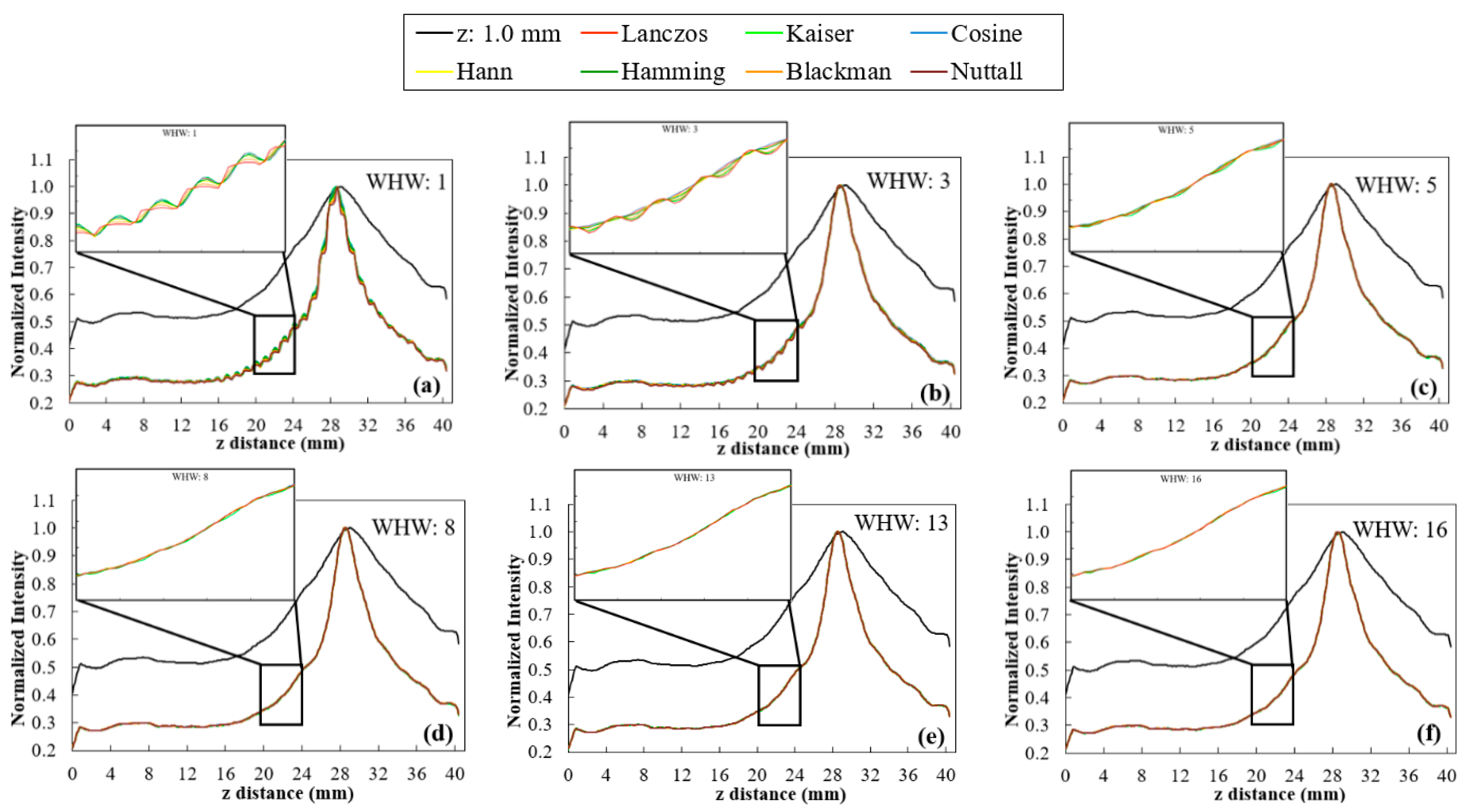
- WHW: In Figure 5, for WHW values above 5, the obtained profiles are very similar. This is translated into the results of Figure 6a, where WHW = 5 produces the highest smoothness in the shortest time. From this value on, there is no significant increase in smoothness, only an increase in the time required for interpolation. On the other hand, in Figure 6b, the FWHM values are very similar for the different WHW values. In this way, the choice is based on the smoothest profile in the shortest possible time. This corresponds to WHW = 5.
- BF(z): According to Figure 7, for BF (z) ≥ 1.5, there is a significant decrease in the variability of the profiles. Through the results in Figure 8a, it is possible to observe that the higher the value of BF (z), the greater the smoothness, reaching a certain convergence for BF (z) ≥ 3. From BF (z) = 1.0 to BF (z) = 2.0, there is a visible decrease in the value of FWHM (Figure 8b), and for BF (z) > 2, these values become very similar. Thus, we choose BF (z) = 2 as the value that represents a better compromise between smoothness, FWHM, and time.
- Interpolator: Nearest and linear interpolators were excluded since the corresponding profiles showed low smoothness when compared to the others. The cubic interpolator was selected to proceed as it presented smoothness and FWHM values comparable to the other interpolations with a similar interpolation time. For the sinc interpolator, considering the results with WHW = 5 and BF (z) = 2, window functions were sorted according to the profile smoothness (in decreasing order). The Hamming window function presented the best correspondent result between the two options (for WHW = 5: Kaiser > Nuttall > Hamming; for BF (z) = 2: Blackman > Lanczos > Hann > Hamming). Since, among them, window functions presented very close results, this selection of a single function to proceed was done only to simplify and concentrate the next results.
3.3. Sampling Distance Study
3.4. Clinical Data
4. Discussion
5. Conclusions
Author Contributions
Funding
Acknowledgments
Conflicts of Interest
Abbreviations
| 2D | Two dimensional |
| 3D | Three dimensional |
| BF | Blur factor |
| CNR | Contrast to noise ratio |
| DBT | Digital breast tomosynthesis |
| DM | Digital mammography |
| FWHM | Full with at half maximum |
| ROI | Region of interest |
| VTK | Visualization toolkit |
| WHW | Window half-width |
References
- Ferlay, J.; Colombet, M.; Soerjomataram, I.; Dyba, T.; Randi, G.; Bettio, M.; Gavin, A.; Visser, O.; Bray, F. Cancer incidence and mortality patterns in Europe: Estimates for 40 countries and 25 major cancers in 2018. Eur. J. Cancer 2018, 103, 356–387. [Google Scholar] [CrossRef] [PubMed]
- Siegel, R.L.; Miller, K.D.; Jemal, A. Cancer statistics, 2020. CA Cancer J. Clin. 2020, 70, 7–30. [Google Scholar] [CrossRef] [PubMed]
- Berry, D.A.; Cronin, K.A.; Plevritis, S.K.; Fryback, D.G.; Clarke, L.; Zelen, M.; Mandelblatt, J.S.; Yakovlev, A.Y.; Habbema, J.D.; Feuer, E.J. Effect of screening and adjuvant therapy on mortality from breast cancer. N. Engl. J. Med. 2005, 353, 1784–1792. [Google Scholar] [CrossRef] [PubMed]
- Independent UK Panel on Breast Cancer Screening. The benefits and harms of breast cancer screening: An independent review. Lancet 2012, 380, 1778–1786. [Google Scholar] [CrossRef]
- Poplack, S.P.; Tosteson, T.D.; Kogel, C.A.; Nagy, H.M. Digital breast tomosynthesis: Initial experience in 98 women with abnormal digital screening mammography. AJR 2007, 189, 616–623. [Google Scholar] [CrossRef]
- Hubbard, R.A.; Kerlikowske, K.; Flowers, C.I.; Yankaskas, B.C.; Zhu, W.; Miglioretti, D.L. Cumulative Probability of False-Positive Recall or Biopsy Recommendation After 10 Years of Screening MammographyA Cohort Study. Ann. Intern. Med. 2011, 155, 481–492. [Google Scholar] [CrossRef]
- Gennaro, G.; Toledano, A.; di Maggio, C.; Baldan, E.; Bezzon, E.; La Grassa, M.; Pescarini, L.; Polico, I.; Proietti, A.; Toffoli, A.; et al. Digital breast tomosynthesis versus digital mammography: A clinical performance study. Eur. Radiol. 2010, 20, 1545–1553. [Google Scholar] [CrossRef]
- Brandt, K.R.; Craig, D.A.; Hoskins, T.L.; Henrichsen, T.L.; Bendel, E.C.; Brandt, S.R.; Mandrekar, J. Can Digital Breast Tomosynthesis Replace Conventional Diagnostic Mammography Views for Screening Recalls Without Calcifications? A Comparison Study in a Simulated Clinical Setting. Am. J. Roentgenol. 2013, 200, 291–298. [Google Scholar] [CrossRef]
- Bonafede, M.M.; Kalra, V.B.; Miller, J.D.; Fajardo, L.L. Value analysis of digital breast tomosynthesis for breast cancer screening in a commercially-insured US population. Clin. Outcomes Res. CEOR 2015, 7, 53–63. [Google Scholar] [CrossRef]
- Gao, Y.; Babb, J.S.; Toth, H.K.; Moy, L.; Heller, S.L. Digital Breast Tomosynthesis Practice Patterns Following 2011 FDA Approval: A Survey of Breast Imaging Radiologists. Acad Radiol. 2017, 24, 947–953. [Google Scholar] [CrossRef]
- Destounis, S.; Santacroce, A.; Arieno, A. DBT as a Screening Tool and a Diagnostic Tool. Curr. Breast Cancer Rep. 2017, 9, 264–271. [Google Scholar] [CrossRef]
- Ramasundara, S.; Tucker, L.; Wallis, M.; Britton, P.; Moyle, P.; Taylor, K.; Sinnatamby, R.; Freeman, A.; Gaskarth, M.; Gilbert, F. Diagnostic implications of digital breast tomosynthesis in symptomatic patients. BCR 2015, 17, P20. [Google Scholar] [CrossRef]
- Svahn, T.M.; Houssami, N.; Sechopoulos, I.; Mattsson, S. Review of radiation dose estimates in digital breast tomosynthesis relative to those in two-view full-field digital mammography. Breast 2015, 24, 93–99. [Google Scholar] [CrossRef] [PubMed]
- Sechopoulos, I. A review of breast tomosynthesis. Part I. The image acquisition process. Med. Phys. 2013, 40, 014301. [Google Scholar] [CrossRef] [PubMed]
- Hofvind, S.; Hovda, T.; Holen, Å.S.; Lee, C.I.; Albertsen, J.; Bjørndal, H.; Brandal, S.H.B.; Gullien, R.; Lømo, J.; Park, D.; et al. Digital Breast Tomosynthesis and Synthetic 2D Mammography versus Digital Mammography: Evaluation in a Population-based Screening Program. Radiology 2018, 287, 787–794. [Google Scholar] [CrossRef]
- Simon, K.; Dodelzon, K.; Drotman, M.; Levy, A.; Arleo, E.K.; Askin, G.; Katzen, J. Accuracy of Synthetic 2D Mammography Compared With Conventional 2D Digital Mammography Obtained With 3D Tomosynthesis. Am. J. Roentgenol. 2019, 212, 1406–1411. [Google Scholar] [CrossRef]
- Van Schie, G.; Wallis, M.G.; Leifland, K.; Danielsson, M.; Karssemeijer, N. Mass detection in reconstructed digital breast tomosynthesis volumes with a computer-aided detection system trained on 2D mammograms. Med. Phys. 2013, 40, 041902. [Google Scholar] [CrossRef]
- Iotti, V.; Giorgi Rossi, P.; Nitrosi, A.; Ravaioli, S.; Vacondio, R.; Campari, C.; Marchesi, V.; Ragazzi, M.; Bertolini, M.; Besutti, G.; et al. Comparing two visualization protocols for tomosynthesis in screening: Specificity and sensitivity of slabs versus planes plus slabs. Eur. Radiol. 2019, 29, 3802–3811. [Google Scholar] [CrossRef]
- Petropoulos, A.E.; Skiadopoulos, S.G.; Karahaliou, A.N.; Messaris, G.A.T.; Arikidis, N.S.; Costaridou, L.I. Quantitative assessment of microcalcification cluster image quality in digital breast tomosynthesis, 2-dimensional and synthetic mammography. Med Biol. Eng. Comput. 2020, 58, 187–209. [Google Scholar] [CrossRef]
- Food and Drug Administration (FDA) U.S. Approval for software option 3DQuoromTM technology-Premarket Approval. Available online: https://www.accessdata.fda.gov/scripts/cdrh/cfdocs/cfpma/pma.cfm?id=P080003S008 (accessed on 25 June 2020).
- 3DQuorum™. Imaging Technology—Improving Radiologist Performance through Artificial Intelligence and SmartSlices (White Paper). Available online: https://www.hologic.com/sites/default/files/downloads/WP-00152_Rev001_3DQuorum_Imaging_Technology_Whitepaper%20%20(1).pdf (accessed on 25 June 2020).
- Venson, J.E.; Albiero Berni, J.C.; Edmilson da Silva Maia, C.; Marques da Silva, A.M.; Cordeiro d’Ornellas, M.; Maciel, A. A Case-Based Study with Radiologists Performing Diagnosis Tasks in Virtual Reality. Stud. Health Technol. Inform. 2017, 245, 244–248. [Google Scholar]
- Suetens, P. Medical image analysis. In Fundamentals of Medical Imaging, 2nd ed.; Cambridge University Press: New York, NY, USA, 2009; pp. 159–189. [Google Scholar]
- O’Connell, A.; Conover, D.L.; Zhang, Y.; Seifert, P.; Logan-Young, W.; Lin, C.-F.L.; Sahler, L.; Ning, R. Cone-Beam CT for Breast Imaging: Radiation Dose, Breast Coverage, and Image Quality. Am. J. Roentgenol. 2010, 195, 496–509. [Google Scholar] [CrossRef] [PubMed]
- Song, H.; Cui, X.; Sun, F. Breast Tissue 3D Segmentation and Visualization on MRI. Int. J. Biomed. Imaging 2013, 2013, 8. [Google Scholar] [CrossRef] [PubMed]
- Jung, Y.; Kim, J.; Feng, D.; Fulham, M. Occlusion and Slice-Based Volume Rendering Augmentation for PET-CT. IEEE J. Biomed. Health Inform. 2017, 21, 1005–1014. [Google Scholar] [CrossRef] [PubMed]
- Alyassin, A.M. Automatic transfer function generation for volume rendering of high-resolution x-ray 3D digital mammography images. In Proceedings of the Medical Imaging 2002, San Diego, CA, USA, February 2002; p. 11. [Google Scholar]
- Alyassin, A.M.; Eberhard, J.W.; Claus, B.E.H.; Kaufhold, J.; González Trotter, D.E.; Kapur, A.; Pakenas, W.P.; Landberg, C.E.; Galbo, C.; Thomas, J.A. 3D Visualization of X-ray Tomosynthesis Digital Mammography Data: Preference Study. In Digital Mammography: IWDM 2002—6th International Workshop on Digital Mammography; Peitgen, H.-O., Ed.; Springer Berlin Heidelberg: Berlin/Heidelberg, Germany, 2003. [Google Scholar]
- Dharanija, R.; Rajalakshmi, T. A Conjunct Analysis for Breast Cancer Detection by Volume Rendering of Low Dosage Three Dimensional Mammogram. In Proceedings of the Progress In Electromagnetics Research Symposium Proceedings, Suzhou, China, 12–16 September 2011; pp. 1361–1365. [Google Scholar]
- Jerebko, A.; Engel, K.; Hofmann, C.; Mertelmeier, T.; Uchiyama, N.; Ongeval, C.V.; Steen, A.V.; Zackrisson, S.; Andersson, I. 3D rendering methods for visualization of clusters of calcifications in digital breast tomosynthesis: A feasibility study. In Proceedings of the ECR 2011, Vienna, Austria, 3–7 March 2011. [Google Scholar]
- Preim, B.; Bartz, D. Visualization in Medicine: Theory, Algorithms, and Applications; Morgan Kaufmann: Burlington, MA, USA, 2007. [Google Scholar]
- Schroeder, W.; Martin, K.; Lorensen, B. The Visualization Toolkit: An Object-oriented Approach to 3D Graphics, 4rd ed.; Kitware: Clifton Park, NY, USA, 2006. [Google Scholar]
- Kitware. The VTK User’s Guide, 11th ed.; Kitware: Clifton Park, NY, USA, 2010. [Google Scholar]
- Siemens. MAMMOMAT Inspiration-Tomosynthesis Option. Available online: https://www.accessdata.fda.gov/cdrh_docs/pdf14/P140011c.pdf (accessed on 25 June 2020).
- VTK. Visualization Toolkit-VTK. Available online: http://www.vtk.org/ (accessed on 25 June 2020).
- STEYX function. Available online: https://support.microsoft.com/en-us/office/steyx-function-6ce74b2c-449d-4a6e-b9ac-f9cef5ba48ab?ui=en-us&rs=en-us&ad=us (accessed on 25 June 2020).
- Thévenaz, P.; Blu, T.; Unser, M. Chapter 28-Image Interpolation and Resampling. In Handbook of Medical Image Processing and Analysis, 2nd Edition; Bankman, I.N., Ed.; Academic Press: Cambridge, MA, USA, 2009; pp. 465–493. [Google Scholar]
- VTK-Interpolators. Visualization Toolkit-VTK-Interpolators. Available online: https://vtk.org/Wiki/VTK/Image_Interpolators (accessed on 25 June 2020).
- Nuttall, A. Some windows with very good sidelobe behavior. IEEE Trans. Acoust. Speech Signal Process. 1981, 29, 84–91. [Google Scholar] [CrossRef]
- Jähne, B. 2.3.3. The Sampling Theorem. In Digital Image Processing: Concepts, Algorithms, and Scientific Applications, 3rd ed.; Springer: Berlin, Germany, 1995. [Google Scholar]


| In Study | |
|---|---|
| Image Interpolators | Linear |
| Cubic | |
| Nearest-neighbor | |
| Image sinc interpolators | Window Function (Lanczos, Kaiser, Cosine, Hann, Hamming, Blackman, Nuttall) |
| Window Half-Width (WHW) | |
| Blur Factor in z-direction (BF(z)) | |
| Interpolator | Linear | Cubic | Nearest |
|---|---|---|---|
| Total time (secs) | 0.45 | 0.50 | 0.45 |
| FWHM90° (mm) | 7.55 | 7.79 | 8.44 |
| Smoothness90º | 64.5 | 72.2 | 37.4 |
| Default | From Our Study | |
|---|---|---|
| Voxel size (mm3) | 0.085 × 0.085 × 1.0 | 0.085 × 0.085 × 0.085 (Hamming with BF (z) = 2) |
| Sampling distance (mm) | 1.0 | 0.025 |
| Total time (s) | 0.23 | 3.05 |
| CNR0° | 7.19 | 22.12 |
| FWHM0° (mm) | 3.67 | 3.52 |
| CNR90° | 6.23 | 39.39 |
| FWHM90° (mm) | 12.38 | 4.06 |
| Smoothness90° | 63.0 | 142.8 |
© 2020 by the authors. Licensee MDPI, Basel, Switzerland. This article is an open access article distributed under the terms and conditions of the Creative Commons Attribution (CC BY) license (http://creativecommons.org/licenses/by/4.0/).
Share and Cite
Mota, A.M.; Clarkson, M.J.; Almeida, P.; Matela, N. Optimization of Breast Tomosynthesis Visualization through 3D Volume Rendering. J. Imaging 2020, 6, 64. https://doi.org/10.3390/jimaging6070064
Mota AM, Clarkson MJ, Almeida P, Matela N. Optimization of Breast Tomosynthesis Visualization through 3D Volume Rendering. Journal of Imaging. 2020; 6(7):64. https://doi.org/10.3390/jimaging6070064
Chicago/Turabian StyleMota, Ana M., Matthew J. Clarkson, Pedro Almeida, and Nuno Matela. 2020. "Optimization of Breast Tomosynthesis Visualization through 3D Volume Rendering" Journal of Imaging 6, no. 7: 64. https://doi.org/10.3390/jimaging6070064
APA StyleMota, A. M., Clarkson, M. J., Almeida, P., & Matela, N. (2020). Optimization of Breast Tomosynthesis Visualization through 3D Volume Rendering. Journal of Imaging, 6(7), 64. https://doi.org/10.3390/jimaging6070064


