Article

Lanthanoid Coordination Polymers Based on Homoditopic Picolinate Ligands: Synthesis, Structure and Magnetic Properties †

by
Verónica Jornet-Mollá
,
Carlos J. Gómez-García
*,
Miquel J. Dolz-Lozano
and
Francisco M. Romero
*
Departament de Química Inorgànica, Universitat de València, C/Dr. Moliner, 50, 46100 Burjassot, Spain
*
Authors to whom correspondence should be addressed.
This work is dedicated to Professors Miguel Julve and Francisco Lloret, two outstanding teachers and brilliant scientists in the field of coordination chemistry. With sincere gratitude for their friendship and support. Sadly, Professor Julve passed away on July 2024. Miguel was gifted with a quick mind, a generous heart and uncommon energy. His wisdom and constant inspiration will never be forgotten.
Magnetochemistry 2025, 11(4), 31; https://doi.org/10.3390/magnetochemistry11040031
Submission received: 17 February 2025 / Revised: 26 March 2025 / Accepted: 28 March 2025 / Published: 7 April 2025

Abstract

:
A ditopic ligand (H2L1), containing picolinate subunits segmented by ethynylene bridges, has been used in the synthesis of a series of isostructural coordination polymers, formulated as [(CH3)2NH2][Ln(L1)2]·H2O·CH3COOH, where Ln = Eu (1), Gd (2), Tb (3), Dy (4) and Ho (5). The single-crystal structures show that these compounds crystallise in the orthorhombic Pna21 space group and form 3D anionic lattices with triangular cavities. AC magnetic susceptibility measurements show that the Gd, Tb and Dy derivatives (24) present a slow relaxation in their magnetisation under an applied DC magnetic field. The detailed study of the AC susceptibility in compounds 2 and 4 shows that they relax following direct and Orbach mechanisms under these conditions. The Dy derivative (4) retains this behaviour in the absence of an external field, relaxing via quantum tunnelling and Orbach mechanisms. Compound 2 is one of the very few reported Gd(III) compounds showing slow relaxation in its magnetisation.

Graphical Abstract

1. Introduction

There is, nowadays, a strong interest in the synthesis and physical properties of lanthanoid coordination polymers (Ln-CPs) [1,2,3]. The reasons for this are manifold: (i) their structural versatility in comparison with transition metals, with higher coordination numbers leading easily to extended frameworks of different dimensionalities, including 3D porous structures [4,5,6,7,8]. (ii) Their fascinating photophysical and magnetic properties, with potential applications in different fields, such as luminescent (bio)sensors and magnetic materials for molecular spintronics and quantum computing [9,10].
Due to the internal character of the 4f electron shell, the magnetism of lanthanoid coordination complexes has traditionally drawn less attention with respect to 3d metal ions. This situation was reversed after the report of single-molecule magnetism (SMM) in the lanthanoid bis-phthalocyaninato anions, [LnPc2] (Ln = Dy, Tb; Pc = phthalocyanine), reported by Ishikawa and coworkers [11,12]. In these compounds, the key to SMM behaviour is the large local magnetic anisotropy that arises from the splitting of the mJ sublevels by the crystal field, giving rise to a high energy barrier for magnetisation and slow spin dynamics [13,14,15]. Most studies concern mononuclear Dy(III) compounds [16,17], but examples of other nuclearities and cations also exist [18,19,20,21,22,23], including Ln-CPs of varying dimensionality [24,25]. In this latter case, the exchange interactions between neighbouring lanthanoid cations can lead to SCM (single-chain magnet) behaviour, especially when dealing with heterometallic 3d–4f systems [26,27,28,29,30]. In most other systems, however, the exchange interactions between lanthanoid ions are rather weak and the slow magnetic relaxation observed is mainly due to single-ion anisotropy effects [31].
Lanthanoid single-ion magnetic anisotropy arises from the interaction of the ground spin-orbit coupled J level with the electrostatic crystal field. This results in a splitting of the mJ states in such a way that the energy gaps between the different ±mJ doublets (resulting in different orientations of the spins) are very sensitive to the coordination environment. Large energy gaps can be obtained by optimizing the strength and symmetry of the ligand field with respect to the shape of the electronic density of the free metal ions. This results in high energy barriers for magnetisation reversal, giving rise to slow relaxation effects and magnetic hysteresis. As described by Long and coworkers, when the shape of the free-ion electron density is oblate, as for Tb3+, Dy3+ and Ho3+ cations, an axial crystal field is needed to maximise the magnetic anisotropy [32]. The family of dysprosium metallocenes, with its ligand electron density concentrated above and below the xy plane, are excellent examples of SMM behaviour [33,34]. Instead, ions such as Er3+, having a prolate electron density, would perform better in an equatorial crystal field. It has been suggested that SMMs based on lanthanoid coordination polymers may be of interest since the lanthanoid coordination sphere can be constrained by the bridging linkers and the crystal symmetry, yielding new materials with improved magnetic properties [24,35]. Further, by using different bridging ligands, different compounds can be obtained that are essentially identical in their first coordination spheres but differ markedly in the way the lanthanoid ions assemble in the crystal lattice, giving some hints about the importance of dipolar interactions in fine-tuning magnetic behaviour [36].
In the quest for interesting bridging ligands, our attention has been focused on chelating and anionic units, which provide thermodynamical stability and charge compensation. Previous examples of coordination polymers containing units of this type involve tetraanionic bis-bidentate dioxidobenzenedicarboxylate ligands [37,38,39] and catecholates [40]. Surprisingly, despite the fact that the picolinate ligand is a universal chelating anionic entity, only a few polytopic picolinate ligands have been reported [41,42,43,44], and their use in metal–organic frameworks are underexplored. We have reported on a family of polytopic picolinate ligands bridged by ethynylene links, which provide rigidity, linear connectivity and electron delocalisation [45,46]. Coordination polymers containing these ligands, in combination with transition metal ions, have also been described. More recently, knowing the ability of picolinate ligands to act as photosensitisers for lanthanoid luminescence [47,48], we reported on Eu(III) CPs based on the ethynylene-bridged bis-picolinate ligand 5,5′-(ethyne-1,2-diyl)dipicolinate (L12−, Figure 1), a strongly luminescent material that can be useful in the detection of several species [49]. We herein describe the whole family of isostructural compounds of formulae [(CH3)2NH2][Ln(L1)2]·H2O·CH3COOH (Ln = Eu (1), Gd (2), Tb (3), Dy (4), Ho (5)), including their synthesis, structural and magnetic characterisation.

2. Materials and Methods

All chemicals and solvents were used as received. Ligand L12− was synthesised as previously described [45].
Synthesis of [(CH3)2NH2][Ln(L1)2]·H2O·CH3COOH (15). A solution of 5,5′-(ethyne-1,2-diyl)dipicolinic acid (15 mg, 0.056 mmol) and Ln(NO3)3·6H2O (0.056 mol) in 4.5 mL of DMF/H2O (8:1 ratio) was placed in a glass vial and stirred for 30 min until a solution was formed. Then, acetic acid was added, and the mixture was placed in an oven at 130 °C for 3 days. After cooling to room temperature at a cooling rate of 0.2 K·min−1, the liquid phase was decanted, and colourless aggregated crystals from compounds 15 were obtained. Details concerning the yields, elemental analyses and IR spectra are given in the Supplementary Materials.
Single crystal X-ray diffraction. The suitable crystals of 15 were coated with paratone N oil, fixed on a small fibre loop and mounted on an Oxford Diffraction Supernova diffractometer equipped with a graphite-monochromated Enhance Mo X-Ray Source (λ = 0.71073 Å) at 120 K. The data collection routines, unit cell refinements and data processing were carried out using the CrysAlisPro v38.46 software package [50]. The structures were solved using SHELXT 2018/2 via the WinGX v2018.3 graphical interface [51] and refined using SHELXL-2018/3 [52]. Although X-ray experiments (powder and single-crystal) unambiguously support the isostructural character of the whole lanthanoid series, satisfactory data for complete structural characterization could only be obtained for the Eu (1) (reported previously) and Tb (3) derivatives. All non-hydrogen atoms were refined anisotropically for 1 (DELU and SIMU restraints were applied to C and O atoms of the acetic acid molecules to allow their anisotropic refinement). For 3, all non-hydrogen atoms were refined anisotropically, except those concerning the acetic acid molecule and O1W. H atoms on carbon atoms were included at calculated positions and refined with a riding model with relative isotropic displacement parameters. Instead, H atoms on solvent molecules and amine H atoms on dimethylammonium cations were found using Fourier difference maps and refined with constrained isotropic thermal parameters, except for the H atoms of O1W. CCDC 2068875 and CCDC 2420909 contain the supplementary crystallographic data for 1 and 3, respectively. These data were provided free of charge by the Cambridge Crystallographic Data Centre.
Powder X-ray Diffraction (PXRD). PXRD measurements for compounds 15 were collected using Cu Kα radiation (λ = 1.54056 Å) at room temperature and in a 2θ range from 2 to 40°. Polycrystalline samples were lightly ground in an agate mortar and filled into a 0.5 mm borosilicate capillary prior to being mounted and aligned on an Empyrean PANalytical powder diffractometer. Simulated diffractograms were obtained from single-crystal X-ray data using the CrystalDiffract software.
Magnetic susceptibility measurements. Variable temperature susceptibility measurements were carried out for compounds 15 in the temperature range of 2–300 K with an applied magnetic field of 100 mT on ground polycrystalline samples, using a Quantum Design (San Diego, CA, USA) MPMS XL-5 SQUID magnetometer. The susceptibility data were corrected for the sample holder and the diamagnetic contributions of the sample using Pascal’s constants [53]. AC susceptibility measurements were performed on the same samples for compounds 25 at low temperatures with different applied DC fields in a frequency range of 10 Hz–10 kHz, using Quantum Design (San Diego, CA, USA) PPMS-9 equipment.
Other characterization techniques. Thermogravimetric analyses were performed on a Mettler-Toledo TGA/SDTA/851e apparatus under an N2 atmosphere at a scan rate of 10 K·min−1. IR transmission measurements were performed from the powdered samples at room temperature in an FT-IR spectrometer (Bruker, alpha II) equipped with an attenuated total reflection (ATR) accessory in the range of 400–4000 cm−1. C, H and N elemental analyses were measured in a CE INSTRUMENTS 1110 EA elemental analyser (SCSIE, Universitat de València).

3. Results and Discussion

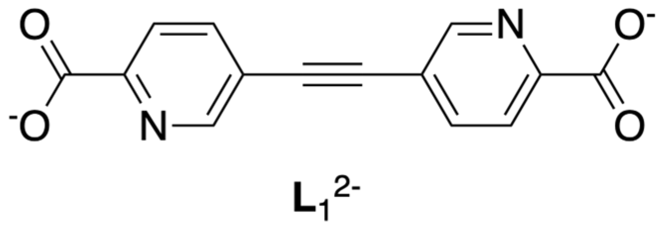
3.1. Synthesis and Characterization

Coordination polymers 15 were prepared by direct reaction between the corresponding lanthanoid nitrate and ligand H2L1 under solvothermal conditions. Although important efforts have been devoted to obtaining single crystals of the erbium derivative, its isolation was not possible. The addition of acetic acid as a modulator was necessary in order to obtain single crystals for X-ray structural analysis. These could also be obtained from the corresponding methyl diesters, Me2L1. In this case, the modulator of choice was formic acid.
In order to confirm the phase purity of the bulk materials, powder X-ray diffraction measurements were performed (Figure S1). The experimental patterns recorded at room temperature for compounds 15 were compared with the simulated diffractogram for the Tb derivative 3 obtained from single-crystal data at 120 K. There is a perfect agreement between both subsets of patterns, discarding the presence of crystalline impurities in the materials.
The thermogravimetric analysis (Figure S2) of the different compounds is very similar, exhibiting a first weight loss between room temperature and 390 K that is associated with the release of water. A second step ascribed to the loss of acetic acid is detected between 425 and 590 K. Above this temperature, the samples start to decompose.

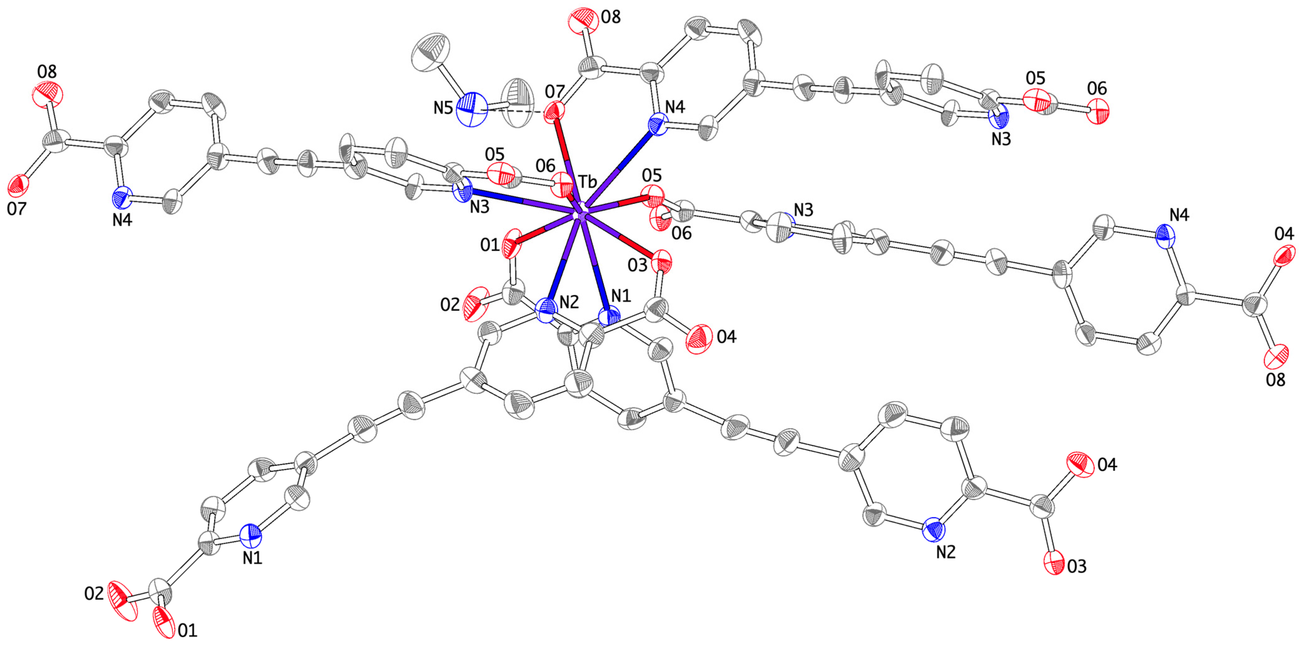
3.2. Structural Properties

Compounds 15 are isostructural and crystallise in the orthorhombic Pna21 space group. Their asymmetric unit contains an Ln3+ cation, two crystallographically independent L12− dianions, one dimethylammonium cation and crystallization solvent molecules (one water molecule and one acetic acid). The Flack parameter is close to zero (Table 1), implying that the model has the correct absolute structure [54].
The presence of dimethylammonium cations is required in order to balance the negative charges of the host framework. They arise from the hydrolysis of dimethylformamide, used as a solvent during the synthesis. In the following, the structure of the Tb compound (3) is described. A comparison with the structure of the Eu derivative (1) is given in Tables S1 and S2.
The terbium cation is located in a general position and exhibits a non-coordinated tricapped trigonal prismatic (TTP) geometry (Figure 2). It is coordinated by four picolinate subunits in a chelating mode. One of these picolinate moieties acts also as a µ-carboxylato bridging ligand and occupies the ninth position in the lanthanoid coordination sphere. The Tb–O distances lie in the 2.33–2.39 Å range, much shorter than the Tb–N1 distances, which comprise between 2.58 and 2.68 Å (Table S1). This is in agreement with the reported values for Ln(III) complexes based on picolinate ligands and is due to the strong oxophilicity of the Ln3+ cations [55]. The triangular faces of the tricapped trigonal prism are defined by O3O6N2 and O1O5O7 atoms, whereas the capping positions over each rectangular face are occupied by N1, N3 and N4 nitrogen atoms. The angle between the trigonal faces is 175.3°, indicating a small deviation from the ideal parallel arrangement.
Continuous shape measure (CShM) calculations using SHAPE 2.1 confirm that the coordination geometries of 1 and 3 are slightly distorted tricapped trigonal prisms (with minimum CShM calculated values corresponding to D3h symmetry, Table S2) [56,57,58]. A CShM calculation of a coordination sphere indicates its deviation from an ideal structure, regardless of its size and orientation, and is given by the following expression:
S G = m i n k = 1 N Q k P k k = 1 N Q k Q 0
The vectors Qk (k = 1, 2…N) indicate the positions of the N donor atoms defining the coordination sphere, and G is a specific and perfect symmetry group with coordinates Pk (k = 1, 2…N). The distances between the vertices of the two objects are calculated until a set of Pk coordinates is found that minimises S(G) (the denominator is a size normalisation factor, Q0 being the coordinate vector of the centre of mass in the investigated structure). The lower the CShM parameter, the better the agreement between the investigated and reference coordination geometries [59]. According to Equation (1), CShM measures must lie within the range of 100 ≥ S ≥ 0. In compounds 1 and 3, values of 3.882 and 3.891, respectively, indicate a close agreement with the ideal TTP geometry (D3h symmetry).
The two independent L12− bis-picolinate ligands adopt different coordination modes. Ligand A acts as a terminal (N1,O1);(N2,O3) bis-chelating ligand, adopting a transoid arrangement of the two picolinate moieties and connecting two Tb3+ complexes separated by the ethynylene spacer. Ligand B behaves also as a ditopic ligand, coordinating to two distant Tb3+ cations related by a translation along the y direction, but one of the picolinate subunits (N3O6) acts as a tridentate ligand in an (N,O);O mode, connecting two lanthanoid metal ions by anti, anti-carboxylate (O5O6) bridges that organise zigzag chains propagating along the c axis. The intrachain terbium–terbium distance is 6.6564(7) Å (Figure 3a). This connectivity results in layers of Tb(III) complexes that run parallel to the bc plane.
The layers are further connected by ligand A, binding two Tb3+ cations separated by a distance of 12.8146(6) Å. This yields a 3D anionic lattice showing triangular cavities (Figure 3b). Dimethylammonium countercations sit within these cavities and are hydrogen-bonded to a coordinating picolinate oxygen atom (H-bond distance N5···O7: 2.741(9) Å) and a water molecule (H-bond distance N5···O1W: 2.704(14) Å). On the other hand, the acetic acid molecule forms a short–strong H-bond with a non-coordinated oxygen atom (H-bond distance O9···O4: 2.486(19) Å).
Both anions deviate considerably from a planar structure. Ligand A adopts a transoid conformation, with a dihedral angle between the two pyridine rings close to 27°. In ligand B, however, the two pyridine rings are almost perpendicular to each other, with a dihedral angle of about 80°. This may be attributed to the fact that this ligand binds to a third Ln3+ cation via the anti, anti-carboxylate (O5O6) bridge. For the same reason, the torsion angle involving the O5 carboxylate oxygen atom (12.59°) is higher than those obtained for the other carboxylate moieties (0.82–8.82°). With respect to the bending of the triple bond, it is similar in both ligands, with a maximum deviation of 8.23° to the ideal –C–C≡C– and –C≡C–C– angles of 180°. Table S3 lists the relevant angles for comparing the crystal structures of 1 and 3.
Although compounds 15 are isostructural, it can be observed that, as the atomic number increases, cell volume decreases. This is a clear signature of the lanthanoid contraction effect (Figure S3).

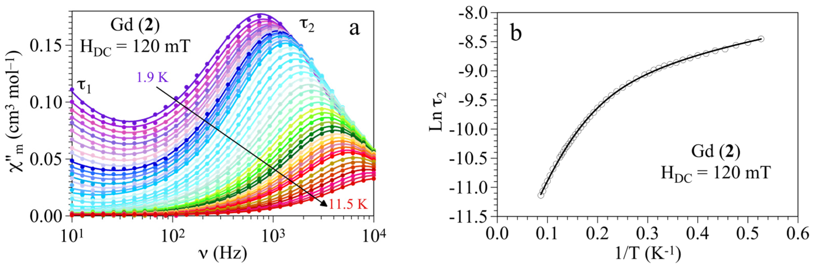
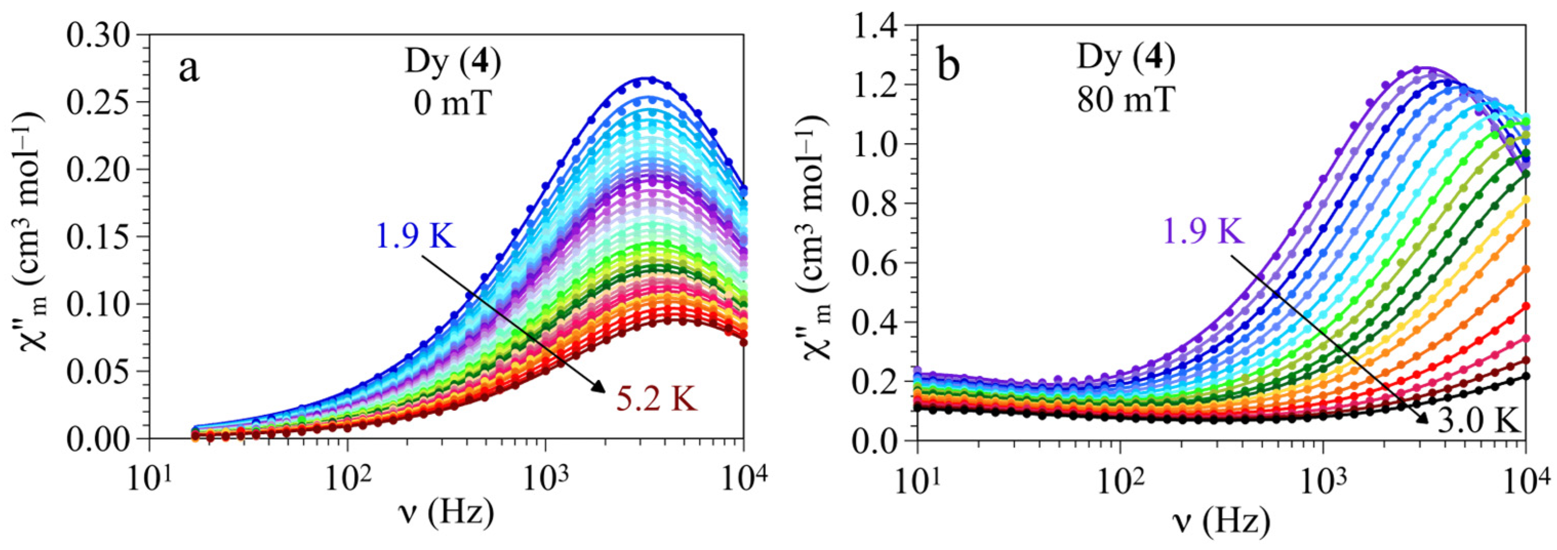
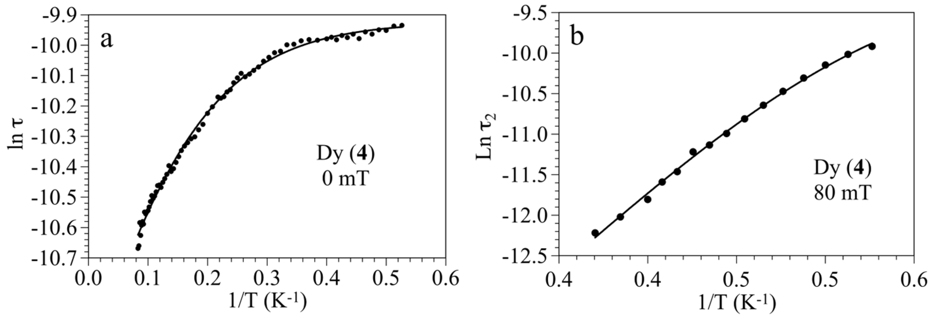
3.3. Magnetic Properties

Magnetic susceptibility measurements were performed on the bulk samples 15, in a static dc magnetic field of 100 mT. The thermal variation in the χmT product (χm = molar magnetic susceptibility) in the 2–300 K temperature range (Figure 4) shows constant values at high temperatures, in agreement with those expected for the Ln3+ ions in their ground states (Table 2) [60]. The exception of this is the europium derivative (1), with a value of χmT at room temperature equal to 1.55 emu·K·mol−1, showing a steady decrease in the χmT signal upon cooling until the signal practically vanishes at 2 K, as expected for the diamagnetic 7F0 ground state of Eu3+ ions. The non-zero value observed at room temperature indicates the thermal population of excited 7FJ (J = 1–6) multiplet levels resulting from the splitting of the 7F ground term by first-order spin-orbit coupling (λ). In fact, the two first excited states (7F1 and 7F2) are quite close to the ground state, and they can be populated at room temperature. Accordingly, we can fit the magnetic susceptibility data for compound 1 to a simple expression that is only dependent on the first-order spin-orbit coupling parameter (λ), as follows:
χ m T = N β 2 3 k T x 24 + 27 x 2 3 2 e x + 135 x 2 5 2 e 3 x + 189 x 7 2 e 6 x + 405 x 9 2 e 10 x + 1485 x 2 11 2 e 15 x + 2457 x 2 13 2 e 21 x 1 + 3 e x + 5 e 3 x + 7 e 6 x + 9 e 10 x + 11 e 15 x + 13 e 21 x
with x = λ/kT. Equation (2) very satisfactorily reproduces the magnetic data for compound 1 with λ = 389(2) cm−1 (solid line in Figure 4). This value is similar to those reported in other EuIII compounds [61,62,63].
The χmT values of the isotropic Gd3+ derivative (2) remain essentially constant in the whole temperature range and decrease abruptly only at temperatures below 10 K. This behaviour indicates that, despite the presence of carboxylate bridges between neighbouring Ln3+ ions, the exchange interactions are very weak [64,65]. In the other compounds (35), the decrease is more gradual and may be ascribed to the progressively thermal depopulation in the highest ground-state Ln3+ sublevels that arise from the splitting of the ground term by the ligand field.
Given the lack of significant interactions, we have fitted the χmT product for compounds 25 to a simple model, including a ZFS contribution (D) and a very weak antiferromagnetic coupling (zj), modelled using the mean-field approximation with the program PHI (solid lines in Figure 4) [66]. The parameters found in the fit are displayed in Table 2.
The good magnetic isolation provided by the picolinate ligands prompted us to measure the AC magnetic susceptibility at low temperatures for 25 in different applied DC fields to look for slow relaxation in the magnetisation (SRM). With the exception of the Ho derivative (5), the compounds show the presence of a frequency-dependent out-of-phase signal (χm) at low temperatures when a DC field is applied (Figures S4–S7). Interestingly, for the Dy derivative (4), we could observe an out-of-phase signal even in the absence of a DC field, although with a lower intensity (Figure S6). The lack of SRM in 23 when no DC field is applied is due to the presence of an efficient relaxation in the magnetization through a quantum tunnelling mechanism. This fast relaxation is suppressed in a DC field. As it can be seen in Figures S4–S6, the intensity of the χm signal initially increases with an increase in the magnetic field and shows two different maxima, one at low frequencies (slow relaxation process with a relaxation time τ1), and a second one at high frequencies (fast relaxation process, τ2). In the Gd (2) and Dy (4) derivatives, we can clearly observe how the maximum of the fast relaxation process (τ2) shifts to lower frequencies and increases in intensity, reaching a minimum frequency and a maximum intensity for DC fields of around 120 mT in 2 and 80 mT in 4 (Figures S4 and S6, respectively). In the Tb derivative (3), the two maxima appear outside the frequency range of our equipment, although we could determine that the maximum intensity is reached for DC values around 140 mT (Figure S5).
The fitting of the frequency dependence of χm to a Debye model for the two relaxation processes (Equation (3), solid lines in Figures S4–S6) gives us the relaxation times (τ) for each applied DC field [67]. Note that, since the maxima of the first relaxation process lie outside the measured frequency range, τ1 values are unreliable in all cases. This applies also to the τ2 value for compound 3. The field dependence of τ2 for compounds 2 and 4 show, as expected, maxima at values around 120 and 80 mT, respectively (Figures S8 and S9). Despite that there are seven adjustable parameters in Equation (3), the isothermal (χT1 and χT2) and adiabatic (χS) susceptibilities, as well as the relaxation times (τ1 and τ2), can be easily estimated from the in-phase signal (χm) values at different frequencies and from the position of the low- and high-frequency maxima, respectively. Note also that, although there is only one crystallographically independent Ln3+ ion in compounds 24, the presence of two different relaxation processes has already been observed in other lanthanoid-based lattices and has been attributed to the presence of relaxation pathways via excited states [68].
χ ω = χ T 1 χ S ω τ 1 1 α 1 c o s π α 1 2 1 + 2 ω τ 1 1 α 1 s i n π α 1 2 + ω τ 1 2 2 α 1 + χ T 2 χ T 1 ω τ 2 1 α 2 c o s π α 2 2 1 + 2 ω τ 2 1 α 2 s i n π α 2 2 + ω τ 2 2 2 α 2
The relaxation times obtained from the fit to the Debye model increase as the DC field increases, reaching maxima at around 120 mT for the Gd derivative (Figure S8) and 80 mT for the Dy compound (Figure S9). This behaviour can be well reproduced with the general model (Equation (4)) [69], using the parameters displayed in Table S4 (solid lines in Figures S8 and S9).
τ 1 = A H n T + B 1 1 + B 2 H 2 + D
In this equation, A, B1, B2 and D are parameters corresponding to the different relaxation mechanisms: direct (A), quantum tunnelling (B1 and B2), and Raman and Orbach (D, since they are field-independent). H is the applied DC magnetic field and n is equal to 4 or 2 for Kramers (Gd and Dy) and non-Kramers (Tb) ions, respectively.
A detailed study of the frequency dependence of χm at different temperatures was performed under a constant DC field set at the values where relaxation time τ2 and signal intensity are the highest. The frequency dependence of χm in an applied DC field of 120 mT for the Gd derivative (2) shows at 1.9 K a maximum of around 740 Hz that shifts to higher frequencies as the temperature increases (Figure 5a). The fit of this data to the Debye model for the two relaxation processes (Equation (3)), gives us the relaxation times at each temperature. These data, displayed in an Arrhenius plot (lnτ vs. 1/T, Figure 5b), show linear dependence at high temperatures for 2, with a clear curvature at lower temperatures. This result suggests the presence of an Orbach thermally activated relaxation mechanism (second term in Equation (5)), with additional Raman and/or direct mechanisms (third and fourth terms, respectively, in Equation (5)) [70]. Attempts to fit the Arrhenius plot for compound 2 to all the possible mechanisms in Equation (5) show that the best fit is obtained including only the Orbach and direct mechanisms (second and fourth terms in Equation (5)), using the parameters displayed in Table S5 (solid line in Figure 5b).
τ 1 = τ Q T 1 + τ 0 1 exp U e f f k T + C T n + A H 4 T
The Tb derivative (3) also shows two different relaxation processes but none of them exhibit a corresponding maximum in the frequency range measured (Figure S10). Therefore, the relaxation times obtained with the Debye model, including the two relaxation processes (Equation (3)), are unreliable and preclude the extraction of relevant parameters from the Arrhenius plot.
Finally, for the Dy derivative (4), we studied the thermal dependence of the out-of-phase signal in both zero and 80 mT DC fields. In both cases, at 1.9 K, we observe maxima in the χm signal at 3200 Hz, with the signal in HDC = 80 mT being stronger (Figure 6). As the temperature is increased under the zero DC field, the maximum remains almost unchanged, in the range of 1.9–5.2 K (Figure 6a), and shifts to higher frequencies above around 5.4 K (Figure S11). For HDC = 80 mT, when the temperature is increased, the maximum shifts very fast to higher frequencies (Figure 6b).
This behaviour indicates that in a zero static field, the predominant mechanism for the relaxation of the magnetisation at low temperatures is quantum tunnelling, which is temperature independent, whereas, at higher temperatures, there is a thermally activated mechanism (Orbach type, see below). As expected, when HDC = 80 mT, the quantum tunnelling is suppressed and the magnetisation relaxes through temperature-dependent mechanisms (direct and Orbach, see below).
As in compound 2, we fitted the data to a Debye model for the two relaxation processes (Equation (3)) to obtain the relaxation times of the fast relaxation process (τ2) at each temperature. The Arrhenius plot (lnτ versus 1/T, Figure 7), in a zero static field, shows an almost temperature-independent behaviour at low temperatures and a curvature as the temperature increases, reaching linear behaviour at higher temperatures. This behaviour suggests the presence of quantum tunnelling and Orbach relaxation mechanisms (first and second terms in Equation (5)). Accordingly, we have fit the data with these two terms to obtain the parameters gathered in Table S5 (solid line in Figure 7a). When HDC = 80 mT, the Arrhenius plot shows linear behaviour with a slight curvature at low temperatures, indicative of the presence of Orbach, direct, and/or Raman mechanisms. Attempts to fit the data with the three possible mechanisms (quantum tunnelling was discarded under the presence of an applied DC field) show that the data can be fitted to Orbach and direct mechanisms, using the parameters shown in Table S5 (solid line in Figure 7b).
The frequency dependence of the in-phase signals (χm) for compounds 24 show an inflexion point corresponding to the maxima in the out-of-phase signals (Figures S12–S16). The Cole–Cole plots (χm vs. χm, Figures S17–S21) show the typical semi-circular shapes in all cases, with one or two semicircles depending on the number of relaxation processes observed in the out-of-phase plots.
It is interesting to note that 2 is one of the few reported Gd(III) lattices showing slow relaxation in its magnetisation (SRM). Thus, although there are hundreds of reports on Dy(III) compounds showing SRM [71,72,73,74], the number of compounds with Gd(III) showing SRM is, by far, much lower. This is due to the fact that the Gd3+ ion has a half-filled 4f7 configuration with a ground multiplet 8S7/2 with no orbital contribution. In the first stage, slow relaxation in Gd(III) complexes has been ascribed to a phonon-bottleneck effect [75]. This picture assumes that the wavelength of the resonant phonon is larger than the separation between Gd3+ ions, a situation that is possible in our case. Indeed, the first reports on SRM for Gd(III) compounds invariably are related to polymeric structures showing relatively short Gd–Gd distances [31,76,77,78], although, recently, well-isolated mononuclear Gd(III) compounds exhibiting SRM have been reported [79] and it has been argued that zero-field splitting in strongly distorted environments could account for the efficiency of the resonant phonon trapping [80]. Thus, the observation of SRM for Gd(III) compounds might be more common than initially expected, as the recent literature provides more examples of this behaviour. An extensive list of materials is given in Table S6 [81,82,83,84,85,86,87,88,89,90,91,92,93,94,95].

4. Conclusions

A segmented ligand L12−, containing two picolinate anions connected by ethynylene bridges, has been used in the synthesis of five isostructural 3D anionic lattices with triangular cavities, formulated as [(CH3)2NH2][Ln(L1)2]·H2O·CH3COOH, where Ln = Eu (1), Gd (2), Tb (3), Dy (4) and Ho (5). AC magnetic measurements show that 24 present slow relaxation when a DC magnetic field is applied, whereas the Dy derivative (4) exhibits slow relaxation in its magnetisation, even in the absence of a static field. The AC susceptibility in compounds 2 and 4 shows that, in both compounds, the dynamic properties follow direct and Orbach mechanisms in a DC field. When this field is switched off, magnetic relaxation in 4 follows quantum tunnelling and Orbach mechanisms. Compound 2 constitutes one of the very rare examples of slow relaxation in a Gd(III) compound.

Supplementary Materials

The following supporting information can be downloaded at: https://www.mdpi.com/article/10.3390/magnetochemistry11040031/s1. Details concerning the synthesis and characterisation of 15; Table S1: relevant bond distances (Å) in the crystal structures of 1 and 3; Table S2: continuous shape measures (CShM) for 1 and 3 using SHAPE 2.1; Table S3: relevant angles (deg) in the crystal structures of 1 and 3; Table S4: magnetic parameters obtained from the fit of the field dependence of the relaxation times at 1.9 K (Equation (4)); Table S5: magnetic parameters obtained from the fit of the Arrhenius plot to the general model (Equation (5)); Figure S1: powder X-ray diffractograms for compounds 15. The simulation of the diffractogram for 3 from single-crystal data is offered for comparison; Figure S2: thermogravimetric analysis for compounds 15; Figure S3: variation in the unit cell volume with increasing atomic numbers (Z) for 15; Figure S4: frequency dependence of the out-of-phase susceptibility (χm) for the Gd derivative (2) at 1.9 K in different applied DC fields; Figure S5: frequency dependence of the out-of-phase susceptibility (χm) for the Tb derivative (3) at 1.9 K in different applied DC fields; Figure S6: frequency dependence of the out-of-phase susceptibility (χm) for the Dy derivative (4) at 1.9 K in different applied DC fields; Figure S7: frequency dependence of the out-of-phase susceptibility (χm) for the Ho derivative (5) at 1.9 K in different applied DC fields; Figure S8: field dependence of the relaxation time for the fast relaxation process (τ2) in the Gd derivative (2) at 1.9 K; Figure S9: field dependence of the relaxation time for the fast relaxation process (τ2) in the Dy derivative (4) at 1.9 K; Figure S10: frequency dependence of χm for the Tb derivative (3) in a DC field of 140 mT at different temperatures; Figure S11: frequency dependence of χm for the Dy derivative (4) in a zero DC field at different temperatures above 5.4 K; Figure S12: frequency dependence of χm for the Gd derivative (2) with HDC = 120 mT, in the temperature range of 1.9–11.5 K; Figure S13: frequency dependence of χm for the Tb derivative (3) with HDC = 140 mT, in the temperature range of 1.9–4.8 K; Figure S14: frequency dependence of χm for the Dy derivative (4) with HDC = 0 mT, in the temperature range of 1.9–5.2 K; Figure S15: frequency dependence of χm for the Dy derivative (4) with HDC = 0 mT, in the temperature range of 5.4–12.0 K; Figure S16: frequency dependence of χm for the Dy derivative (4) with HDC = 80 mT, in the temperature range of 1.9–3.0 K; Figure S17: Cole-Cole plot for the Gd derivative (2) with HDC = 120 mT, in the temperature range of 1.9–11.5 K; Figure S18: Cole-Cole plot for the Tb derivative (3) with HDC = 140 mT, in the temperature range of 1.9–4.8 K; Figure S19: Cole-Cole plot for the Dy derivative (4) with HDC = 0 mT, in the temperature range of 1.9–5.2 K; Figure S20: Cole-Cole plot for the Dy derivative (4) with HDC = 0 mT, in the temperature range of 5.4–12.0 K; Figure S21: Cole-Cole plot for the Dy derivative (4) with HDC = 80 mT, in the temperature range of 1.9–3.0 K.

Author Contributions

Conceptualization, F.M.R.; formal analysis, V.J.-M., C.J.G.-G., M.J.D.-L. and F.M.R.; investigation, V.J.-M., C.J.G.-G., M.J.D.-L. and F.M.R.; writing—original draft preparation, C.J.G.-G. and F.M.R.; writing—review and editing, V.J.-M., C.J.G.-G., M.J.D.-L. and F.M.R. All authors have read and agreed to the published version of the manuscript.

Funding

This research forms part of the Advanced Materials program and was funded by the Spanish MCIN, with funding from the European Union NextGeneration EU (PRTR-C17.I1) and the Generalitat Valenciana (project MFA-2022-057). We also express thanks for Grant PID2021-125907NB-I00, funded by the MCIN/AEI/10.13039/501100011033, as well as “ERDF A way of making Europe” and project CIPROM-2022-060 from the Generalitat Valenciana, for financial support.

Institutional Review Board Statement

Not applicable.

Informed Consent Statement

Not applicable.

Data Availability Statement

The data in this contribution will be provided upon request to the corresponding author.

Conflicts of Interest

The authors declare no conflicts of interest.

References

  1. Yan, B. Lanthanide-functionalized metal–organic framework hybrid systems to create multiple luminescent centers for chemical sensing. Acc. Chem. Res. 2017, 50, 2789–2798. [Google Scholar] [CrossRef] [PubMed]
  2. Shi, Y.; Song, M.-M.; Tao, D.-L.; Bo, Q.-B. Diverse lanthanide coordination polymers with 3,3′-dimethylcyclopropane-1,2-dicarboxylate ligand: Synthesis, crystal structure, and properties. ACS Omega 2018, 3, 12122–12131. [Google Scholar] [CrossRef]
  3. Feng, X.; Shang, Y.; Zhang, H.; Li, R.; Wang, W.; Zhang, D.; Wang, L.; Li, Z. Enhanced luminescence and tunable magnetic properties of lanthanide coordination polymers based on fluorine substitution and phenanthroline ligand. RSC Adv. 2019, 9, 16328–16338. [Google Scholar] [CrossRef]
  4. Ruiz-Muelle, A.B.; García- García, A.; García-Valdivia, A.A.; Oyarzabal, I.; Cepeda, J.; Seco, J.M.; Colacio, E.; Rodríguez-Diéguez, A.; Fernández, I. Design and synthesis of a family of 1D-lanthanide-coordination polymers showing luminescence and slow relaxation of the magnetization. Dalton Trans. 2018, 47, 12783–12794. [Google Scholar] [CrossRef] [PubMed]
  5. Liu, J.-H.; Zhang, R.-T.; Zhang, J.; Zhao, D.; Li, X.-X.; Sun, Y.-Q.; Zheng, S.-T. A series of 3D porous lanthanide-substituted polyoxometalate frameworks based on rare hexadecahedral {Ln6W8O28} heterometallic cage-shaped clusters. Inorg. Chem. 2019, 58, 14734–14740. [Google Scholar] [CrossRef]
  6. Zhang, S.; Shi, W.; Li, L.; Duan, E.; Cheng, P. Lanthanide coordination polymers with “fsy-type” topology based on 4, 4′-azobenzoic acid: Syntheses, crystal structures, and magnetic properties. Inorg. Chem. 2014, 53, 10340–10346. [Google Scholar] [CrossRef]
  7. Ma, F.; Sun, R.; Sun, A.-H.; Xiong, J.; Sun, H.-L.; Gao, S. Regulating the structural dimensionality and dynamic properties of a porous dysprosium coordination polymer through solvent molecules. Inorg. Chem. Front. 2020, 7, 930–938. [Google Scholar] [CrossRef]
  8. Blais, C.; Calvez, G.; Suffren, Y.; Daiguebonne, C.; Paranthoen, C.; Bazin, E.; Freslon, S.; Bernot, K.; Guillou, O. Luminance and brightness: Application to lanthanide-based coordination polymers. Inorg. Chem. 2022, 48, 19588–19596. [Google Scholar] [CrossRef]
  9. Bogani, L.; Wernsdorfer, W. Molecular spintronics using single-molecule magnets. Nat. Mater. 2008, 7, 179–186. [Google Scholar] [CrossRef]
  10. Urdampilleta, M.; Nguyen, N.V.; Cleuziou, J.P.; Klyatskaya, S.; Ruben, M.; Wernsdorfer, W. Molecular quantum spintronics: Supramolecular spin valves based on single-molecule magnets and carbon nanotubes. Int. J. Mol. Sci. 2011, 12, 6656–6667. [Google Scholar] [CrossRef]
  11. Ishikawa, N.; Sugita, M.; Ishikawa, T.; Koshihara, S.; Kaizu, Y. Lanthanide double-decker complexes functioning as magnets at the single-molecular level. J. Am. Chem. Soc. 2003, 125, 8694–8695. [Google Scholar] [CrossRef] [PubMed]
  12. Ishikawa, N.; Sugita, M.; Ishikawa, T.; Koshihara, S.; Kaizu, Y. Mononuclear lanthanide complexes with a long magnetization relaxation time at high temperatures: A new category of magnets at the single-molecular level. J. Phys. Chem. B 2004, 108, 11265–11271. [Google Scholar] [CrossRef]
  13. Woodruff, D.N.; Winpenny, R.E.P.; Layfield, R.A. Lanthanide single-molecule magnets. Chem. Rev. 2013, 113, 5110–5148. [Google Scholar] [CrossRef]
  14. Ungur, L.; Chibotaru, L.F. Strategies toward high-temperature lanthanide-based single-molecule magnets. Inorg. Chem. 2016, 55, 10043–10056. [Google Scholar] [CrossRef] [PubMed]
  15. Bernot, K. Get under the umbrella: A comprehensive gateway for researchers on lanthanide-based single-molecule magnets. Eur. J. Inorg. Chem. 2023, 26, e202300336. [Google Scholar] [CrossRef]
  16. Zhang, P.; Guo, Y.-N.; Tang, J. Recent advances in dysprosium-based single molecule magnets: Structural overview and synthetic strategies. Coord. Chem. Rev. 2013, 257, 1728–1763. [Google Scholar] [CrossRef]
  17. AlDamen, M.A.; Clemente-Juan, J.M.; Coronado, E.; Martí-Gastaldo, C.; Gaita-Ariño, A. Mononuclear lanthanide single-molecule magnets based on polyoxometalates. J. Am. Chem. Soc. 2008, 130, 8874–8875. [Google Scholar] [CrossRef] [PubMed]
  18. Meihaus, K.R.; Long, J.R. Magnetic blocking at 10 K and a dipolar-mediated avalanche in salts of the bis(η8-cyclooctatetraenide) complex [Er(COT)2]. J. Am. Chem. Soc. 2013, 135, 17952–17957. [Google Scholar] [CrossRef]
  19. Le Roy, J.J.; Jeletic, M.; Gorelsky, S.I.; Korobkov, I.; Ungur, L.; Chibotaru, L.F.; Murugesu, M. An organometallic building block approach to produce a multidecker 4f single-molecule magnet. J. Am. Chem. Soc. 2013, 135, 3502–3510. [Google Scholar] [CrossRef]
  20. Tang, J.; Hewitt, I.; Madhu, N.T.; Chastanet, G.; Wernsdorfer, W.; Anson, C.E.; Benelli, C.; Sessoli, R.; Powell, A.K. Dysprosium triangles showing single-molecule magnet behavior of thermally excited spin states. Angew. Chem. Int. Ed. 2006, 45, 1729–1733. [Google Scholar] [CrossRef]
  21. Wang, J.; Sun, C.-Y.; Zheng, Q.; Wang, D.-Q.; Chen, Y.-T.; Ju, J.-F.; Sun, T.-M.; Cui, Y.; Ding, Y.; Tang, Y.-F. Lanthanide single-molecule magnets: Synthetic strategy, structures, properties and recent advances. Chem. Asian J. 2023, 18, e202201297. [Google Scholar] [PubMed]
  22. Dolinar, B.S.; Alexandropoulos, D.I.; Vignesh, K.R.; James, T.; Dunbar, K.R. Lanthanide triangles supported by radical bridging ligands. J. Am. Chem. Soc. 2018, 140, 908–911. [Google Scholar] [PubMed]
  23. Boulon, M.-E.; Cucinotta, G.; Luzon, J.; Degl’Innocenti, C.; Perfetti, M.; Bernot, K.; Calvez, G.; Caneschi, A.; Sessoli, R. Magnetic anisotropy and spin-parity effect along the series of lanthanide complexes with DOTA. Angew. Chem. Int. Ed. 2013, 52, 350–354. [Google Scholar] [CrossRef]
  24. Wan, Q.; Wakizaka, M.; Yamashita, M. Single-ion magnetism behaviors in lanthanide (III) based coordination frameworks. Inorg. Chem. Front. 2023, 10, 5212–5224. [Google Scholar]
  25. Huang, G.; Fernandez-Garcia, G.; Badiane, I.; Camarra, M.; Freslon, S.; Guillou, O.; Daiguebonne, C.; Totti, F.; Cador, O.; Guizouarn, T.; et al. Magnetic slow relaxation in a metal–organic framework made of chains of ferromagnetically coupled single-molecule magnets. Chem. Eur. J. 2018, 24, 6983–6991. [Google Scholar] [CrossRef] [PubMed]
  26. Bernot, K.; Bogani, L.; Caneschi, A.; Gatteschi, D.; Sessoli, R. A family of rare-earth-based single chain magnets: Playing with anisotropy. J. Am. Chem. Soc. 2006, 128, 7947–7956. [Google Scholar]
  27. Gheorghe, R.; Madalan, A.M.; Costes, J.-P.; Wernsdorfer, W.; Andruh, M. A heterotrimetallic 3d–3d′–4f single chain magnet constructed from anisotropic high-spin 3d–4f nodes and paramagnetic spacers. Dalton Trans. 2010, 39, 4734–4736. [Google Scholar]
  28. Paulovič, J.; Cimpoesu, F.; Ferbinteanu, M.; Hirao, K. Mechanism of ferromagnetic coupling in copper (II)-gadolinium (III) complexes. J. Am. Chem. Soc. 2004, 126, 3321–3331. [Google Scholar]
  29. Singh, S.K.; Beg, M.F.; Rajaraman, G. Role of magnetic exchange interactions in the magnetization relaxation of {3d–4f} single-molecule magnets: A theoretical perspective. Chem. Eur. J. 2016, 22, 672–680. [Google Scholar]
  30. Dey, A.; Acharya, J.; Chandrasekhar, V. Heterometallic 3d–4f complexes as single-molecule magnets. Chem. Asian J. 2019, 14, 4433–4453. [Google Scholar]
  31. Girginova, P.I.; Pereira, L.C.J.; Coutinho, J.T.; Santos, I.C.; Almeida, M. Slow magnetic relaxation in lanthanide ladder type coordination polymers. Dalton Trans. 2014, 43, 1897–1905. [Google Scholar] [CrossRef] [PubMed]
  32. Rinehart, J.D.; Long, J.R. Exploiting single-ion anisotropy in the design of f-element single-molecule magnets. Chem. Sci. 2011, 2, 2078–2085. [Google Scholar] [CrossRef]
  33. Guo, F.-S.; Day, B.M.; Chen, Y.-C.; Tong, M.-L.; Mansikkamäki, A.; Layfield, R.A. Magnetic hysteresis up to 80 kelvin in a dysprosium metallocene single-molecule magnet. Science 2018, 362, 1400–1403. [Google Scholar] [CrossRef]
  34. Tarannum, I.; Moorthy, S.; Singh, S.K. Understanding electrostatics and covalency effects in highly anisotropic organometallic sandwich dysprosium complexes [Dy(CmRm)2] (where R = H, SiH3, CH3 and m = 4 to 9): A computational perspective. Dalton Trans. 2023, 52, 15576–15589. [Google Scholar] [CrossRef] [PubMed]
  35. Liu, K.; Zhang, X.; Meng, X.; Shi, W.; Cheng, P.; Powell, A.K. Constraining the coordination geometries of lanthanide centers and magnetic building blocks in frameworks: A new strategy for molecular nanomagnets. Chem. Soc. Rev. 2016, 45, 2423–2439. [Google Scholar] [CrossRef]
  36. Zhang, X.; Vieru, V.; Feng, X.; Liu, J.-L.; Zhang, Z.; Na, B.; Shi, W.; Wang, B.-W.; Powell, A.K.; Chibotaru, L.F.; et al. Influence of guest exchange on the magnetization dynamics of dilanthanide single-molecule-magnet nodes within a metal–organic framework. Angew. Chem. Int. Ed. 2015, 54, 9861–9865. [Google Scholar] [CrossRef]
  37. Caskey, S.R.; Wong-Foy, A.G.; Matzger, A.J. Dramatic tuning of carbon dioxide uptake via metal substitution in a coordination polymer with cylindrical pores. J. Am. Chem. Soc. 2008, 130, 10870–10871. [Google Scholar] [CrossRef] [PubMed]
  38. Bloch, E.D.; Murray, L.J.; Queen, W.L.; Chavan, S.; Maximoff, S.N.; Bigi, J.P.; Krishna, R.; Peterson, V.K.; Grandjean, F.; Long, G.J.; et al. Selective binding of O2 over N2 in a redox–active metal–organic framework with open iron(II) coordination sites. J. Am. Chem. Soc. 2011, 133, 14814–14822. [Google Scholar] [CrossRef]
  39. Kapelewski, M.T.; Geier, S.J.; Hudson, M.R.; Stück, D.; Mason, J.A.; Nelson, J.N.; Xiao, D.J.; Hulvey, Z.; Gilmour, E.; FitzGerald, S.A.; et al. M2(m-dobdc) (M = Mg, Mn, Fe, Co, Ni) metal–organic frameworks exhibiting increased charge density and enhanced H2 binding at the open metal sites. J. Am. Chem. Soc. 2014, 136, 12119–12129. [Google Scholar] [CrossRef]
  40. Hmadeh, M.; Lu, Z.; Liu, Z.; Gándara, F.; Furukawa, H.; Wan, S.; Augustyn, V.; Chang, R.; Liao, L.; Zhou, F.; et al. New porous crystals of extended metal-catecholates. Chem. Mater. 2012, 24, 3511–3513. [Google Scholar] [CrossRef]
  41. Park, B.G.; Pink, M.; Lee, D. Fluorogenic N,O-chelates built on a C2-symmetric aryleneethynylene platform: Spectroscopic and structural consequences of conformational preorganization and ligand denticity. J. Organomet. Chem. 2011, 696, 4039–4045. [Google Scholar] [CrossRef]
  42. Kodanko, J.J.; Lippard, S.J. Synthesis and characterization of a ditriflate-bridged, diiron(II) complex with syn-N-donor ligands: [Fe2(μ-OTf)2(PIC2DET)2](BARF)2. Inorg. Chim. Acta 2008, 361, 894–900. [Google Scholar]
  43. Kodanko, J.J.; Xu, D.; Song, D.; Lippard, S.J. Iron substitution for sodium in a carboxylate-bridged, heterodinuclear sodium–iron Complex. J. Am. Chem. Soc. 2005, 127, 16004–16005. [Google Scholar] [CrossRef]
  44. Severance, R.C.; Rountree, E.S.; Smith, M.D.; zur Loye, H.-C. Ligand-based luminescence in lead-containing complexes: The effect of conjugated organic ligands on fluorescence. Solid State Sci. 2012, 14, 1512–1519. [Google Scholar]
  45. Jornet-Mollá, V.; Romero, F.M. Synthesis of rigid ethynyl-bridged polytopic picolinate ligands for MOF applications. Tetrahedron Lett. 2015, 56, 6120–6122. [Google Scholar] [CrossRef]
  46. Jornet-Mollá, V.; Martín-Mezquita, C.; Giménez-Saiz, C.; Romero, F.M. Zinc (II) picolinate-based coordination polymers as luminescent sensors of Fe3+ ions and nitroaromatic compounds. Inorg. Chim. Acta 2022, 538, 120993. [Google Scholar] [CrossRef]
  47. Bünzli, J.-C.G.; Piguet, C. Taking advantage of luminescent lanthanide ions. Chem. Soc. Rev. 2005, 34, 1048–1077. [Google Scholar]
  48. Lamture, J.B.; Zhou, Z.H.; Kumar, A.S.; Wensel, T.G. Luminescence properties of terbium (III) complexes with 4-substituted dipicolinic acid analogs. Inorg. Chem. 1995, 34, 864–869. [Google Scholar] [CrossRef]
  49. Jornet-Mollá, V.; Dreessen, C.; Romero, F.M. Robust lanthanoid picolinate-based coordination polymers for luminescence and sensing applications. Inorg. Chem. 2021, 60, 10572–10584. [Google Scholar] [CrossRef]
  50. CrysAlisPro v38.46; Oxford Diffraction Ltd.: Abingdon, UK, 2017.
  51. Farrugia, L.J. WinGX suite for small-molecule single-crystal crystallography. J. Appl. Crystallogr. 1999, 32, 837–838. [Google Scholar] [CrossRef]
  52. Sheldrick, G.M. Crystal structure refinement with SHELXL. Acta Cryst. 2015, C71, 3–8. [Google Scholar]
  53. Bain, G.A.; Berry, J.F. Diamagnetic corrections and Pascal’s constants. J. Chem. Educ. 2008, 85, 532–536. [Google Scholar] [CrossRef]
  54. Flack, H.D. On enantiomorph-polarity estimation. Acta Cryst. 1983, A39, 876–881. [Google Scholar] [CrossRef]
  55. Wu, Y.-P.; Xu, G.-W.; Dong, W.-W.; Zhao, J.; Li, D.-S.; Zhang, J.; Bu, X. Anionic lanthanide MOFs as a platform for iron-selective sensing, systematic color tuning, and efficient nanoparticle catalysis. Inorg. Chem. 2017, 56, 1402–1411. [Google Scholar] [CrossRef]
  56. Casanova, D.; Alemany, P.; Bofill, J.M.; Alvarez, S. Shape and symmetry of heptacoordinate transition-metal complexes: Structural trends. Chem. Eur. J. 2003, 9, 1281–1295. [Google Scholar] [CrossRef]
  57. Alvarez, S.; Alemany, P.; Casanova, D.; Cirera, J.; Llunell, M.; Avnir, D. Shape maps and polyhedral interconversion paths in transition metal chemistry. Coord. Chem. Rev. 2005, 249, 1693–1708. [Google Scholar] [CrossRef]
  58. Llunell, M.; Casanova, D.; Cirera, J.; Bofill, J.M.; Alemany, P.; Alvarez, S. SHAPE, version 2.1; Universitat de Barcelona: Barcelona, Spain, 2013. [Google Scholar]
  59. Alvarez, S.; Avnir, D.; Llunell, M.; Pinsky, M. Continuous symmetry maps and shape classification. The case of six-coordinated metal compounds. New J. Chem. 2002, 26, 996–1009. [Google Scholar] [CrossRef]
  60. Sorace, L.; Gatteschi, D. Electronic Structure and Magnetic Properties of Lanthanide Molecular Complexes. In Lanthanides and Actinides in Molecular Magnetism; Wiley-VCH: Hoboken, NJ, USA, 2015; pp. 1–26. [Google Scholar]
  61. de Oliveira Maciel, J.W.; Lemes, M.A.; Valdo, A.K.; Rabelo, R.; Martins, F.T.; Queiroz Maia, L.J.; de Santana, R.C.; Lloret, F.; Julve, M.; Cangussu, D. Europium(III), terbium(III), and gadolinium(III) oxamato-based coordination polymers: Visible luminescence and slow magnetic relaxation. Inorg. Chem. 2021, 60, 6176–6190. [Google Scholar] [CrossRef]
  62. Vostrikova, K.E.; Sukhikh, T.S.; Lavrov, A.N. The synthesis, crystal structure, and magnetic properties of mono-scorpionate Eu(III) complexes. Inorganics 2023, 11, 418. [Google Scholar] [CrossRef]
  63. Benmansour, S.; Cerezo-Navarrete, C.; Gómez-García, C.J. Slow relaxation of the magnetisation in a two-dimensional metal–organic framework with a layered square lattice. Magnetochemistry 2025, 11, 1. [Google Scholar] [CrossRef]
  64. AlDamen, M.A.; Cardona-Serra, S.; Clemente-Juan, J.M.; Coronado, E.; Gaita-Ariño, A.; Martí-Gastaldo, C.; Luis, F.; Montero, O. Mononuclear lanthanide single molecule magnets based on the polyoxometalates [Ln(W5O18)2]9− and [Ln(β2-SiW11O39)2]13− (LnIII = Tb, Dy, Ho, Er, Tm, and Yb). Inorg. Chem. 2009, 48, 3467–3479. [Google Scholar] [CrossRef]
  65. Kalita, P.; Malakar, A.; Goura, J.; Nayak, S.; Herrera, J.M.; Colacio, E.; Chandrasekhar, V. Mononuclear lanthanide complexes assembled from a tridentate NNO donor ligand: Design of a DyIII single-ion magnet. Dalton Trans. 2019, 48, 4857–4866. [Google Scholar]
  66. Chilton, N.F.; Anderson, R.P.; Turner, L.D.; Soncini, A.; Murray, K.S. PHI: A powerful new program for the analysis of anisotropic monomeric and exchange-coupled polynuclear d- and f-block complexes. J. Comput. Chem. 2013, 34, 1164–1175. [Google Scholar]
  67. Dolai, M.; Ali, M.; Titiš, J.; Boča, R. Cu(II)–Dy(III) and Co(III)–Dy(III) based single molecule magnets with multiple slow magnetic relaxation processes in the Cu(II)–Dy(III) complex. Dalton Trans. 2015, 44, 13242–13249. [Google Scholar] [PubMed]
  68. Blagg, R.J.; Ungur, L.; Tuna, F.; Speak, J.; Comar, P.; Collison, D.; Wernsdorfer, W.; McInnes, E.J.L.; Chibotaru, L.; Winpenny, R.E.P. Magnetic relaxation pathways in lanthanide single-molecule magnets. Nature Chem. 2013, 5, 673–678. [Google Scholar]
  69. Wang, M.-H.; Tsai, M.-Y.; Su, Y.-C.; Chiu, S.-T.; Lin, P.-H.; Long, J. Zero-field single-molecule magnet behavior in a series of dinuclear dysprosium(III) complexes based on benzothiazolyl-based ligands and β-diketonates. Cryst. Growth Des. 2024, 24, 422–431. [Google Scholar]
  70. Demir, S.; Zadrozny, J.M.; Long, J.R. Large spin-relaxation barriers for the low-symmetry organolanthanide complexes [Cp*2Ln(BPh4)] (Cp*=pentamethylcyclopentadienyl; Ln=Tb, Dy). Chem. Eur. J. 2014, 20, 9524–9529. [Google Scholar]
  71. Zhu, Z.; Guo, M.; Li, X.; Tang, J. Molecular magnetism of lanthanide: Advances and perspectives. Coord. Chem. Rev. 2019, 378, 350–364. [Google Scholar]
  72. Parmar, V.S.; Mills, D.P.; Winpenny, R.E.P. Mononuclear dysprosium alkoxide and aryloxide single-molecule magnets. Chem. Eur. J. 2021, 27, 7625–7645. [Google Scholar] [CrossRef]
  73. Zabala-Lekuona, A.; Manuel Seco, J.; Colacio, E. Single-molecule magnets: From Mn12-ac to dysprosium metallocenes, a travel in time. Coord. Chem. Rev. 2021, 441, 213984. [Google Scholar]
  74. Ashebr, T.G.; Li, H.; Ying, X.; Li, X.-L.; Zhao, C.; Liu, S.; Tang, J. Emerging trends on designing high-performance dysprosium(III) single-molecule magnets. ACS Mater. Lett. 2022, 4, 307–319. [Google Scholar] [CrossRef]
  75. Orendáč, M.; Sedláková, L.; Čizmár, E.; Orendáčova, A.; Feher, A.; Zvyagin, S.A.; Wosnitza, J.; Zhu, W.H.; Wang, Z.M.; Gao, S. Spin relaxation and resonant phonon trapping in [Gd2(fum)3(H2O)4]⋅3H2O. Phys. Rev. B 2010, 81, 214410. [Google Scholar] [CrossRef]
  76. Rossin, A.; Giambastiani, G.; Peruzzini, M.; Sessoli, R. Amine-templated polymeric lanthanide formates: Synthesis, characterization, and applications in luminescence and magnetism. Inorg. Chem. 2012, 51, 6962–6968. [Google Scholar] [CrossRef] [PubMed]
  77. Orts-Arroyo, M.; Rabelo, R.; Carrasco-Berlanga, A.; Moliner, N.; Cano, J.; Julve, M.; Lloret, F.; De Munno, G.; Ruiz-García, R.; Mayans, J.; et al. Field-induced slow magnetic relaxation and magnetocaloric effects in an oxalato-bridged gadolinium(III)-based 2D MOF. Dalton Trans. 2021, 50, 3801–3805. [Google Scholar] [CrossRef] [PubMed]
  78. Orts-Arroyo, M.; Sanchis-Perucho, A.; Moliner, N.; Castro, I.; Lloret, F.; Martínez-Lillo, J. One-dimensional gadolinium(III) complexes based on alpha-and beta-amino acids exhibiting field-induced slow relaxation of magnetization. Inorganics 2022, 10, 32. [Google Scholar] [CrossRef]
  79. Chen, Y.C.; Peng, Y.-Y.; Liu, J.-L.; Tong, M.-L. Field-induced slow magnetic relaxation in a mononuclear Gd (III) complex. Inorg. Chem. Commun. 2019, 107, 107449. [Google Scholar] [CrossRef]
  80. Mayans, J.; Escuer, A. Correlating the axial zero field splitting with the slow magnetic relaxation in GdIII SIMs. Chem. Commun. 2021, 57, 721–724. [Google Scholar] [CrossRef]
  81. Liu, B.; Wang, B.; Wang, Z. Static field induced magnetic relaxations in dinuclear lanthanide compounds of [phen2Ln2(HCOO)4(HCOO)2−2x (NO3)2x ] (1, Ln = Gd and x = 0.52; 2, Ln = Er and x = 0.90; phen = 1,10-phenanthroline). Sci. China Chem. 2012, 55, 926–933. [Google Scholar] [CrossRef]
  82. Martínez-Pérez, M.J.; Cardona-Serra, S.; Schlegel, C.; Moro, F.; Alonso, P.J.; Prima-García, H.; Clemente-Juan, J.M.; Evangelisti, M.; Gaita-Ariño, A.; Sesé, J.; et al. Gd-based single-ion magnets with tunable magnetic anisotropy: Molecular design of spin qubits. Phys. Rev. Lett. 2012, 108, 247213. [Google Scholar] [CrossRef]
  83. Arauzo, A.; Lazarescu, A.; Shova, S.; Bartolomé, E.; Cases, R.; Luzón, J.; Bartolomé, J.; Turta, C. Structural and magnetic properties of some lanthanide (Ln = Eu(III), Gd(III) and Nd(III)) cyanoacetate polymers: Field-induced slow magnetic relaxation in the Gd and Nd substitutions. Dalton Trans. 2014, 43, 12342–12356. [Google Scholar] [CrossRef]
  84. Holmberg, R.J.; Anh Ho, L.T.; Ungur, L.; Korobkov, I.; Chibotaru, L.F.; Murugesu, M. Observation of unusual slow-relaxation of the magnetisation in a Gd-EDTA chelate. Dalton Trans. 2015, 44, 20321–20325. [Google Scholar] [CrossRef] [PubMed]
  85. Yoshida, T.; Cosquer, G.; Izuogu, D.C.; Ohtsu, H.; Kawano, M.; Lan, Y.; Wernsdorfer, W.; Nojiri, H.; Breedlove, B.K.; Yamashita, M. Field-induced slow magnetic relaxation of GdIII complex with a Pt−Gd heterometallic bond. Chem. Eur. J. 2017, 23, 4551–4556. [Google Scholar] [CrossRef] [PubMed]
  86. Izuogu, D.C.; Yoshida, T.; Zhang, H.; Cosquer, G.; Katoh, K.; Ogata, S.; Hasegawa, M.; Nojiri, H.; Damjanović, M.; Wernsdorfer, W.; et al. Slow magnetic relaxation in a palladium–gadolinium complex induced by electron density donation from the palladium ion. Chem. Eur. J. 2018, 24, 9285–9294. [Google Scholar] [CrossRef] [PubMed]
  87. da Cunha, T.T.; Barbosa, V.M.M.; Oliveira, W.X.C.; Pinheiro, C.B.; Pedroso, E.F.; Nunes, W.C.; Pereira, C.L.M. Slow magnetic relaxation in mononuclear gadolinium(III) and dysprosium(III) oxamato complexes. Polyhedron 2019, 169, 102–113. [Google Scholar] [CrossRef]
  88. Vaz, R.C.A.; Esteves, I.O.; Oliveira, W.X.C.; Honorato, J.; Martins, F.T.; Marques, L.F.; dos Santos, G.L.; Freire, R.O.; Jesus, L.T.; Pedroso, E.F.; et al. Mononuclear lanthanide(III)-oxamate complexes as new photoluminescent field-induced single-molecule magnets: Solid-state photophysical and magnetic properties. Dalton Trans. 2020, 49, 16106–16124. [Google Scholar] [CrossRef]
  89. Handzlik, G.; Magott, M.; Arczyński, M.; Sheveleva, A.M.; Tuna, F.; Sarewicz, M.; Osyczka, A.; Rams, M.; Vieru, V.; Chibotaru, L.F.; et al. Magnetization dynamics and coherent spin manipulation of a propeller Gd(III) complex with the smallest helicene ligand. J. Phys. Chem. Lett. 2020, 11, 1508–1515. [Google Scholar] [CrossRef]
  90. Brzozowska, M.; Handzlik, G.; Zychowicz, M.; Pinkowicz, D. Low-coordinate dinuclear dysprosium(III) single molecule magnets utilizing LiCl as bridging moieties and tris(amido)amine as blocking ligands. Magnetochemistry 2021, 7, 125. [Google Scholar] [CrossRef]
  91. Tkáč, V.; Tarasenko, R.; Doroshenko, A.; Kavečansky, V.; Čizmár, E.; Orendáčová, A.; Smolko, R.; Černák, J.; Orendáč, M. Reciprocating thermal behavior in a magnetic field induced slow spin relaxation of [Gd2(H2O)6(C2O4)3]∙2.5H2O. Solid State Sci. 2023, 136, 107105. [Google Scholar] [CrossRef]
  92. Masuda, Y.; Sakata, S.; Kayahara, S.; Irie, N.; Kofu, M.; Kono, Y.; Sakakibara, T.; Horii, Y.; Kajiwara, T. Slow magnetic relaxation of linear trinuclear M(II)–Gd(III)–M(II) complexes with D3 point group symmetry (M(II) = Zn(II) and Mg(II)). J. Phys. Chem. C 2023, 127, 3295–3306. [Google Scholar] [CrossRef]
  93. Kumar, S.; Gabarró Riera, G.; Arauzo, A.; Hruby, J.; Hill, S.; Bogani, L.; Rubio-Zuazo, J.; Jover, J.; Bartolomé, E.; Sañudo, E.C. On-surface magnetocaloric effect for a van der Waals Gd(III) 2D MOF grown on Si. J. Mater. Chem. A 2024, 12, 6269–6279. [Google Scholar] [CrossRef]
  94. da Silveira, C.O.C.; Oliveira, W.X.C.; da Silva Júnior, E.N.; Alvarenga, M.E.; Martins, F.T.; Gatto, C.C.; Pinheiro, C.B.; Pedroso, E.F.; Silva, J.P.O.; Marques, L.F.; et al. Photoluminescence and magnetic properties of isostructural europium(III), gadolinium(III) and terbium(III) oxamate-based coordination polymers. Dalton Trans. 2024, 53, 14995–15009. [Google Scholar] [CrossRef] [PubMed]
  95. Orts-Arroyo, M.; Ten-Esteve, A.; Ginés-Cárdenas, S.; Cerdá-Alberich, L.; Martí-Bonmatí, L.; Martínez-Lillo, J. A gadolinium(III) complex based on pyridoxine molecule with single-ion magnet and magnetic resonance imaging properties. Int. J. Mol. Sci. 2024, 25, 2112. [Google Scholar] [CrossRef] [PubMed]
Figure 1. The segmented picolinate-based ligand L12−.
Magnetochemistry 11 00031 g001
Figure 2. Thermal ellipsoid plot of the Tb3+ coordination environment in 3.
Magnetochemistry 11 00031 g002
Figure 3. (a) View of the crystal structure of 3 along the x direction, showing the layers defined by ligand B (green). (b) Projection of the crystal structure of 3 onto the xz plane, showing the connections between layers mediated by ligand A (pink) and the presence of triangular voids containing dimethylammonium cations (blue). H atoms have been omitted for clarity.
Magnetochemistry 11 00031 g003aMagnetochemistry 11 00031 g003b
Figure 4. Thermal variation in the χmT product for compounds 15. The solid lines are the fits to a simple model with weak intermolecular interactions (see text).
Magnetochemistry 11 00031 g004
Figure 5. (a) Frequency dependence of χm for the Gd derivative (2) in a DC field of 120 mT at different temperatures. The solid lines are the best fit for the Debye model for the two processes (Equation (3)). (b) Arrhenius plot of the relaxation times for 2 in a DC field of 120 mT. The solid line is the best fit for a model with direct and Orbach relaxation mechanisms (Equation (5)).
Magnetochemistry 11 00031 g005
Figure 6. (a) Frequency dependence of χm for the Dy derivative (4) at different temperatures in zero DC field. (b) Same plot in a DC field of 80 mT. The solid lines are the best fit for the Debye model for the two processes (Equation (3)).
Magnetochemistry 11 00031 g006
Figure 7. (a) Arrhenius plot of the relaxation times for the Dy derivative (4) in a zero static field. (b) The same plot in a DC field of 80 mT. The solid lines are the best fits to the model (see text).
Magnetochemistry 11 00031 g007
Table 1. X-ray crystal data for 1 and 3.
13
chemical formulaC32H26EuN5O11C32H26N5O11Tb
a (Å)19.6440(2)19.70340(10)
b (Å)12.35500(10)12.32080(10)
c (Å)13.21490(10)13.14510(10)
α (deg)90.0090.00
β (deg)90.0090.00
γ (deg)90.0090.00
V3)3207.28(5)3191.13(4)
T (K)119.7(8)120.00(10)
Z44
Mr (g/mol)808.54815.50
crystal systemorthorhombicorthorhombic
space groupPna21 (No. 33)Pna21 (No. 33)
crystal dimensions (mm)0.124    0.105    0.0730.180    0.136    0.122
µ (Mo ) (mm−1)2.0252.286
λ (Å)0.710730.71073
density (Mg/m3)1.6741.697
index ranges for h, k, l−25/24, −16/16, −16/17−25/25, −16/15, −17/17
θ range (deg)3.298 to 28.1073.307 to 27.710
goodness-of-fit on F21.0821.115
reflns collected118251116487
independent reflns (Rint)7383 (0.0975)7172 (0.0435)
data/restraints/parameters7383/33/4537172/6/422
R1, wR2 [I > 2σ(I)] 10.0389, 0.07650.0308, 0.0714
R1, wR2 (all data) 10.0627, 0.08830.0356, 0.0747
absolute structure parameter−0.039(6)−0.026(4)
1 R1 = ∑(|Fo| − |Fc|)/∑|Fo|; wR2 = [∑[w(Fo2Fc2)2]/ ∑[wFo4]]1/2.
Table 2. Experimental and expected χmT values (emu·K·mol−1) at 300 K and magnetic parameters extracted from the fit of the DC magnetic data for 15.
12345
Experimental χmT1.557.8612.0513.6514.04
Expected χmT0 (1.50)7.8711.8214.0514.04
λ (cm−1)389(2)
Experimental g 2.023(4)1.528(2)1.301(2)1.195(2)
Expected g 2.01.51.331.20
|D| (cm−1) 0.27(3)0.15(1)1.35(6)1.14(2)
zj (cm−1) −0.018(2)−0.091(2)−0.032(1)−0.079(1)
Disclaimer/Publisher’s Note: The statements, opinions and data contained in all publications are solely those of the individual author(s) and contributor(s) and not of MDPI and/or the editor(s). MDPI and/or the editor(s) disclaim responsibility for any injury to people or property resulting from any ideas, methods, instructions or products referred to in the content.

