Article

Gamma Radiation (60Co) Induces Mutation during In Vitro Multiplication of Vanilla (Vanilla planifolia Jacks. ex Andrews)

by
María Karen Serrano-Fuentes
1,
Fernando Carlos Gómez-Merino
1,
Serafín Cruz-Izquierdo
2,
José Luis Spinoso-Castillo
1 and
Jericó Jabín Bello-Bello
3,*
1
Colegio de Postgraduados, Campus Córdoba, Carretera Córdoba Veracruz, Amatlán de los Reyes Km 348, Veracruz 94946, Mexico
2
Colegio de Postgraduados, Campus Montecillo, Carretera México-Texcoco, Montecillo, Texcoco Km 36.5, Texcoco 56230, Mexico
3
CONACYT—Colegio de Postgraduados, Campus Córdoba, Carretera Córdoba Veracruz, Amatlán de los Reyes Km 348, Veracruz 94946, Mexico
*
Author to whom correspondence should be addressed.
Horticulturae 2022, 8(6), 503; https://doi.org/10.3390/horticulturae8060503
Submission received: 11 May 2022 / Revised: 31 May 2022 / Accepted: 2 June 2022 / Published: 5 June 2022

Abstract

:
In vitro mutagenesis is an alternative to induce genetic variation in vanilla (Vanilla planifolia Jacks. ex Andrews), which is characterized by low genetic diversity. The objective of this study was to induce somaclonal variation in V. planifolia by gamma radiation and detect it using inter-simple sequence repeat (ISSR) molecular markers. Shoots previously established in vitro were multiplied in Murashige and Skoog culture medium supplemented with 2 mg·L−1 BAP (6-benzylaminopurine). Explants were irradiated with different doses (0, 20, 40, 60, 80 and 100 Gy) of 60Co gamma rays. Survival percentage, number of shoots per explant, shoot length, number of leaves per shoot, and lethal dose (LD50) were recorded after 60 d of culture. For molecular analysis, ten shoots were used for each dose and the donor plant as a control. Eight ISSR primers were selected, and 43 fragments were obtained. The percentage of polymorphism (% P) was estimated. A dendrogram based on Jaccard’s coefficient and the neighbor joining clustering method was obtained. Results showed a hormetic effect on the explants, promoting development at low dose (20 Gy) and inhibition and death at high doses (60–100 Gy). The LD50 was observed at the 60 Gy. Primers UBC-808, UBC-836 and UBC-840 showed the highest % P, with 42.6%, 34.7% and 28.7%, respectively. Genetic distance analysis showed that treatments without irradiation and with irradiation presented somaclonal variation. The use of gamma rays during in vitro culture is an alternative to broaden genetic diversity for vanilla breeding.

1. Introduction

Vanilla (Vanilla planifolia Jacks. ex Andrews), of the family Orquidaceae, is cultivated for its fruits for the extraction of vanillin, one of the most valuable spices in the food, cosmetic and pharmaceutical industries [1,2]. Despite its economic importance, V. planifolia is classified in the category B2ab (iii, v) “Endangered” in The International Union for Conservation of Nature (IUCN) Red List (http://www.iucnredlist.org accessed on 22 June 2021) version 3.1. In Mexico, V. planifolia is a classified species with special protection due to severe fragmentation of its habitat [3].
Vanilla is propagated asexually by cuttings and is manually pollinated, obtaining pods that contain seeds with low or no germination [1,4]. Commercial propagation by cuttings limits the genetic diversity of this species, causing susceptibility to pests and diseases and loss of tolerance to abiotic factors [5], leading to premature fruit drop. Therefore, expanding the genetic diversity of V. planifolia is important in breeding programs [6].
An alternative to induce genetic variability is by in vitro mutagenesis techniques [7,8,9]. Genetic variations obtained from in vitro culture are also called somaclonal variations [10]. Somaclonal variations obtained by in vitro mutagenesis with gamma radiation are free of regulatory constraints and allow the regeneration of genetic variations in a short period of time at low cost, added to which this system provides easy manipulation of explants in confined and controlled spaces under aseptic conditions [11,12,13]. Mutagenesis with cobalt 60 (60Co) has high penetration potential, poses no risk to the environment and can be used to irradiate cells, tissues, organs and whole plants [14,15]. In vitro mutagenesis with 60Co has been used in the breeding of San Francisco lily (Laelia autumnalis) [16], rice (Oryza sativa L.) [17], ginger (Zingiber officinale Rosc.) [8], potato (Solanum tuberosum L.) [18], and tomato (Lycopersicon esculentum L.) [19].
Molecular marker analysis is an important tool to estimate somaclonal variation [20,21,22]. Among molecular markers, inter simple sequence repeats (ISSRs) are characterized by being dominant, reproducible, inexpensive and they do not require prior knowledge of the genome [23]. Recently, ISSRs have been studied to analyze somaclonal variation in broadleaf plantain (Plantago major) [24], sugarcane (Saccharum spp.) [25], tulip (Tulipa suaveolens) [26], arracacha (Arracacia xanthorrhiza) [27] and Disanthus cercidifolius Maxim., an ornamental shrub [28]. In V. planifolia, ISSRs have been previously used by Bello-Bello et al. [29], Ramírez-Mosqueda et al. [30] and Pastelín-Solano et al. [31]. The aim of this study was to induce somaclonal variation in V. planifolia by means of gamma irradiation with 60Co and detect it using ISSR molecular markers.

2. Materials and Methods

2.1. Plant Material

The mother plant was collected from a commercial plantation in Veracruz, Mexico (19.39°81′00″ N, −96.76°22′62″ W). The accession was identified by the National Agro-Alimentary Health, Safety and Quality Service (SENASICA, certificate No. LAB 30044001/2018) and handling authorization was approved by the Mexico program of the Ministry of the Environment and Natural Resources (permission No. SGPA/DGVS/2868/19). The accession has been deposited in a public in vitro germplasm bank at the Plant Tissue Culture Laboratory of Colegio de Postgraduados, Veracruz, Mexico. In this study, all procedures performed were in compliance with the IUCN Policy Statement on Research Involving Species at Risk of Extinction and the Convention on the Trade in Endangered Species of Wild Fauna and Flora.

2.2. In Vitro Establishment and Multiplication

Young, 20–30 cm long vanilla mother seedling stems containing three buds were used for in vitro establishment under greenhouse conditions. Leaves were removed from the stems and 2 cm long nodal segments containing one bud were cut. The nodes were used as explants and washed with running water and two drops of Tween 20 (Sigma-Aldrich, Chemical Company, St. Louis, MO, USA) for 30 min. Subsequently, the explants were immersed in a 0.1 mg·L−1 fungicide solution of 50 WP Captan-ultra (Arysta Life Science México S.A. de C.V. Coah., MX) for 30 min. The explants were submerged in a 5% (w/v) solution of NaClO (Cloralex, Industrias Alen, S.A. de C.V, NL, MX) (6% a.i.). Finally, they were immersed in a 1.3% (w/v) mercuric chloride (HgCl2) solution for 15 min and rinsed five times with sterile distilled water. The explants were cultured individually in 22 × 150 mm test tubes containing 10 mL MS [32] medium, supplemented with 2 mg·L−1 6-benzylaminopurine (BAP) (Sigma-Aldrich, St. Louis, MO, USA) and 30 g·L−1 sucrose. The Murashige and Skoog (MS) medium was adjusted to a pH of ±5.8 with 1 N sodium hydroxide (NaOH), and 0.25% (w/v) phytagel (Sigma-Aldrich) was used. The material was sterilized in an autoclave at 120 °C for 20 min. Cultures were incubated at 24 °C and the photoperiod was 16 h with a white LED light (460 and 560 nm) at an irradiance of 45 ± 5 μmol m−2 s−1. After two weeks of culture, the explants were transferred to 500 mL jars with 30 mL MS multiplication medium supplemented with 2 mg·L−1 BAP under the aforementioned light and temperature conditions. Three subcultures were performed in 60-day periods prior to the 60Co irradiation treatments.

2.3. Gamma Irradiation of In Vitro Explants

In vitro regenerated shoots were obtained, at the differentiation during multiplication stage, of approximately 2 cm in length and were irradiated at the National Institute for Nuclear Research, located in the city of Toluca, Mexico, with a Transelektro irradiator (LG1-01, Budapest, HU) using gamma rays with 60Co. The explants were treated with doses of 0, 20, 40, 40, 60, 80 and 100 Gy, with an exposure time of 2.1, 4.2, 6.3, 8.3, and 10.4 min, respectively. Shoot irradiation was carried out in glass Petri dishes containing MS medium without growth regulators. Twelve explants were used, with four explants per petri dish. Subsequently, the irradiated explants were immediately transferred to MS multiplication medium to avoid the possible effects of denaturation of the medium components. The explants were cultured for 60 d in the multiplication medium described above, under the aforementioned light and temperature conditions. The variables to be measured were survival percentage, number of shoots per explant, shoot length and number of leaves per shoot. The lethal dose (LD50) was calculated by observing survival percentage of explants at 60 days.

2.4. Data and Molecular Analysis

A completely random design was used for the gamma irradiation and in vitro multiplication. All treatments were performed in triplicate. Data were subjected to an analysis of variance and Tukey test (p < 0.05) using IBM SPSS statistical software v21. Percentage data were transformed with the formula Y = arcsine (√(×/100)), where × is the percentage value.
A binary matrix was made with the ISSR fragment bands and recorded as present (1) or absent (0). For each primer, the percentage of polymorphism was calculated. In addition, a cluster analysis was performed using Jaccard’s similarity coefficient based on similarity between sets of samples relative to matches, where 1 is similarity and 0 is divergence, and the neighbor joining (NJ) agglomeration model based on minimum evolution [33]. The donor plant was set as an outgroup. Neighbor joining clustering was performed after 1000 Bootstrap replicates. The resulting cluster was expressed as a dendrogram using the PAST software v3.04.

2.5. DNA Extraction and ISSR Analysis

The donor plant and non-irradiated explants were used as controls. Leaf samples from the donor plant and ten randomly selected shoots per irradiation dose were used in all experiments. For DNA isolation, the Stewart and Via [34] method was performed. The amount and purity of the DNA was determined by a spectrophotometer (Nanodrop 2000, Thermo Scientific, Waltham, MA, USA). The DNA samples used were those obtained with an A260/230 ratio of 1.8–2.2. The DNA was verified on a 1.5% (w/v) agarose gel stained with 3 mg·mL−1 ethidium bromide (Sigma-Aldrich) at a concentration of 0.1 mg·mL−1 using TAE 1X as buffer. To detect the polymorphism of the V. planifolia genomic DNA, 14 ISSR primers were evaluated. Eight primers were selected based on its reproducibility of the banding patterns (Table 1).
The PCR reactions were brought to a volume of 25 µL, with 30 ng template DNA, 1.5 U GoTaq DNA Polymerase (Promega, Madison, WI, USA), 1X Buffer (10 mM Tris-HCL and 50 mM KCL), 2.5 mM MgCl2, 0.2 mM dNTPs and 0.5 µM primer. The products were amplified in an Engine System thermal cycler (PTC-200, BIO-RAD, Watertown, MA). A program starting with one cycle for 4 min at 94 °C was used, followed by 35 cycles for 50 s at 94 °C, 45–53 °C according to the primer for 50 s and 72 °C for 90 s. Finally, an extension at 72 °C for 10 min.
The amplification fragments were separated by electrophoresis on 2.5% (w/v) agarose gels in a 1X TAE buffer solution at 90 V for 1.5 h. The gels were stained with 3 mg·mL−1 ethidium bromide. A 1 kb Plus DNA ladder (Promega, Madison, WI, USA) was used. Finally, the gels were photographed using a gel documentation system (ChemiDocXRS, Bio-Rad, Hercules, CA, USA).

3. Results

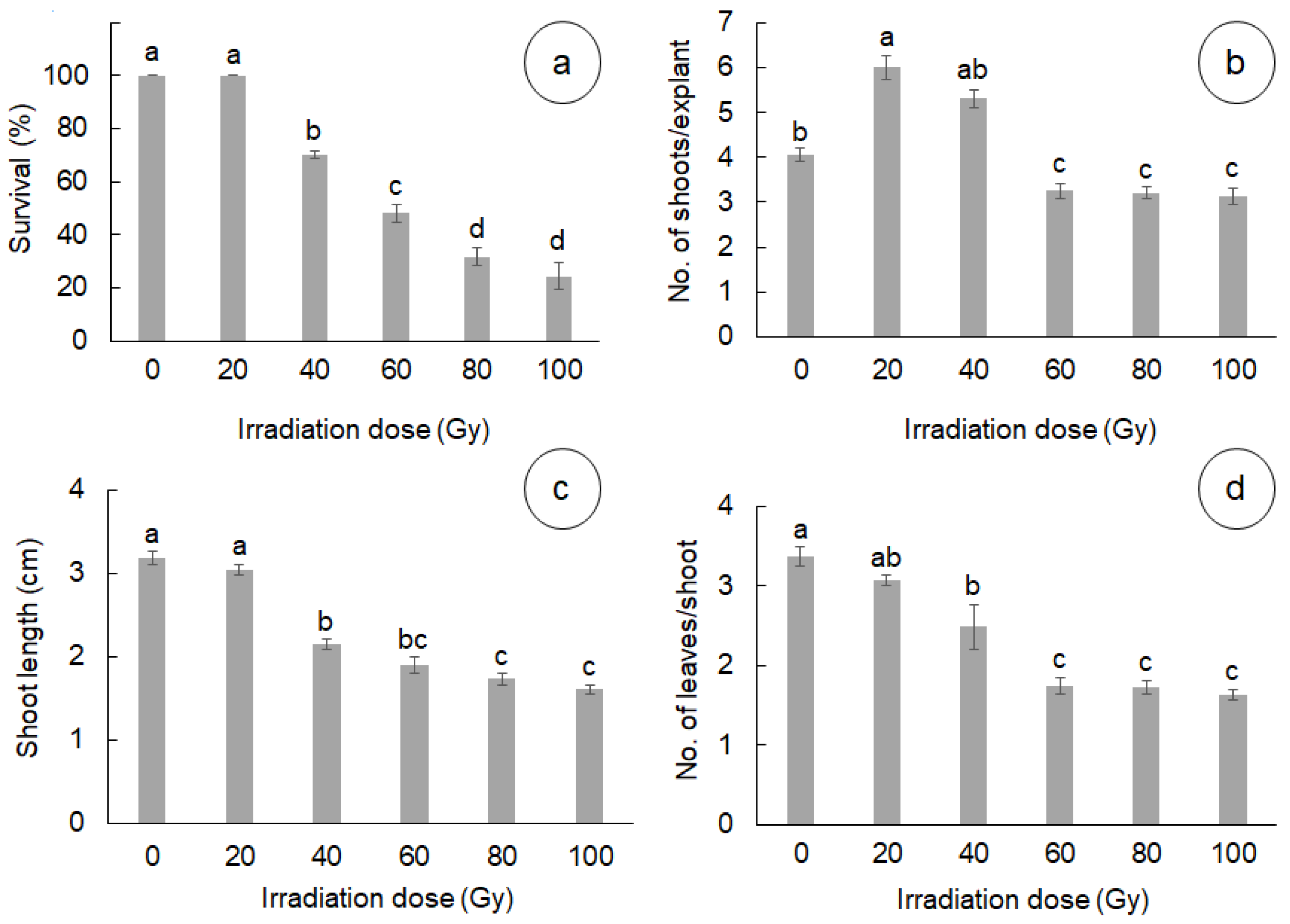
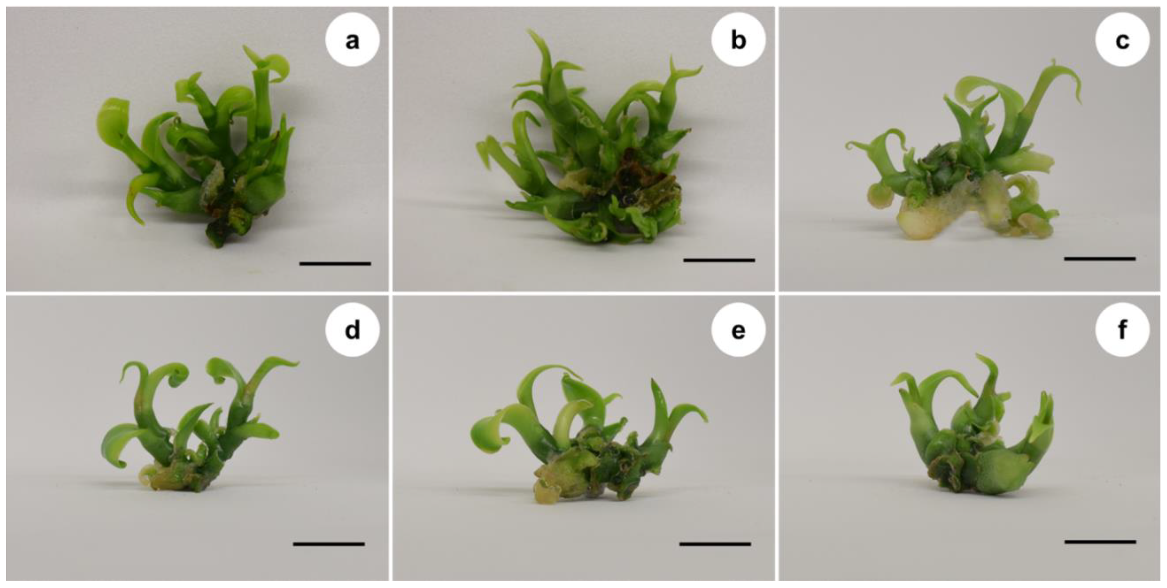
3.1. Effect of Gamma Radiation on In Vitro Survival and Development

The different doses of gamma irradiation showed a significant effect on survival percentage, number of shoots per explant, and length and number of leaves per shoot (Figure 1). The LD50 was observed at the 60 Gy, this dose reduced survival of explants by 52%. The highest survival percentage was observed at the 0 and 20 Gy doses, with 100% survival, whereas the lowest survival percentages were obtained at the 80 and 100 Gy doses, with 31 and 24% survival, respectively (Figure 1a). Regarding the number of shoots per explant, the highest number of new shoots was obtained at the 20 and 40 Gy doses, with 6 and 5.3 shoots per explant, respectively, whereas the lowest number of shoots was observed at the 60, 80 and 100 Gy doses, obtaining only 3 shoots per explant (Figure 1b). The longest shoot length was obtained at the 0 and 20 Gy doses, with 3.1 and 3 cm in length, respectively. The shortest length was observed at the 60, 80 and 100 Gy doses, with 1.6 to 1.8 cm in height (Figure 1c). The highest number of leaves was observed at the 0 and 20 Gy doses, with 3.3 and 3 leaves per shoot, whereas the lowest number of leaves per shoot was observed at 60, 80 and 100 Gy, with 1.6 to 1.7 leaves per shoot on average (Figure 1d). In addition, the administration of different doses of gamma irradiation had an effect on the multiplication rate in vanilla shoot development (Figure 2).

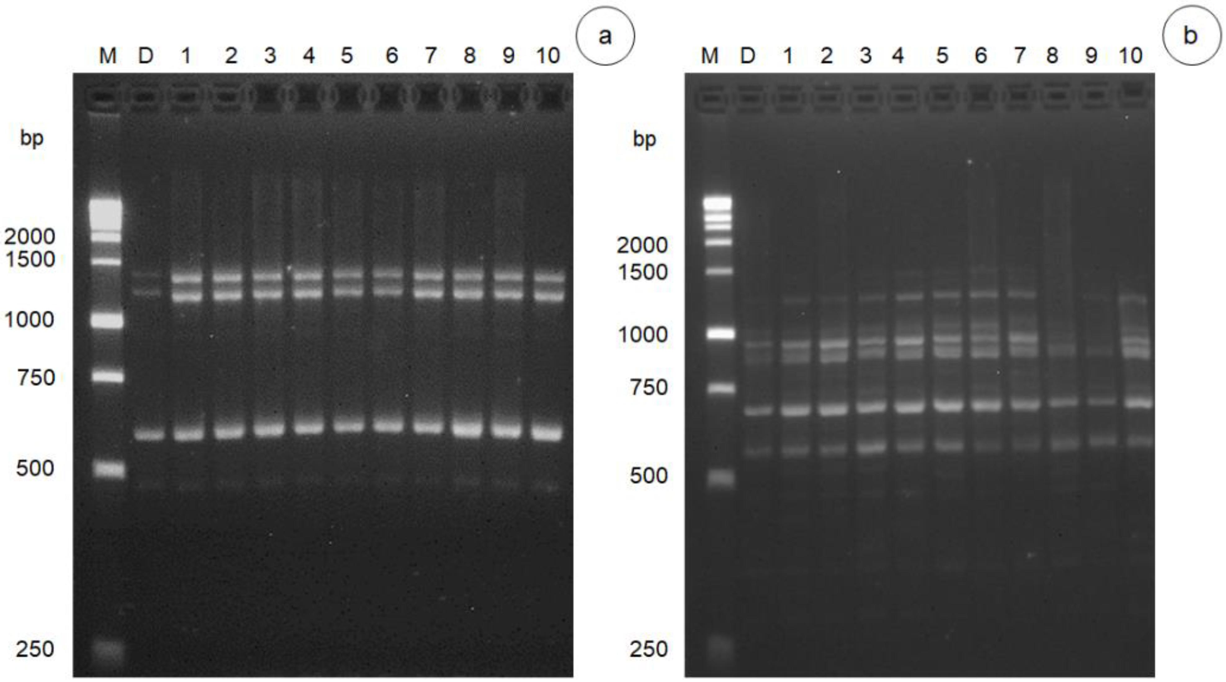
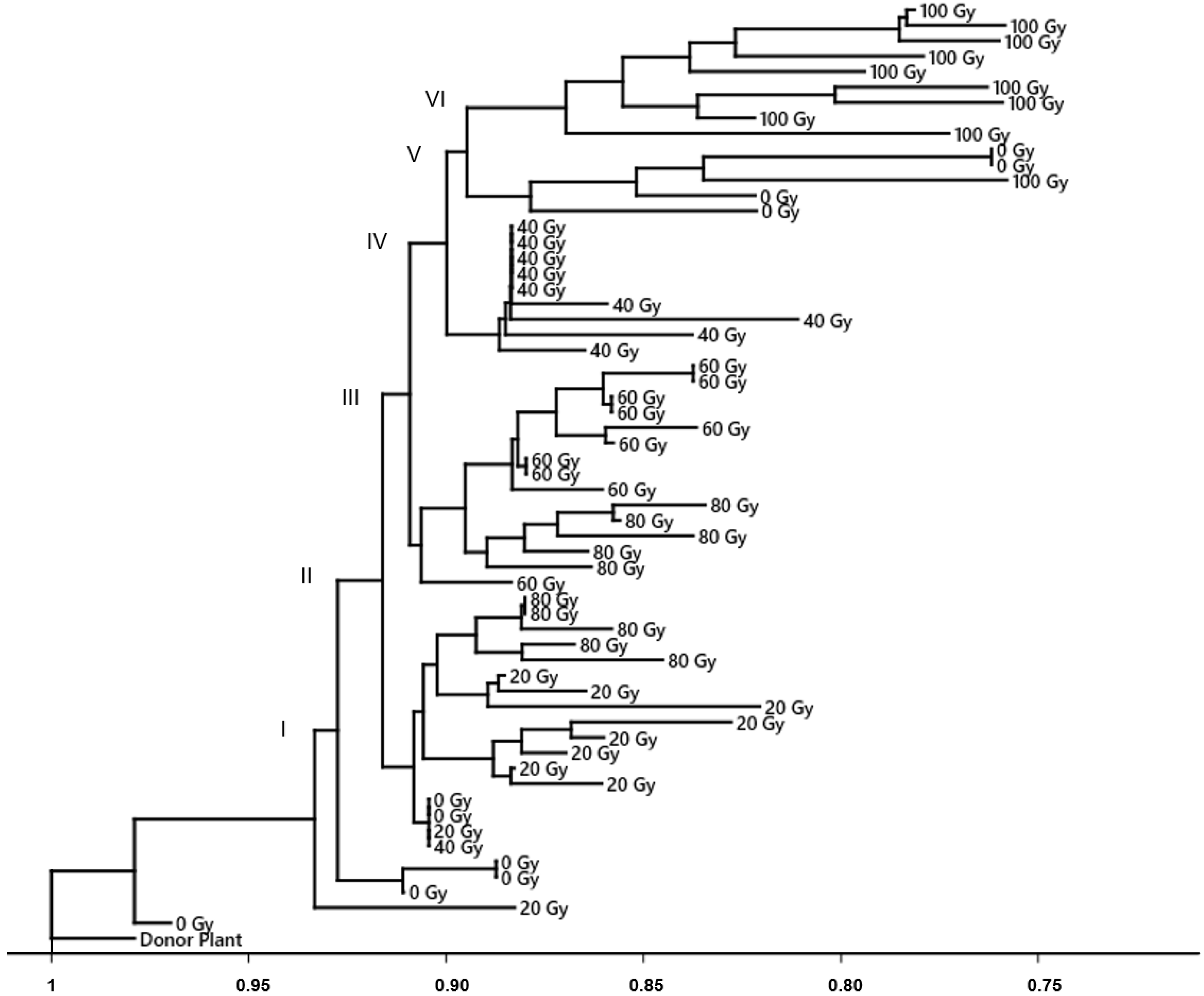
3.2. DNA Polymorphism of Gamma Radiation on Somaclonal Variation

The ISSR analysis revealed the presence of monomorphic and polymorphic bands between the control and the different irradiation doses with respect to the donor plant (Figure 3 and Supplementary Figure S1). The eight selected ISSR primers amplified a total of 43 bands ranging from 250 to 2000 bp. The primers that showed the highest percentage of polymorphism were UBC-808 with 42.6% (9 bands), followed by UBC-836 with 34.7% (6 bands) and UBC-840 with 28.7% (6 bands). On the other hand, the primers that showed less than 15% polymorphism were UBC-825 (6 bands), T06 (4 bands) and UBC-809 (7 bands). Primers T05 (1 band) and UBC-812 (4 bands) revealed the presence of monomorphic fragments (Table 1).
The dendrogram based on the neighbor joining model showed no similarity in the irradiation doses evaluated with respect to the donor plant. According to Bootstrap probabilities, similarity distances and branch lengths, the first group was considered to be the donor plant, whereas the second group comprised the doses of 0, 20, 40, 60, 80 and 100 Gy. The second group was divided into six subgroups, where subgroup six had the greatest similarity distance (0.76), formed by nine individuals of 100 Gy doses. The shortest distance was observed in the first and second subgroups with a distance of 0.88 and 0.86, respectively, with one individual of 0 and 20 Gy, respectively. For the rest of the subgroups, no clustering trend was observed with respect to the gamma radiation doses evaluated (Figure 4).

4. Discussion

This study showed that gamma irradiation has an effect on survival and development during in vitro multiplication of V. planifolia. The mortality of the explants at doses higher than 60 Gy (DL50) could be explained by the radiosensitivity of explants exposed to gamma rays. The sensitivity of explants to gamma radiation depends on tissue type, size, degree of development and water content [35]. Ionizing radiation interacts with atoms and molecules to produce free radicals in cells. These radicals can damage the structure of biomolecules such as carbohydrates, lipids, proteins, enzymes and nucleic acids, affecting the primary metabolism of plants [36,37]. In addition, irradiation can affect biochemical processes such as photosynthesis, respiration, krebs cycle and the metabolism of biomolecules. According to Hasbullah et al. [38] and Hernández-Muñoz et al. [35], irradiation can also affect cell division and cause damage to chromosomes and DNA.
The increase in the number of shoots at a dose of 20 Gy could be associated with a hormetic effect. According to Calabrese [39], the hormetic effect is characterized by beneficial or stimulation of development at low doses; and toxicity, inhibition or death at high doses. According to Jalal et al. [40], reactive oxygen species (ROS) are associated with hormesis because they are signaling molecules that trigger different physiological, biochemical and molecular processes in plant development. In this study, doses higher than 60 Gy caused the death of the explants and a reduction in the number of shoots per explant, shoot length and number of leaves per shoot. The reduced development and increased mortality rate at high doses could be associated with a longer time exposure to 60Co. The high dosage of gamma ray causes production and accumulation of ROS, which are toxic to plant tissues [41,42]”.
In this regard, Oliveira et al. [41] stated that the excess accumulation of free radicals resulting from water radiolysis produces negative effects on structural and functional biomolecules causing alterations in cellular metabolism, while Liu et al. [42] noted that high doses of gamma radiation can induce oxidative stress; this stress generates the formation of ROS that affects cell division and leads to apoptosis.
The mechanisms of the hormetic effect have yet to be fully elucidated; however, Iavicoli et al. [43] stated that hormesis is an adaptive preconditioning response to a stress of greater magnitude based on an evolutionary event. The hormetic effect has been observed in other in vitro mutagenesis studies with 60Co in golden-flowered vetchling (Lathyrus chrysanthus Boiss) [44], San Francisco lily (Laelia autumnalis) [16], shoreline purslane (Sesuvium portulacastrum) [45] and more recently in rice (Oryza sativa L.) [17]. These results suggest that gamma irradiation with 60Co could promote in vitro morphogenesis in explants in a dormant state in recalcitrant species.
The ISSR markers were able to detect somaclonal variations between individuals and the different doses of gamma irradiation evaluated. In this regard, Khan et al. [46] stated that ISSR markers produce multiple bands at the same locus, are highly reproducible and do not need prior information from the plant genome. In this study, primers UBC-808, UBC-836 and UBC-840 revealed the highest percentage of polymorphism and can be used for future analysis of somaclonal variation or genetic diversity in V. planifolia.
In general, individuals irradiated with doses of 100 Gy showed the least genetic similarity; however, for the rest of the doses evaluated, no clustering trend was observed. In vanilla, other studies involving somaclonal variation analysis using ISSR markers have observed that this species tends to be genetically unstable upon in vitro regeneration [29,31,47]. Ramírez-Mosqueda and Iglesias-Andreu [47] reported somaclonal variation during indirect organogenesis, with 71.66% polymorphism. Bello-Bello et al. [29] found an increase in the percentage of polymorphism with increasing concentrations of plant nanoparticles (AgNPs) in the culture medium during the growth of V. planifolia, with 25% polymorphism at a concentration of 200 mg·L−1 AgNPs. Pastelín-Solano et al. [31] demonstrated that the number of subcultures during direct organogenesis is an important factor in the increase in somaclonal variation, obtaining % P greater than 15% from subculture number six. On the other hand, other studies found no somaclonal variation in V. planifolia [30,48,49,50]. Sreedhar et al. [49] did not observe somaclonal variation during long-term growth using ISSR and RAPD markers. Gantait et al. [48] did not observe somaclonal variation during direct organogenesis using ISSR markers. Ramírez-Mosqueda et al. [30] in variegated plants obtained during direct organogenesis in temporary immersion obtained 0% polymorphism using ISSR markers. Recently, Manokari et al. [50] through direct organogenesis demonstrated 0% polymorphism using markers based on start codon targeted (SCoT) polymorphism.
Somaclonal variation during in vitro culture can originate through various aspects such as: explant type, regeneration pathway, subculture number, culture duration, growth regulator type, genotype and ploidy level [22,23,24,25,26,27,28,29,30,31,32,33,34,35,36,37,38,39,40,41,42,43,44,45,46,47,48,49,50,51]. However, somaclonal variation can be induced by chemical and physical mutagenic agents. Gamma irradiation using 60Co can generate different types of mutations, namely deletions and insertions, translocations and base substitutions [52,53]. According to Jain [54], mutations produced by somaclonal variability are very similar to those produced spontaneously or by mutagenesis methods.
The somaclonal variation obtained in non-irradiated explants could be explained by the genetic nature of V. planifolia. In this regard, Nair and Ravindra [55], and Bory et al. [56] observed in vanilla somatic associations and anomalies in the number of chromosomes, being lower than the reported 2n = 32. This could explain why, during in vitro regeneration of vanilla, higher somaclonal variation is expected compared to other species that do not show somatic association or anomalies in the ploidy level. The somaclonal variation found in irradiated treatments, in addition to the genetic nature of the species, could be due to the high penetrating potential of gamma rays and mainly to the breaking of the chemical bonds in the DNA double strand, eliminating nucleotides or replacing them with new ones [14]. In this study, DNA mutations can probably affect homeotic genes with effects on the ability to regenerate new shoots.
Predieri [57] and Bairu et al. [51] state that in vitro culture increases the efficiency of mutagenic treatments due to the manipulation of explants in constant cell division under controlled conditions without biotic or abiotic factors that interfere with the mutagenic treatment. The effect of in vitro mutagenesis using 60Co to broaden genetic variation for breeding purposes has been studied in San Francisco lily (Laelia autumnalis) [16], rice (Oryza sativa L.) [17], ginger (Zingiber officinale Rosc.) [8], potato (Solanum tuberosum L.) [18], and tomato (Lycopersicon esculentum L.) [19].
The species V. planifolia has low genetic diversity due to the cuttings-based asexual reproduction [58]. This commercial propagation method limits the diversity of the species. In vanilla, somaclonal variation is an alternative to broaden the genetic base of this species and generate new alleles [31] that can address the inbreeding depression of this species. The increase in the genetic diversity of vanilla is an important factor contributing to tolerance to abiotic and resistance to biotic factors caused by different climate change scenarios to avoid its extinction. Gamma radiation is a very useful mutagenesis method to generate genetic variations for the improvement of this species. In addition, future studies are required to analyze flowering stage and ripe fruits with morphological and biochemical markers to find possible phenotype variation.

5. Conclusions

In this study, it was observed that gamma radiation has a hormetic effect on explants, promoting the formation of new shoots at low dose (20 Gy) and inhibition of sprouting and death at high doses (60–100 Gy). Furthermore, in vitro regeneration via direct organogenesis and the different doses of gamma irradiation evaluated with 60Co were shown to have an effect on somaclonal variation. The analysis of NJ clustering and Jaccard’s genetic distance showed that the treatment without irradiation and the treatments with irradiation present genetic divergence from the donor plant. ISSR markers were shown to be efficient in detecting somaclonal variation. These results support the possibility of using gamma rays during in vitro culture to increase genetic diversity and undertake a vanilla breeding program.

Supplementary Materials

The following supporting information can be downloaded at: https://www.mdpi.com/article/10.3390/horticulturae8060503/s1, Figure S1: Uncropped blot of Figure 3a, Figure S2: Uncropped blot of Figure 3b.

Author Contributions

Conceptualization, M.K.S.-F. and J.J.B.-B.; designed the experiments, analyzed the data, conducted data interpretation and drafted the manuscript, M.K.S.-F.; conducted all the experimental work, F.C.G.-M., S.C.-I., J.L.S.-C. and J.J.B.-B.; contributed to the conceptualization of the experiment and revising the manuscript. All authors have read and agreed to the published version of the manuscript.

Funding

This research received no external funding.

Institutional Review Board Statement

Not applicable.

Informed Consent Statement

Not applicable.

Data Availability Statement

All data in this study can be found in the manuscript or in the Supplementary Materials.

Conflicts of Interest

The authors declare no conflict of interests.

References

  1. Gantait, S.; Kundu, S. In vitro biotechnological approaches on Vanilla planifolia Andrews: Advancements and opportunities. Acta Physiol. Plant. 2017, 39, 196. [Google Scholar] [CrossRef]
  2. Arya, S.S.; Rookes, J.E.; Cahill, D.M.; Lenka, S.K. Vanillin: A review on the therapeutic prospects of a popular flavouring molecule. Adv. Tradit. Med. 2021, 21, 1–17. [Google Scholar] [CrossRef]
  3. SEMARNAT (Secretaría de Medio Ambiente y Recursos Naturales). Norma Oficial Mexicana (NOM-059-ECOL-2001) de Protección Especial de Especies Nativas de México de Flora y Fauna Silvestres; Diario Oficial de la Federación: Mexico City, Mexico, 2010; pp. 2–56.
  4. Wan Abdullah, W.M.A.N.; Low, L.; Mumaiyizah, S.B.; Chai, Q.Y.; Loh, J.Y.; Ong-Abdullah, J.; Lai, K.S. Effect of lignosulphonates on Vanilla planifolia shoot multiplication, regeneration and metabolism. Acta Physiol. Plant. 2020, 42, 107. [Google Scholar] [CrossRef]
  5. Li, J.; Demesyeux, L.; Brym, M.; Chambers, A.H. Development of species-specific molecular markers in Vanilla for seedling selection of hybrids. Mol. Biol. Rep. 2020, 47, 1905–1920. [Google Scholar] [CrossRef]
  6. Hu, Y.; Resende, M.F.R.; Bombarely, A.; Brym, M.; Bassil, E.; Chambers, A.H. Genomics-based diversity analysis of Vanilla species using a Vanilla planifolia draft genome and Genotyping-By-Sequencing. Sci. Rep. 2019, 9, 3416. [Google Scholar] [CrossRef]
  7. Li, F.; Shimizu, A.; Nishio, T.; Tsutsumi, N.; Kato, H. Comparison and Characterization of Mutations Induced by Gamma-Ray and Carbon-Ion Irradiation in Rice (Oryza sativa L.) Using Whole-Genome Resequencing. G3 Genes Genom. Genet. 2019, 9, 3743–3751. [Google Scholar] [CrossRef] [Green Version]
  8. Sharma, V.; Thakur, M.; Tomar, M. In vitro selection of gamma irradiated shoots of ginger (Zingiber officinale Rosc.) against Fusarium oxysporum f.sp. zingiberi and molecular analysis of the resistant plants. Plant Cell Tissue Organ Cult. 2020, 143, 319–330. [Google Scholar] [CrossRef]
  9. IAEA. Organismo Internacional de Energía Atómica. Inducción de Mutaciones. Available online: https://www.iaea.org/es/temas/induccion-de-mutaciones (accessed on 14 June 2021).
  10. Larkin, P.J.; Scowcroft, W.R. Somaclonal variation—A novel source of variability from cell cultures for plant improvement. Theor. Appl. Genet. 1981, 60, 197–214. [Google Scholar] [CrossRef]
  11. Waugh, R.; Leader, D.J.; Mc Callum, N.; Cadwell, D. Harvesting the potential of induced biological diversity. Trends Plant Sci. 2006, 11, 71–79. [Google Scholar] [CrossRef]
  12. Ghani, M.; Sharma, S.K. Induction of powdery mildew resistance in gerbera (Gerbera jamesonii) through gamma irradiation. Physiol. Mol. Biol. Plants 2019, 25, 159–166. [Google Scholar] [CrossRef]
  13. Salava, H.; Thula, S.; Mohan, V.; Kumar, R.; Maghuly, F. Application of Genome Editing in Tomato Breeding: Mechanisms, Advances, and Prospects. Int. J. Mol. Sci. 2021, 22, 682. [Google Scholar] [CrossRef] [PubMed]
  14. Oladosu, Y.; Rafii, M.Y.; Abdullah, N.; Hussin, G.; Ramli, A.; Rahim, H.A.; Miah, G.; Usman, M. Principle and application of plant mutagenesis in crop improvement: A review. Biotechnol. Biotechnol. Equip. 2016, 30, 1–16. [Google Scholar] [CrossRef] [Green Version]
  15. Spinoso-Castillo, J.L.; Escamilla-Prado, E.; Aguilar-Rincón, C.H.; Morales-Ramos, V.; García-de los Santos, G.; Corona-Torres, T. Physiological response of seeds of three coffee varieties to gamma rays (60Co). Rev. Chapingo Ser. Hortic. 2021, 27, 101–112. [Google Scholar] [CrossRef]
  16. Hernández-Muñoz, S.; Pedraza-Santos, M.E.; López, P.A.; De La Cruz-Torres, E.; Martínez-Palacios, A.; Fernández-Pavía, S.P.; Chávez-Bárcenas, A.T. Estimulación de la germinación y desarrollo in vitro de Laelia autumnalis con rayos gamma. Rev. Fitotec. Mex. 2017, 40, 271–283. [Google Scholar] [CrossRef]
  17. Abdelnour-Esquivel, A.; Pérez, J.; Rojas, M.; Vargas, W.; Gatica-Arias, A. Use of gamma radiation to induce mutations in rice (Oryza sativa L.) and the selection of lines with tolerance to salinity and drought. Vitr. Cell. Dev. Biol. Plant 2020, 56, 88–97. [Google Scholar] [CrossRef]
  18. Mohamed, E.A.; Osama, E.; Manal, E.; Samah, A.; Salah, G.; Hazem, K.M.; Nabil, E. Impact of gamma irradiation pretreatment on biochemical and molecular responses of potato growing under salt stress. Chem. Biol. Technol. Agric. 2021, 8, 35. [Google Scholar] [CrossRef]
  19. Özge, Ç.; Alp, A.; Sinan, M.; Çimen, A. Comparison of tolerance related proteomic profiles of two drought tolerant tomato mutants improved by gamma radiation. J. Biotechnol. 2021, 330, 35–44. [Google Scholar] [CrossRef]
  20. Pardo, A.; Hernández, A.; Méndez, N.; Alvarado, G. Análisis genético, mediante marcadores RAPD, de microbulbos de ajo conservados e irradiados in vitro. Bioagro 2015, 27, 143–150. [Google Scholar]
  21. Muñoz-Miranda, L.A.; Rodríguez-Sahagún, A.; Acevedo, H.G.J.; Cruz-Martínez, V.O.; Torres-Morán, M.I.; Lépiz-Ildefonso, R.; Aarland, R.C.; Castellanos-Hernández, O.A. Evaluation of somaclonal and ethyl methane sulfonate-induced genetic variation of Mexican oregano (Lippia graveolens HBK). Agronomy 2019, 9, 166. [Google Scholar] [CrossRef] [Green Version]
  22. Ranghoo-Sanmukhiya, V.M. Somaclonal Variation and Methods Used for its Detection. In Propagation and Genetic Manipulation of Plants; Siddique, I., Ed.; Springer Nature: Singapore, 2021. [Google Scholar] [CrossRef]
  23. Zietkiewicz, E.; Rafalski, A.; Labuda, D. Genome Fingerprinting by Simple Sequence Repeat (SSR)-Anchores Polimerase Chain Reaction Amplification. Genomics 1994, 20, 176–183. [Google Scholar] [CrossRef]
  24. Ghorbanpour, M.; Khadivi-Khub, A. Somaclonal variation in callus samples of Plantago major using inter-simple sequence repeat marker. Caryologia 2015, 68, 19–24. [Google Scholar] [CrossRef]
  25. Martínez-Estrada, E.; Caamal-Velázquez, J.H.; Salinas-Ruiz, J.; Bello-Bello, J.J. Assessment of somaclonal variation during sugarcane micropropagation in temporary immersion bioreactors by intersimple sequence repeat (ISSR) markers. Vitr. Cell. Dev. Biol. Plant 2017, 53, 553–560. [Google Scholar] [CrossRef]
  26. Kritskaya, T.A.; Kashin, A.S.; Kasatkin, M.Y. Micropropagation and Somaclonal Variation of Tulipa suaveolens (Liliaceae) in vitro. Russ. J. Mar. Biol. 2019, 50, 209–215. [Google Scholar] [CrossRef]
  27. Vitamvas, J.; Viehmannova, I.; Cepkova, P.H.; Mrhalova, H.; Eliasova, K. Assessment of somaclonal variation in indirect morphogenesis-derived plants of Arracacia xanthorrhiza. Pesqui. Agropecu. Bras. 2019, 54, e00301. [Google Scholar] [CrossRef] [Green Version]
  28. Ulvrova, T.; Vitamvas, J.; Cepkova, P.H.; Eliasova, K.; Janovska, D.; Bazant, V.; Viehmannova, I. Micropropagation of an ornamental shrub Disanthus cercidifolius Maxim. and assessment of genetic fidelity of regenerants using ISSR and flow cytometry. Plant Cell Tissue Organ Cult. 2021, 144, 555–566. [Google Scholar] [CrossRef]
  29. Bello-Bello, J.J.; Spinoso-Castillo, J.L.; Arano-Avalos, S.; Martínez-Estrada, E.; Arellano-García, M.E.; Pestryakov, A.; Toledano-Magaña, Y.; García-Ramos, J.C.; Bogdanchikova, N. Cytotoxic, genotoxic, and polymorphism effects on Vanilla planifolia Jacks ex Andrews after long-term exposure to Argovit® silver nanoparticles. Nanomaterials 2018, 8, 754. [Google Scholar] [CrossRef] [Green Version]
  30. Ramírez-Mosqueda, M.A.; Iglesias-Andreu, L.G.; Favián-Vega, E.; Telxeira da Silva, J.A.; Leyva-Ovalle, O.R.; Murguía-González, J. Morphogenetic stability of variegated Vanilla planifolia Jacks. plants micropropagated in a temporary immersion system (TIB®). Rend. Lincei Sci. Fis. Nat. 2019, 30, 603–609. [Google Scholar] [CrossRef]
  31. Pastelín-Solano, M.C.; Salinas, R.J.; González, A.M.T.; Castañeda, C.O.; Galindo, T.M.E.; Bello-Bello, J.J. Evaluation of in vitro shoot multiplication and ISSR marker based assessment of somaclonal variants at different subcultures of vanilla (Vanilla planifolia Jacks). Physiol. Mol. Biol. Plants 2019, 25, 561–567. [Google Scholar] [CrossRef]
  32. Murashige, T.; Skoog, F. A Revised medium for rapid growth and bio assays with tobacco tissue cultures. Physiol. Plant. 1962, 15, 473–497. [Google Scholar] [CrossRef]
  33. Saitou, N.; Nei, M. The neighbor-joining method: A new method for reconstructing phylogenetic trees. Mol. Biol. Evol. 1987, 4, 406–425. [Google Scholar] [CrossRef]
  34. Stewart, C.N.; Via, L.E. A rapid CTAB DNA isolation technique useful for RAPD fingerprinting and other PCR applications. Biotechniques 1993, 14, 748–750. [Google Scholar] [PubMed]
  35. Hernández-Muñoz, S.; Pedraza-Santos, M.E.; López, P.A.; Gómez-Sanabria, J.M.; Morales-García, J.L. Mutagenesis in the improvement of ornamental plants. Rev. Chapingo Ser. Hortic. 2019, 25, 151–167. [Google Scholar] [CrossRef]
  36. Chahal, G.S.; Gosal, S.S. Principles and Procedures of Plant Breeding; Alpha Science International Ltd.: Oxford, UK, 2002; pp. 399–412. [Google Scholar]
  37. Magdy, A.M.; Fahmy, E.M.; Abd EL-Rahman, M.A.A.; Awad, G. Improvement of 6-gingerol production in ginger rhizomes (Zingiber officinale Roscoe) plants by mutation breeding using gamma irradiation. Appl. Radiat. Isot. 2020, 162, 109193. [Google Scholar] [CrossRef] [PubMed]
  38. Hasbullah, N.A.; Taha, R.M.; Saleh, A.; Mahmad, N. Irradiation effect on in vitro organogenesis, callus growth and plantlet development of Gerbera jamesonii. Hortic. Bras. 2012, 30, 252–257. [Google Scholar] [CrossRef] [Green Version]
  39. Calabrese, E.J. Hormesis: Path and progression to significance. Int. J. Mol. Sci. 2018, 19, 2871. [Google Scholar] [CrossRef] [Green Version]
  40. Jalal, A.; Oliveira, J.J.C.; Ribeiro, J.S.; Fernandes, G.C.; Mariano, G.G.; Trindade, V.D.R.; Reis, A.R. Hormesis in plants: Physiological and biochemical responses. Ecotoxicol. Environ. Saf. 2021, 207, 111225. [Google Scholar] [CrossRef]
  41. Oliveira, N.M.; de Medeiros, A.D.; de Lima Nogueira, M.; Arthur, V.; de Araújo Mastrangelo, T.; da Silva, C.B. Hormetic effects of low-dose gamma rays in soybean seeds and seedlings: A detection technique using optical sensors. Comput. Electron. Agric. 2021, 187, 106251. [Google Scholar] [CrossRef]
  42. Liu, H.; Li, H.; Yang, G.; Yuan, G.; Ma, Y.; Zhang, T. Mechanism of early germination inhibition of fresh walnuts (Juglans regia) with gamma radiation uncovered by transcriptomic profiling of embryos during storage. Postharvest Biol. Technol. 2021, 172, 111380. [Google Scholar] [CrossRef]
  43. Iavicoli, I.; Leso, V.; Fontana, L.; Calabrese, E.J. Nanoparticle exposure and hormetic dose–responses: An update. Int. J. Mol. Sci. 2018, 19, 805. [Google Scholar] [CrossRef] [Green Version]
  44. Beyaz, R.; Kahramanogullari, C.T.; Yildiz, C.; Darcin, E.S.; Yildiz, M. The effect of gamma radiation on seed germination and seedling growth of Lathyrus chrysanthus Boiss. under in vitro conditions. J. Environ. Radioact. 2016, 162, 129–133. [Google Scholar] [CrossRef]
  45. Kapare, V.; Satdive, R.; Fulzele, D.P.; Malpathak, N. Impact of Gamma Irradiation Induced Variation in Cell Growth and Phytoecdysteroid Production in Sesuvium portulacastrum. J. Plant Growth Regul. 2017, 36, 919–930. [Google Scholar] [CrossRef]
  46. Khan, M.M.H.; Rafii, M.Y.; Ramlee, S.I.; Jusoh, M.; Al Mamun, M.; Halidu, J. DNA fingerprinting, fixation-index (Fst), and admixture mapping of selected Bambara groundnut (Vigna subterranea [L.] Verdc.) accessions using ISSR markers system. Sci. Rep. 2021, 11, 14527. [Google Scholar] [CrossRef] [PubMed]
  47. Ramírez-Mosqueda, M.A.; Iglesias-Andreu, L.G. Indirect organogenesis and assessment of somaclonal variation in plantlets of Vanilla planifolia Jacks. Plant Cell Tissue Organ Cult. 2015, 123, 657–664. [Google Scholar] [CrossRef]
  48. Sreedhar, R.V.; Venkatachalam, L.; Bhagyalakshmi, N. Genetic fidelity of long-term micropropagated shoot cultures of vanilla (Vanilla planifolia Andrews) as assessed by molecular markers. Biotechnol. J. 2007, 2, 1007–1013. [Google Scholar] [CrossRef]
  49. Gantait, S.; Mandal, N.; Bhattacharyya, S.; Das, P.K.; Nandy, S. Mass multiplication of Vanilla planifolia with pure genetic identity confirmed by ISSR. Int J. Plant Dev. Biol 2009, 3, 18–23. [Google Scholar]
  50. Manokari, M.; Priyadharshini, S.; Jogam, P.; Dey, A.; Shekhawat, M.S. Meta-topolin and liquid medium mediated enhanced micropropagation via ex vitro rooting in Vanilla planifolia Jacks. ex Andrews. Plant Cell Tissue Organ Cult. 2021, 146, 69–82. [Google Scholar] [CrossRef]
  51. Bairu, M.W.; Aremu, A.O.; Van Staden, J. Variación somaclonal en plantas: Causas y métodos de detección. Plant Growth Regul. 2011, 63, 147–173. [Google Scholar] [CrossRef]
  52. Yang, G.; Luo, W.; Zhang, J.; Yan, X.; Du, Y.; Zhou, L.; Li, W.; Wand, H.; Chen, Z.; Guo, T. Genome-wide comparisons of mutations induced by carbon-ion beam and gamma-rays irradiation in rice via resequencing multiple mutants. Front. Plant Sci. 2019, 10, 1514. [Google Scholar] [CrossRef] [Green Version]
  53. Holme, I.B.; Gregersen, P.L.; Brinch-Pedersen, H. Induced genetic variation in crop plants by random or targeted mutagenesis: Convergence and differences. Front. Plant Sci. 2019, 10, 1468. [Google Scholar] [CrossRef]
  54. Jain, S.M. Tissue culture-derived variation in crop improvement. Euphytica 2001, 118, 153–166. [Google Scholar] [CrossRef]
  55. Nair, R.R.; Ravindran, P.N. Somatic association of chromosomes and other mitotic abnormalities in Vanilla planifolia (Andrews). Caryologia 1994, 47, 65–73. [Google Scholar] [CrossRef]
  56. Bory, S.; Catrice, O.; Brown, S.; Leitch, I.J.; Gigant, R.; Chiroleu, F.; Grisoni, M.; Duval, M.F.; Besse, P. Natural polyploidy in Vanilla planifolia (Orchidaceae). Genome 2008, 51, 816–826. [Google Scholar] [CrossRef] [PubMed]
  57. Predieri, S. Mutation induction and tissue culture in improving fruits. Plant Cell Tissue Organ Cult. 2001, 64, 185–210. [Google Scholar] [CrossRef]
  58. Villanueva-Viramontes, S.; Hernández-Apolinar, M.; Fernández-Concha, G.C.; Dorantes-Euán, A.; Dzib, G.R.; Martínez-Castillo, J. Wild Vanilla planifolia and its relatives in the Mexican Yucatan Peninsula: Systematic analyses with ISSR and ITS. Bot. Sci. 2017, 95, 169–187. [Google Scholar] [CrossRef] [Green Version]
Figure 1. Effect of gamma radiation on in vitro survival and development of vanilla (Vanilla planifolia Jacks. ex Andrews). (a) Survival percentage, (b) shoots per explant, (c) shoot length and (d) leaves per shoot at 60 d of culture. Values represent the mean ± standard error. Means with different letters are significantly different (Tukey, p < 0.05).
Horticulturae 08 00503 g001
Figure 2. Effect of different doses of gamma irradiation on in vitro shoot development of vanilla (Vanilla planifolia Jacks. ex Andrews) at 60 days of culture; (af) 0, 20, 40, 60, 80 and 100 Gy, respectively. Black bar = 1 cm.
Horticulturae 08 00503 g002
Figure 3. Electrophoresis pattern with ISSR markers of ten individuals (1–10) of vanilla (Vanilla planifolia Jacks. ex Andrews) exposed to gamma radiation with respect to donor plant (D). (a) Dose 60 Gy, primer UBC-812 (b) dose 20 Gy, primer UBC-840. M = molecular weight marker and bp = base pairs.
Horticulturae 08 00503 g003
Figure 4. Neighbor joining dendrogram based on Jaccard’s coefficient, calculated for each gamma irradiation dose with respect to the donor plant of vanilla (Vanilla planifolia Jacks. ex Andrews). The values under line represent: 1 similarity, and 0 divergence. I-VI: subgroups with different similarity distances.
Horticulturae 08 00503 g004
Table 1. Inter simple sequence repeat (ISSR) molecular markers selected to evaluate the somaclonal variation of vanilla (Vanilla planifolia Jacks. ex Andrews) at different doses of gamma irradiation with 60Co.
PrimersSequence (5′–3′)Annealing Temperature
(°C)
BandsRange
(bp)
Polymorphism (%)
UBC-809AGAGAGAGAGAGAGAGG457250–15004
T06AGAGAGAGAGAGAGAGT504400–7503
UBC-840GAGAGAGAGAGAGAGAYT526500–150028.7
UBC-836AGAGAGAGAGAGAGAGYA506400–150034.7
UBC-812GAGAGAGAGAGAGAGAA504500–15000
UBC-825ACACACACACACACACT506500–200014.9
UBC-808AGAGAGAGAGAGAGAGC539400–100042.6
T05CGTTGTGTGTGTGTGTGT531750 0
bp = base pair; Y = C or T.
Publisher’s Note: MDPI stays neutral with regard to jurisdictional claims in published maps and institutional affiliations.

