Article

Development of a Highly Sensitive Loop-Mediated Isothermal Amplification Incorporated with Flocculation of Carbon Particles for Rapid On-Site Diagnosis of Blood Disease Bacterium Banana

by
Mohammad Malek Faizal Azizi
1,
Han Yih Lau
1,*,
Norliza Abu Bakar
1,*,
Sohana Romeli
1,
Muhammad Fairuz Mohd Yusof
1,
Rafidah Badrun
1 and
Nur Sulastri Jaffar
2
1
Biotechnology and Nanotechnology Research Centre, Malaysian Agricultural Research and Development Institute (MARDI), Persiaran MARDI-UPM, Serdang 43400, Selangor, Malaysia
2
Horticulture Research Centre, Malaysian Agricultural Research and Development Institute (MARDI), Persiaran MARDI-UPM, Serdang 43400, Selangor, Malaysia
*
Authors to whom correspondence should be addressed.
Horticulturae 2022, 8(5), 406; https://doi.org/10.3390/horticulturae8050406
Submission received: 5 April 2022 / Revised: 30 April 2022 / Accepted: 2 May 2022 / Published: 5 May 2022

Abstract

:
Bananas are one of the most crucial fruit crops worldwide and significantly contribute to food security in developing countries. However, blood disease of bananas caused by Ralstonia syzygii subspecies celebensensis has become a threat to banana production. Rapid and accurate diagnosis of BDB for on-site detection is pivotal at an early stage for an effective disease control strategy. This study developed LAMP with specific primers targeting BDB, followed by a flocculation assay for visualising positive amplification in the LAMP assay. The assay was sensitive to picogram amounts of gDNA (0.5 pg). LAMP assay on BDB gDNA showed flocculation, but negative results on Fusarium oxysporus cubense and Ralstonia solanacearum confirming the specificity of the assays. Field testing conducted at MARDI headquarters and Taman Pertanian Universiti discovered that the LAMP-flocculation assays were successful in detecting BDB on symptomatic samples as well as on samples from a healthy plot with no symptom observed at the sampling stage, revealing that this assay can detect BDB at an early infection stage. The validation results showed that the LAMP-flocculation assay was comparable with the PCR technique. This newly developed technique is highly specific and sensitive for the early detection of BDB for the adoption of precautionary control measures.

1. Introduction

Bananas (Musa spp.) are a key fruit crop, providing a major staple food source for millions of people in developing countries. Asia is the largest banana producer worldwide with an annual production of 64,730.74 million metric tonnes, followed by Africa, South America, Central America, the Caribbean, Oceania, Europe, and North America in 2020 [1]. Banana production has become a source of income for smallholder farmers and a prominent global commodity [2]. Although bananas are one of the essential fruits in Africa, Asia, and Latin America, only 13% of the total bananas produced are exported to international markets, implying the importance of the fruit for local markets and food security [3].
However, banana bacterial wilt diseases have been considered as the major obstacles to banana production, causing lowering yields and productivity. In the early 1900s, the blood disease of bananas (BDB) caused by Ralstonia syzygii subspecies celebesensis was first confined on Salayar Island near Sulawesi, Indonesia [4]. Afterwards, the disease was widely spread on local cooking banana cultivars. It started in Southern Sulawesi, which was formerly known as Celebes by 1920 [5,6] and spread further throughout the island until its emergence in Java in the late 1980s [7]. The BDB was subsequently first discovered in the province of Perak and more recently in the region of Selangor, Malaysia [3,8]. Ralstonia syzygii subsp. celebesensis is typically soil-borne and can be non-specifically transmitted by pollinating insects, which mechanically transfer the bacteria from an infected to a healthy banana inflorescence [9]. Local transmission of BDB was hypothesised as occurring via contaminated tools and equipment, infected plant material (suckers and fruit), infested soil, water, bats, and birds. Long-distance transmission of the disease is associated with human activities through the movement of infected plant materials [3,9]. The BDB pathogen, R. syzygii, is closely related to Ralstonia solanacearum and belongs to phylotype IV within the R. solanacearum group, most closely resembling the R. solanacearum strains from Indonesia [10,11].
BDB exhibits similar symptoms to Moko banana disease caused by R. solanacearum, which originated in Central America (phylotypes IIA-6, IIB-3, and IIB-4, historically recognised as Race 2). However, unlike R. solanacearum causing Moko/Bugtok disease, BDB is not pathogenic to Heliconia spp. or to solanaceous hosts [11]. As the disease progresses, the symptoms include wilting, yellowing, and necrosis of mature leaves, red or brown vascular staining through the centre of the pseudostem and peduncle, bacterial ooze, and characteristic reddish-brown fruit rot [8,11].
Scientists have widely proposed the methods of DNA-based molecular diagnostics due to the reliability, sensitivity, and specificity in detecting plant pathogens. Currently, polymerase chain reaction (PCR) is the most popular and reliable molecular technique for the detection of BDB [12]. However, PCR-based procedures are time-consuming and laborious to perform outside the laboratory due to the limits imposed by the heavy equipment that ensures the cyclic reactions occur at instinct temperatures. Therefore, it is not a suitable technique for on-site detection. Loop-mediated isothermal amplification (LAMP) is an alternative to PCR that uses DNA repair enzymes instead of high-temperature heating in DNA denaturation [13]. This allows rapid DNA amplification at a low temperature (60–65 °C) and PCR-like sensitivity, making it ideal for point-of-care (POC) applications. The portable heating block used to perform the LAMP assay maintains the required temperature and is ideal for on-site testing [14]. However, equipment-free detection after amplification is required to detect a signal with the naked eye without specialised devices or tools. In the laboratory, the most regularly used technique for analysing amplification products is agarose gel electrophoresis with dyes such as ethidium bromide followed by visualisation under ultraviolet light [14]. However, this technique is not suitable for on-site detection.
Bridging flocculation is a well-established phenomenon in colloid chemistry and is employed in a plethora of colloidal separation processes (e.g., to clarify contaminated water). In the 1950s, Ruehrwein and Ward [15] first described the phenomenon, which was explained in the 1960s by Healy and La Mer [16] as the result of the surface adsorption of polymers that are long enough to cross-link numerous particles and hence (reversibly) flocculate from solution. To rely on this phenomenon, our study developed a point-of-care diagnostic diagnostic approach using LAMP and carbon particle bridging flocculation for binary (Yes/No) detection of BDB. LAMP-positive DNA samples with lengthy DNA are expected to create flocculation, which can be observed using the naked eye assay (Figure 1). The combination of these assays can make a significant contribution to the banana industry as an enabler tool for rapid on-site diagnosis of important BDB.

2. Materials and Methods

2.1. Pathogen Collection and Culture Conditions

Fusarium oxysporum f.sp. cubense (FocTR4) and Ralstonia syzygii subsp. celebesensis (Rsc) (GenBank No. CP019911.1) were obtained from the Research Centre of Horticulture, Malaysian Agriculture Research and Development Institute (MARDI). The Ralstonia solanacearum (Rs) strain PS107 was obtained from the Research Centre of Biodiversity and Environmental, MARDI. Both bacterial isolates were subcultured onto Kelman’s tetrazolium salt (Kelman’s TZC) agar medium (Merck KGaA, Darmstadt, Germany) [17] and incubated for three days at 28 °C. Meanwhile, the Foc isolate was subcultured into potato dextrose agar (PDA) (Thermo Scientific™ Oxoid™, Basingstoke, England) and incubated for one week at room temperature.
One or two colonies of Rsc/Rs were inoculated in 50 mL Luria Broth (LB) media (Thermo Scientific™ Oxoid™, Basingstoke, England), and the Cultures were grown in an incubator shaker at 28 °C with 200 rpm shaking overnight. The overnight cultures were centrifuged at 6000× g for 5 min and the supernatants were discarded. The pellets were stored for DNA extraction. A small block of agar containing Foc hyphae was placed on a PDA media plate and incubated for seven days at room temperature until the plate was fully covered with the hyphae. The hyphae of Foc on the media surface were then scrapped out and kept in a 2-mL microcentrifuge tube for DNA extraction. DNA extracted from Foc and Rs was used in the LAMP assay sensitivity study. However, Ralstonia syzygii subsp. celebesensis (Rsc) (GenBank No. CP019911.1) was used as a positive control in all the assays in this study.

2.2. DNA Extraction

Both bacterial isolates were cultured on Kelman’s broth for 16 to 18 h at 37 °C. The magnetic bead DNA extraction method was used to extract the total genomic DNA from all strains of bacteria. Banana leaf sample (~300 mg) was used for plant genomic DNA extraction by adding 200 ul of lysis buffer (50 mM Tris-HCl pH 8.0, 1.5 M guanidium-HCl, 2% w/v PVP40 and 1% v/v Triton-X). Plant tissue was ground in a 1.5 mL-tube with a plastic pestle in the presence of lysis buffer. Nucleic acids were then extracted using a modified Solid Phase Reversible Immobilisation (SPRI) protocol [18,19] provided by the manufacturer (Thermo Scientific™, Basingstoke, England). Approximately 10 μL of the cleared lysate was added with 1.8 volumes of SPRI beads into a binding buffer (10 mM Tris-HCl pH 8.0, 20% PEG8000, 2.5 M NaCl) and incubated for five minutes. A magnetic stand was used to separate the DNA bound beads from the lysate and washed twice with 100% isopropanol and twice with 80% ethanol before being eluted in 10 μL of water. All chemicals were obtained from Merck KGaA, Darmstadt, Germany unless stated otherwise.

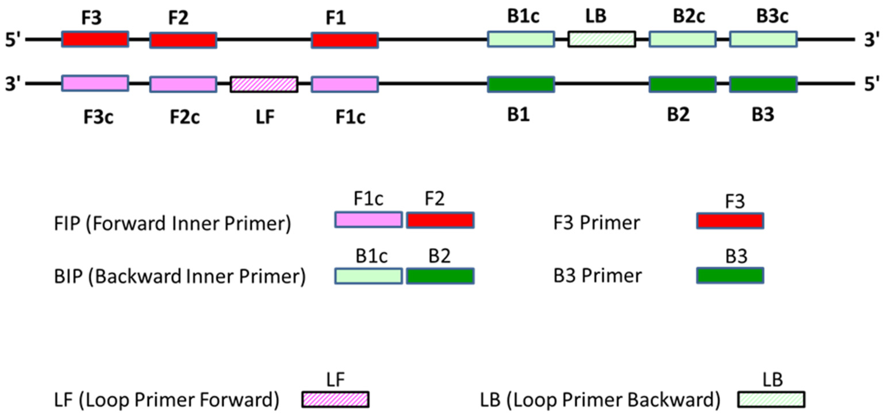
2.3. Design of LAMP Primer

Six LAMP primers were designed based on the sequence A2-HR MARDI chromosome of R. syzygii subsp. celebesensis (GenBank No. CP019911.1). The sequence of the LAMP primers used in this study is shown in Figure 2 from the 5′-end as F3, F2, F1 and B3, B2, B1. A forward inner primer (FIP) consists of F2 and F1c (the complementary sequence of F1), and a backward inner primer (BIP) is a combination of B3 and B1c (the complementary sequence of B1) (Figure 2). In addition, the forward loop primer (FL) is designed based on the complementary sequence between F1 and F2, while the backward loop primer (BL) is designed based on the complementary sequence between B1 and B2. The sequences of the LAMP primers are shown in Table 1.

2.4. LAMP Reaction

LAMP primer was prepared by combining all six primers (Table 1) into a single solution containing 40 µM FIP, 40 µM BIP, 5 µM F3, 5 µM B3, 10 µM LF, 10 µM LB. Extracted nucleic acid was used to perform 25 µL reactions (1X ThermoPol Buffer, 6 mM MgSO4, 1.4 mM each dNTP, 8U Bst DNA polymerase, 3 µL of primer mix) (New England BioLab, Ipswich, MA, USA.) at 65 °C for 40 min (concentration of gDNA depended on the experimental design). The sensitivity of the LAMP assay was evaluated using different concentrations of BDB genomic DNA from 0.0001 ng to 10 ng. The specificity test was performed using gDNA from R. solanacearum and Foc. After the amplification, verification of the LAMP reaction was conducted by using gel electrophoresis (1.5% agarose gel) (Merck KGaA, Darmstadt, Germany) containing FloroSafe DNA stain (1st Base, Singapore) in 1X TAE buffer (Merck KGaA, Darmstadt, Germany). Gels were then inspected in Gel doc to visualise DNA amplification.

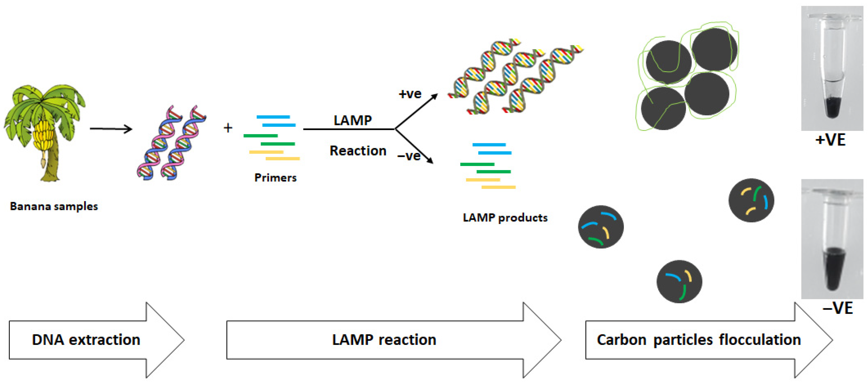
2.5. Analysis of LAMP Product Using Carbon Particles Flocculation

The flocculation solution contains activated charcoal with the size of 100–400 mesh (Sigma, St. Louis, MO, USA) and powdered diatomaceous earth. Active charcoal and diatomaceous earth were mixed in a 50 mL solution (50 mM Tris (pH 8), 10 mM spermine and 1% (w/v) PEG8000) with 400 mg activated charcoal and 600 mg diatomaceous earth. A volume of 20 μL of flocculation solution was then added to at least 20 µL of LAMP reaction and the tube was gently agitated to mix the solutions. Positive samples with successfully amplified long DNA in LAMP showed the particles flocculated and settled on the bottom of the tube within 20 s. However, for unsuccessfully amplified LAMP, negative samples with short oligonucleotides remained suspended in black.

2.6. Inoculation Test

Inoculation tests were performed on a three-month old tanduk cultivar (Musa acuminata × Musa balbisiana (AAB) cv. ‘Tanduk’ was purchased from a nursery at Horticulture, MARDI and acclimated in a greenhouse at 27–32 °C. Before inoculation, the inoculum bacterial suspension of BDB was prepared with a concentration of 108 colony-forming units (CFU)/mL. The experiment was arranged as a completely randomised block design (RCBD) with four replicates. Each seedling was inoculated with 5 mL of bacterial suspension by injection at the pseudostem section. Sterile distilled water was used as the negative control treatment. The inoculated plants were placed in the greenhouse until the symptoms were observed five days after inoculation. Leaves, stalks, and leaf midribs of the infected plants were collected. The samples were then subjected to the LAMP assay and carbon flocculation assay.

2.7. Field Test

Infected samples were collected from two banana plantations that were BDB hotspots, MARDI headquarters, and Taman Pertanian Universiti, UPM, Selangor. Samples were classified visually as healthy (no symptoms) and unhealthy (BDB symptoms). At MARDI headquarters, sample collections were conducted on leaves, stems, and stalks from four banana trees that exhibited BDB symptoms and on a symptomless banana tree (P1*, P2*, P3* and P4*). At Taman Pertanian Universiti, UPM, samples of stems, leaves, and stalks were collected from a total number of 13 banana trees in which two of the trees were from the healthy banana plot that was 500 m away from the infected banana plot. All samples were then subjected to a carbon flocculation assay to detect the presence of BDB in the samples. The samples from the healthy plot were labelled as H1 and H2. The samples from the infected plot were labelled as P1, P2, P3, P4, P5, P6, P7, P8, P9, P9, P10, and P11.

2.8. Validation

Validation of the developed diagnostic method from the samples was performed by PCR. Amplification of DNA targets was performed in a 25 µL reaction containing 1 µL of extracted genomic DNA (1 ng), 1 unit of KAPA Hotstart DNA polymerase, 1X PCR buffer (Roche Diagnostics, Risch-Rotkreuz, Switzerland), 0.2 mM each dNTP and 0.25 µM for each universal forward and reverse primer (Table 1). PCR reaction was conducted in a Bio-Rad thermocycler (Bio-Rad Laboratories, Inc., Herculas, CA, USA) using the following conditions: denaturation at 95 °C for 5 min followed by 30 cycles of 95 °C for 30 s, 58 °C for 30 s and 72 °C for 30 s and final elongation at 72 °C for 1 min. The products were validated by gel electrophoresis using 1.5% agarose gels (Merck KGaA, Darmstadt, Germany) containing FloroSafe DNA stain (1st Base, Singapore) in 1X TAE buffer (Merck KGaA, Darmstadt, Germany). The gel was then visualised in the Gel Doc.

3. Results

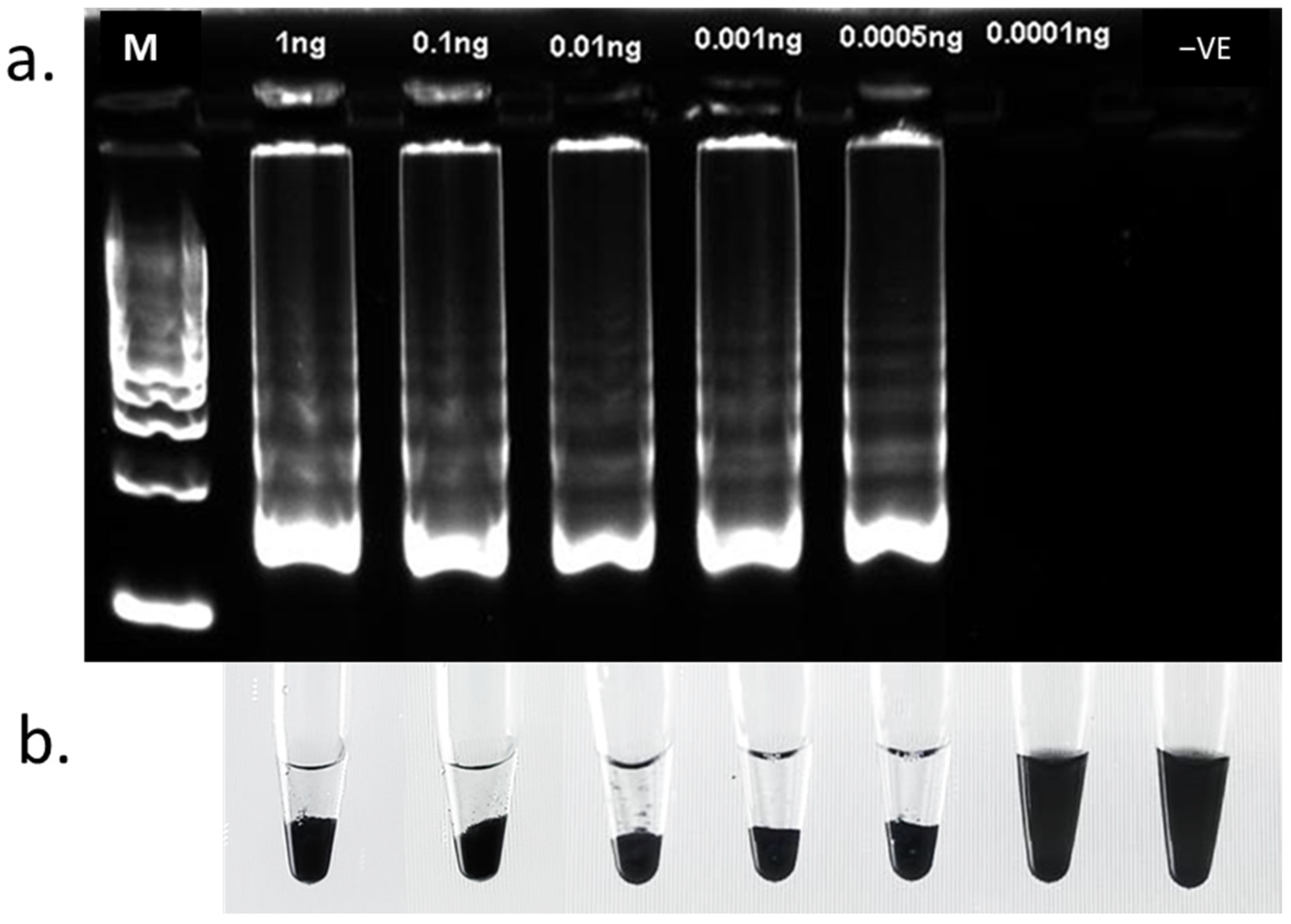
3.1. LAMP Assay Sensitivity
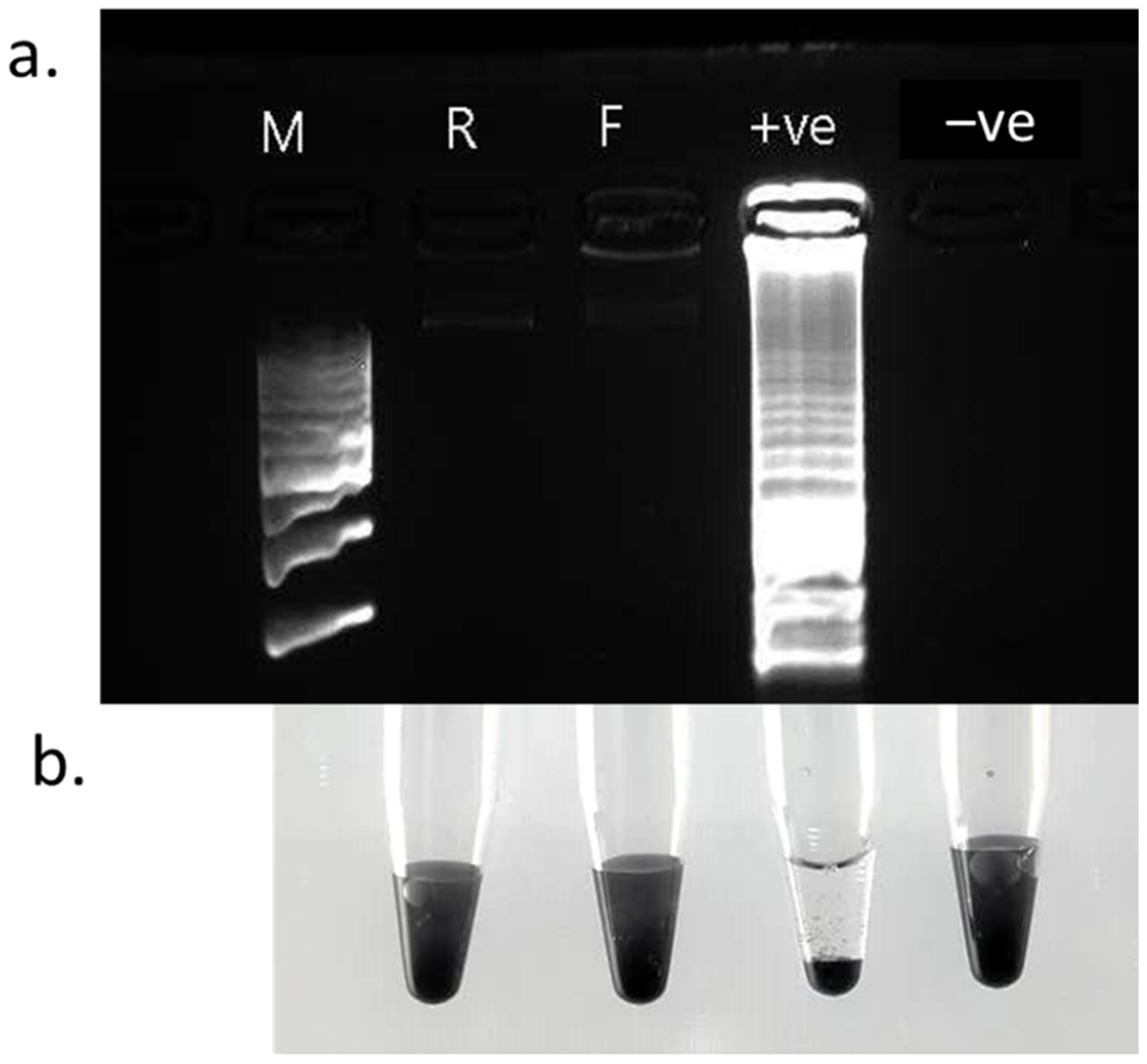
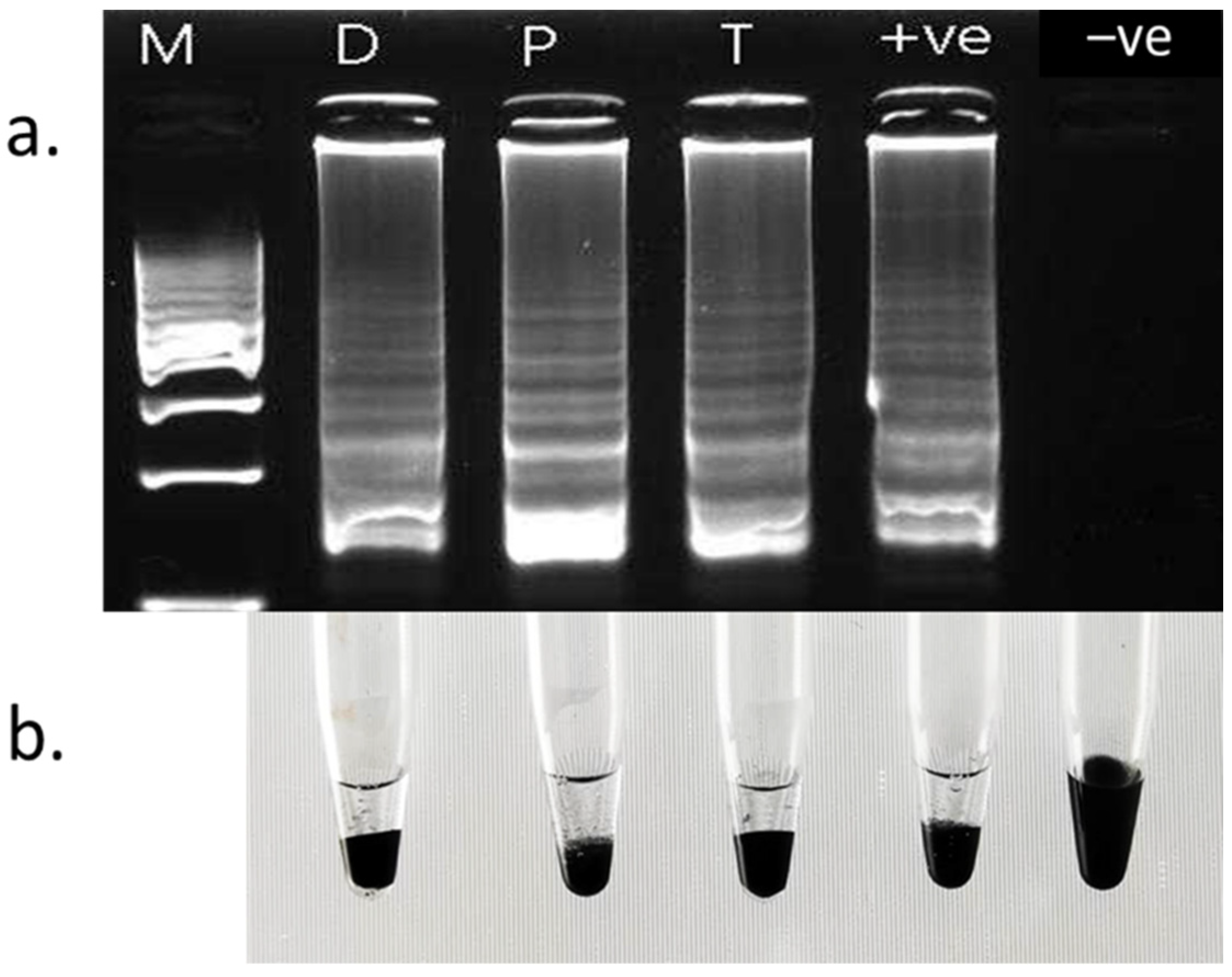
The sensitivity of the LAMP assay was assessed with 0.0001 ng to 10 ng of BDB genomic DNA. The LAMP reaction products were then subjected to 1% gel electrophoresis to confirm the amplifications (Figure 3). Based on the result, the sensitivity of the LAMP assay was 0.0005 ng.

3.2. Specificity

The specificity of the LAMP assay was studied using different gDNA from R. solanacearum and Foc (Figure 4). The result shows that amplification can be observed on the BDB sample but not on the other two pathogens. Therefore, the LAMP assay has high specificity toward BDB. The activated charcoal was flocculated and accumulated at the bottom of the tube in a positive sample (BDB), showing that the designed LAMP primer was highly specific to BDB. However, in R. solanacearum, Foc, and negative samples, the solution remained black, indicating a negative reaction.

3.3. Inoculation Test of BDB
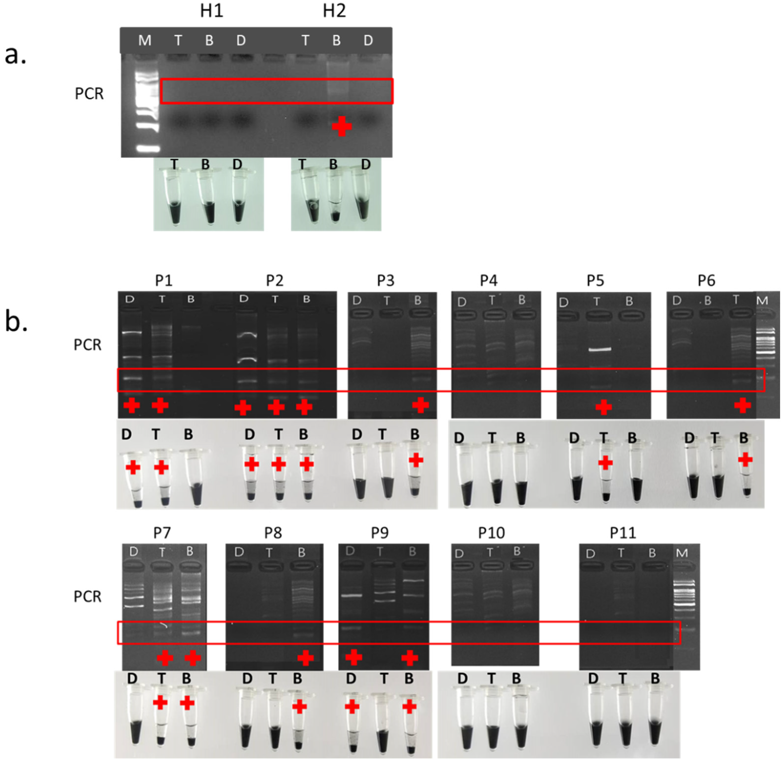
The LAMP assay and carbon flocculation assay were performed on the samples of leaves, leave midrib, and stalks five days post-inoculation. The LAMP products of leaves (D), leaf midribs (P), stalks (T), and a positive control (R. syzygii subsp. celebesensis, GenBank No. CP019911.1) were separated on an agarose gel for confirmation of DNA amplification in the positive LAMP reactions (Figure 5). However, no amplification was observed in the negative control (no template). Likewise, the carbon flocculation assay showed that the DNA particles of D, P, T, and the positive control were flocculated and settled on the bottom of the tube within 20 s, thus revealing successfully amplified long DNA in LAMP. However, charcoal in the negative control remained suspended, displaying an unsuccessfully amplified LAMP assay.

3.4. Field Trial

Symptomatic samples P1*, (B = stem, T = stalk, and D = leaves), P2* (B), and P4* (B) of banana trees from the sampling plot at MARDI headquarters showed positive amplification in LAMP assay and positive reaction in carbon flocculation assay (Figure 6). A positive control R. syzygii subsp. celebesensis (GenBank No. CP019911.1) displayed a positive LAMP amplification and a positive reaction in carbon flocculation assay by exhibiting flocculated charcoal accumulated at the bottom of the tube. These results revealed that the positive reaction of carbon flocculation assay had a resemblance to LAMP assay amplification. However, no LAMP amplification and no flocculation occurred in the P3* samples (B, T, and D) and P4* samples (T and D). Meanwhile, a positive reaction was detected in eight out of 11 sets of samples (B, T, and D) from the infected plot at Taman Pertanian Universiti in the carbon flocculation assay, indicating that approximately 73% of the plot was infected by BDB. However, BDB was also detected via a positive reaction in the flocculation assay on H2 sample (B) from a healthy plot.

3.5. Validation

Validation of both developed diagnostic methods was performed by PCR. DNA of the samples from the infected (P1, P2, P3, P4, P5, P6, P7, P8, P10, and P11) and healthy plots (H1 and H2) (Figure 6b) of Taman Pertanian Universiti were extracted and analysed using PCR (Figure 7). As displayed in Figure 7, a PCR product of the expected size was 280 bp. The results of PCR were the same as the results of LAMP and carbon flocculation assays, thus proving that the LAMP and carbon flocculation assays are as reliable as PCR due to the same results from the amplification of all samples.

4. Discussion

Early detection of the BDB pathogen could very well contribute to the successful management of the disease. BDB is a bacterial infection that causes severe blood disease in bananas. Because of the similarity of the symptoms, the disease is typically referred to as bacterial wilt, which is always caused by R. solanacearum. Due to its persistence and broad host range, this bacterium is well-known as one of the most important phytopathogenic bacteria in the world [8]. A sensitive detection protocol is critically required for early detection of pathogens so that they can be rogued and minimise the spread of the pathogens. DNA detection is currently the most sensitive and reliable method for detecting BDB. DNA extraction and PCR are the most common steps in the methods used to generate this information. As a result, many published protocols necessitate the use of special equipment, expensive reagents, and extensive user training, making them prohibitively expensive for large-scale screenings, such as those required to survey a collection of nursery stock [20]. This study aimed to demonstrate the pivotal diagnostic of LAMP incorporated with carbon flocculation assay in the early detection of BDB pathogen. To our knowledge, this study is the first to report on the deployment of LAMP assay incorporated with the carbon flocculation assay technique for the detection of BDB.
The LAMP assay is a better alternative to the conventional PCR method owing to it being more reliable, cost-effective, highly sensitive, specific, and rapid than PCR [20]. It was used in this study to attempt early detection of BDB in bananas [21,22]. It is primarily the diagnosis and mitigation of crop damage that would benefit from early detection. However, to enhance the rapidity of detection of the amplified product, the deployment of DNA-induced flocculation as a readout method has been proven to be more practical, faster, and easier to perform and is well-suited for low-resource and field applications [23].
Six LAMP primers designed to target the A2-HR MARDI chromosome of R. syzygii subsp. celebesensis (GenBank No. CP019911.1) showed high sensitivity in the LAMP assay. The sensitivity of the LAMP assay is high as it detects amounts of 0.5 pg gDNA from R. syzygii subsp. celebesensis. The detection limit of the LAMP assay is sufficient to avoid interference from the sample’s DNA and low enough to cause flocculation during amplification [23]. Thus, the development of flocculation solutions was visualised on BDB, but not on Foc and R. solanacearum. This indicates that the combination of the LAMP assay with the carbon flocculation assay was specific to detect BDB.
However, increasing the efficiency of the LAMP assay for on-site diagnostics is critical to the development of a method that requires minimal instrumentation. The most often-used method for analysing amplification products in the laboratory is agarose gel electrophoresis in the presence of dyes such as ethidium bromide. Besides agarose gel electrophoresis, a variety of colour-based methods have been developed. One such method involves adding gold nanoparticles (AuNP) with covalently attached probes to the completed reaction, which causes the colour of the AuNPs to change in the presence of the specific amplicon [23,24,25,26,27,28,29]. In an alternative technique, the DNA intercalating fluorescent dye SYBR Green I is added to the reaction, which turns the solution orange in the absence of DNA amplification and yellow/green in the presence of amplified DNA [23,30,31,32]. To achieve a significant difference between the positive and negative DNA amplification results, a high quantity of SYBR must be given after the amplification is complete, as it would otherwise inhibit the reaction [23,33]. In this study, the use of a carbon flocculation assay in the LAMP assay aids the visualisation of the positive amplification of the pathogen genome without performing gel electrophoresis or using any colour-based methods. This method deploys amplified DNA as a trigger for suspended particle flocculation. Previously, the use of DNA-induced flocculation as a readout method was described [23,34,35,36]. This flocculation assay has been successfully implemented in detecting bacteria, viruses and fungi in humans, plants, and animals. On the other hand, this study takes a radically different approach, which results in a significantly faster and easier system to perform, making it ideal for low-resource and field applications.
For field application, resource-constrained laboratories and farmer advisory service personnel can deploy the LAMP detection technique incorporated with carbon flocculation assay for DNA visualisation to make an accurate and timely diagnosis of BDB, thus alerting farmers to take early control measures to halt the disease’s spread. These assays can detect BDB even earlier than conventional PCR methods, making it useful for recommending precautionary or pre-symptomatic disease control measures [21]. In the glasshouse study, the LAMP assay incorporated with carbon flocculation assay successfully detected BDB on the symptomatic samples (leaves, leaf midribs, and stalks) five days after inoculation. In the field-testing study at MARDI headquarters, the LAMP assay incorporated with carbon flocculation assay successfully detected BDB on all symptomatic samples except for P3* and P4* (stalks and leaves) after BDB infection. Interestingly, BDB was detected on a healthy sample, P2* (stem), even though no symptoms were visible. Meanwhile, at Taman Pertanian Universiti, BDB was detected on eight out of 11 sets of infected samples. Likewise, BDB was detected on the sample, H2 (stem) from a healthy plot. The efficacy of the techniques has been verified via the PCR method, thus revealing that the LAMP assay incorporated with carbon flocculation assay can detect not only BDB at a severe stage of infection but also BDB at an early stage of infection, which may help farmers take preventive control measures to reduce the chance of outbreaks of disease. The particles in the flocculation solution have a large surface area for interaction with DNA molecules due to their high porosity and irregular shape. Additionally, their low densities ensure that they will remain suspended for extended periods unless flocculation is induced. The charcoal particles create a dark, opaque solution that enables positive and negative samples to be easily distinguished in any light environment, while the diatomaceous earth particles help aggregate the fine charcoal particles and reduce the time required for them to settle at the tube’s bottom [23].
The efficacy of the LAMP assay incorporated with carbon flocculation assay was validated with conventional PCR. The validation has revealed that the sensitivity of the LAMP assay incorporated with the carbon flocculation assay was comparable to the PCR technique. In comparison to PCR, this technique has a faster reaction time and almost identical or better sensitivity and specificity [23]. The flocculation assay technique has been proven to work with both PCR and LAMP and can theoretically be implemented in any amplification technology as long as the amplicon size is large enough to induce the bridging flocculation mechanism.

5. Conclusions

The development of diagnostic tools that are suitable for use on-site by diagnostic facilities with limited resources is critical for agricultural support services. LAMP is an effective diagnostic tool for resource-constrained environments, and its uncomplicated visual format facilitates detection. Additionally, a sophisticated laboratory environment is not required, and the machine used to run the LAMP reactions is less expensive than real-time PCR machines. Additionally, it is battery-operated, making it ideal for remote areas with limited access to electricity. It has a rapid sample processing time and involves less training than PCR-based diagnostics. The combination of LAMP and flocculation assays has proven to be effective in detecting BDB, and it can be deployed with any amplification technology as long as the amplicon size is large enough to induce the bridging flocculation mechanism. Owing to the flocculation reaction being extremely robust and adapting well to changes in operating procedures and environmental conditions, it is ideal for on-site implementation. Besides, this method is accessible to non-specialists with limited technical training or resources and can be easily integrated into existing diagnostic platforms for the detection of plant diseases.

Author Contributions

Conceptualisation, H.Y.L. and N.A.B.; Methodology, H.Y.L., N.A.B., M.F.M.Y. and R.B.; Data curation, M.M.F.A., H.Y.L., N.A.B., S.R., M.F.M.Y., R.B. and N.S.J.; Software; H.Y.L.; visualization, M.M.F.A.; writing—original draft preparation, M.M.F.A.; writing—review and editing, H.Y.L. and N.A.B.; supervision and funding acquisition, H.Y.L. and N.A.B. All authors have read and agreed to the published version of the manuscript.

Funding

This study was supported by PRB 405, MARDI Development Grant.

Institutional Review Board Statement

Not applicable.

Informed Consent Statement

Not applicable.

Acknowledgments

We would like to thank the Malaysian Agricultural Research and Development Institute (MARDI) for the research funding, PRB 405, MARDI Development Grant.

Conflicts of Interest

The authors declare that the research was conducted in the absence of any commercial or financial relationships that could be construed as a potential conflict of interest.

References

  1. Shahbandeh, M. World Production of Bananas in 2020, by Region. Available online: https://www.statista.com/statistics/264003/production-of-bananas-worldwide-by-region/ (accessed on 30 March 2022).
  2. Ploetz, R.C.; Kema, G.H.J.; Ma, L.J. Impact of Diseases on Export and Smallholder Production of Banana. Annu. Rev. Phytopathol. 2015, 53, 269–288. [Google Scholar] [CrossRef]
  3. Blomme, G.; Dita, M.; Jacobsen, K.S.; Vicente, L.P.; Molina, A.; Ocimati, W.; Poussier, S.; Prior, P. Bacterial Diseases of Bananas and Enset: Current State of Knowledge and Integrated Approaches toward Sustainable Management. Front. Plant Sci. 2017, 8, 1290. [Google Scholar] [CrossRef] [PubMed]
  4. Eden-Green, S.J. Diversity of Pseudomonas solanacearum and Related Bacteria in South Asia: New Direction of Moko Disease. In Bacterial Disease and its Causative Agent Pseudomonas solanacearum; Hayward, A.C., Hartman, G.L., Eds.; CAB International: Wellington, New Zealand, 1994; pp. 25–34. [Google Scholar]
  5. Gäumann, E. Onderzoekingen over de Bloedziekte Der Bananen Op Celebes, I. Meded. Inst. Plantenziekten 1921, 50, 1–47. [Google Scholar]
  6. Stover, R.H.; Espinoza, A. Blood Disease of Bananas in Sulawesi. Fruits 1992, 47, 611–613. [Google Scholar]
  7. Thwaites, R.; Eden-Green, S.J.; Black, R. Diseases Caused by Bacteria. In Diseases of Banana, Abacá and Enset; Jones, D.R., Ed.; CAB International: Wallingford, New Zealand, 2000; pp. 213–239. [Google Scholar]
  8. Teng, S.K.; Aziz, N.A.A.; Mustafa, M.; Laboh, R.; Ismail, I.S.; Sulaiman, S.R.; Azizan, A.A.; Devi, S. The Occurrence of Blood Disease of Banana in Selangor, Malaysia. Int. J. Agric. Biol. 2016, 18, 92–97. [Google Scholar] [CrossRef]
  9. Buddenhagen, I. Blood Bacterial Wilt of Banana: History, Field Biology and Solution. Acta Hortic. 2009, 828, 57–68. [Google Scholar] [CrossRef]
  10. Taghavi, M.; Hayward, C.; Sly, L.I.; Fegan, M. Analysis of the Phylogenetic Relationships of Strains of Burkholderia Solanacearum, Pseudomonas Syzygii, and the Blood Disease Bacterium of Banana Based on 16S RRNA Gene Sequences. Int. J. Syst. Bacteriol. 1996, 46, 10–15. [Google Scholar] [CrossRef]
  11. Remenant, B.; de Cambiaire, J.C.; Cellier, G.; Jacobs, J.M.; Mangenot, S.; Barbe, V.; Lajus, A.; Vallenet, D.; Medigue, C.; Fegan, M.; et al. Ralstonia Syzygii, the Blood Disease Bacterium and Some Asian, R. Solanacearum Strains Form a Single Genomic Species despite Divergent Lifestyles. PLoS ONE 2011, 6, e24356. [Google Scholar] [CrossRef]
  12. Sahetapy, B.; Maryana, N.; Manuwoto, S.; Mutaqin, K.H.; Latumahina, F. Test of Blood Disease Bacterium (Bdb) Transmission By Potential Insect Vectors. J. Hama Dan Penyakit Tumbuh. Trop. 2020, 20, 71–77. [Google Scholar] [CrossRef]
  13. Notomi, T.; Okayama, H.; Masubuchi, H.; Yonekawa, T.; Watanabe, K.; Amino, N.; Hase, T. Loop-Mediated Isothermal Amplification of DNA. Nucleic Acids Res. 2000, 28, e63. [Google Scholar] [CrossRef] [Green Version]
  14. Ivanov, A.V.; Safenkova, I.V.; Zherdev, A.V.; Dzantiev, B.B. The Potential Use of Isothermal Amplification Assays for In-Field Diagnostics of Plant Pathogens. Plants 2021, 10, 2424. [Google Scholar] [CrossRef] [PubMed]
  15. Ruehrwein, R.A.; Ward, D.W. Mechanism of Clay Aggregation by Polyelectrolytes. Soil Sci. 1952, 73, 485–492. [Google Scholar] [CrossRef]
  16. Healy, T.W.; La Mer, V.K. The Energetics of Flocculation and Redispersion by Polymers. J. Colloid Sci. 1964, 19, 323–332. [Google Scholar] [CrossRef]
  17. Kelman, A. The Relationship of Pathogenicity of Pseudomonas Solanacearum to Colony Appearance in a Tetrazolium Medium. Phytopathology 1954, 44, 693–695. [Google Scholar]
  18. Rohland, N.; Reich, D. Cost-effective, High-throughput DNA Sequencing Libraries for Multiplexed Target Capture. Genome Res. 2012, 22, 939–946. [Google Scholar] [CrossRef] [PubMed] [Green Version]
  19. Deangelis, M.M.; Wang, D.G.; Hawkins, T.L. Solid-Phase Reversible Immobilization for the Isolation of PCR Products. Nucleic Acids Res. 1995, 23, 4742–4743. [Google Scholar] [CrossRef] [PubMed]
  20. Wang, H.; Turechek, W.W. A Loop-Mediated Isothermal Amplification Assay and Sample Preparation Procedure for Sensitive Detection of Xanthomonas Fragariae in Strawberry. PLoS ONE 2016, 11, e0147122. [Google Scholar] [CrossRef]
  21. Prasannakumar, M.K.; Buela Parivallal, P.; Manjunatha, C.; Mahesh, H.B.; Pramesh, D.; Narayan, K.S.; Gopal, V.B.; Priyanka, K.; Puneeth, M.E.; Rangaswamy, K.T. Loop-Mediated Isothermal Amplification Assay for Pre-Symptomatic Stage Detection of Xanthomonas axonopodis pv. punicae Infection in Pomegranate. Australas. Plant Pathol. 2020, 49, 467–473. [Google Scholar] [CrossRef]
  22. Ordonez, N.; Salacinas, M.; Mendes, O.; Seidi, M.F.; Meijer, H.J.G.; Schoen, C.D.; Kema, G.H.J. A Loop-mediated Isothermal Amplification LAMP Assay Based on Unique Markers Derived from Genotyping by Sequencing Data for Rapid in Planta Diagnosis of Panama Disease caused by Tropical Race 4 in Banana. Plant Pathol. 2019, 68, 1682–1693. [Google Scholar] [CrossRef] [Green Version]
  23. Mason, M.G.; Botella, J.R. A Simple, Robust and Equipment-Free DNA Amplification Readout in Less than 30 Seconds. RSC Adv. 2019, 9, 24440–24450. [Google Scholar] [CrossRef] [Green Version]
  24. Valentini, P.; Fiammengo, R.; Sabella, S.; Gariboldi, M.; Maiorano, G.; Cingolani, R.; Pompa, P.P. Gold-Nanoparticle-Based Colorimetric Discrimination of Cancer-Related Point Mutations with Picomolar Sensitivity. ACS Nano 2013, 7, 5530–5538. [Google Scholar] [CrossRef] [PubMed]
  25. Zhang, Y.; Hu, J.; Zhang, C.Y. Sensitive Detection of Transcription Factors by Isothermal Exponential Amplification-Based Colorimetric Assay. Anal. Chem. 2012, 84, 9544–9549. [Google Scholar] [CrossRef] [PubMed]
  26. Chomean, S.; Wangmaung, N.; Sritongkham, P.; Promptmas, C.; Ittarat, W. Genotyping of α-Thalassemias by the Colorimetric Nanogold Probes. Clin. Chim. Acta 2014, 437, 197–202. [Google Scholar] [CrossRef] [PubMed]
  27. Chomean, S.; Wangmaung, N.; Sritongkham, P.; Promptmas, C.; Mas-oodi, S.; Tanyong, D.; Ittarat, W. Molecular Diagnosis of α-Thalassemias by the Colorimetric Nanogold. Analyst 2014, 139, 813–822. [Google Scholar] [CrossRef] [PubMed]
  28. Ma, C.; Wang, W.; Mulchandani, A.; Shi, C. A Simple Colorimetric DNA Detection by Target-Induced Hybridization Chain Reaction for Isothermal Signal Amplification. Anal. Biochem. 2014, 457, 19–23. [Google Scholar] [CrossRef]
  29. Muangchuen, A.; Chaumpluk, P.; Suriyasomboon, A.; Ekgasit, S. Colorimetric Detection of Ehrlichia Canis via Nucleic Acid Hybridization in Gold Nano-Colloids. Sensors 2014, 14, 14472–14487. [Google Scholar] [CrossRef] [Green Version]
  30. Njiru, Z.K.; Mikosza, A.S.J.; Armstrong, T.; Enyaru, J.C.; Ndung’u, J.M.; Thompson, A.R.C. Loop-Mediated Isothermal Amplification (LAMP) Method for Rapid Detection of Trypanosoma Brucei Rhodesiense. PLoS Negl. Trop. Dis. 2008, 2, e147. [Google Scholar] [CrossRef] [PubMed]
  31. Hong, M.; Zha, L.; Fu, W.; Zou, M.; Li, W.; Xu, D. A Modified Visual Loop-Mediated Isothermal Amplification Method for Diagnosis and Differentiation of Main Pathogens from Mycobacterium Tuberculosis Complex. World J. Microbiol. Biotechnol. 2012, 28, 523–531. [Google Scholar] [CrossRef]
  32. Zhang, X.; Zhang, H.; Pu, J.; Qi, Y.; Yu, Q.; Xie, Y.; Peng, J. Development of a Real-Time Fluorescence Loop-Mediated Isothermal Amplification Assay for Rapid and Quantitative Detection of Fusarium Oxysporum f. sp. Cubense Tropical Race 4 in Soil. PLoS ONE 2013, 8, e82841. [Google Scholar] [CrossRef] [Green Version]
  33. Fischbach, J.; Xander, N.C.; Frohme, M.; Glökler, J.F. Shining a Light on LAMP Assays- a Comparison of LAMP Visualization Methods Including the Novel Use of Berberine. Biotechniques 2015, 58, 189–194. [Google Scholar] [CrossRef] [Green Version]
  34. Leslie, D.C.; Li, J.; Strachan, B.C.; Begley, M.R.; Finkler, D.; Bazydlo, L.A.L.; Barker, N.S.; Haverstick, D.M.; Utz, M.; Landers, J.P. New Detection Modality for Label-Free Quantification of DNA in Biological Samples via Superparamagnetic Bead Aggregation. J. Am. Chem. Soc. 2012, 134, 5689–5696. [Google Scholar] [CrossRef] [PubMed]
  35. Lin, C.; Zhang, Y.; Zhou, X.; Yao, B.; Fang, Q. Naked-Eye Detection of Nucleic Acids through Rolling Circle Amplification and Magnetic Particle Mediated Aggregation. Biosens. Bioelectron. 2013, 47, 515–519. [Google Scholar] [CrossRef] [PubMed]
  36. Wee, E.J.H.; Lau, H.Y.; Botella, J.R.; Trau, M. Re-Purposing Bridging Flocculation for on-Site, Rapid, Qualitative DNA Detection in Resource-Poor Settings. Chem. Commun. 2015, 51, 5828–5831. [Google Scholar] [CrossRef] [PubMed]
Figure 1. A schematic diagram of the LAMP-flocculation assay.
Horticulturae 08 00406 g001
Figure 2. A schematic diagram of the LAMP primer design used in this experiment.
Horticulturae 08 00406 g002
Figure 3. Sensitivity of the LAMP assay (a) and carbon particle flocculation assay (b). LAMP assay using 0.0001 ng to 10 ng of purified target DNA from BDB. Amounts of DNA template were as follows: Lane 1: 1 ng; Lane 2: 0.1 ng; Lane 3: 0.01 ng; Lane 4: 0.001; Lane 5: 0.0005 ng; Lane 6: 0.0001 ng; Lane 7: −ve negative control; Lane M: 100 bp DNA marker. Each figure represents 3 experimental replicates.
Horticulturae 08 00406 g003
Figure 4. Specificity assessment of the LAMP assay for Ralstonia syzygii subspecies celebesensis genomic DNA by visualisation on agarose gel stained (a) and carbon flocculation assay with activated charcoal (b). R = Ralstonia solanacearum; F = Fusarium oxysporum cubense; +ve = positive control (R. syzygii subspecies celebesensis); −ve = negative control (without DNA template); M = 100 bp DNA marker. Each figure represents at least 3 experimental replicates.
Horticulturae 08 00406 g004
Figure 5. Analysis of LAMP amplification products by gel electrophoresis (a) and carbon flocculation assay (b). +ve = positive control using R. syzygii subsp. celebesensis (GenBank No. CP019911.1). −ve = negative control without DNA template. LAMP reactions were subsequently loaded to agarose gel and separated by electrophoresis. M = 100 bp DNA marker. Flocculation results showed positive reaction for the samples of D, P, T, and +ve with DNA particles flocculated and settled on the bottom of the tube but no reaction in negative control. D = leaves, P = leaf midrib, and T = stalk. Each figure represents least 3 experimental replicates.
Horticulturae 08 00406 g005
Figure 6. Assessment of the developed LAMP and carbon flocculation assays at two BDB hotspots: (a) MARDI headquarters; and (b) Taman Pertanian Universiti. (+) represents BDB detected in the sample. Each figure represents at least 3 experimental replicates.
Horticulturae 08 00406 g006
Figure 7. Validation of field applicability of samples LAMP assay and carbon flocculation assay by comparing the LAMP and carbon flocculation results using PCR: (a) samples from healthy plot (H1 and H2); and (b) samples from infected plot (P1, P2, P3, P4, P5, P6, P7, P8, P9, P10, P11). M = 100 bp DNA marker; D = leaves; T = stalk; and B = stem. (+) represents BDB detected in the sample and the red box indicates the size of amplified PCR band (280 bp). Each figure represents at least 3 experimental replicates.
Horticulturae 08 00406 g007
Table 1. Sequences of LAMP and PCR primers involved in this study.
PrimerPrimer Sequence (5′-3′)
F3AACTGGAGTGCTTGAAGC
B3CTCTGTGGCGATTGTCTAC
FIPGGACCTGTCACTCGAACCATGCTGCGGACTCCACATTAC
BIPGCTCGTCCGTCTCGTTGAGGGTTGCTTACCTTAGAGACTC
LFGGCGTTACACCACATCCA
LBGGTAATAGCCTCGCTCCG
PCR FwdGTCGCCGTCAACTCACTTTCC
PCR RevGTCGCCGTCAGCAATGCGGAATCG
Publisher’s Note: MDPI stays neutral with regard to jurisdictional claims in published maps and institutional affiliations.

