Article

Detection of a Point Mutation (G143A) in Cyt b of Corynespora cassiicola That Confers Pyraclostrobin Resistance

1
College of Plant Protection, Shandong Agricultural University, Tai’an 271018, China
2
College of Plant Protection, Northwest A&F University, Yangling 712100, China
*
Authors to whom correspondence should be addressed.
Horticulturae 2021, 7(6), 155; https://doi.org/10.3390/horticulturae7060155
Submission received: 9 May 2021 / Revised: 14 June 2021 / Accepted: 16 June 2021 / Published: 18 June 2021
(This article belongs to the Section Plant Pathology and Disease Management (PPDM))

Abstract

:
Point mutation G143A in the cytochrome b (Cyt b) protein commonly confers resistance to quinone outside inhibitor (QoI) fungicides in phytopathogenic fungi, including Corynespora cassiicola, which causes cucumber target spot disease. However, the effect of G143A on the binding between the QoI fungicide and the Cyt b protein, and the use of LAMP (loop-mediated isothermal amplification) to detect this point mutation had not been reported previously in C. cassiicola. In this study, the sensitivity of 131 C. cassiicola isolates—collected from Shandong province, China in 2019 and 2020—to pyraclostrobin was determined. The EC50 values ranged from 1.67 to 8.82 μg/mL, and sequencing results showed that all C. cassiicola isolates contained the G143A mutation. Molecular docking results suggested that G143A significantly alters the affinity of pyraclostrobin to the Cyt b protein. Following development of three LAMP primer pairs, the best reaction condition for LAMP analysis was 65 °C for 60 min, and the detection limit was 0.01 ng/μL of DNA containing the point mutation. In conclusion, the G143A mutation conferring pyraclostrobin resistance is widespread in C. cassiicola from Shandong province, and the LAMP method can be used to monitor QoI resistance in C. cassiicola caused by the G143A mutation in the field.

1. Introduction

Cucumber target spot is a worldwide fungal disease in cucumber production [1,2]. It is caused by Corynespora cassiicola, which can infect a wide range of hosts, including nematode cysts and the human skin [3,4,5,6]. In addition to cucumber, it can also cause disease on leaves, stems, roots, flowers, and fruits in up to 530 plant species [7]. A saprophytic, endophytic lifestyle complicates C. cassiicola management [8].
Chemical control is the main method to limit cucumber target spot disease in China. At present, the domestically registered agrichemicals for the prevention and control of this disease include quinone outside inhibitors (QoI) (pyraclostrobin, trifloxystrobin, azoxystrobin), succinate dehydrogenase inhibitors (SDHI) (pydiflumetofen, fluopyram, fluxapyroxad), and sterol biosynthesis inhibitors (SBI) (difenoconazole, tebuconazole, prochloraz) (http://www.chinapesticide.org.cn/ (accessed on 9 May 2021)). However, the long-term and frequent use of these chemical fungicides has caused C. cassiicola populations to gradually develop fungicide resistance [2,9,10,11,12,13,14]. Thus, timely detection is of particular importance for fungicide resistance management and guidance on the scientific use of fungicides in the field.
Traditional in vitro methods, such as mycelial growth rate and spore germination methods, are widely used for determining fungicide sensitivity (https://www.frac.info/ (accessed on 9 May 2021)). Often, highly resource-intensive methods limit the number of isolates that can be assayed, and some pathogens are difficult to culture in vitro. Thus, the isolation, culture, identification, and preservation of target pathogens is time-consuming and labor-intensive. Based on a clear resistance mechanism (point mutation in a target protein), many rapid and progressive molecular tools, such as AS-PCR (allele-specific PCR), PCR-RFLP (PCR-restriction fragment length polymorphism), qPCR, droplet digital PCR, and pyrosequencing, have been developed or used to detect fungicide-resistant populations in different plant pathogens [15,16,17,18,19]. However, sophisticated and expensive instruments and complicated test techniques limit the application of these methods in small laboratories.
LAMP (loop-mediated isothermal amplification) is a constant-temperature nucleic acid amplification technology, using Bst (Bacillus stearothermophilus) DNA polymerase developed by Japanese workers [20,21]. Compared with PCR methods, LAMP is more rapid and sensitive, and the results are more easily analyzed. The products of LAMP can be analyzed by agarose gel electrophoresis, turbidity, or fluorescence. This method has been extensively used to identify pathogenic microorganisms (viruses, fungi, bacteria, oomycetes) [22,23,24,25]. From 2014, LAMP assays were gradually developed to monitor SBI, QoI, SDHI, and MBC (methyl benzimidazole carbamate) fungicide resistance in different plant pathogens [26,27,28,29,30]. Although QoI resistance and the point mutation G143A in the Cyt b protein have been widely detected in C. cassiicola [12], the effect of this point mutation on the binding between pyraclostrobin and Cyt b, and an LAMP assay for this point mutation in C. cassiicola have not been reported. Thus, the aim of the current study was to (1) investigate the pyraclostrobin sensitivity of C. cassiicola isolated from infected cucumber leaves collected from Shandong province during 2019 and 2020; (2) confirm the relationship between point mutation G143A and pyraclostrobin resistance; and (3) develop an LAMP method for detecting G143A in C. cassiicola isolates.

2. Materials and Methods

2.1. Pathogens and Plants

C. cassiicola isolates, 131 in total, were obtained from cucumber leaves with typical symptoms from 8 cities in Shandong province during 2019 and 2020. Pyraclostrobin-sensitive isolates Cc4 and Cc10 were kindly provided by Associate Professor Yabing Duan of Nanjing Agricultural University.
Cucumber seeds (cv. Youliangwang) were sown in plastic pots (10 × 10 × 10 cm, 2 seeds per pot) filled with a peat/vermiculite mixture (2:1 v/v). All seedlings were grown under greenhouse conditions (27 ± 2 °C, 80% relative humidity, and 12/12 h photoperiod) until the four-leaf stage.
For mycelium collection, C. cassiicola, Didymella bryoniae, and Botrytis cinerea were maintained on PDA (potato dextrose agar), and Phytophthora melonis was inoculated on WKB (white kidney bean) agar [31]. For detached cucumber leaf inoculation, a mycelial plug of four of the above pathogens was placed on the upper surface of leaves for 4 days at 25 °C.

2.2. Determination of Pyraclostrobin Sensitivity

Pyraclostrobin (98%, active ingredient) was provided by Shandong United Pesticide Industry Co. Ltd. (Taian, China). A stock solution (104 μg/mL) was prepared with DMSO (dimethyl sulfoxide). Sensitivity of C. cassiicola to pyraclostrobin was investigated using the mycelial growth rate method. Agar disks (diameter 5 mm) containing C. cassiicola mycelium were inoculated onto AEA (alkyl ester agar medium) plates containing a series of concentrations (0.2, 0.5, 1, 5, 10, 20, 40, 80 μg/mL) of pyraclostrobin and 50 μg/mL of SHAM (salicylhydroxamic acid). C. cassiicola was incubated at 25 °C for 6 days in darkness, and then colony diameters were measured. Each treatment had three replicates, and the experiment was repeated twice. EC50 values were calculated according to a previous study [12].

2.3. Genomic DNA Extraction and Analysis of the Cyt b Gene

Mycelium (100 mg) or the infected cucumber leaf sample (100 mg), which was frozen in liquid nitrogen, was ground in a ball mill for 2 min. (1) A total of 800 µL of 3% CTAB was added to each tube, followed by 1 h incubation at 60 °C in a water bath. (2) A total of 800 µL phenol–chloroform–isoamyl alcohol (25:24:1, v:v) was added. The sample was mixed gently by inversion and then spun at 12,000 rpm for 10 min. (3) The supernatant was transferred into a new centrifuge tube, and 650 µL of chloroform–isoamyl alcohol (24:1, v:v) was added. (4) The supernatant was transferred into a new centrifuge tube after centrifuging at 12,000 rpm for 10 min. (5) A 0.6-fold isoamyl alcohol was added and incubated for 30 min at −20 °C. (6) Tubes were centrifuged at 12,000 rpm for 10 min. (7) The liquid was decanted, and the precipitate was washed with ice-cold 70% ethanol. This step was done twice. (8) Tubes were placed into a Concentrator Plus (Eppendorf, Hamburg, Germany) with the caps open. The Concentrator Plus was run at 42 °C for approximately 10 min. (9) Dry precipitate was resuspended in 50 µL of ddH2O. The concentration of genomic DNA was tested with a NanoDrop ONE spectrophotometer (ThermoScientific, Waltham, MA, USA). The Cyt b gene was amplified using Primers CcCytb-F/CcCytb-R (Table 1) [12] and M5 Taq PCR Mix with Blue Dye (Mei5 Biotechnology, Co., Ltd., Beijing, China). PCR products were sequenced by Tsingke Biotechnology Co., Ltd. (Xian, China), and sequences were analyzed using DNAMAN 6.0 Software.

2.4. Molecular Docking of Pyraclostrobin in Cyt b of C. cassiicola

An in silico model of the C. cassiicola Qo-site was built by comparative modelling using MODELLER v9.19, with the yeast structure (PDB 1KB9) [32] as a template, that exhibits 57.89% sequence identity with the sequence from C. cassiicola. To evaluate the stereochemical quality of the CcCyt b 3D model, a Ramachandran map was generated using PROCHECK. Verify3D was used to analyze the compatibility of the CcCyt b model with its own amino acid sequence. The structure of the substrate (pyraclostrobin) was constructed using AutoDock 4.2.6 and optimized using the MOPAC program. To exclude unreasonable spatial structures and make the binding model more stable, energy optimization was performed using an Amber14 force field. Autodock results were analyzed with AutoDock Tools 1.5.6., and illustrations of the 3D models were generated using Ligplot 1.4.5 and Pymol 1.8.6.

2.5. LAMP Primer Design

The LAMP primer was designed on the website Primer Explorer (v5) (http://primerexplorer.jp/e/ (accessed on 8 February 2021)) according to the mutation GGT (Gly) → GCT (Ala) at the 143 amino acid codon of Cyt b, and the mutation site was located at the 3′ end of the forward inner primer (FIP). Three pairs of primers with a mismatch at the penultimate position were designed to improve the specificity of the LAMP primers (Table 1, Figure 1).

2.6. LAMP Reaction Mixtures

WarmStart Colorimetric LAMP 2× Master Mix (New England Biolabs, Beijing, China) was used in this study. The LAMP reaction mixture (25 μL) included a 12.5 μL WarmStart Colorimetric LAMP 2× Master Mix (New England Biolabs), 1.6 μM FIP and BIP, 0.2 μM F3 and B3, 0.4 μM loop F and loop B, and a 1 μL DNA template. The results were double confirmed through visual color changes (from pink to yellow) and 2% agarose gel electrophoresis.

2.7. Optimization of LAMP Reaction Time and Temperature

A series of constant temperatures (i.e., 61, 63, 65, 67, 69 °C), and a series of reaction times (i.e., 30, 45, 60, 75, and 90 min) were set to investigate the optimal reaction time and temperature.

2.8. LAMP Assay Specificity

To test the specificity of the LAMP assay, pyraclostrobin-sensitive C. cassiicola isolates (Cc4 and Cc10), pyraclostrobin-resistant C. cassiicola isolates (Cc21, Cc23, Cc31, Cc35, Cc42, Cc53, Cc71, Cc66, Cc96, Cc123), D. bryoniae, B. cinerea, and P. melonis, isolated from diseased cucumber plants, were selected. Genomic DNA of wild-type Cc4, Cc10, and ddH2O were used as negative controls and blank, respectively. The LAMP assay was performed at 65 °C for 60 min. Experiments were repeated three times.

2.9. LAMP Assay Detection Limit

A concentration series (100, 10, 1, 0.1, 0.01, 0.001, and 0.0001 ng/μL) of DNA template was used to determine the detection limit of the LAMP assay for the G143A mutation in C. cassiicola.

3. Results

3.1. Pyraclostrobin Sensitivity of C. cassiicola Isolates and Sequence of Cyt b Gene

The EC50 values of pyraclostrobin for the 131 C. cassiicola isolates ranged from 1.67 to 8.82 μg/mL, with a mean of 4.74 μg/mL (Table 2). There was no correlation between the fungicide sensitivity and source of isolates (Table 2). Sequencing of the Cyt b gene showed that there was a G143A mutation in the Cyt b gene in all C. cassiicola isolates.

3.2. Pyraclostrobin Docking in the Cyt b of C. cassiicola

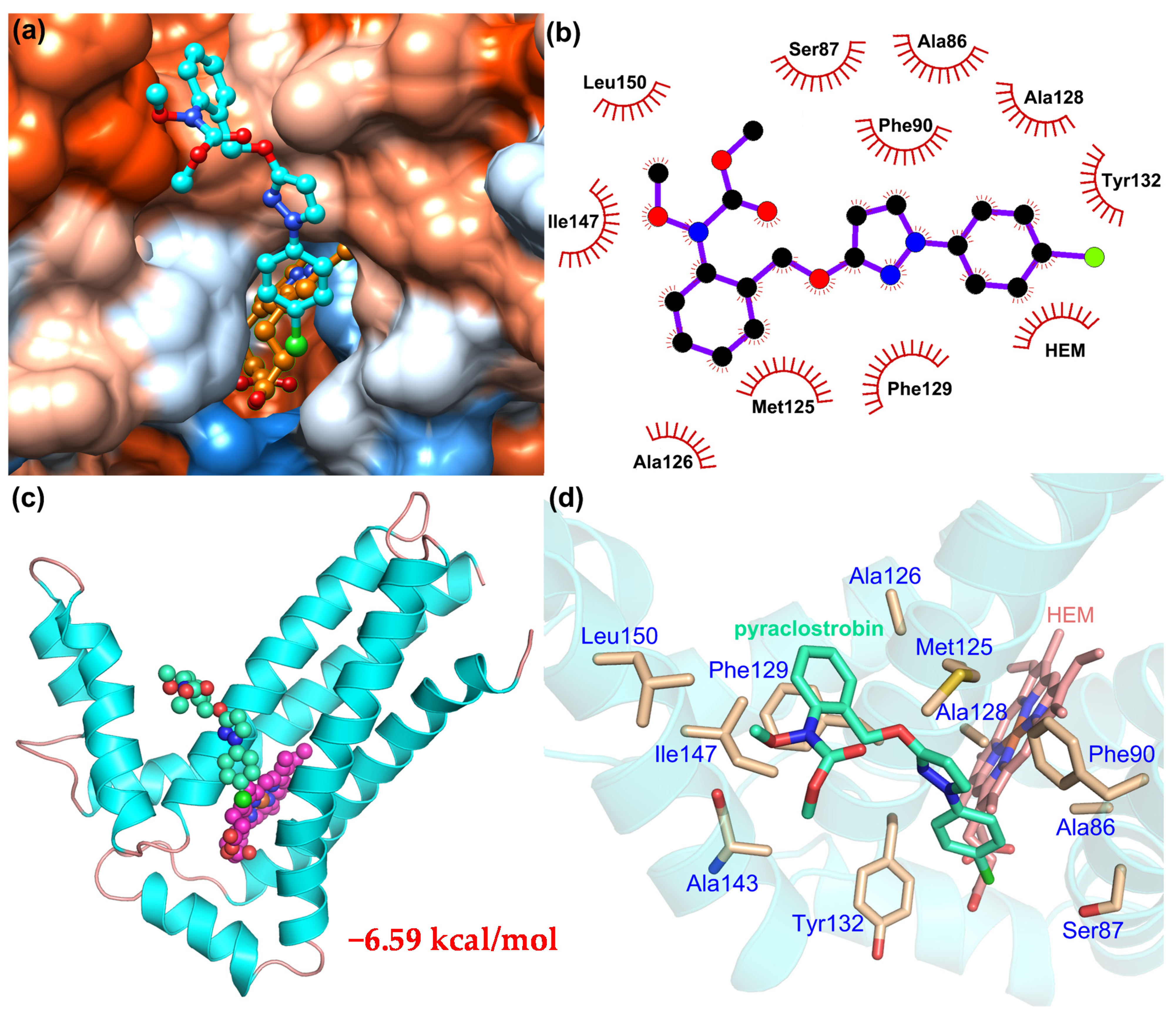
Docking results showed that pyraclostrobin can be bound to the vicinity of heme and form a more stable structure (Figure 2 and Figure 3). For wild-type CcCyt b, pyraclostrobin binds in a hydrophobic pocket composed of 14 amino acid residues, including Ser83, Ser86, Ser87, Phe90, Met125, Ala128, Phe129, Tyr132, Val133, Met139, Ser140, Gly143, Ala144, and Ile147. Among them, Phe90, Ala128, Phe129, Ala144, and Ile147 have strong hydrophobicity, and hydrophobic effects can enhance the affinity between pyraclostrobin and CcCytb (−7.35 kcal/mol) (Figure 2). For mutated CcCyt b with G143A, pyraclostrobin is incorporated in a hydrophobic pocket consisting of 10 amino acid residues, including Ser86, Ser87, Phe90, Met125, Ala126, Ala128, Phe129, Tyr132, Ile147, and Leu150. The number of residues recognized by pyraclostrobin is reduced. Because of the G143A mutation, the side chain is changed to a methyl group. Due to the effect of the spatial position, the direction of pyraclostrobin is changed, resulting in a changed molecular conformation. Thus, the binding capacity of pyraclostrobin and CcCyt b with G143A was weakened (−6.59 kcal/mol) (Figure 3).

3.3. LAMP Reaction Optimization

The two primer sets, containing FIP2 or FIP3, amplified the target Cyt b gene with the G143A mutation, according to a color change from pink to orange (Figure 4a). Thus, the primer set F3, B3, FIP3, and BIP, and corresponding genomic DNA were used for further optimization of the LAMP reaction. When the LAMP reaction was performed at 65 °C, the color change (pink to yellow) and intensity of the ladder-like band were significant (Figure 4b). Thus, the optimal reaction temperature was 65 °C, the same as the recommended temperature. In addition, the reaction time was also optimized at 65 °C. The target gene could be amplified within 45–75 min, and the color change (pink to yellow) and ladder-like band were most distinct after 60 and 75 min, respectively (Figure 4c). Thus, the LAMP reaction condition for the tested primer set was incubation at 65 °C for 60 to 75 min.

3.4. Detection Limit of LAMP Assay and Conventional PCR

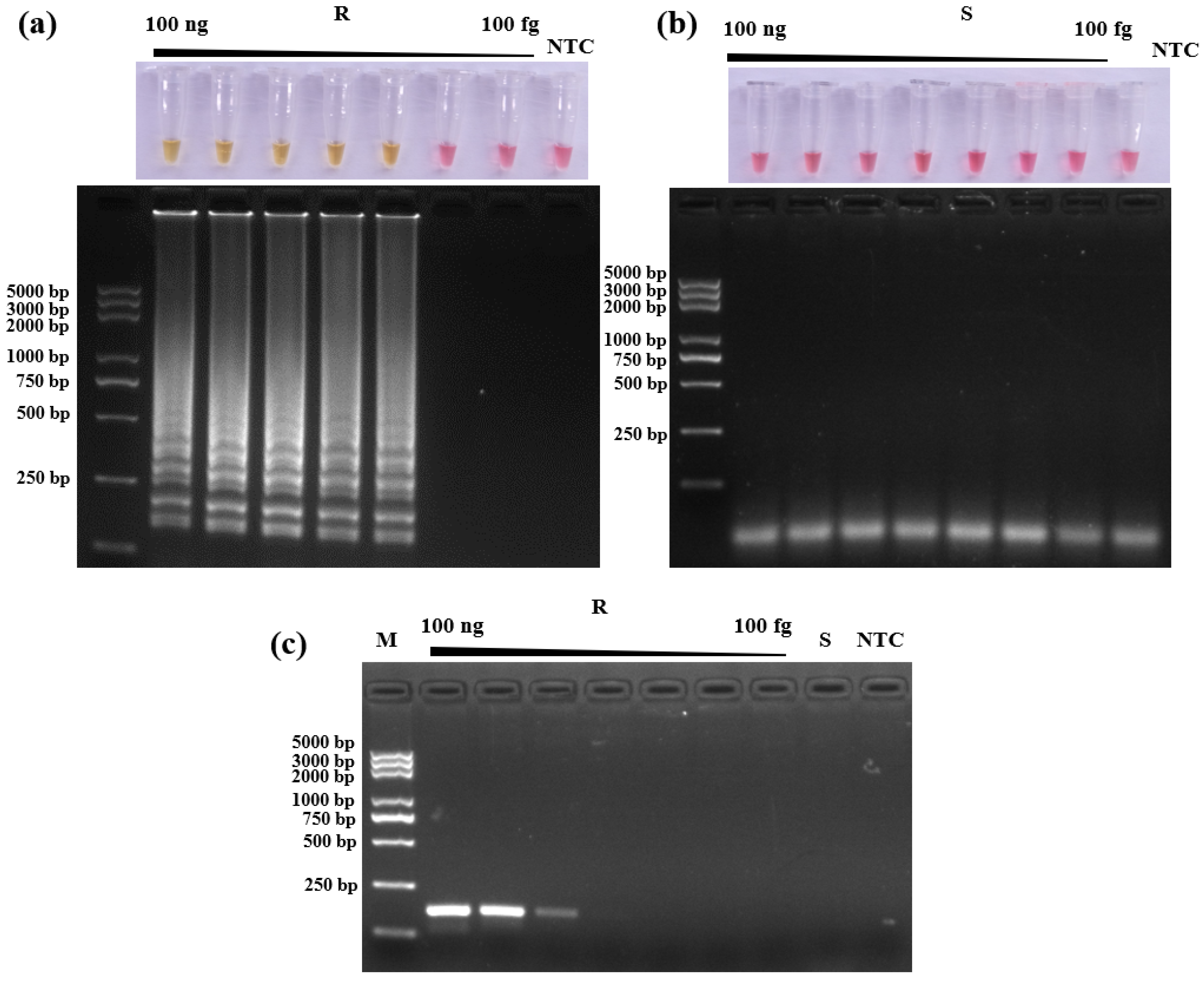
The lowest detection limit of the LAMP reaction was determined using the primer set (F3, B3, FIP3, and BIP) and tenfold serial dilutions of genomic DNA containing the G143A mutation from 100 ng/μL. The detection limit of LAMP is 0.01 ng/µL, and the detection limit of conventional PCR is 1 ng/µL (Figure 5).

3.5. LAMP Method Specificity

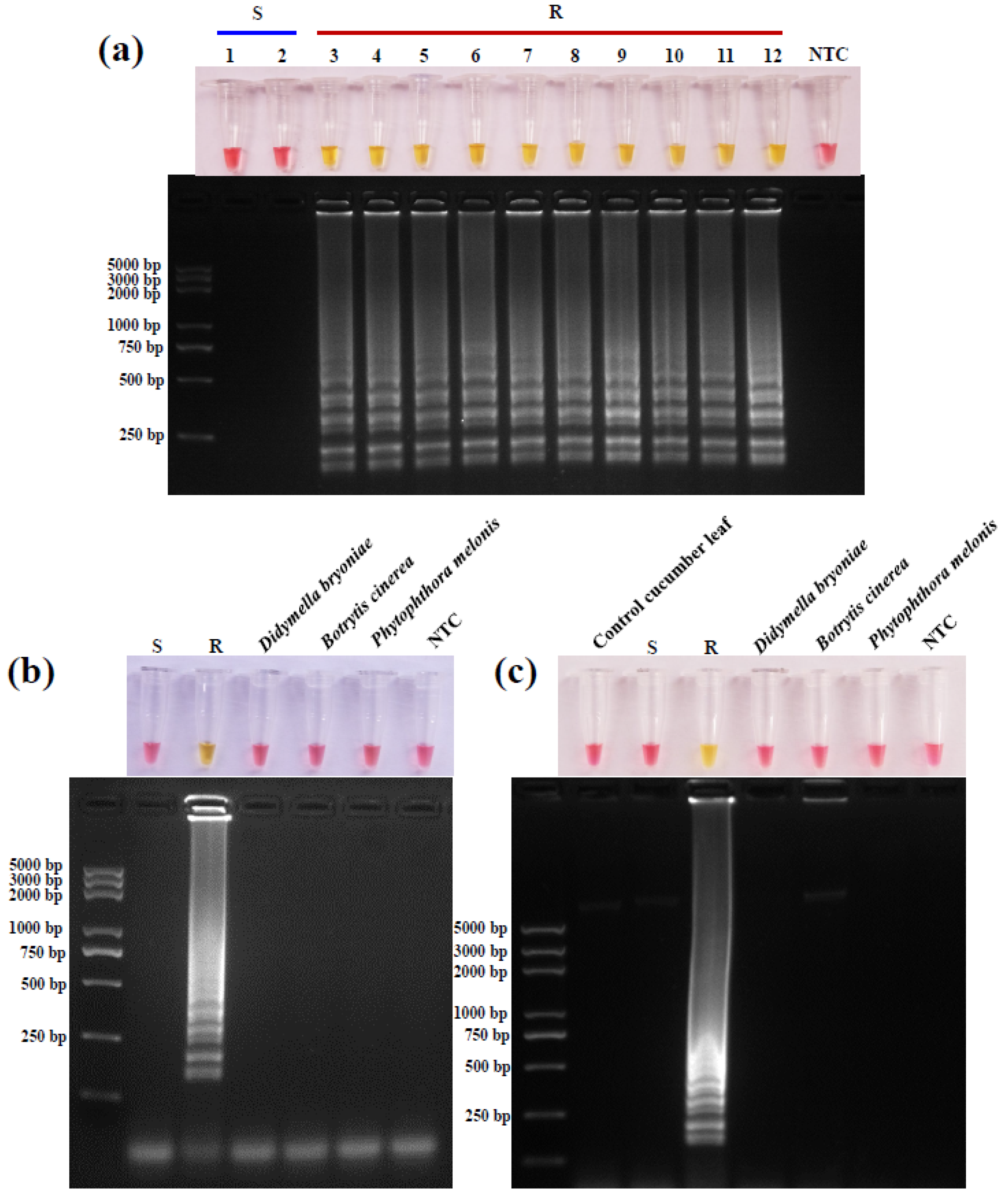
Genomic DNA was extracted from mycelia or infected cucumber leaves of several common pathogens of cucumber. The LAMP reaction results were positive only for C. cassiicola isolates containing the G143A mutation, and negative for pyraclostrobin-sensitive C. cassiicola isolates and D. bryoniae, B. cinerea, and P. melonis, irrespective of the DNA sampled from mycelia (Figure 6a,b) or infected leaves (Figure 6c).

4. Discussion

Duan et al. reported that all 619 C. cassiicola isolates (obtained in 2017 and 2018) from greenhouse-cultivated cucumbers in China contained the G143A mutation in Cyt b [12]. Thus, QoI resistance is a very serious problem in C. cassiicola populations from cucumber in China. Continuous resistance monitoring is of high significance for rational application of QoI in the protection and control of cucumber target spot disease. In the current study, all 131 C. cassiicola isolates obtained from Shandong province collected in 2019 and 2020 contained the G143A mutation. The EC50 values of pyraclostrobin for field isolates collected in this study were 1.67–8.82 μg/mL, similar to the data (0.51–7.01) for eight G143A-containing C. cassiicola strains for pyraclostrobin reported by Duan et al. [12]. Compared with the two QoI-sensitive wild-type C. cassiicola isolates (Cc4 and Cc10), which had a low range of EC50 values varying from 0.006 to 0.01 μg/mL (Table 2), all 131 C. cassiicola isolates had high levels of QoI resistance.
Apart from the relatively high inherent resistance risks of C. cassiicola and QoI, long-term continuous use is likely the main cause of current QoI resistance in C. cassiicola. The first QoI—trifloxystrobin—was registered to control cucumber target spot disease in 2014, and pyraclostrobin was registered in 2018 (http://www.chinapesticide.org.cn/ (accessed on 9 May 2021)). However, QoIs have been registered in China to control cucumber downy mildew, powdery mildew, and anthracnose for approximately 15 years. The symptoms of cucumber target spot are similar to those of downy mildew, bacterial angular leaf spot, and anthracnose, and it is not easy to distinguish them. The use of fungicides for downy mildew, bacterial angular leaf spot, and anthracnose during production is often not appropriate. This might be an important reason for the development of QoI resistance in C. cassiicola in China, but the actual field control effect of pyraclostrobin to C. cassiicola should be evaluated further.
It has been reported that QoI resistance of Podosphaera xanthii and Pseudoperonospora cubensis was observed to decline under no QoI fungicide selection pressure [33,34]. Furthermore, heteroplasmy (the coexistence of wild-type and mutated genes) was also found in QoI-resistant isolates of C. cassiicola, Mycovellosiella nattrassii, Colletotrichum gloeosporioides, and Podosphaera leucotricha [35,36]. Thus, it is recommended to suspend or postpone the use of QoI fungicides in the prevention and control of cucumber target spot disease.
G143A, F129L, and G137R are the three most widely reported main causes of QoI resistance. Many reports have shown that F129L and G137R mediate only low to moderate resistance levels and might have larger fitness penalties [37,38,39,40]. Interestingly, F129L and G137R were not found in any reported QoI-resistant C. cassiicola isolates obtained from cucumber, and F129L was found only in three moderately QoI-resistant C. cassiicola isolates obtained from tomato [12,13,36]. Thus, G143A is the most widespread mutation in QoI-resistant pathogens. Docking results showed that the G143A mutation causes a change in the binding pattern of pyraclostrobin and CcCyt b, and the number of amino acid residues that are recognized around the recognition is reduced. Hydrophobic interaction is reduced, leading to lowered binding ability of pyraclostrobin and the active center of CcCyt b–G143A, thereby conferring high resistance. Thus, rapid detection methods should be developed for G143A in Cyt b of C. cassiicola.
For traditional fungicide sensitivity determination, a mycelial growth inhibition assay, spore germination assay, and detached leaflet assay have been widely used (https://www.frac.info/ (accessed on 9 May 2021)). These methods require large amounts of medium and space, and are time consuming. Recently, a simple, accurate, and fast microtiter method was used to monitor fungicide sensitivity in C. cassiicola [14]. However, this method also requires the isolation and culturing of pathogen isolates. More rapid molecular detection methods have been developed to detect fungicide resistance. For G143A in C. cassiicola, PCR-RFLP was developed by Ishii et al. [36], but more recent molecular methods based on PCR or sequencing require sophisticated equipment and technical experience. Thus, further development of LAMP offers the best option for on-site detection, because this method only requires a heated chamber and samples. In this study, the WarmStart® Colorimetric LAMP 2X Master Mix (DNA & RNA) Kit was used. In this kit, an enhanced version of Bst DNA Polymerase (WarmStart® Bst 2.0 DNA Polymerase) and a visible pH indicator are mixed together to provide a rapid visual detection. To a certain extent, this approach can solve the problem of false positives. Our results showed that the LAMP system established for G143A in C. cassiicola based on this kit had high specificity and sensitivity (10 times better than conventional PCR), and the reaction takes only 1 h. Thus, the LAMP system established in this study can be used for rapid detection of G143A in C. cassiicola.

5. Conclusions

All tested isolates of C. cassiicola from Shandong province during 2019 and 2020 were resistant to pyraclostrobin and contained the G143A mutation in Cyt b. Docking results demonstrated that G143A mutation results in a change in the binding pattern of pyraclostrobin and CcCyt b, and the binding ability of pyraclostrobin with the active center of CcCyt b–G143A is lowered. The LAMP assay with a WarmStart Colorimetric LAMP 2× Master Mix kit was specific, rapid, and sensitive for detecting the G143A mutation in C. cassiicola. It will provide rapid and valid information for fungicide resistance management.

Author Contributions

Conceptualization, L.J., F.L., W.M. and X.L. (Xili Liu); methodology, X.L. (Xiuhuan Li) and C.L.; software, X.L. (Xiuhuan Li) and C.L.; validation, W.M. and X.L. (Xili Liu); formal analysis, X.L. (Xiuhuan Li) and C.L.; investigation, X.L. (Xiuhuan Li), G.L. and C.L.; resources, J.Z. and L.J.; data curation, X.L. (Xiuhuan Li), G.L. and C.L.; writing—original draft preparation, X.L. (Xiuhuan Li); writing—review and editing, L.J., W.M., F.L. and X.L. (Xili Liu); visualization, X.L. (Xili Liu); supervision, W.M. and X.L. (Xili Liu); project administration, W.M. and X.L. (Xili Liu); funding acquisition, W.M. and X.L. (Xili Liu). All authors have read and agreed to the published version of the manuscript.

Funding

This research was funded by National Key Research and Development Program of China, grant number 2016YFD0201305, 2017YFD0200307, and 2016YFD0200500.

Institutional Review Board Statement

Not applicable.

Informed Consent Statement

Not applicable.

Data Availability Statement

Not applicable.

Acknowledgments

The authors would like to thank Yabing Duan for his permission and providing the pyraclostrobin-sensitive Corynespora cassiicola Cc4, Cc10.

Conflicts of Interest

The authors declare no conflict of interest.

References

  1. Miyamoto, T.; Ishii, H.; Seko, T.; Kobori, S.; Tomita, Y. Occurrence of Corynespora cassiicola isolates resistant to boscalid on cucumber in Ibaraki Prefecture, Japan. Plant Pathol. 2010, 58, 1144–1151. [Google Scholar] [CrossRef]
  2. Li, T.; Xiu, Q.; Zhang, J.; Wang, J.X.; Duan, Y.B.; Zhou, M.G. Pharmacological characteristics and efficacy of fluazinam against Corynespora cassiicola, causing cucumber target spot in greenhouses. Plant Dis. 2020, 104, 2449–2454. [Google Scholar] [CrossRef]
  3. Carris, L.M.; Glawe, D.A.; Gray, L.E. Isolation of the soybean pathogens Corynespora cassiicola and Phialophora gregata from cysts of Heterodera glycines in Illinois. Mycologia 1986, 78, 503–506. [Google Scholar] [CrossRef]
  4. Dixon, L.J.; Schlub, R.L.; Pernezny, K.; Datnoff, L.E. Host specialization and phylogenetic diversity of Corynespora cassiicola. Phytopathology 2009, 99, 1015–1027. [Google Scholar] [CrossRef] [PubMed] [Green Version]
  5. Lv, G.X.; Ge, Y.P.; Shen, Y.N.; Li, M.; Zhang, X.; Chen, H.; Deng, S.; de Hoog, G.S.; Liu, W.D. Phaeohyphomycosis caused by a plant pathogen, Corynespora cassiicola. Med. Mycol. 2011, 49, 657–661. [Google Scholar] [CrossRef] [Green Version]
  6. Yamada, H.; Takahashi, N.; Hori, N.; Asano, Y.; Mochizuki, K.; Ohkusu, K.; Nishimura, K. Rare case of fungal keratitis caused by Corynespora cassiicola. J. Infect Chemother. 2013, 19, 1167–1169. [Google Scholar] [CrossRef]
  7. Smith, L.J. Host Range, Phylogenetic, and Pathogenic Diversity of Corynespora cassiicola (Berk. & Curt.) Wei. Ph.D. Thesis, University of Florida, Gainesville, FL, USA, 2008. [Google Scholar]
  8. Déon, M.; Scomparin, A.; Tixier, A.; Mattos, C.R.R.; Leroy, T.; Seguin, M.; Roeckel-Drevet, P.; Pujade-Renaud, V. First characterization of endophytic Corynespora cassiicola isolates with variant cassiicolin genes recovered from rubber trees in Brazil. Fungal Divers 2012, 54, 87–99. [Google Scholar] [CrossRef] [Green Version]
  9. Date, H.; Kataoka, E.; Tanina, K.; Sasaki, S.; Inoue, K.; Nasu, H.; Kasuyama, S. Sensitivity of Corynespora cassicola, causal agent of Corynespora leaf spot of cucumber, to thiophanate-methyl, diethofencarb and azoxystrobin. Jpn. J. Phytopathol. 2004, 70, 10–13. [Google Scholar] [CrossRef]
  10. Miyamoto, T.; Ishii, H.; Stammler, G.; Koch, A.; Ogawara, T.; Tomita, Y.; Fountaine, J.M.; Ushio, S.; Seko, T.; Kobori, S. Distribution and molecular characterization of Corynespora cassiicola isolates resistant to boscalid. Plant Pathol. 2010, 59, 873–881. [Google Scholar] [CrossRef]
  11. Ishii, H.; Miyamoto, T.; Ushio, S.; Kakishima, M. Lack of cross-resistance to a novel succinate dehydrogenase inhibitor, fluopyram, in highly boscalid-resistant isolates of Corynespora cassiicola and Podosphaera xanthii. Pest Manag. Sci. 2011, 67, 474–482. [Google Scholar] [CrossRef]
  12. Duan, Y.; Xin, W.; Lu, F.; Li, T.; Li, M.; Wu, J.; Wang, J.; Zhou, M. Benzimidazole- and QoI-resistance in Corynespora cassiicola populations from greenhouse-cultivated cucumber: An emerging problem in China. Pestic. Biochem. Phys. 2019, 153, 95–105. [Google Scholar] [CrossRef]
  13. Mackenzie, K.J.; Xavier, K.V.; Wen, A.; Timilsina, S.; Adkison, H.M.; Dufault, N.S.; Vallad, G.E. Widespread QoI fungicide resistance revealed among Corynespora cassiicola tomato isolates in Florida. Plant Dis. 2019, 104, 893–903. [Google Scholar] [CrossRef] [PubMed]
  14. Xavier, S.A.; de Mello, F.E.; da Silva, H.P.; Canteri, M.G.; Koga, L.J.; Lopes, I.O.N.; Godoy, C.V. Microtiter method to monitor Corynespora cassiicola and sensitivity of the pathogen to carbendazim, prothioconazole and pyraclostrobin. Crop Prot. 2021, 144, 105554. [Google Scholar] [CrossRef]
  15. Dufour, M.C.; Fontaine, S.; Montarry, J.; Corio-Costet, M.F. Assessment of fungicide resistance and pathogen diversity in Erysiphe necator using quantitative real-time PCR assays. Pest Manag. Sci. 2011, 67, 60–69. [Google Scholar] [CrossRef]
  16. Furuya, S.; Suzuki, S.; Kobayashi, H.; Saito, S.; Takayanagi, T. Rapid method for detecting resistance to a QoI fungicide in Plasmopara viticola populations. Pest Manag. Sci. 2010, 65, 840–843. [Google Scholar] [CrossRef] [PubMed]
  17. Hashimoto, M.; Aoki, Y.; Saito, S.; Suzuki, S. Characterisation of heteroplasmic status at codon 143 of the Botrytis cinerea cytochrome b gene in a semi-quantitative AS-PCR assay. Pest Manag. Sci. 2015, 71, 467–477. [Google Scholar] [CrossRef]
  18. Gobeil-Richard, M.; Tremblay, D.M.; Beaulieu, C.; der Heyden, H.V.; Carisse, O. A pyrosequencing-based method to quantify genetic substitutions associated with resistance to succinate dehydrogenase inhibitor fungicides in Botrytis spp. populations. Pest Manag. Sci. 2016, 72, 566–573. [Google Scholar] [CrossRef]
  19. Massi, F.; Torriani, S.F.F.; Borghi, L.; Toffolatti, S.L. Fungicide resistance evolution and detection in plant pathogens: Plasmopara viticola as a case study. Microorganisms 2021, 9, 119. [Google Scholar] [CrossRef]
  20. Notomi, T.; Okayama, H.; Masubuchi, H.; Yonekawa, T.; Watanabe, K.; Amino, N.; Hase, T. Loop-mediated isothermal amplification of DNA. Nucleic Acids Res. 2000, 28, E63. [Google Scholar] [CrossRef] [Green Version]
  21. Nagamine, K.; Hase, T.; Notomi, T. Accelerated reaction by loop-mediated isothermal amplification using loop primers. Mol. Cell Probes 2002, 16, 223–229. [Google Scholar] [CrossRef]
  22. Fukuta, S.; Nimi, Y.; Ohishi, K.; Yoshimura, Y.; Anai, N.; Hotta, M.; Fukaya, M.; Kato, T.; Oya, T.; Kanbe, M. Development of reverse transcription loop-mediated isothermal amplification (RT-LAMP) method for detection of two viruses and chrysanthemum stunt viroid. Annu. Rep. Kansai Plant Prot. Soc. 2005, 47, 31–36. [Google Scholar] [CrossRef]
  23. Safavieh, M.; Ahmed, M.U.; Ng, A.; Zourob, M. High-throughput real-time electrochemical monitoring of LAMP for pathogenic bacteria detection. Biosens. Bioelectron. 2014, 58, 101–106. [Google Scholar] [CrossRef] [PubMed]
  24. Niessen, L. Current state and future perspectives of loop-mediated isothermal amplification (LAMP)-based diagnosis of filamentous fungi and yeasts. Appl. Microbiol. Biotechnol. 2015, 99, 553–574. [Google Scholar] [CrossRef] [PubMed]
  25. Feng, W.; Hieno, A.; Kusunoki, M.; Suga, H.; Kageyama, K. LAMP detection of four plant-pathogenic oomycetes and its application in lettuce fields. Plant Dis. 2018, 103, 298–307. [Google Scholar] [CrossRef] [Green Version]
  26. Hu, X.R.; Dai, D.J.; Wang, H.D.; Zhang, C.Q. Rapid on-site evaluation of the development of resistance to quinone outside inhibitors in Botrytis cinerea. Sci. Rep. 2017, 7, 13861. [Google Scholar] [CrossRef]
  27. Chen, S.; Schnabel, G.; Yuan, H.; Luo, C. LAMP detection of the genetic element ‘Mona’ associated with DMI resistance in Monilinia fructicola. Pest Manag. Sci. 2019, 75, 779–786. [Google Scholar] [CrossRef]
  28. Fan, F.; Yin, W.X.; Li, G.Q.; Lin, Y.; Luo, C.X. Development of a LAMP method for detecting SDHI fungicide resistance in Botrytis cinerea. Plant Dis. 2018, 102, 1612–1618. [Google Scholar] [CrossRef] [Green Version]
  29. Vielba-Fernández, A.; de Vicente, A.; Pérez-García, A.; Fernández-Ortuño, D. Monitoring methyl benzimidazole carbamate-resistant isolates of the cucurbit powdery mildew pathogen, Podosphaera xanthii, using loop-mediated isothermal amplification. Plant Dis. 2019, 103, 1515–1524. [Google Scholar] [CrossRef]
  30. Zhu, J.; Zhang, L.; Li, H.; Gao, Y.; Mu, W.; Liu, F. Development of a LAMP method for detecting the N75S mutant in SDHI-resistant Corynespora cassiicola. Anal. Biochem. 2020, 597, 113687. [Google Scholar] [CrossRef]
  31. Chen, L.; Zhu, S.; Lu, X.; Pang, Z.; Cai, M.; Liu, X. Assessing the risk that Phytophthora melonis can develop a point mutation (V1109L) in CesA3 conferring resistance to carboxylic acid amide fungicides. PLoS ONE 2012, 7, e42069. [Google Scholar] [CrossRef]
  32. Lange, C. Specific roles of protein-phospholipid interactions in the yeast cytochrome bc1 complex structure. Eur. Mol. Biol. Organ. 2001, 20, 6591–6600. [Google Scholar] [CrossRef] [PubMed] [Green Version]
  33. Sierotzki, H.; Kraus, N.; Assemat, P.; Stanger, C.; Cleere, S.; Windass, J.; Gisi, U. Evaluation of resistance to QoI fungicides in Plasmopara viticola populations in Europe. In Modern Fungicides and Antifungal Compounds IV; Dehne, H.W., Gisi, U., Kuck, K.H., Russell, P.E., Lyr, H., Eds.; BCPC Publishing: Alton, UK, 2005; pp. 73–80. [Google Scholar]
  34. Genet, J.L.; Jaworska, G.; Deparis, F. Effect of dose rate and mixtures of fungicides on selection for QoI resistance in populations of Plasmopara viticola. Pest Manag. Sci. 2006, 62, 188–194. [Google Scholar] [CrossRef] [PubMed]
  35. Lesemann, S.S.; Schimpke, S.; Dunemann, F.; Deising, H.B. Mitochondrial heteroplasmy for the cytochrome b gene controls the level of strobilurin resistance in the apple powdery mildew fungus Podosphaera leucotricha (Ell.&Ev.) E.S. Salmon. J. Plant Dis. Protect. 2006, 113, 259–266. [Google Scholar]
  36. Ishii, H.; Yano, K.; Date, H.; Furuta, A.; Sagehashi, Y.; Yamaguchi, T.; Sugiyama, T.; Nishimura, K.; Hasama, W. Molecular characterization and diagnosis of QoI resistance in cucumber and eggplant fungal pathogens. Phytopathology 2007, 97, 1458–1466. [Google Scholar] [CrossRef] [PubMed] [Green Version]
  37. Esser, L.; Quinn, B.; Li, Y.F.; Zhang, M.; Elberry, M.; Yu, L.; Yu, C.A.; Xia, D. Crystallographic studies of quinol oxidation site inhibitors: A modified classification of inhibitors for the cytochrome bc1 complex. J. Mol. Biol. 2004, 341, 281–302. [Google Scholar] [CrossRef]
  38. Sierotzki, H.; Frey, R.; Wullschleger, J.; Palermo, S.; Karlin, S.; Godwin, J.; Gisi, U. Cytochrome b gene sequence and structure of Pyrenophora teres and P. tritici-repentis and implications for QoI resistance. Pest Manag. Sci. 2007, 63, 225–233. [Google Scholar] [CrossRef]
  39. Sierotzki, H. Respiration Inhibitors: Complex III. In Fungicide Resistance in Plant Pathogens; Ishii, H., Hollomon, D.W., Eds.; Springer Publishing: Tokyo, Japan, 2015; pp. 119–144. [Google Scholar]
  40. Klosowski, A.C.; de Mio, L.L.M.; Miessner, S.; Rodrigues, R.; Stammler, G. Detection of the F129L mutation in the cytochrome b gene in Phakopsora pachyrhizi. Pest Manag. Sci. 2016, 72, 1211–1215. [Google Scholar] [CrossRef] [PubMed]
Figure 1. LAMP primers for the G143A mutation of Cyt b in Corynespora cassiicola. (a) Sequence alignment of sensitive and resistant Cyt b alleles. The sequences used as LAMP primers are indicated by vertical lines. For each mutation, the F2 primer ends with its corresponding G143A mutant nucleotide. (b) Schematic diagram of LAMP primers used in this work. The internal primer FIP is composed of F1c and F2, whereas BIP is composed of B1 and B2c. The primers ending with “c” are complementary to the primers without “c”.
Horticulturae 07 00155 g001
Figure 2. Binding mode of pyraclostrobin and CcCyt b protein. (a) Binding mode of pyraclostrobin, and hydrophobic and hydrophilic surfaces of CcCyt b protein. Blue and orange represent hydrophilic and hydrophobic regions, respectively. (b) Two-dimensional interaction of pyraclostrobin and CcCyt b, whereby red zigzag represents hydrophobic effect. (c) Binding position of pyraclostrobin and CcCyt b protein. (d) Interaction of pyraclostrobin and the amino acids of CcCyt b.
Horticulturae 07 00155 g002
Figure 3. Binding mode of pyraclostrobin and CcCyt b protein with G134A. (a) Binding mode of pyraclostrobin and hydrophobic and hydrophilic surfaces of CcCyt b–G143A protein. Blue and orange represent hydrophilic and hydrophobic regions, respectively. (b) Two-dimensional interaction of pyraclostrobin and CcCyt b–G143A, whereby red zigzag represents hydrophobic effect. (c) Binding position of pyraclostrobin and CcCyt b–G143A protein. (d) Interaction of pyraclostrobin and the amino acids of CcCyt b–G143A.
Horticulturae 07 00155 g003
Figure 4. Optimization of LAMP reaction conditions for G143A in Corynespora cassiicola. (a) Selection of FIP primer of LAMP reaction. Optimization of the LAMP reaction temperature (b) and time (c) on the basis of color change (upper) and corresponding gel electrophoresis detection (lower). Reaction temperature gradient was set to 61, 63, 65, 67, and 69 °C, and the reaction time gradient was 30, 45, 60, 75, and 90 min. “S”, pyraclostrobin-sensitive isolate Cc4; “R”, pyraclostrobin-resistant isolate Cc66.
Horticulturae 07 00155 g004
Figure 5. Sensitivity test of the LAMP reaction (a,b) and conventional PCR (c) for G143A in Corynespora cassiicola. LAMP reaction products of pyraclostrobin-resistant isolate (a) and -sensitive isolate (b) were evaluated on the basis of color change (upper) and corresponding gel electrophoresis detection (lower); conventional PCR products were assessed by 2% gel electrophoresis (c). The template genomic DNA concentrations used in reaction tubes from left to right were 100 ng, 10ng, 1 ng, 0.1 ng, 0.01 ng, 0.001 ng, and 100 fg. “S”, pyraclostrobin-sensitive C. cassiicola isolate Cc4; “R”, pyraclostrobin-resistant C. cassiicola isolate Cc66. “NTC”, negative treatment control with dH2O as reaction template.
Horticulturae 07 00155 g005
Figure 6. Specificity of LAMP assay for the G143A mutation in the Cyt b gene of Corynespora cassiicola or other causal agents (Didymella bryoniae, Botrytis cinerea, and Phytophthora melonis) that cause common cucumber diseases. Genomic DNA of mycelia (a,b) or infected cucumber leaves (c) of four important pathogens. “S”, pyraclostrobin-sensitive C. cassiicola isolates (Cc4, Cc10); “R”, pyraclostrobin-resistant C. cassiicola isolates (Cc21, Cc23, Cc31, Cc35, Cc42, Cc53, Cc71, Cc66, Cc96, Cc123). “NTC”, negative control using dH2O as reaction template.
Horticulturae 07 00155 g006
Table 1. Primers used for LAMP and partial Cyt b gene amplification from Corynespora cassiicola.
Primer NameSequence (5′-3′) aUse
F3CCTGGGTTATGTTTTACCATACForward outer primer for LAMP
B3AAGTGCATTAGTGCTAAAGCBackward outer primer for LAMP
BIPTTGAGGAGGTTTCTCTGTTAACAATGCTAATACGAAAGGTAAAACGAAATBackward inner primer for LAMP
FIP1ACTCAAGGGATGGCACTCATTAGGTCAAATGTCCTTATGACCForward inner primers to detect the G143A mutants of Cyt b gene in C. cassiicola for LAMP
FIP2ACTCAAGGGATGGCACTCATTAGGTCAAATGTCCTTATGAAC
FIP3ACTCAAGGGATGGCACTCATTAGGTCAAATGTCCTTATGATC
FGTCAAATGTCCTTATGATCTo amplify partial fragments (163 bp) of Cyt b gene of C. cassiicola
RGCTAATACGAAAGGTAAAACGAAAT
CcCytb-FGCGAATTCCTATTTAGTTGATTCPCR primers to amplify the partial Cytb from C. cassiicola [12]
CcCytb-RGGTTACCTGATCCAGCTGTATC
a Nucleotides with underline were mutant in the Cyt b gene sequence from pyraclostrobin-sensitive and -resistant C. cassiicola isolates. Nucleotides in boxes are mismatches manually introduced to detect G143A resistant genotypes of C. cassiicola.
Table 2. Sensitivity of Corynespora cassiicola isolates collected from Shandong province to pyraclostrobin.
SourcePyraclostrobin
N Range of EC50 (μg/mL)Mean ± SD (μg/mL)
Lanling County, Linyi City251.98–8.685.09 ± 2.25 ns
Xin County, Liaochen City161.67–6.944.74 ± 1.58
Daiyue District, Taian City221.74–8.745.09 ± 1.79
Laicheng Distrist, Laiwu City41.95–4.693.71 ± 2.20
Pingyuan County, Dezhou City121.68–8.823.92 ± 2.04
Shouguang City, Weifang City261.99–8.005.32 ± 1.92
Jiyang County, Jinan City121.70–6.213.68 ± 1.71
Zhoucun County, Zibo City141.88–8.694.43 ± 1.95
Cumulative1311.67–8.824.74 *
Duan’s lab §20.006–0.010.008
N = Number of isolates. SD = standard deviation. ns not significant data (p = 0.05). * The mean EC50 value of pyraclostribin to 131 Corynespora cassiicola isolates. § Two pyraclostrobin-sensitive isolates Cc4 and Cc10 were kindly provided by Associate Professor Yabing Duan of Nanjing Agricultural University.
Publisher’s Note: MDPI stays neutral with regard to jurisdictional claims in published maps and institutional affiliations.