Article

Dynamic Carotenoid Profiles and Function Analysis of the RrPSY1 Gene in Rosa rugosa Flowers

1
College of Horticulture and Landscape Architecture, Yangzhou University, Yangzhou 225009, China
2
National Key Laboratory for the Development and Utilization of Forest Food Resources, Nanjing Forestry University, Nanjing 210037, China
*
Author to whom correspondence should be addressed.
These authors contributed equally to this work.
Horticulturae 2025, 11(9), 1137; https://doi.org/10.3390/horticulturae11091137
Submission received: 15 August 2025 / Revised: 15 September 2025 / Accepted: 16 September 2025 / Published: 18 September 2025

Abstract

Rosa rugosa is an important ornamental and edible species that is valued for its floral colors and essential oils in the cosmetic and pharmaceutical industries. Carotenoids, beyond their health-promoting roles, function as accessory pigments that influence petal coloration, flower quality, and stress responses. However, their accumulation patterns and molecular biosynthesis in R. rugosa remain poorly understood. Here, UPLC-APCI-MS/MS analysis across three developmental stages (bud, semi-open, and full bloom) revealed stage-specific carotenoid accumulation, with phytoene and phytofluene markedly increasing at the semi-open stage. In total, 11 carotenoids were identified, comprising four carotenes and seven xanthophylls. Differential accumulation of metabolites (DAMs) analysis indicated shifts in compounds, including (E/Z)-phytoene, phytofluene, and β-carotene across stages. Genetic complementation assays in Escherichia coli and transient overexpression in rose petals confirmed that RrPSY1 functions as a phytoene synthase. qRT-PCR results showed its upregulation under salt treatment, suggesting a role in enhancing stress tolerance through carotenoid-mediated antioxidant protection. Furthermore, sub-cellular localization experiments confirmed plastid targeting of RrPSY1. Together, these findings clarify the role of RrPSY1 in carotenoid biosynthesis and provide a foundation for future studies on metabolic regulation and biosynthesis of carotenoids in R. rugosa.

1. Introduction

Carotenoids are naturally occurring lipophilic pigments that are widely distributed in plants, algae, and certain microorganisms [1]. They are responsible for the yellow, orange, and red pigmentation observed in many photosynthetic organisms [2]. More than 1100 carotenoids have been cataloged in databases (http://carotenoiddb.jp/, accessed on 30 January 2024) [3]. Based on their structure, carotenoids are divided into two classes: xanthophylls, which contain oxygen functional groups, and carotenes, which are hydrocarbon molecules that are devoid of oxygen [4]. Beyond their central roles in photosynthesis and photoprotection, and as precursors for phytohormones such as abscisic acid (ABA) and strigolactones (SLs), carotenoids have specialized functions in flowers. They contribute to coloration and pigmentation patterns, enhancing visual cues that attract pollinators. In petals, carotenoids also act as accessory pigments, protecting tissues from excessive light and oxidative stress [5,6]. From a nutritional perspective, carotenoids are valued as provitamin A precursors and for maintaining visual health; lutein and zeaxanthin, for example, protect against age-related eye disorders [7,8,9].
In plants, carotenoid biosynthesis proceeds via the plastid-localized methylerythritol 4-phosphate (MEP) pathway [10] (Figure 1). This pathway produces isopentenyl diphosphate (IPP) and dimethylallyl diphosphate (DMAPP), which are converted into geranylgeranyl diphosphate (GGPP) by geranylgeranyl diphosphate synthase (GGPPS) [11]. The first committed and rate-limiting step in carotenoid biosynthesis is catalyzed by phytoene synthase (PSY, EC 2.5.1.32), which condenses two GGPP molecules to form phytoene, a 15-cis isomer [12]. PSY is a key enzyme in the carotenoid biosynthetic pathway. The first plant PSY gene to be characterized was pTOM5, a tomato fruit-specific isoform that strongly influences carotenoid accumulation in tomato fruit [13,14]. Tomato PSY2, in contrast, provides precursors for ABA biosynthesis [15]. PSYs have been extensively studied across plant species and applied in metabolic engineering to enhance carotenoid content for nutritional improvement [16,17,18,19]. Its enzymatic activity requires Mn2+ as a cofactor and stabilized by chaperone proteins that support structural integrity and catalytic function [20].
Rosa rugosa, native to East Asia, is valued for its essential oil, widely used in cosmetics and pharmaceuticals [21,22]. Its petals are also edible [21,22]. Rose flowers are rich in terpenoids and phenylpropanoids, as well as other metabolites [23,24]. Among these, carotenoids represent an important but relatively understudied group compared with other metabolites. Whereas anthocyanin accumulation has been extensively investigated in roses, the contribution of carotenoids, particularly in R. rugosa, to coloration and stress response remains poorly understood [25]. Previous studies of carotenoids in roses have been limited and largely descriptive [26]. In particular, the spatiotemporal dynamics of carotenoid accumulation and the molecular mechanisms underlying their biosynthesis in R. rugosa have not been elucidated. We hypothesized that carotenoids in R. rugosa petals undergo stage-specific changes during flower development and contribute to stress responses. To test this, we employed high-sensitivity UPLC-APCI-MS/MS metabolite profiling across three floral stages: bud (S1), semi-open (S2), and full bloom (S3), chosen to represent distinct phases of petal development. We further examined the function of RrPSY1, using E. coli genetic complementation, transient overexpression in rose petals, subcellular localization, and stress-induced expression analysis. These findings address a critical gap in understanding carotenoid biosynthesis in R. rugosa and provide a foundation for future efforts to enhance edible rose varieties.

2. Materials and Methods

2.1. Plant Materials

The experimental material consisted of R. rugosa “Zi Zhi”, which was cultivated at the Yangzhou University nursery (32.391° N, 119.419° E) in Yangzhou, Jiangsu Province, China. Flower samples displaying uniform morphological characteristics were selected, rapidly frozen in liquid nitrogen, and preserved at −80 °C for subsequent analysis.

2.2. Carotenoid Extraction and Analysis

Carotenoid extraction and quantification were performed according to previously established protocols at Metware Biotechnology Co., Ltd. (Wuhan, China), with modifications for rose petal tissue [27]. Approximately 50 mg sample of tissue was extracted with a hexane/acetone/ethanol mixture (1:1:1, v/v) by vortexing for 20 min. Following centrifugation (12,000× g, 5 min, 4 °C), the supernatant was subjected to triple liquid–liquid partitioning with saturated NaCl, dried under N2, and saponified overnight in MTBE/10% KOH-MeOH (1:1). The dried extract was reconstituted in MeOH/MTBE (1:1), filtered through a 0.22 μm membrane, and analyzed using a UPLC-APCI-MS/MS system (UPLC, ExionLC™ AD; MS, Applied Biosystems 6500 Triple Quadrupole, Sciex, Singapore). The sensitivity of the method was validated by determining the limit of detection (LOD) and limit of quantification (LOQ) for each carotenoid (Supplementary Table S1). Metabolites with Variable Importance in Projection (VIP) ≥ 1.0 in the orthogonal partial least squares-discriminant analysis (OPLS-DA) model were considered significant. For univariate analysis, thresholds were set at fold change (FC) ≥ 2.0 or ≤0.5 and p < 0.05. All quantifications were based on three independent biological replicates.

2.3. Salt Treatment and qRT-PCR Analysis

Two groups of R. rugosa plants, each with three biological replicates, were established. The salt stress group was treated with 100 mM NaCl for 2 h, whereas the control group (CK) was treated with deionized water for the same duration [28]. For developmental expression analysis, petals were sampled at three stages: bud (S1), semi-open bloom (S2), and full bloom (S3). For salt stress treatment, leaves and petals at S2 were sampled separately, frozen instantaneously in liquid nitrogen, and stored at −80 °C [29]. RNA was extracted using the Takara MiniBEST Plant RNA Extraction Kit (TaKaRa, Beijing, China), following the manufacturer’s instructions. First-strand cDNA was synthesized from 2 μg of total RNA, using the PrimeScript™ II 1st-strand cDNA Synthesis Kit (TaKaRa, Beijing, China). The quantitative real-time PCR (qRT-PCR) was performed using HiScript III RT SuperMix and ChamQ SYBR Color qPCR Master Mix on a Bio-Rad CFX96 system, with three independent biological replicates per sample. Relative gene expression levels were normalized using 5.8S as the internal reference gene and were calculated using the 2−ΔΔCt method based on Ct values obtained via CFX Manager software 1.3 [30]. The primer sequences are provided in Supplementary Table S2.

2.4. Identification and Bioinformatic Analysis of RrPSY1

The RrPSY1 gene was identified by a BLASTP search against the R. rugosa proteome. Conserved domains were validated using Pfam (https://pfam.xfam.org/ 15 March 2024) [31]. Physicochemical properties were assessed using the ExPASy online tool (https://www.expasy.org/ 15 March 2024). Multiple sequence alignment and phylogenetic tree construction of PSY proteins were conducted in MEGA (version 11) using the neighbor-joining method with 500 bootstrap replicates [32]. The analyzed PSY protein sequences analyzed included: Arabidopsis thaliana AtPSY (AAA32836), Solanum lycopersicum SlPSY1 (ABM45873), SlPSY2 (ABU40771), and SlPSY3 (Solyc01g005940), Oryza sativa OsPSY1 (AAS18307), OsPSY2 (AK073290), and OsPSY3 (DQ356431), Malus domestica MdPSY1 (KT189149), MdPSY2 (KT189150), and MdPSY4 (KT189152). The open reading frame (ORF) of RrPSY1 was amplified with PrimeSTAR Max DNA polymerase, cloned into pEASY-Blunt vectors (TransGen Biotech, Beijing, China), and transformed into E. coli Trans1-T1 competent cells for sequencing by Sangon Biotech. Amino acid sequences of all PSY proteins used are detailed in Supplementary Table S3.

2.5. Pigment Complementation Assay

The RrPSY1 gene was cloned into the pEASY-E1 expression vector and co-transformed with pAC-85b (available at https://www.addgene.org/Francis_X_Cunningham_Jr/, 1 April 2023) into E. coli BL21 (DE3) cells (TransGen Biotech, Beijing, China). Positive colonies were selected and cultured overnight in LB medium with carbenicillin (100 μg/mL) and chloramphenicol (34 μg/mL) at 37 °C. The culture was diluted into the 50 mL LB medium and grown at 37 °C to OD600 = 0.5, followed by induction with 0.3 mM isopropyl β-thiogalactopyranoside (IPTG). After overnight incubation at 22 °C, 1 mL aliquots were centrifuged (8000× g, 60 s), resuspended in 100 μL H2O, and extracted with 400 μL ice-cold 90% acetone for 5 min. Following centrifugation (15,000× g, 5 min), carotenoids were quantified as described previously [33].

2.6. Subcellular Localization Assay

Subcellular localization of RrPSY1 was predicted with TargetP-2.0 (https://services.healthtech.dtu.dk/services/TargetP-2.0/. 8 April 2024). The ORF of RrPSY1 was inserted into the pCAMBIA 1300-35S-sGFP vector employing the ClonExpress II One Step Cloning Kit (Vazyme, Nanjing, China). The resultant construct, pCAMBIA 1300-35S-RrPSY1-sGFP, was then transformed into Agrobacterium tumefaciens strain GV3101 and transiently infiltrated into the leaves of Nicotiana benthamiana leaves [34]. GFP fluorescence was visualized 48–72 h post-infiltration using a Zeiss LSM 880 confocal microscope at 488 nm.

2.7. Transient Overexpression Transformation in Rose

The full-length RrPSY1 gene was constructed into the pCAMBIA 1300-35S-E9 vector and introduced into the A. tumefaciens strain GV3101. The culture was diluted 1:50 into a fresh LB medium and was grown at 28 °C with shaking until OD600 reached 0.8–1.2. Cells were harvested by centrifugation at 5000× g for 10 min, and the pellet was resuspended in infiltration buffer (10 mM MES, pH 5.6, 100 µM Acetosyringone) to final OD600 of 0.5, which was then transiently infiltrated into Rosa hybrida petals for 30 min. After discarding the bacterial suspension, the petals were washed with 1/2 MS solution, then placed on a Petri plate containing the MS medium, supplemented with 2 mg/L ABA, 3 mg/L 6-BA and 50 mM acetosyringone. The plates were incubated in the dark at 24 °C for 72 h [35,36]. For chlorophyll and carotenoid extraction, 50 mg of plant material was homogenized in 80% acetone. For RT-qPCR, 2 μg total RNA of petals were used for analysis. The concentrations of chlorophyll and carotenoids were calculated according to a previously described method [27]. As a negative control, rose petal discs were infiltrated with Agrobacterium carrying the empty pCAMBIA1300-35S-E9 vector.

2.8. Statistical Analysis

Gene expression and carotenoid data were analyzed by one-way analysis of variance (ANOVA) using SPSS Statistics 28.0. Assumptions of normality and homogeneity of variances were verified with the Shapiro–Wilk and Levene’s tests. Where significant differences were observed (p < 0.05), Dunnett’s multiple comparison test was applied for comparisons among multiple groups, while independent t-tests were used for pairwise comparisons (* p < 0.05, ** p < 0.01).

3. Results

3.1. Dynamic Carotenoid Profiles in R. rugosa Petals During Flower Development

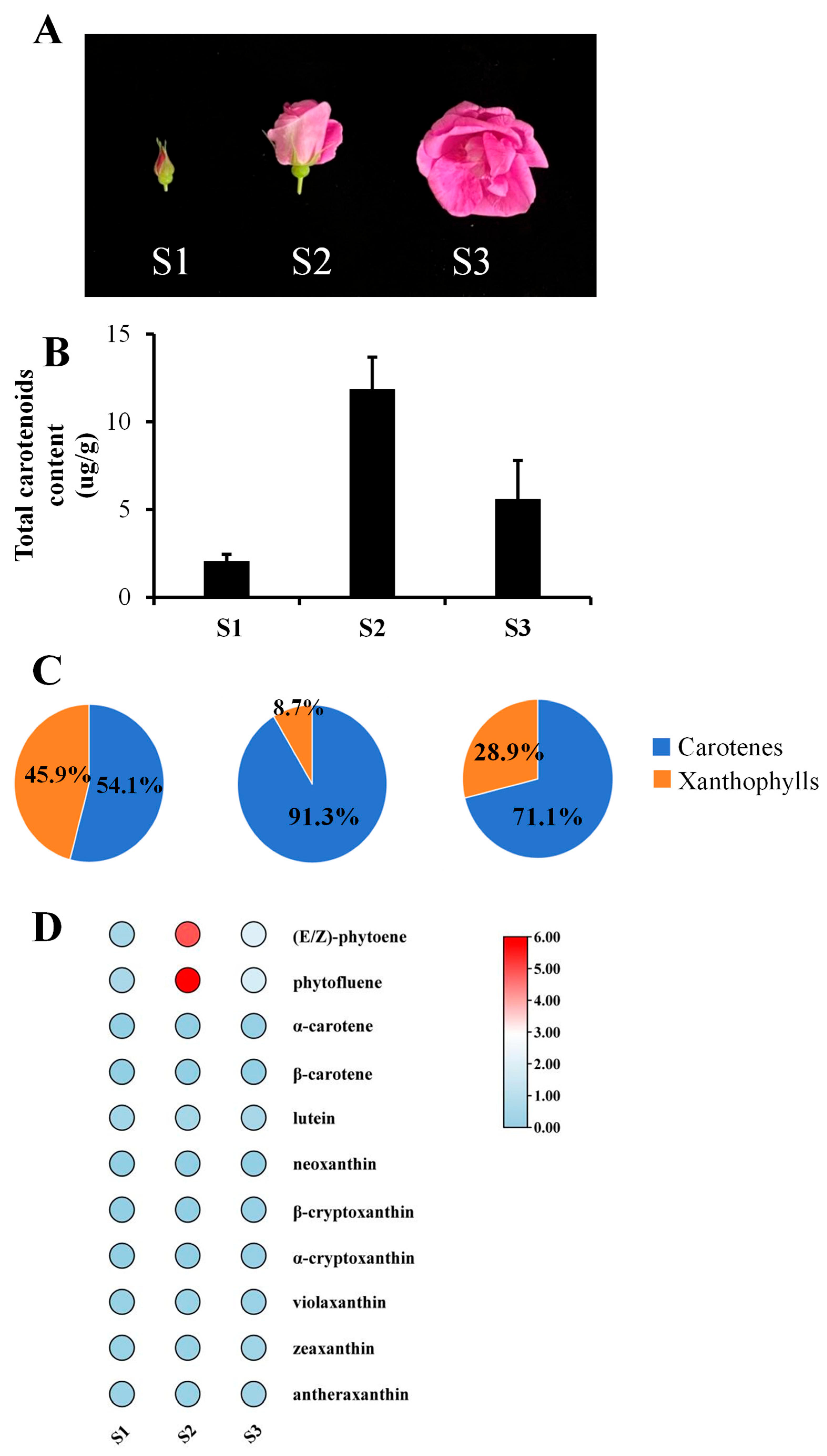
To investigate carotenoid content and developmental alterations in R. rugosa petals, three stages: bud (S1), the semi-open bloom (S2), and the full bloom (S3), were analyzed using the UPLC-APCI-MS/MS (Figure 2A). A total of 11 carotenoids were identified across the three stages (Supplementary Table S1). These included four carotenes (hydrocarbon compounds) and seven xanthophylls (oxygenated derivatives). The total carotenoid content exhibited significant dynamic changes, with a marked increase at S2 followed by a decrease at S3 (Figure 2B). Carotenes accounted for 72.3% of the total pool, whereas xanthophylls comprised 27.7%. The relative proportions shifted during development: carotenes accounted for 54.1%, 91.3%, and 71.1% at S1, S2, and S3, respectively, whereas xanthophylls accounted for 45.9%, 8.7%, and 28.9% at these stages (Figure 2C). Quantitative analysis demonstrated that phytofluene (6.88 μg/g) and phytoene (5.89 μg/g) were the most abundant carotenoids at S2. Both belong to the class of colorless carotenes. Other carotenoids were present at low concentrations (0 to 0.2 μg/g), with only minor variation across stages (Figure 2D).
Differential accumulations of metabolites (DAMs) between developmental stages were analyzed (Supplementary Table S4). A comparison of S3 to S1 revealed that most xanthophylls were downregulated. In contrast, the early pathway precursors phytoene and phytofluene were significantly upregulated. The six metabolites that were found to be downregulated were α-carotene, β-carotene, β-cryptoxanthin, antheraxanthin, zeaxanthin, and α-cryptoxanthin. The two metabolites that were found to be upregulated were phytoene and phytofluene.

3.2. Identification and Phylogenetic Analysis of the RrPSY1 Gene in R. rugosa

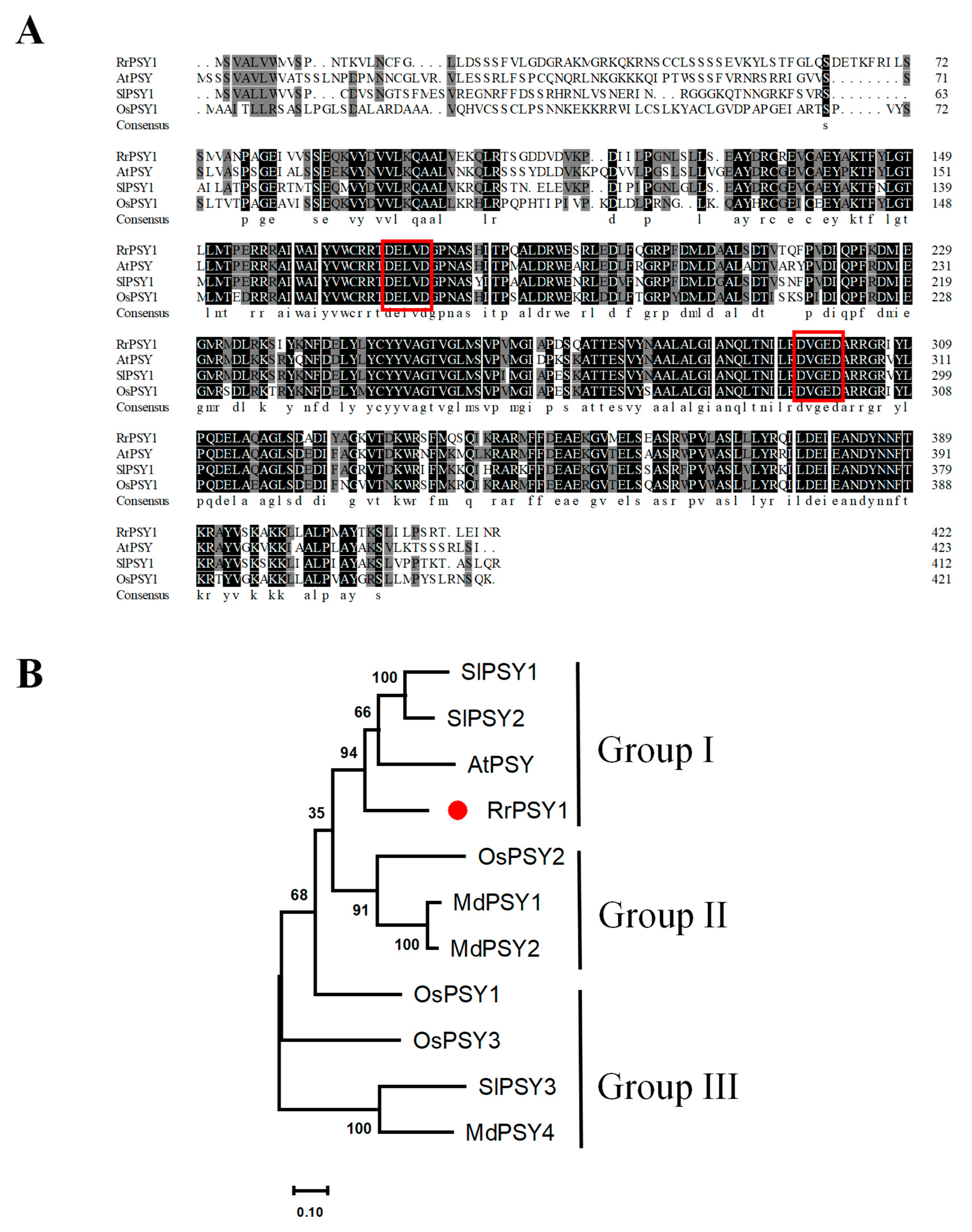
The high accumulation of phytoene and phytofluene during floral development (particularly at S2), suggested an important role for RrPSY1, which catalyzes the conversion of GGPP to phytoene (Figure 1). Our previous protein interaction assays confirmed that RrGGPPS1 physically interacts with RrPSY1 (evm.modle.Chr4.3473), implicating its function in carotenoid biosynthesis in R. rugosa [27]. The coding sequence length (CDS) of RrPSY1 is 1272 bp, encoding a protein with a predicted molecular weight of 47.7 kDa and an isoelectric point (pI) of 5.82. Gene structure analysis revealed six exons. Sequence alignment analysis revealed two conserved aspartate-rich motifs (DXXXD), which serve as substrate-Mg2+-binding sites (Figure 3A) [37].
To determine the evolutionary relationship of RrPSY1, we constructed a phylogenetic tree using MEGA11 software, incorporating PSY proteins from Arabidopsis thaliana, rice (Oryza sativa), maize (Zea mays), and tomato (Solanum lycopersicum). The analysis demonstrated RrPSY1 clusters within Group I, alongside tomato SlPSY1, SlPSY2, and Arabidopsis AtPSY1 (Figure 3B).

3.3. Subcellular Localization of RrPSY1

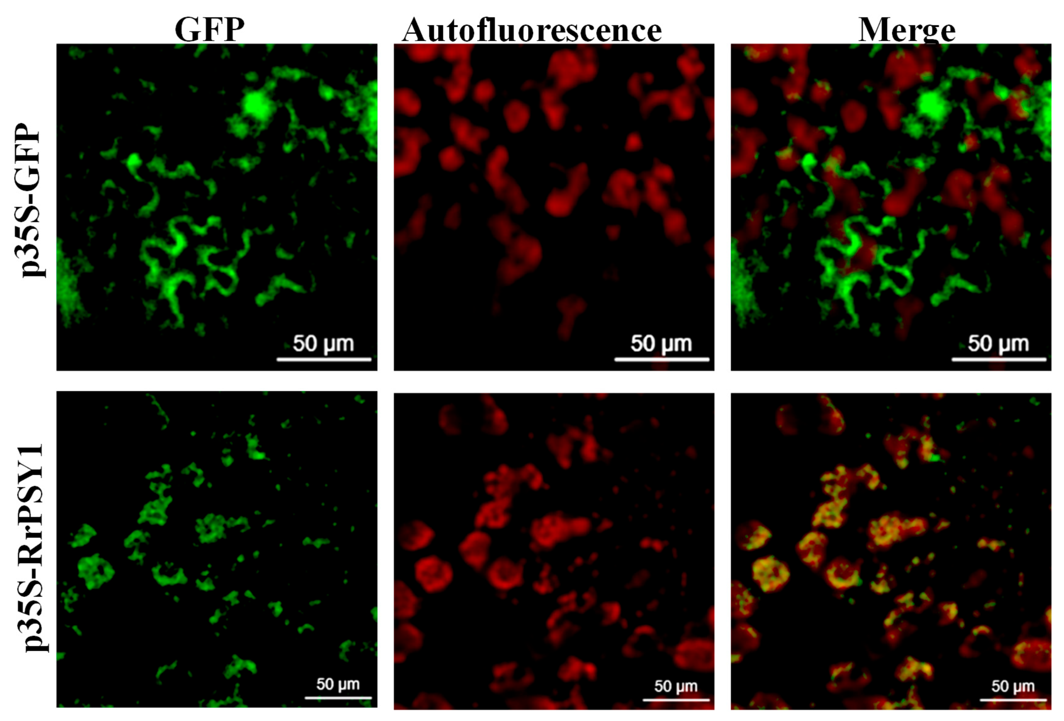
To investigate the subcellular localization of RrPSY1, an in silico prediction was performed using the TargetP-2.0. The analysis indicated a high probability (0.5952) of an N-terminal chloroplast transit peptide in RrPSY1 protein. To validate this prediction, a subcellular localization vector was constructed, and transient expression was carried out in tobacco (Nicotiana benthamiana) epidermal cells. Confocal microscopy showed that RrPSY1 is localized to plastids (Figure 4).

3.4. In Vitro Functional Characterization of RrPSY1

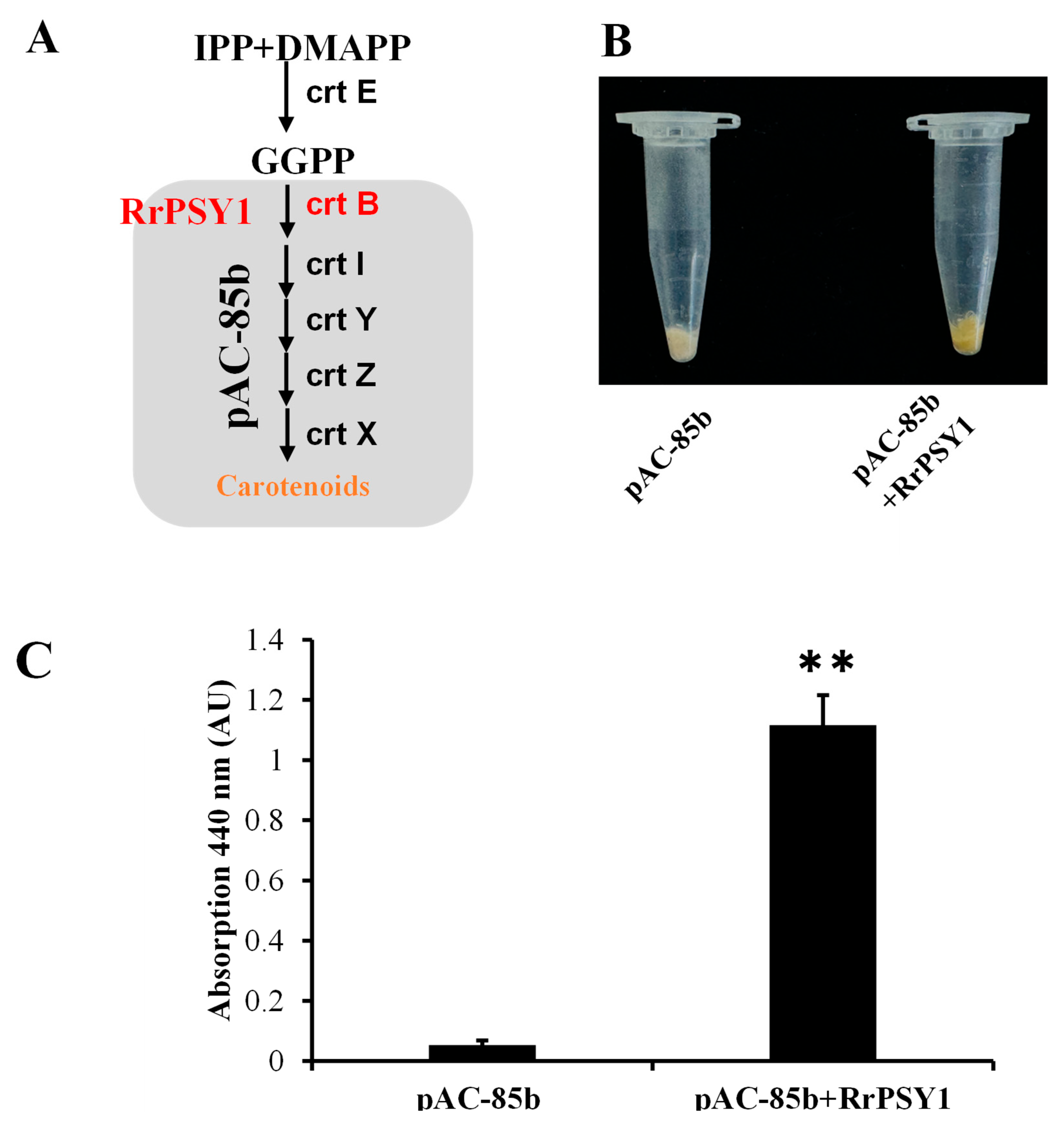
The enzymatic function of RrPSY1 was assessed using a color complementation assay in E. coli. The pAC-85b plasmid harbors four genes (CrtE, CrtY, CrtI, and CrtB) involved in β-carotene biosynthesis, but CrtB, PSY gene is non-functional in this system (Figure 5A). Co-transformation of E. coli with RrPSY1 and pAC-85b restored the pathway, while transformation with the empty pAC-85b vector served as a negative control. The RrPSY1-expressing colonies exhibited yellow pigmentation due to β-carotene accumulation (Figure 5B). Spectrophotometric analysis confirmed a distinct absorption peak at 440 nm, characteristic of carotenoids in the RrPSY1 + pAC-85b co-transformants, verifying the catalytic activity of RrPSY1 (Figure 5C).

3.5. Transient Overexpression of RrPSY1 in Rose Petals

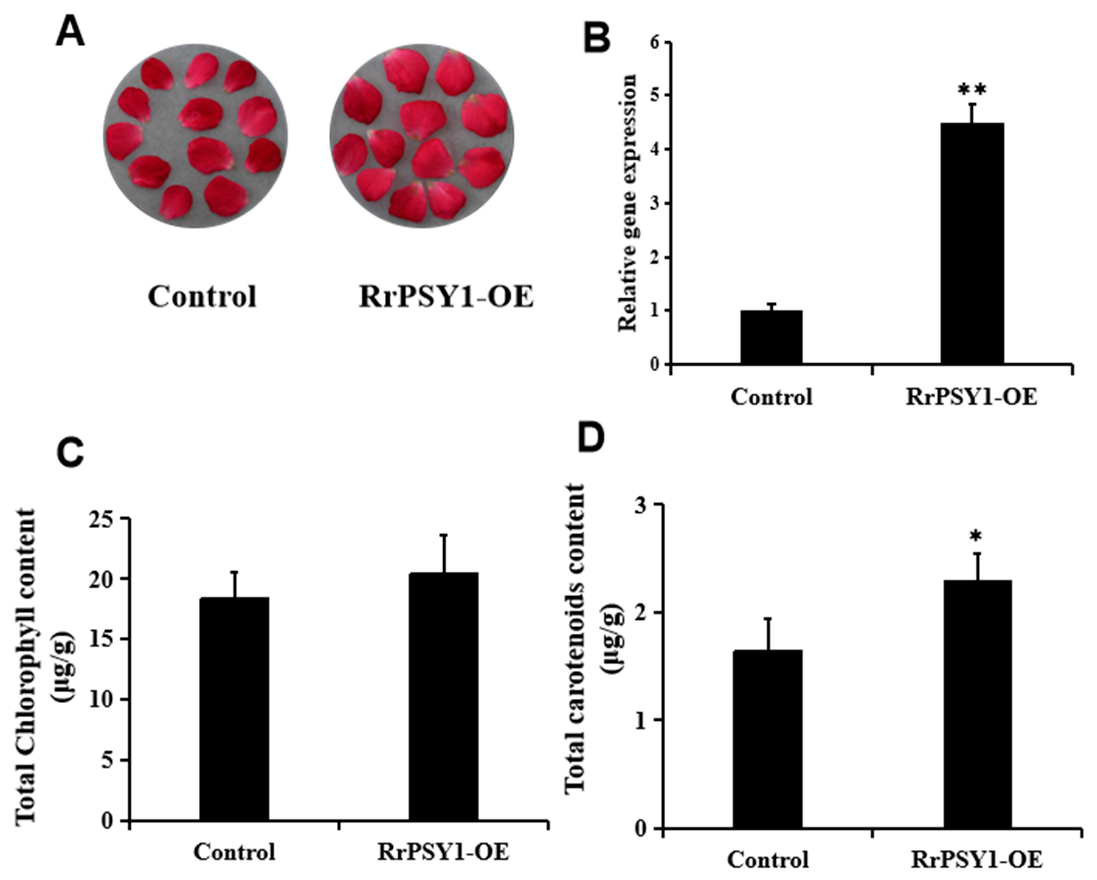
To investigate the function of RrPSY1 in planta, transient overexpression assays were performed in R. hybrida petal discs. No visible phenotypic changes were observed between transformed and control petals (Figure 6A). This is likely due to multi-level regulatory control of carotenoid biosynthesis, in which PSY activity, although essential, is not the sole limiting factor. qRT-PCR analysis confirmed a fourfold increase in RrPSY1 transcript abundance (Figure 6B). Furthermore, while the total contents of chlorophyll a and chlorophyll b remained unchanged, carotenoid content showed a slight, but measurable, increase in overexpressing petals (Figure 6C,D).

3.6. Expression Analysis of RrPSY1 Under Salt Stress

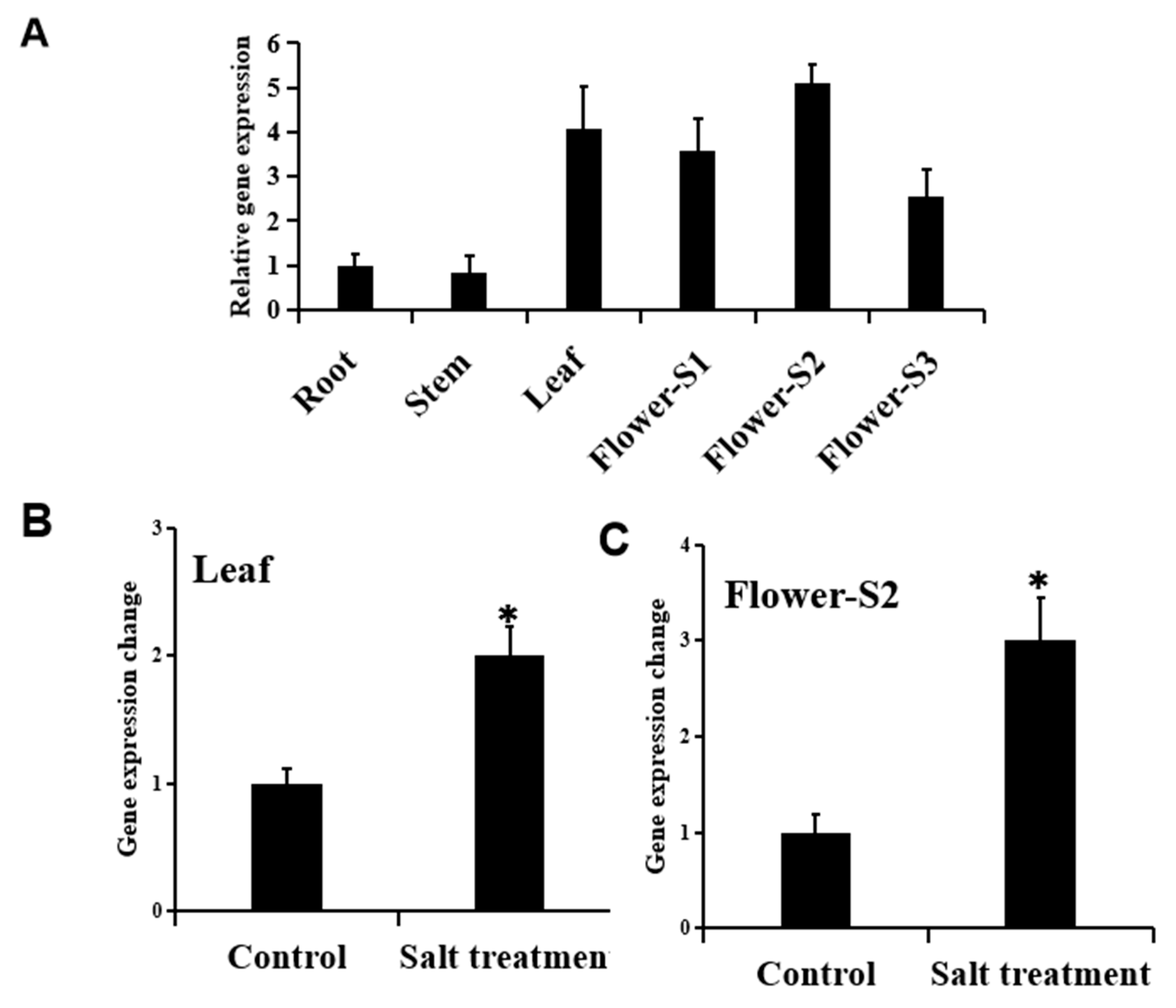
The spatiotemporal expression pattern of RrPSY1 was assessed by qRT-PCR across different R. rugosa tissues and developmental stages (Figures S1–S3). Expression was predominantly detected in leaves and floral tissues, with transcript abundance peaking at S2 (Figure 7A). Because salinity impairs plant growth and productivity [38], and carotenoids contribute to stress tolerance [39], the expression of RrPSY1 under salt stress was examined. Under salinity stress, the RrPSY1 gene was significantly induced in leaves (2-fold) (Figure 7B) and petals (2.5-fold) (Figure 7C), indicating that RrPSY1 is responsive to salt stress in R. rugosa.

4. Discussion

In this study, three developmental stages of R. rugosa petals were analyzed to characterize dynamic changes in carotenoid metabolites (Figure 2). Although there were only four carotenes (4) compared to seven xanthophylls (7), carotenes accounted for 72.3% of the total carotenoids, while xanthophylls constituted 27.7%. This metabolic profile suggests a potential bottleneck in the carotenoid biosynthetic pathway downstream of carotene formation, prior to the xanthophyll generation in petals. At S2, phytoene and phytofluene, both colorless carotenes, were the predominant metabolites. These findings align with the observation that rose coloration is largely determined by anthocyanins. Despite their lack of pigmentation, phytoene and phytofluene have been reported to confer health-promoting benefits [40,41]. Similarly, a previous study identified ten carotenoids across six rose cultivars, with yellow-flowered cultivars exhibiting the highest carotenoid content [26,42]. In the present study, carotenoid levels in R. rugosa petals were comparable to those in pink rose cultivars [26], but were considerably lower than those in rose hips, where concentrations 48.84 μg/g [27].
During both petal and rose hip development, total carotenoid levels increased. However, rose hips exhibited substantial compositional changes in carotenoids, while fewer DAMs were observed in petals (Figure 2) [27]. Notably, phytoene and phytofluene accumulated in rose petals, whereas lycopene dominated in rose hips. This organ-specific difference likely reflects divergent functional requirements: in fruits, carotenoids contribute to pigmentation that attracts animals for seed dispersal, whereas in petals, coloration is primarily anthocyanin-dependent.
RrPSY1 exhibits a conserved exon-intron structure, consistent with PSY genes in multiple plant species [43]. The presence of the DXXXD domain, a hallmark of isoprenoid synthases, is essential for catalytic activity. In cassava roots, mutations near this motif markedly reduce PSY1 activity and efficiency (Figure 3) [44,45]. Sub-cellular localization analysis confirmed that RrPSY1 resides in plastids (Figure 4), consistent with the plastid localization of its upstream partner, RrGGPPS1. Although localization assays employed the constitutive 35S promoter, which may not fully reflect endogenous expression, the observed plastid co-localization, combined with prior evidence of their physical interaction [27], strongly supports their coordinated role in carotenoid biosynthesis. Such transient multi-enzyme complexes are a recurring feature of plant-specialized metabolism, including alkaloids, flavonoids, and terpenoid biosynthesis [46].
The salt-induced expression of RrPSY1 may contribute to carotenoid-derived apocarotenoid phytohormones, notably ABA and SLs, which regulate stress responses in Medicago truncatula and Solanum lycopersicum [6,47]. We therefore propose that the observed induction of RrPSY1 under salinity represents an adaptive mechanism to support the production of protective hormones.
Beyond PSY, carotenoid biosynthesis is regulated at multiple levels, including MYB transcription factors, OR proteins, and ubiquitin-related pathways [48,49,50,51,52,53]. Although their contribution in R. rugosa remains unknown, these regulators may influence petal carotenoid accumulation. Another interesting example is that overexpression of NUDX23 has been shown to upregulate PSY and GGPPS expression and enhance carotenoid production [54], while NUDX1 gene in roses hydrolyzes GPP to generate GP, thereby directing flux toward monoterpene (geraniol) biosynthesis [55]. Given that isoprenoid precursors are shared between carotenoid and monoterpene branches, NUDX activity may indirectly shape carotenoid metabolism by influencing the metabolic partitioning. Future research should therefore focus on the regulatory networks integrating carotenoid metabolism with broader isoprenoid biosynthesis in R. rugosa.

5. Conclusions

This study demonstrates stage-specific carotenoid accumulation in R. rugosa petals (S1–S3) during flower development. Functional validation confirmed that RrPSY1 localizes to plastids and functions as a key rate-limiting enzyme in carotenoid biosynthesis. Furthermore, RrPSY1 expression was significantly upregulated in both leaves and petals under salt stress, suggesting a role in carotenoid metabolism and stress adaptation. Additional metabolite analyses are required to determine whether this transcriptional response translates into increased carotenoid accumulation. Despite these advances, two major limitations remain: (1) The absence of an efficient genetic transformation system as R. rugosa restricts in-planta functional validation by gene knockout or stable overexpression. Future work should therefore combine transcriptomic and metabolomic analyses to provide a more comprehensive understanding of carotenoid biosynthesis and its regulation. (2) The transcriptional and post-translational mechanisms controlling RrPSY1 expression remain unresolved. These findings deepen our understanding of carotenoid metabolism in ornamental flowers and establish a foundation for future genetic improvement. Targeted enhancement of provitamin A carotenoids (e.g., β-carotene) and health promoting xanthophylls (e.g., lutein) has potential not only for nutritional improvement, but also for ornamental applications, including petal color diversification and enhancement of flower quality [56].

Supplementary Materials

The following supporting information can be downloaded at: https://www.mdpi.com/article/10.3390/horticulturae11091137/s1. Supplementary Table S1: Carotenoid composition and content in R. rugosa flowers during S1, S2, and S3 stages. Supplementary Table S2: The primers used in this study. Supplementary Table S3: The amino acid sequences sequence of RrPSY1 and other PSYs from various plants. Supplementary Table S4: Differentially accumulated metabolites (DAMs) detected in S1, S2 and S3 comparison groups.

Author Contributions

Methodology, Y.Y. and Y.C. (Yazheng Cao); Software, Y.C. (Yazheng Cao) and Y.C. (Yudie Chen); Validation, Y.Y., Y.C. (Yazheng Cao) and Y.C. (Yudie Chen); Formal analysis, Y.Y., Y.C. (Yudie Chen) and G.W.; Investigation, G.W.; Resources, X.L. and Y.X.; Data curation, Y.X.; Writing—original draft, H.H. and G.W.; Writing—review & editing, H.H., K.Z. and G.W.; Supervision, L.F. and G.W.; Funding acquisition, L.F. and G.W. All authors have read and agreed to the published version of the manuscript.

Funding

This study is supported by the National Natural Science Foundation of China [Grant No. 32372738], the Natural Science Foundation of Jiangsu Province [BK20231331], and the College Student Innovation Training Program of Jiangsu Province.

Institutional Review Board Statement

Not applicable.

Informed Consent Statement

Not applicable.

Data Availability Statement

Data is contained within the article and supplementary material.

Acknowledgments

Language refinement and clarity improvements were facilitated through DeepSeek tools (https://www.deepseek.com/). The authors thoroughly reviewed and refined all content, assuming full responsibility for the final manuscript.

Conflicts of Interest

The authors declare no conflicts of interest.

References

  1. Sun, T.; Rao, S.; Zhou, X.; Li, L. Plant carotenoids: Recent advances and future perspectives. Mol. Hortic. 2022, 2, 3. [Google Scholar] [CrossRef]
  2. Rodriguez-Concepcion, M.; Avalos, J.; Bonet, M.L.; Boronat, A.; Gomez-Gomez, L.; Hornero-Mendez, D.; Limon, M.C.; Meléndez-Martínez, A.J.; Olmedilla-Alonso, B.; Palou, A.; et al. A global perspective on carotenoids: Metabolism, biotechnology, and benefits for nutrition and health. Prog. Lipid Res. 2018, 70, 62–93. [Google Scholar] [CrossRef] [PubMed]
  3. Yabuzaki, J. Carotenoids Database: Structures, chemical fingerprints and distribution among organisms. Database 2017, 2017, bax004. [Google Scholar] [CrossRef]
  4. Rao, A.V.; Rao, L.G. Carotenoids and human health. Pharmacol. Res. 2007, 55, 207–216. [Google Scholar] [CrossRef]
  5. Hou, X.; Rivers, J.; León, P.; McQuinn, R.P.; Pogson, B.J. Synthesis and Function of Apocarotenoid Signals in Plants. Trends Plant Sci. 2016, 21, 792–803. [Google Scholar] [CrossRef]
  6. Moreno, J.C.; Mi, J.; Alagoz, Y.; Al-Babili, S. Plant apocarotenoids: From retrograde signaling to interspecific communication. Plant J. 2021, 105, 351–375. [Google Scholar] [CrossRef]
  7. Tang, G. Bioconversion of dietary provitamin A carotenoids to vitamin A in humans. Am. J. Clin. Nutr. 2010, 91, 1468S–1473S. [Google Scholar] [CrossRef]
  8. Van Hoang, D.; Pham, N.M.; Lee, A.H.; Tran, D.N.; Binns, C.W. Dietary Carotenoid Intakes and Prostate Cancer Risk: A Case-Control Study from Vietnam. Nutrients 2018, 10, 70. [Google Scholar] [CrossRef]
  9. Aziz, E.; Batool, R.; Akhtar, W.; Rehman, S.; Shahzad, T.; Malik, A.; Shariati, M.A.; Laishevtcev, A.; Plygun, S.; Heydari, M.; et al. Xanthophyll: Health benefits and therapeutic insights. Life Sci. 2020, 240, 117104. [Google Scholar] [CrossRef]
  10. Rodríguez-Concepción, M.; Boronat, A. Elucidation of the methylerythritol phosphate pathway for isoprenoid biosynthesis in bacteria and plastids. A metabolic milestone achieved through genomics. Plant Physiol. 2002, 130, 1079–1089. [Google Scholar] [CrossRef] [PubMed]
  11. Barja, M.V.; Rodriguez-Concepcion, M. Plant geranylgeranyl diphosphate synthases: Every (gene) family has a story. aBIOTECH 2021, 2, 289–298. [Google Scholar] [CrossRef] [PubMed]
  12. Dogbo, O.; Laferriére, A.; D’Harlingue, A.; Camara, B. Carotenoid biosynthesis: Isolation and characterization of a bifunctional enzyme catalyzing the synthesis of phytoene. Proc. Natl. Acad. Sci. USA 1988, 85, 7054–7058. [Google Scholar] [CrossRef]
  13. Slater, A.; Maunders, M.J.; Edwards, K.; Schuch, W.; Grierson, D. Isolation and characterisation of cDNA clones for tomato polygalacturonase and other ripening-related proteins. Plant Mol. Biol. 1985, 5, 137–147. [Google Scholar] [CrossRef]
  14. Bartley, G.E.; Viitanen, P.V.; Bacot, K.O.; Scolnik, P.A. A tomato gene expressed during fruit ripening encodes an enzyme of the carotenoid biosynthesis pathway. J. Biol. Chem. 1992, 267, 5036–5039. [Google Scholar] [CrossRef]
  15. Gupta, P.; Rodriguez-Franco, M.; Bodanapu, R.; Sreelakshmi, Y.; Sharma, R. Phytoene synthase 2 in tomato fruits remains functional and contributes to abscisic acid formation. Plant Sci. 2022, 316, 111177. [Google Scholar] [CrossRef]
  16. Sun, T.; Yuan, H.; Cao, H.; Yazdani, M.; Tadmor, Y.; Li, L. Carotenoid Metabolism in Plants: The Role of Plastids. Mol. Plant 2018, 11, 58–74. [Google Scholar] [CrossRef]
  17. Wang, Z.; Zhang, L.; Dong, C.; Guo, J.; Jin, L.; Wei, P.; Li, F.; Zhang, X.; Wang, R. Characterization and functional analysis of phytoene synthase gene family in tobacco. BMC Plant Biol. 2021, 21, 32. [Google Scholar] [CrossRef]
  18. Li, F.; Vallabhaneni, R.; Yu, J.; Rocheford, T.; Wurtzel, E.T. The maize phytoene synthase gene family: Overlapping roles for carotenogenesis in endosperm, photomorphogenesis, and thermal stress tolerance. Plant Physiol. 2008, 147, 1334–1346. [Google Scholar] [CrossRef]
  19. Ampomah-Dwamena, C.; Driedonks, N.; Lewis, D.; Shumskaya, M.; Chen, X.; Wurtzel, E.T.; Espley, R.V.; Allan, A.C. The Phytoene synthase gene family of apple (Malus x domestica) and its role in controlling fruit carotenoid content. BMC Plant Biol. 2015, 15, 185. [Google Scholar] [CrossRef]
  20. Fraser, P.D.; Schuch, W.; Bramley, P.M. Phytoene synthase from tomato (Lycopersicon esculentum) chloroplasts—Partial purification and biochemical properties. Planta 2000, 211, 361–369. [Google Scholar] [CrossRef]
  21. Bian, Y.; Pan, J.; Gao, D.; Feng, Y.; Zhang, B.; Song, L.; Wang, L.; Ma, X.; Liang, L. Bioactive metabolite profiles and quality of Rosa rugosa during its growing and flower-drying process. Food Chem. 2024, 450, 139388. [Google Scholar] [CrossRef]
  22. Chroho, M.; Bouymajane, A.; Oulad El Majdoub, Y.; Cacciola, F.; Mondello, L.; Aazza, M.; Zair, T.; Bouissane, L. Phenolic Composition, Antioxidant and Antibacterial Activities of Extract from Flowers of Rosa damascena from Morocco. Separations 2022, 9, 247. [Google Scholar] [CrossRef]
  23. Kanani, M.; Chamani, E.; Shokouhian, A.A.; Torabi-Giglou, M. Plant secondary metabolism and flower color changes in damask rose at different flowering development stages. Acta. Physiol. Plant 2021, 43, 55. [Google Scholar] [CrossRef]
  24. Xu, Y.; Li, M.; Ding, H.; Ma, Y.; Yang, Y.; Feng, L. Comparative effects of humic acid biostimulation on soil properties, growth, and fragrance of Rosa rugosa. Ind. Crops Prod. 2025, 225, 120444. [Google Scholar] [CrossRef]
  25. Ogata, J.; Kanno, Y.; Itoh, Y.; Tsugawa, H.; Suzuki, M. Anthocyanin biosynthesis in roses. Nature 2005, 435, 757–758. [Google Scholar] [CrossRef]
  26. Wan, H.; Yu, C.; Han, Y.; Guo, X.; Luo, L.; Pan, H.; Zheng, T.; Wang, J.; Cheng, T.; Zhang, Q. Determination of Flavonoids and Carotenoids and Their Contributions to Various Colors of Rose Cultivars (Rosa spp.). Front. Plant Sci. 2019, 10, 123. [Google Scholar] [CrossRef]
  27. Wei, G.; Chen, Y.; Wang, M.; Xi, Y.; Xu, Y.; Hussain, H.; Zhu, K.; Xu, Y.; Bai, M.; Wang, J.; et al. Integrative application of metabolomics and transcriptomics provides new insights into carotenoid biosynthesis during Rosa rugosa hips ripening. Food Biosci. 2024, 60, 104422. [Google Scholar] [CrossRef]
  28. Xu, Y.; Shi, Y.; Zhang, W.; Zhu, K.; Feng, L.; Wang, J. C2H2 Zinc Finger Protein Family Analysis of Rosa rugosa Identified a Salt-Tolerance Regulator, RrC2H2-8. Plants 2024, 13, 3580. [Google Scholar] [CrossRef]
  29. Wei, G.; Chen, Y.; Wang, J.; Feng, L. Molecular cloning and characterization of farnesyl diphosphate synthase from Rosa rugosa Thunb associated with salinity stress. PeerJ 2024, 12, e16929. [Google Scholar] [CrossRef]
  30. Livak, K.J.; Schmittgen, T.D. Analysis of relative gene expression data using real-time quantitative PCR and the 2−ΔΔCT Method. Methods 2001, 25, 402–408. [Google Scholar] [CrossRef] [PubMed]
  31. Chen, F.; Su, L.; Hu, S.; Xue, J.-Y.; Liu, H.; Liu, G.; Jiang, Y.; Du, J.; Qiao, Y.; Fan, Y.; et al. A chromosome-level genome assembly of rugged rose (Rosa rugosa) provides insights into its evolution, ecology, and floral characteristics. Hortic. Res. 2021, 8, 141. [Google Scholar] [CrossRef]
  32. Kumar, S.; Stecher, G.; Li, M.; Knyaz, C.; Tamura, K. MEGA X: Molecular Evolutionary Genetics Analysis across Computing Platforms. Mol. Biol. Evol. 2018, 35, 1547–1549. [Google Scholar] [CrossRef]
  33. Jang, S.J.; Jeong, H.B.; Jung, A.; Kang, M.Y.; Kim, S.; Ha, S.H.; Kwon, J.K.; Kang, B.C. Phytoene synthase 2 can compensate for the absence of PSY1 in the control of color in Capsicum fruit. J. Exp. Bot. 2020, 71, 3417–3427. [Google Scholar] [CrossRef] [PubMed]
  34. Hua, B.; Wu, J.; Han, X.; Bian, X.; Xu, Z.; Sun, C.; Wang, R.; Zhang, W.; Liang, F.; Zhang, H.; et al. Auxin homeostasis is maintained by sly-miR167-SlARF8A/B-SlGH3.4 feedback module in the development of locular and placental tissues of tomato fruits. New Phytol. 2024, 241, 1177–1192. [Google Scholar] [CrossRef] [PubMed]
  35. Conart, C.; Bomzan, D.P.; Huang, X.Q.; Bassard, J.E.; Paramita, S.N.; Saint-Marcoux, D.; Rius-Bony, A.; Hivert, G.; Anchisi, A.; Schaller, H.; et al. A cytosolic bifunctional geranyl/farnesyl diphosphate synthase provides MVA-derived GPP for geraniol biosynthesis in rose flowers. Proc. Natl. Acad. Sci. USA 2023, 120, e2221440120. [Google Scholar] [CrossRef] [PubMed]
  36. He, G.; Zhang, R.; Jiang, S.; Wang, H.; Ming, F. The MYB transcription factor RcMYB1 plays a central role in rose anthocyanin biosynthesis. Hortic. Res. 2023, 10, uhad080. [Google Scholar] [CrossRef]
  37. Zhai, S.; Li, G.; Sun, Y.; Song, J.; Li, J.; Song, G.; Li, Y.; Ling, H.; He, Z.; Xia, X. Genetic analysis of phytoene synthase 1 (Psy1) gene function and regulation in common wheat. BMC Plant Biol. 2016, 16, 228. [Google Scholar] [CrossRef]
  38. Muchate, N.S.; Nikalje, G.C.; Rajurkar, N.S.; Suprasanna, P.; Nikam, T.D. Plant Salt Stress: Adaptive Responses, Tolerance Mechanism and Bioengineering for Salt Tolerance. Bot. Rev. 2016, 82, 371–406. [Google Scholar] [CrossRef]
  39. Uarrota, V.G.; Stefen, D.L.V.; Leolato, L.S.; Gindri, D.M.; Nerling, D. Revisiting Carotenoids and Their Role in Plant Stress Responses: From Biosynthesis to Plant Signaling Mechanisms During Stress. In Antioxidants and Antioxidant Enzymes in Higher Plants; Gupta, D.K., Palma, J.M., Corpas, F.J., Eds.; Springer International Publishing: Cham, Switzerland, 2018; pp. 207–232. [Google Scholar]
  40. Mapelli-Brahm, P.; Meléndez-Martínez, A.J. The colourless carotenoids phytoene and phytofluene: Sources, consumption, bioavailability and health effects. Curr. Opin. Food Sci. 2021, 41, 201–209. [Google Scholar] [CrossRef]
  41. Engelmann, N.J.; Clinton, S.K.; Erdman, J.W., Jr. Nutritional aspects of phytoene and phytofluene, carotenoid precursors to lycopene. Adv. Nutr. 2011, 2, 51–61. [Google Scholar] [CrossRef]
  42. Wan, H.; Yu, C.; Han, Y.; Guo, X.; Ahmad, S.; Tang, A.; Wang, J.; Cheng, T.; Pan, H.; Zhang, Q. Flavonols and Carotenoids in Yellow Petals of Rose Cultivar (Rosa ‘Sun City’): A Possible Rich Source of Bioactive Compounds. J. Agric. Food Chem. 2018, 66, 4171–4181. [Google Scholar] [CrossRef]
  43. Lisboa, M.P.; Canal, D.; Filgueiras, J.P.C.; Turchetto-Zolet, A.C. Molecular evolution and diversification of phytoene synthase (PSY) gene family. Genet. Mol. Biol. 2022, 45, e20210411. [Google Scholar] [CrossRef]
  44. Pandit, J.; Danley, D.E.; Schulte, G.K.; Mazzalupo, S.; Pauly, T.A.; Hayward, C.M.; Hamanaka, E.S.; Thompson, J.F.; Harwood, H.J., Jr. Crystal structure of human squalene synthase. A key enzyme in cholesterol biosynthesis. J. Biol. Chem. 2000, 275, 30610–30617. [Google Scholar] [CrossRef] [PubMed]
  45. Welsch, R.; Arango, J.; Bär, C.; Salazar, B.; Al-Babili, S.; Beltrán, J.; Chavarriaga, P.; Ceballos, H.; Tohme, J.; Beyer, P. Provitamin A accumulation in cassava (Manihot esculenta) roots driven by a single nucleotide polymorphism in a phytoene synthase gene. Plant Cell 2010, 22, 3348–3356. [Google Scholar] [CrossRef] [PubMed]
  46. Zhang, Y.; Fernie, A.R. Metabolons, enzyme–enzyme assemblies that mediate substrate channeling, and their roles in plant metabolism. Plant Commun. 2021, 2, 100081. [Google Scholar] [CrossRef]
  47. Stauder, R.; Welsch, R.; Camagna, M.; Kohlen, W.; Balcke, G.U.; Tissier, A.; Walter, M.H. Strigolactone Levels in Dicot Roots Are Determined by an Ancestral Symbiosis-Regulated Clade of the PHYTOENE SYNTHASE Gene Family. Front. Plant Sci. 2018, 9, 255. [Google Scholar] [CrossRef] [PubMed]
  48. Sagawa, J.M.; Stanley, L.E.; LaFountain, A.M.; Frank, H.A.; Liu, C.; Yuan, Y.-W. An R2R3-MYB transcription factor regulates carotenoid pigmentation in Mimulus lewisii flowers. New Phytol. 2016, 209, 1049–1057. [Google Scholar] [CrossRef]
  49. Meng, Y.; Wang, Z.; Wang, Y.; Wang, C.; Zhu, B.; Liu, H.; Ji, W.; Wen, J.; Chu, C.; Tadege, M.; et al. The MYB Activator WHITE PETAL1 Associates with MtTT8 and MtWD40-1 to Regulate Carotenoid-Derived Flower Pigmentation in Medicago truncatula. Plant Cell 2019, 31, 2751–2767. [Google Scholar] [CrossRef]
  50. Stanley, L.; Yuan, Y.W. Transcriptional Regulation of Carotenoid Biosynthesis in Plants: So Many Regulators, So Little Consensus. Front. Plant Sci. 2019, 10, 1017. [Google Scholar] [CrossRef]
  51. Wang, P.; Wang, Y.; Wang, W.; Chen, T.; Tian, S.; Qin, G. Ubiquitination of phytoene synthase 1 precursor modulates carotenoid biosynthesis in tomato. Commun. Biol. 2020, 3, 730. [Google Scholar] [CrossRef]
  52. Zhou, X.; Welsch, R.; Yang, Y.; Álvarez, D.; Riediger, M.; Yuan, H.; Fish, T.; Liu, J.; Thannhauser, T.W.; Li, L. Arabidopsis OR proteins are the major posttranscriptional regulators of phytoene synthase in controlling carotenoid biosynthesis. Proc. Natl. Acad. Sci. USA 2015, 112, 3558–3563. [Google Scholar] [CrossRef] [PubMed]
  53. Sun, T.; Wang, P.; Rao, S.; Zhou, X.; Wrightstone, E.; Lu, S.; Yuan, H.; Yang, Y.; Fish, T.; Thannhauser, T.; et al. Co-chaperoning of chlorophyll and carotenoid biosynthesis by ORANGE family proteins in plants. Mol. Plant 2023, 16, 1048–1065. [Google Scholar] [CrossRef]
  54. Rao, S.; Cao, H.; O’Hanna, F.J.; Zhou, X.; Lui, A.; Wrightstone, E.; Fish, T.; Yang, Y.; Thannhauser, T.; Cheng, L.; et al. Nudix hydrolase 23 post-translationally regulates carotenoid biosynthesis in plants. Plant Cell 2024, 36, 1868–1891. [Google Scholar] [CrossRef]
  55. Magnard, J.L.; Roccia, A.; Caissard, J.C.; Vergne, P.; Sun, P.; Hecquet, R.; Dubois, A.; Hibrand-Saint, O.L.; Jullien, F.; Nicole, F.; et al. Biosynthesis of monoterpene scent compounds in roses. Science 2015, 349, 81–83. [Google Scholar] [CrossRef] [PubMed]
  56. Zheng, X.; Giuliano, G.; Al-Babili, S. Carotenoid biofortification in crop plants: citius, altius, fortius. Biochim. Biophys. Acta Mol. Cell Biol. Lipids 2020, 1865, 158664. [Google Scholar] [CrossRef] [PubMed]
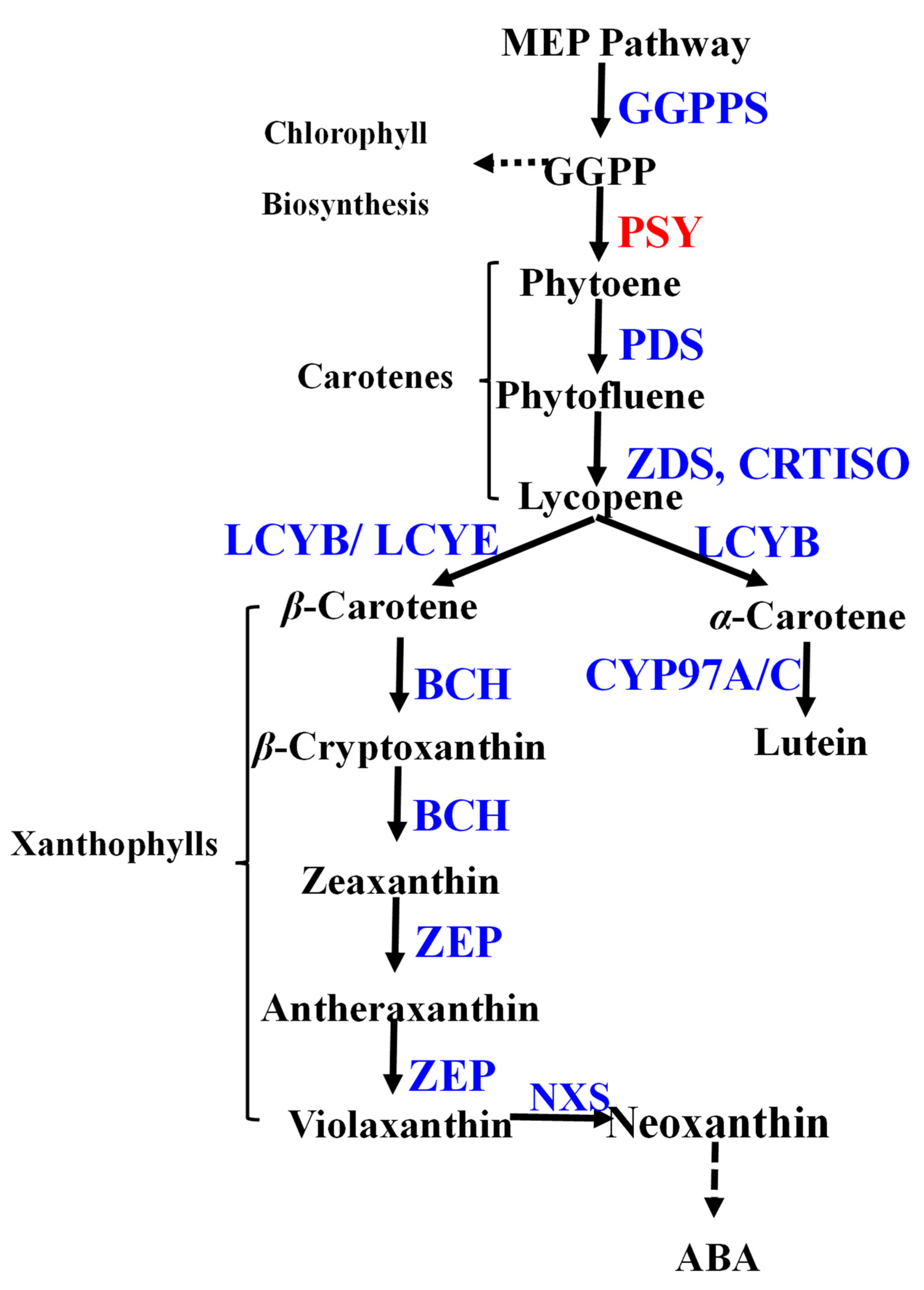
Figure 1. Simplified carotenoid metabolic pathway in plants. Carotenoids are generally derived from the MEP pathway. Dashed lines indicate multiple steps. PSY (highlighted in red) was investigated in this study. Abbreviations in blue: GGPPS, geranylgeranyl diphosphate synthase; PSY, phytoene synthase; PDS, phytoene desaturase; ZDS, ζ-carotene isomerase; CRTISO, carotenoid isomerase; LCYB, lycopene β-cyclase; LCYE, lycopene ε-cyclase; CYP97A, cytochrome P450 carotene β-hydroxylase; CYP97C, cytochrome P450 carotene ε-hydroxylase; BCH, β-carotene hydrolase; ZEP, zeaxanthin epoxidase; NXS, neoxanthin synthase.
Horticulturae 11 01137 g001
Figure 2. Dynamic changes in carotenoid accumulation during R. rugosa petal development. (A) Petal developmental stages of R. rugosa petals: the bud (S1), the semi-open bloom (S2), and the full bloom (S3). (B) Total carotenoid content at S1, S2, and S3. Bar chart values represent means ± SD. Metabolite concentrations are shown as absolute amounts (µg/g FW) to reflect quantitative accumulation. (C) Developmental shift in the relative proportion of carotenes (blue) and xanthophylls (orange) during development. (D) Hierarchical clustering heatmap of carotenoid components across three petal developmental stages.
Horticulturae 11 01137 g002
Figure 3. Bioinformatic analysis of RrPSY1 reveals conserved motifs and evolutionary relationships. (A) Multiple sequence alignment of RrPSY1 and PSY proteins from representative plant species. The full list of sequences used is provided in Supplementary Table S2. Black and gray indicate 100%, and 75% identical residues, respectively. The two highly conserved first and second aspartate-rich regions (DD (XX)2-4D) are presented in red box. (B) Phylogenetic tree analysis of PSY proteins from various plants. RrPSY1 (marked by red dot) clusters within the well-defined Group I of dicot PSY proteins, closely related to SlPSY1 and SlPSY2 from tomato and AtPSY from Arabidopsis. The scale bar indicates sequence divergence, where 0.10 represents a 10% difference between sequences.
Horticulturae 11 01137 g003
Figure 4. Subcellular localization of RrPSY1 in Nicotiana benthamiana epidermal cells. GFP fluorescence (green) was detected by confocal laser scanning microscopy, with chlorophyll autofluorescence (red) and merged channels shown for localization analysis. Transient expression of the 35S::GFP (empty vector) control construct, showing diffuse GFP fluorescence throughout the cytoplasm. The RrPSY1-GFP fusion protein exhibited a punctate signal that co-localizes with the chloroplasts. Scale bars = 50 μm.
Horticulturae 11 01137 g004
Figure 5. Functional characterization of RrPSY1 enzymatic activity by genetic complementation in E. coli. (A) Schematic of the heterologous carotenoid pathway reconstituted in E. coli for RrPSY1 functional complementation. Heterologous expression of RrPSY1 in E. coli harboring carotenogenic plasmid pAC-85b. An E. coli strain only carrying pAC-85b alone served as the negative control. Expression of a functional PSY gene is required to catalyze the synthesis of phytoene from GGPP, thus completing the pathway and enabling carotenoid accumulation. (B) Cell pellets obtained by centrifugation were photographed in clear microcentrifuge tubes. The negative control strain is white due to the blocked pathway. In contrast, the strain co-expressing RrPSY1 and pAC-85b produces yellow pigmentation. (C) Spectrophotometric quantification of carotenoids, showing a carotenoid content-specific absorption peak at 440 nm. Asterisks indicate significant differences calculated using t test (p = 0.027).
Horticulturae 11 01137 g005
Figure 6. Transient overexpression of RrPSY1 in rose petals. (A) Phenotypic comparison of control petals (left) and petals transiently overexpressing RrPSY1 (right) after incubation. (B) qRT-PCR analysis of RrPSY1 transcript levels in transformed petals compared to controls. (C) Total chlorophyll content in the transformed petal discs and (D) total carotenoid content in the transformed petal discs. Data represent mean ± SD (n = 3 biological replicates). Asterisks indicate significant differences calculated using t test p = 0.0047 in Figure 6B and p = 0.032 in Figure 6D.
Horticulturae 11 01137 g006
Figure 7. Spatiotemporal expression and salt stress responsiveness of RrPSY1 in R. rugosa. (A) Tissue-specific RrPSY1 expression across organs and developmental stages. Gene expression was quantified by qRT-PCR and is presented relative to the expression level in root tissue (set as 1.0). (B) qRT-PCR analysis of RrPSY1 in leaf under salinity stress. Expression levels are presented relative to the respective control group (set as 1.0). (C) qRT-PCR analysis of RrPSY1 in S2 under salinity stress. Asterisks indicate significant differences calculated using t test p = 0.026 in Figure 7B and p = 0.018 in Figure 7C.
Horticulturae 11 01137 g007
Disclaimer/Publisher’s Note: The statements, opinions and data contained in all publications are solely those of the individual author(s) and contributor(s) and not of MDPI and/or the editor(s). MDPI and/or the editor(s) disclaim responsibility for any injury to people or property resulting from any ideas, methods, instructions or products referred to in the content.