Article

Characteristics and Transcriptome Analysis of Anther Abortion in Male Sterile Celery (Apium graveolens L.)

1
College of Horticulture, Sichuan Agricultural University, Chengdu 611130, China
2
College of Horticulture, Nanjing Agricultural University, Nanjing 210095, China
3
Institute of Horticulture, Guizhou Academy of Agricultural Sciences, Guiyang 550006, China
*
Authors to whom correspondence should be addressed.
Horticulturae 2025, 11(8), 901; https://doi.org/10.3390/horticulturae11080901
Submission received: 16 June 2025 / Revised: 24 July 2025 / Accepted: 30 July 2025 / Published: 3 August 2025

Abstract

To elucidate the molecular mechanisms underlying anther abortion in celery male sterile lines, this study investigates the morphological differences of floral organs and differential gene expression patterns between two lines at the flowering stage. Using the male sterile line of celery ‘QCBU-001’ and the fertile line ‘Jinnan Shiqin’ as materials, anther structure was analyzed by paraffin sections, and related genes were detected using transcriptome sequencing and qRT-PCR. The results indicated that the anther locules were severely shrunken at maturity in the sterile lines. The callose deficiency led to abnormal development of microspores, preventing the formation of mature pollen grains and ultimately leading to complete anther abortion. The transcriptome results revealed that 3246 genes were differentially expressed in sterile and fertile lines, which were significantly enriched in pathways such as starch and sucrose metabolism and phenylpropanoid biosynthesis. Additionally, differential expression patterns of transcription factor families (MYB, bHLH, AP2, GRAS, and others) suggested their potential involvement in regulating anther abortion. Notably, the expression level of callose synthase gene AgGSL2 was significantly downregulated in sterile anthers, which might be an important cause of callose deficiency and pollen sterility. This study not only provides a theoretical basis for elucidating the molecular mechanism underlying male sterility in celery but also lays a foundation for the utilization and improvement of male sterile lines in vegetable hybrid breeding.

1. Introduction

Plant male sterility is a widespread phenomenon in plants in which stamens fail to form normal pollen. The molecular mechanisms underlying male sterility can be classified into the following categories: abnormal meiosis, aberrant callose metabolism, defective tapetum development, impaired pollen wall formation, abnormal anther dehiscence, and other sterility types [1]. Male sterile lines are an ideal parental material in crop hybrid breeding. Using male sterile lines to produce hybrid seeds can significantly reduce breeding costs, increase breeding efficiency, and yield a significant hybrid advantage. According to studies, hybrid crops yield 20–50% more than conventional types, resulting in significant economic benefits for agriculture [2,3,4]. Currently, numerous vegetable crops, such as tomatoes [5], Chinese cabbage [6], sweet peppers [7], and radishes [8], have produced male sterile lines and successfully used them in hybrid dominance breeding. This approach offers superior seed sources for raising quality, resistance, adaptability, and yield.
Anther development is a complex process regulated by multiple genes [9,10]. Continued research has identified various genes and metabolic pathways that cause infertility in numerous plants. Altered expression of transcription factors often leads to abnormal pollen development and male sterility [11]. According to Shan et al., the transcription factor TDF1 of the MYB family may be crucial in controlling the tapetum’s late function and development during wheat anther development [12]. Bian et al. found that the bHLH transcription factor NtMYC2a regulates carbohydrate metabolism during pollen maturation in tobacco [13]. Moreover, AP2, a member of the AP2/ERF transcription family, plays a complex role in flower development by regulating the expression of many floral organ genes [14]. Carbohydrate metabolism is a basic metabolic pathway for plant growth and development, and disorders of sugar metabolism can lead to abnormal pollen development [15]. Duan et al. found that the silencing of SlSUS3 in the tomato SlSUS3-RNAi line led to insufficient carbon and energy supply, impacting pollen viability and germination rate [16]. According to Liu et al., the downregulation of the genes for phenylalanine ammonia-lyase (PAL), peroxidase (POD), and tyrosine aminotransferase (TAT) can impact the synthesis of lignin, sporopollenin, and flavonoids, leading to impaired pollen wall formation and abortion in temperature-sensitive cytoplasmic male sterile wheat lines [17]. Callose formation during anther development is a critical step in the meiosis of pollen mother cells and significantly affects plant fertility. The Arabidopsis callose synthases AtGSL1, AtGSL2, AtGSL5, AtGSL8, and AtGSL10 were all identified to be associated with pollen development [18,19,20]. OsGSL5 in rice is responsible for callose wall synthesis and pollen outer wall formation [21].
Transcriptome sequencing is a high-throughput sequencing technique that has been effectively used recently to investigate the metabolic processes and gene regulatory roles of plants. Transcriptome sequencing in tobacco has revealed that abnormalities in amino acid biosynthesis, amino sugar and nucleotide sugar metabolism, and starch and sucrose metabolism pathways may disrupt nutrient supply, thereby impairing stamen maturation [22]. In male sterile rice lines, differentially expressed genes (DEGs) were significantly enriched in phenylpropanoid metabolism, fatty acid metabolism, and secondary metabolic pathways. These changes induce male sterility by disrupting flavonoid and lignin synthesis and altering lipid distribution [23]. According to the Kyoto encyclopedia of genes and genomes (KEGG) study of the male sterile line HZ1A of Capsicum annuum indicated that lipid metabolic pathways, oxidative phosphorylation, and phenylpropanoid biosynthesis may regulate male sterility during anther development [24].
Celery (Apium graveolens L.) is a biennial herbaceous plant belonging to the Apiaceae family, known for its rich nutrient profile and distinctive flavor [25]. Its flower organs are small, with few seeds per fruit, and it is a cross-pollinated vegetable crop. The artificial emasculation might cause harm to the flower organs. Celery hybrid breeding began relatively late, and the molecular mechanism underlying anther abortion in male sterile celery lines remains unclear, warranting in-depth investigation. There is an urgent need to address the lack of male sterile celery materials and the challenges associated with hybrid breeding. To date, there are only four cases of celery male sterile materials reported in the world literature [26,27,28,29]. For instance, Tan et al. identified a maternally inherited male-sterile celery line ‘QCBU-001’ through radiation mutagenesis. This line produces well-developed fleshy anthers incapable of pollen formation but can effectively receive foreign pollen to form seeds, making it suitable for celery hybrid breeding [29]. Previous research has primarily focused on the morphology and physiology of male sterility in celery [30], with limited exploration of sterility genes. Understanding male sterility and its abortive mechanisms requires further investigation.
The fertile celery variety ‘Jinnan Shiqin’ selected in this study exhibits vigorous growth, strong adaptability, low fiber content, and rich flavor. It is widely cultivated across China throughout the year and is a major cultivar with high market recognition [31]. The sterile line ‘QCBU-001’ was derived from the offspring of radiation-mutated ‘Jinnan Shiqin’. It is a typical cytoplasmic male sterility (CMS) material with stably heritable male sterility (100% sterility rate), and hybrid seeds can be produced via pollination with fertile lines [29]. Therefore, in this study, we took the anthers of the celery male sterile line ‘QCBU-001’ and the fertile line ‘Jinnan Shiqin’ as the research objects. Morphological comparisons were performed to identify differences in the external morphology of anthers between the two lines. Paraffin sectioning was used to examine the internal anatomy of mature anthers, while transcriptome sequencing helped uncover genes associated with male sterility. qRT-PCR was employed to verify the expression of key genes, thereby elucidating their functions in regulating anther development. The results of this study will provide a basis for the creation and research of superior male sterile lines of celery via genetic engineering.

2. Materials and Methods

2.1. Plant Material

This study used the male sterile line of celery ‘QCBU-001’ (St) and the fertile line ‘Jinnan Shiqin’ (Fe) as materials. All the plant materials used were cultivated in the breeding base of Sichuan Agricultural University. The flowers for each sample (1.0 g) of the two lines were collected at maturity for testing. The anthers during the maturity pollen period were stripped on ice, frozen in cold nitrogen, and stored at −80 °C for spare. Three biological replicates were set up for the two samples.

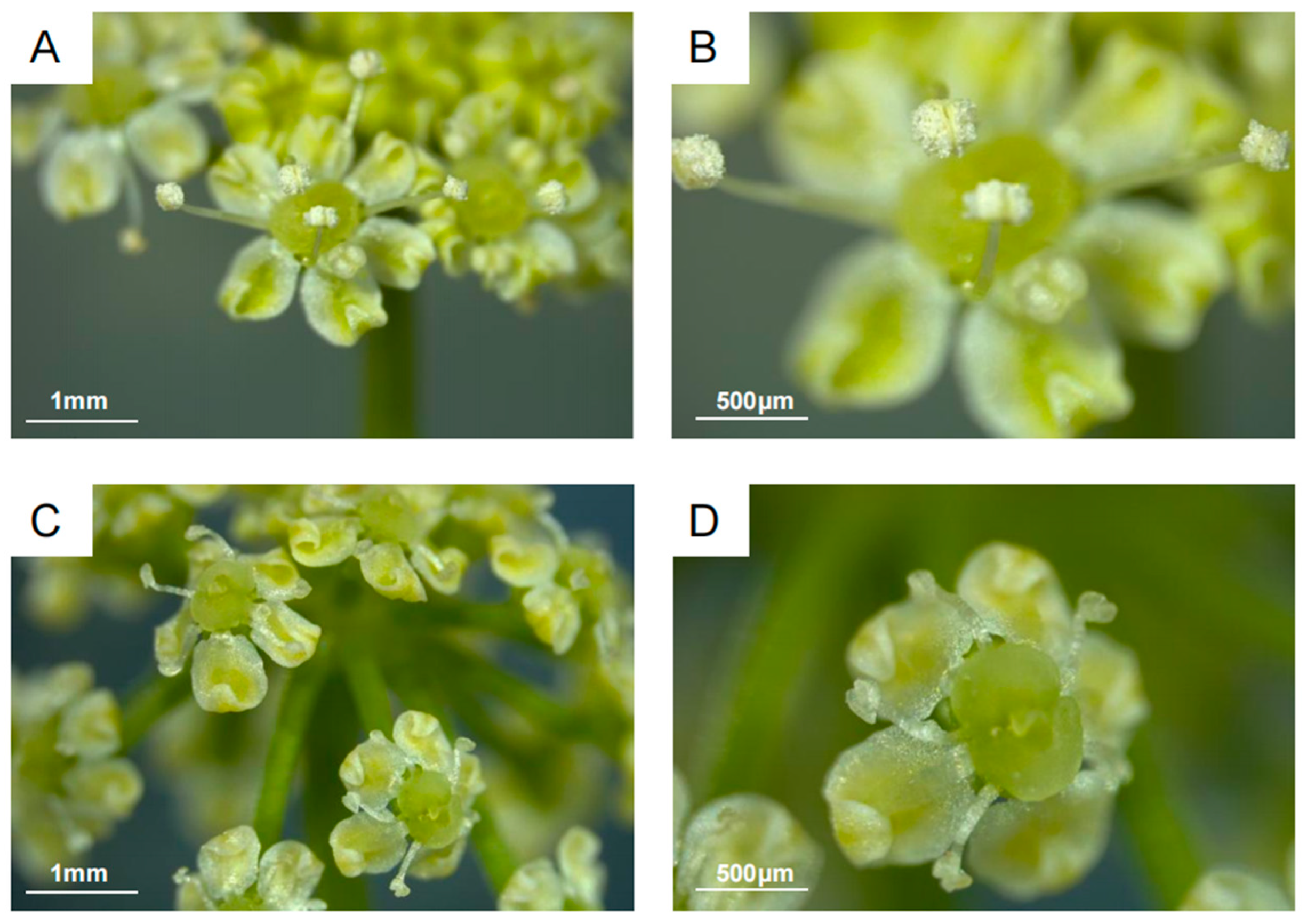
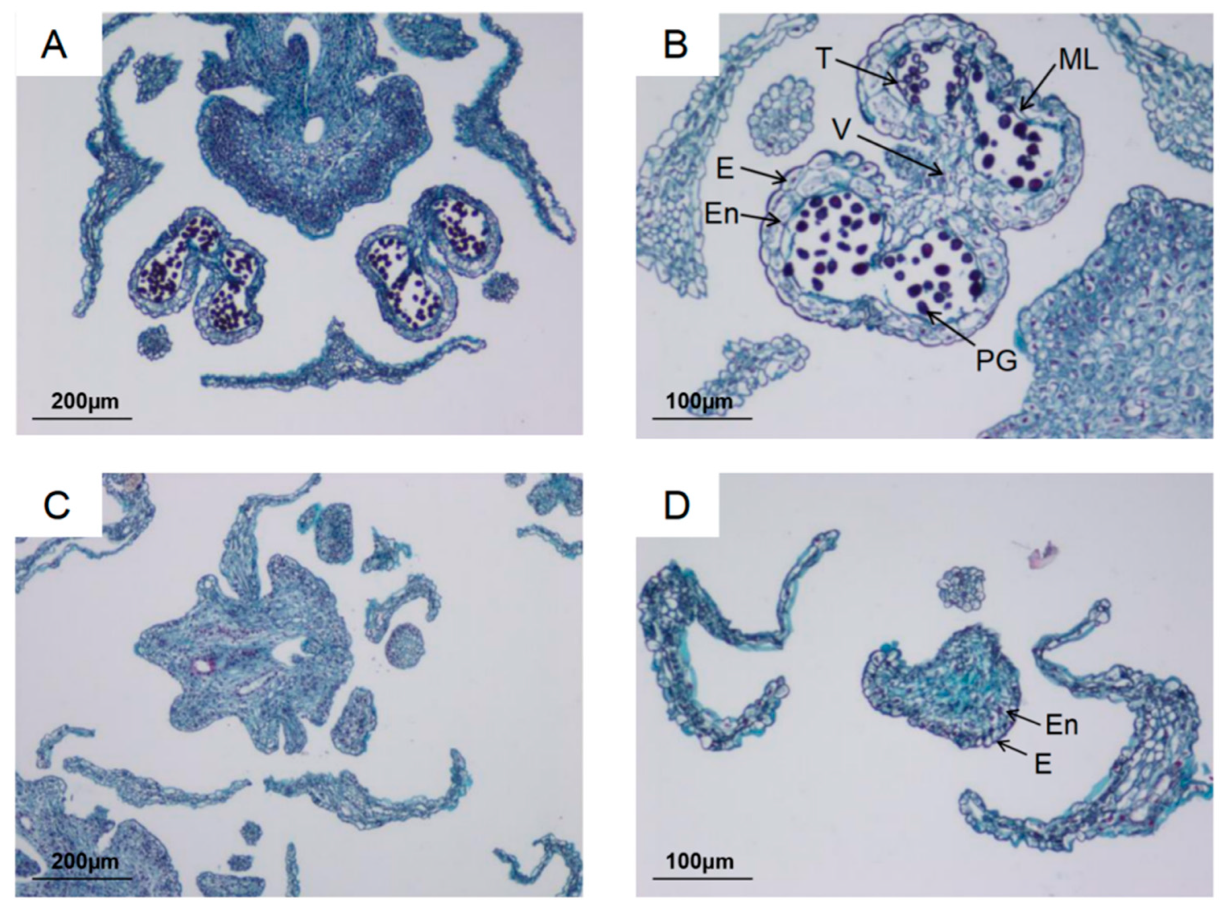
2.2. Phenotypic Characters of Floral Organs and Anther Histology of Celery

Fertile and sterile flower buds were collected at full bloom to observe the morphological characteristics of the floral organs. Fresh anthers from fertile and sterile celery were removed and placed into two 8 mL portions of 50% FAA fixative (V formalin: V glacial acetic acid: V 50% alcohol = 1:1:18) for 24 h. Slices were made using the conventional paraffin sectioning method [32]. After sectioning, the morphological structure of the anthers was examined under an optical microscope (Nikon Eclipse Ci, Tokyo, Japan), and photographs were taken.

2.3. Total RNA Library Construction, Transcriptome Sequencing, and Assembly

Total RNA was extracted from the anthers of the sterile and fertile lines using the total RNA kit according to the manufacturer’s instructions (Tiangen, Beijing, China). The purity and integrity were tested using the NanoPhotometer spectrophotometer and Agilent 2100 bioanalyzer, respectively. Oligo (dT) was used to enrich the mRNA with a polyA tail. The mRNA was then fragmented, and the first strand of cDNA was synthesized with random oligonucleotides and reverse transcriptase. RNaseH and dNTPs were added to synthesize the second strand of cDNA in the DNA polymerase I system. The purified double-stranded cDNA was amplified by PCR, and the products were further purified to obtain libraries for Illumina HiSeq 4000 sequencing. The transcriptome data were submitted to the China National center for Bioinformatton (CNCB) under the accession number PRJCA043534. The library construction and transcriptome sequencing were performed by Shanghai Sangon Biological Engineering Co. (Shanghai, China).
Raw reads were quality-filtered using Trimmomatic to remove adapters, unknown bases, and low-quality sequences, retaining only high-quality sequences (clean reads) for subsequent analysis.

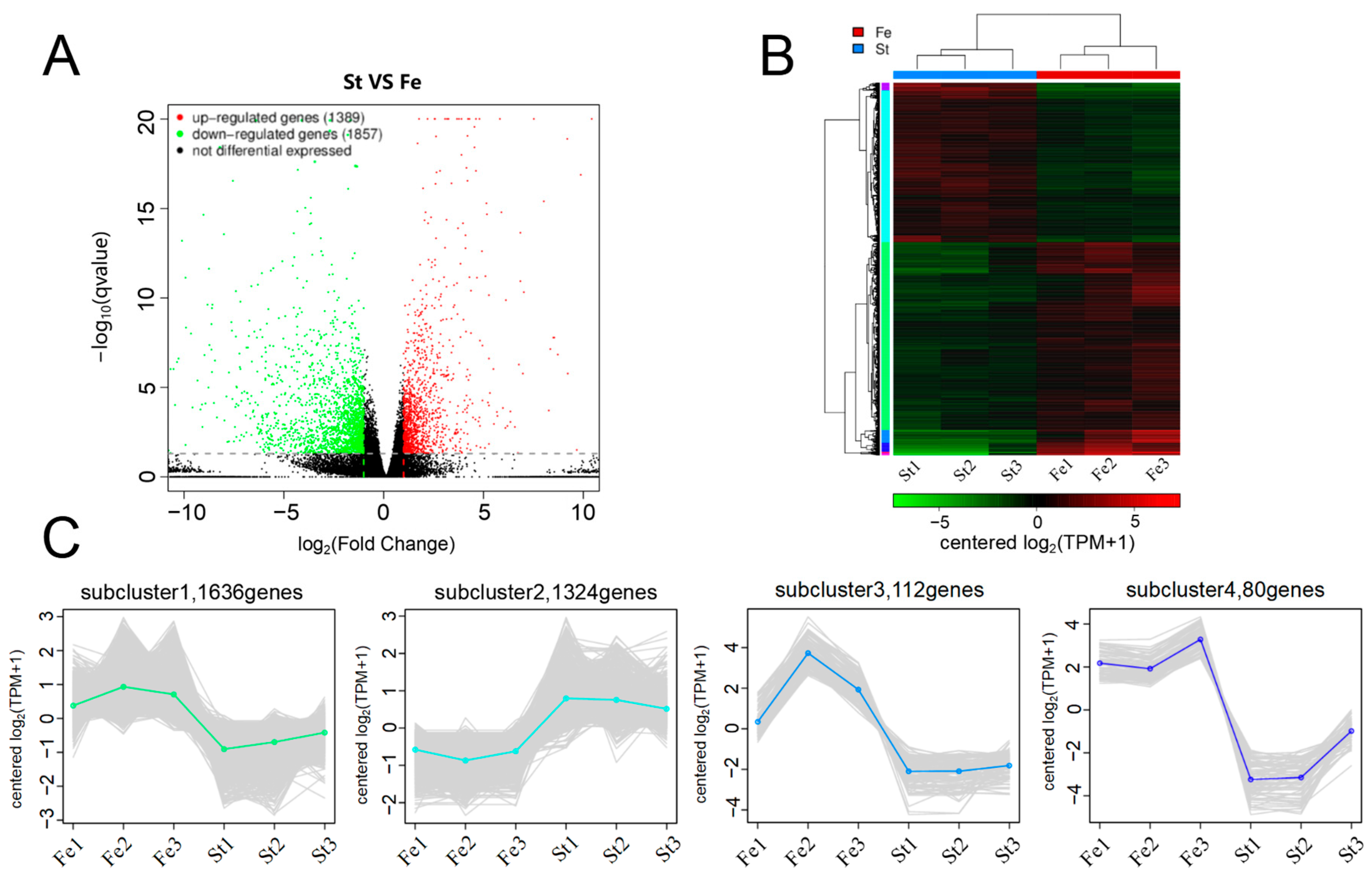
2.4. Differentially Expressed Genes (DEGs) Analysis

The samples were analyzed for expression differences using DESeq to obtain the set of DEGs between the two samples. The relative expression difference multiplicity of genes, q-value < 0.05 and |FoldChange| > 2, was used as the criterion to screen the DEGs [33].
The obtained DEGs were sequence-aligned with gene ontology (GO) and KEGG databases using BLAST software (version 2.60) to obtain functional information about the genes.
Transcription factors within the DEGs of celery were identified using iTAK software (http://itak.feilab.net/cgi-bin/itak/index.cgi) (accessed on 10 October 2024). The transcription factor families of these DEGs were screened and categorized in combination with Plant TFDB (http://planttfdb.cbi.pku.edu.cn) (accessed on 13 October 2024), a plant transcription factor database [34]. Additionally, a clustering heatmap was drawn using TBtools software (version 2.225) after calculating the expression levels of these genes using log2 (FPKM+1).

2.5. Identification and Analysis of AgGSL Gene Family Members

The celery protein sequences were downloaded from two published celery genome databases (http://apiaceae.njau.edu.cn/celerydb [35] and http://celerydb.bio2db.com/ [36]) (accessed on 16 October 2024) to establish a local BLAST database. The glucan synthase-like (GSL) conserved structural domain seed file (PF02364) was downloaded from the Pfam database (http://pfam.xfam.org/) (accessed on 16 October 2024). Using the HMMER software, the GSL protein sequences were identified from the celery protein sequences, allowing for the removal of duplicates and redundant information. NCBI-CDD and Pfam (http://pfam.xfam.org) (accessed on 16 October 2024) were used for further confirmation of GSL structural domains. Physicochemical properties of celery GSL protein sequences were analyzed using the ExPASy website (http://web.expasy.org/protparam/) (accessed on 16 October 2024).
Celery genomic information and promoter sequences 1500 bp upstream of the start codon of the AgGSLs gene were obtained from the celery genome database. Furthermore, the cis-acting elements were identified using the PlantCARE web-based software (https://bioinformatics.psb.ugent.be/webtools/plantcare/html/) (accessed on 24 October 2024).

2.6. qRT-PCR Validation of DEGs

Based on the results of transcriptome sequencing and differential gene screening, specific primers were designed using Primer Premier (version 6.0; Table S3). The fluorescence quantitative reaction system was prepared according to the instructions of the manufacturer (Beijing All Style Gold Biotechnology Co., Ltd., Beijing, China). The experiments were performed on the Bio-Rad Real-time PCR System and Bio-Rad CFX Manager. The relative expression of the genes was calculated using the 2–ΔΔCT method, with the celery AgActin gene as an internal reference. Significance analysis was performed using the Statistical Package for the Social Sciences software (version 23.0), and graphs were plotted using GraphPad Prism software (version 9.5).

3. Results

3.1. Analysis of Differences in Phenotypic Characters and Histological Analysis Between Fertile and Sterile Flowers

The morphology of the celery floral organs (Figure 1) revealed that the normal filaments were significantly elongated, while the filaments in the sterile lines were severely shriveled and exhibited considerably less elongation than those in fertile flowers. In the fertile lines, we observed plump spherical anthers with glossy light yellow color and pollen dispersal. However, in the sterile lines, the anthers were small and shriveled, with deformed shapes and uneven dark yellow surfaces, which did not undergo bursting and did not release pollen.
A comparative cytological analysis of anther structure was performed using paraffin sections from fertile and sterile lines at maturity. As illustrated in Figure 2, the anther wall layers of fertile celery anthers, from the outside to the inside, are the epidermis, fibrous layer, middle layer, and tapetum in sequence. In contrast to fertile anthers (Figure 2A,B), the tapetum of sterile anthers (Figure 2C,D) is completely disintegrated, with only the epidermis and a significantly thickened fibrous layer remaining. Fertile anthers exhibited normal microspore development. The pollen sacs contained abundant mature pollen grains, exhibiting typical morphology and structure. Conversely, sterile anthers displayed severe structural deformities characterized by pronounced atrophy of all four locules. The internal tissues of sterile anthers completely collapsed, preventing proper microspore separation and release. Notably, no mature pollen grains were observed in sterile anthers at maturity, indicating complete pollen abortion and male sterility. This developmental failure could be caused by aberrant tapetum development, which interferes with the normal generation and distribution of callose during microspore development, disrupting microsporogenesis and eventually leading to abortive pollen formation.

3.2. Sequencing Quality and Gene Expression Analysis

Sequencing raw data after filtering (Table S1), 49,834,684–70,286,840 reads were used for comparison analysis. The base sequencing quality values, Q30, were all above 95%, and the GC content was at 43.65–44.29%. Using similarity and principal component analysis (Figure S1), each sample was differentiated and clustered. The results indicated high similarity between replicated samples, demonstrating good reproducibility and meeting the testing requirements. In summary, the sequencing results can cover most of the expressed genes, and the quantity and quality of the transcriptome data are sufficiently robust for further analysis in the next step.
The gene expression results were analyzed using DESeq, and the differential gene volcano plot illustrated the distribution of upregulated and downregulated genes. In this plot, red indicates upregulated genes, while green represents downregulated genes. As illustrated in Figure 3A, 3246 DEGs were detected between male sterile (St) and fertile (Fe) lines of celery, including 1389 upregulated and 1857 downregulated genes, with significant differences in the transcriptomes of fertile and sterile lines. The number of downregulated expressed genes was about 1.3 times the number of upregulated expressed genes, indicating that genes in celery male sterile lines control their fertility through downregulated expression. Cluster analysis of the differential gene sets indicated that most DEGs were expressed in sterile lines at lower levels than in fertile lines, as illustrated in Figure 3B. As displayed in Figure 3C, subclusters 1, 3, and 4 contained 1636, 112, and 80 genes, respectively, all of which were downregulated for expression in sterile lines relative to fertile lines. Conversely, subcluster 2 (1324 genes) exhibited upregulated expression in sterile lines relative to fertile lines. The results suggest that sterile lines can be used to explore the mechanism of male sterility in celery by regulating the expression profiles of specific DEGs, thereby providing a foundation for subsequent analysis of key regulatory genes.

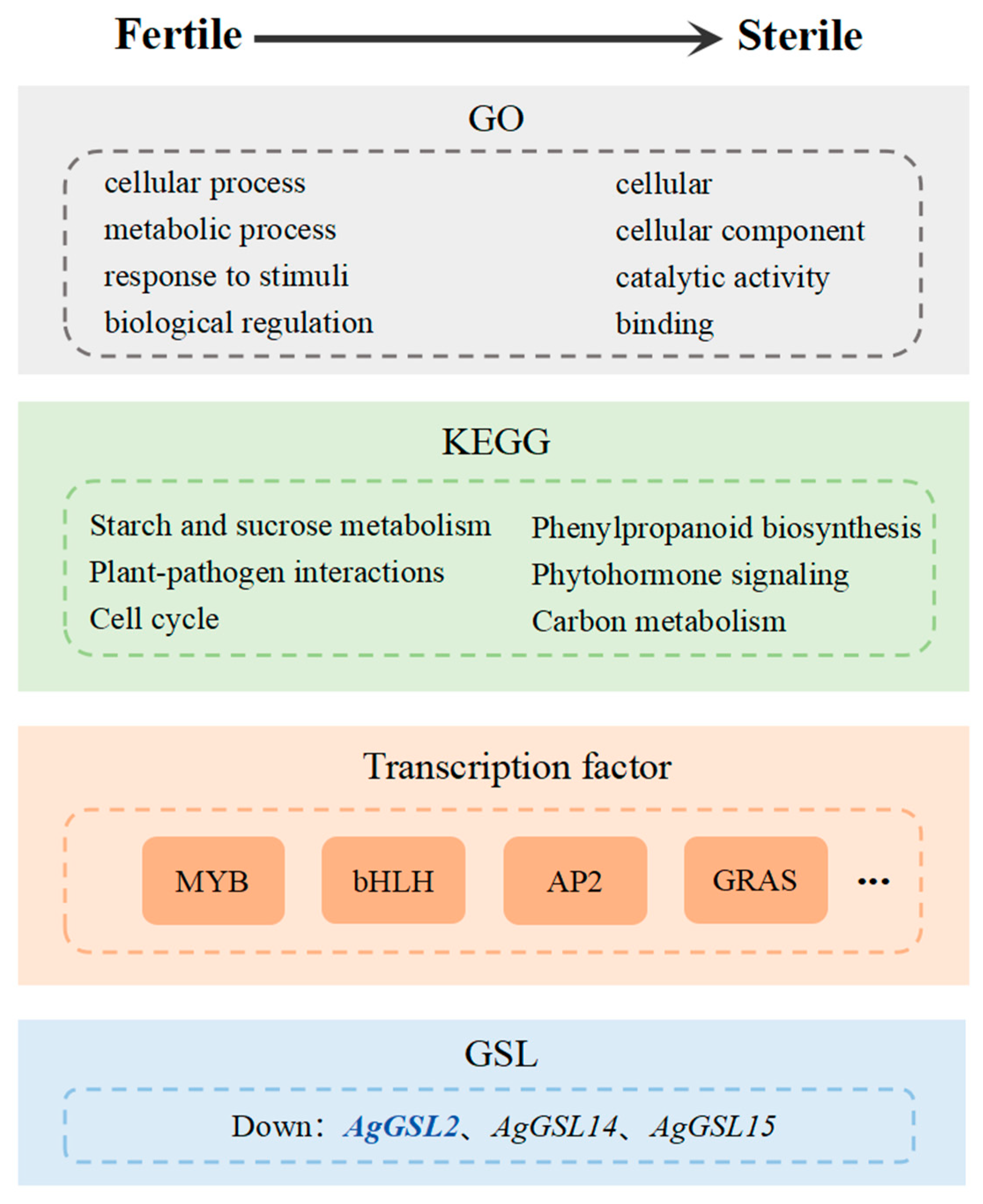
3.3. GO Functional Annotation and KEGG Pathway Enrichment Analysis of DEGs

To thoroughly investigate the biological functions of sterile and fertile differential genes, we performed GO functional annotation and KEGG pathway enrichment analysis. Our analysis of the anther transcriptome sequencing data revealed 1596 DEGs distributed across 61 functional categories associated with biological processes, cellular components, and molecular functions (Figure 4A). Among them, the secondary functions of cellular process (GO:0009987), metabolic process (GO:0008152), response to stimuli (GO:0050896), and biological regulation (GO:0065007) in biological processes accounted for a relatively large number of DEGs, which were 1061, 901, 682, and 637, respectively. In cellular components, the most annotated DEGs were related to the category of cellular (GO:0005623) and cellular component (GO:0044464), each with 1280 DEGs. The number of DEGs in molecular functions such as catalytic activity (GO:0003824) and binding function (GO:0005488) was higher, with 672 and 494, respectively. According to KEGG annotation (Figure 4B, Table S2), DEGs were mainly involved in pathways such as starch and sucrose metabolism, plant–pathogen interactions, cell cycle, phytohormone signaling, carbon metabolism, and phenylpropanoid biosynthesis. Notably, the starch and sucrose metabolism pathways exhibited the greatest enrichment and the highest number of DEGs, indicating their significance in triggering subsequent changes in DEGs in other pathways.

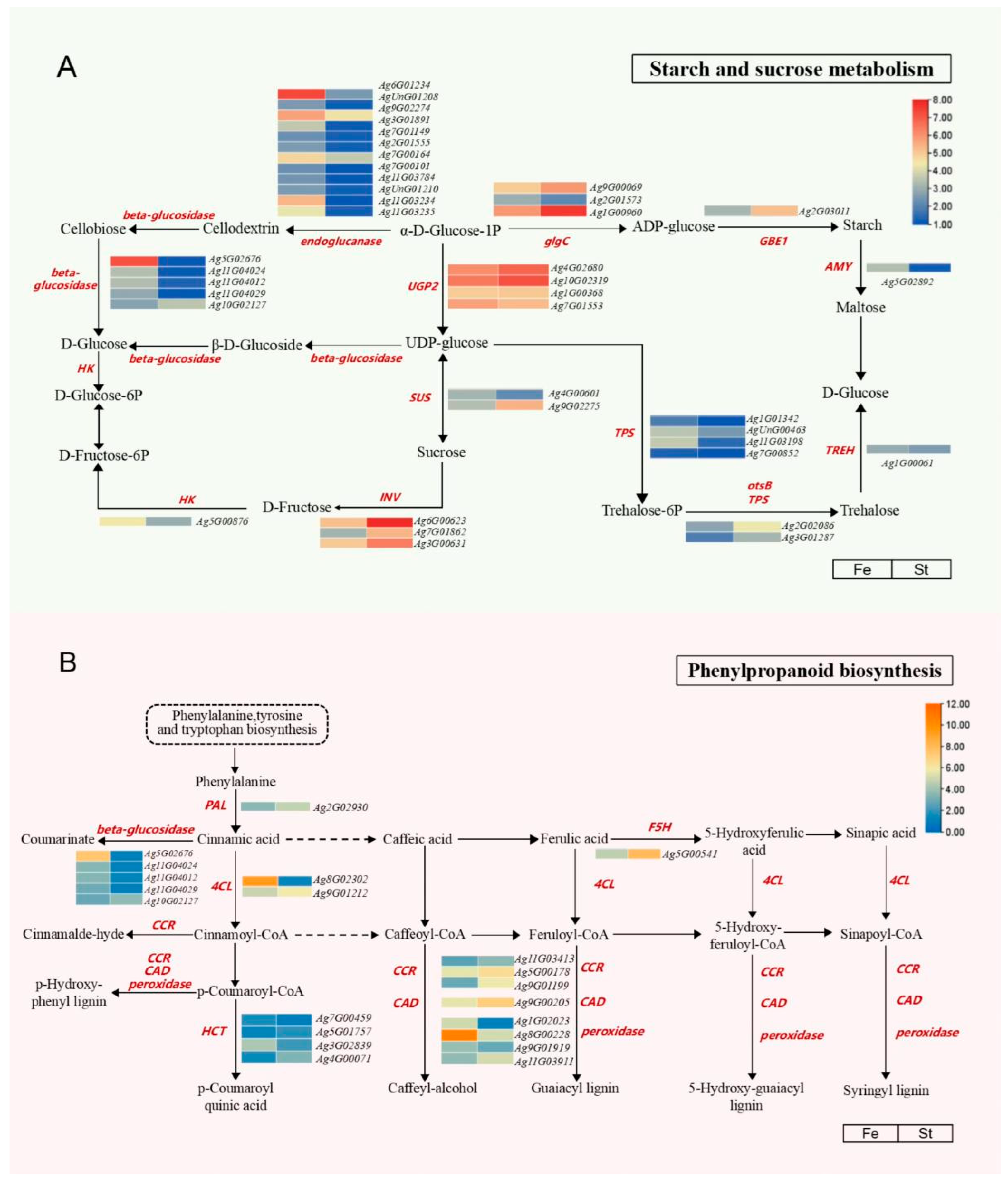
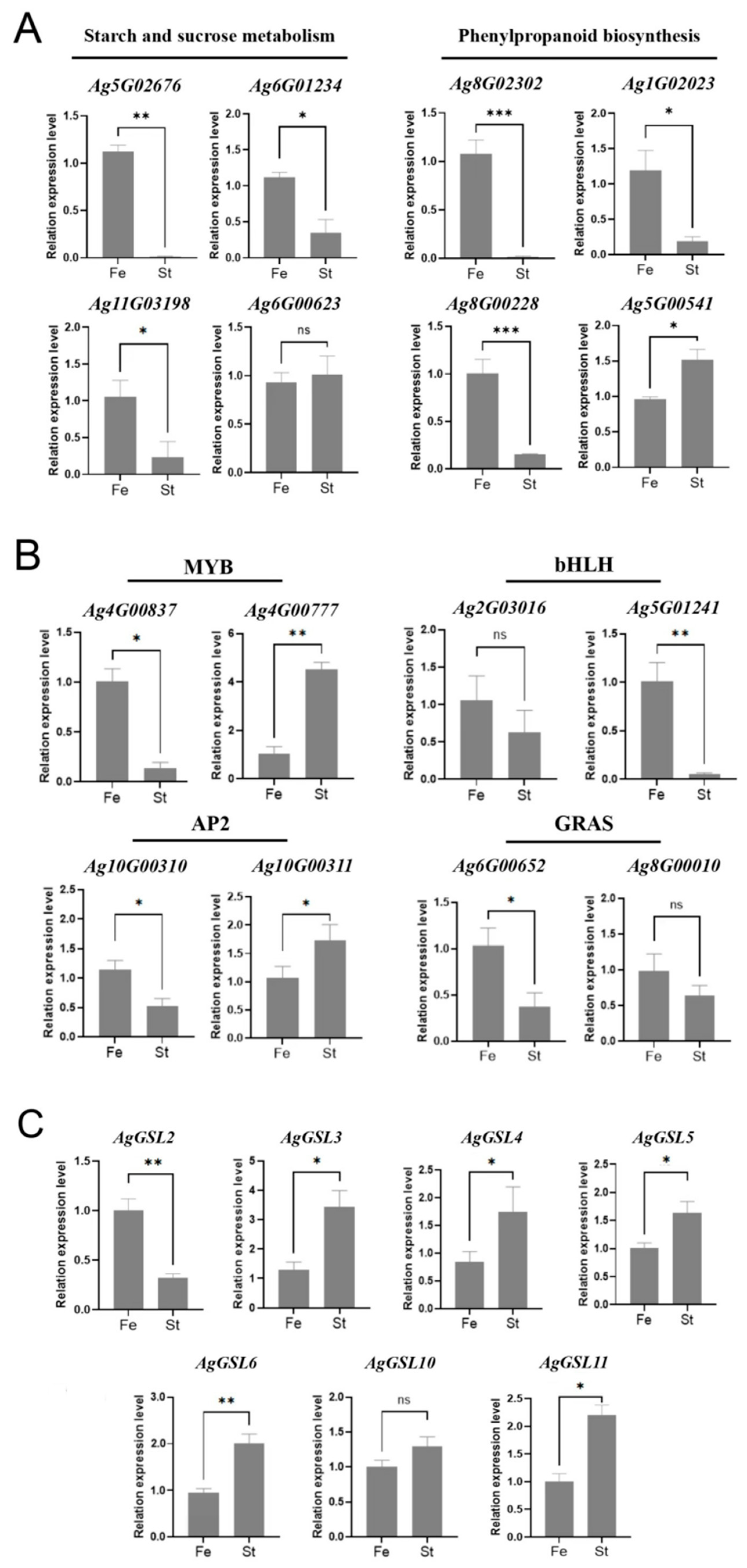
3.4. Analysis of Starch and Sucrose Metabolism and Phenylpropanoid Biosynthesis Pathways and Gene Expression

Significant differences have been identified between sterile and fertile lines in the starch and sucrose pathways, as well as the phenylpropanoid biosynthesis pathway, through a combination of metabolomic and transcriptomic analyses. As illustrated in Figure 5A, in the starch and sucrose pathway (only 12 enzymes, 39 enzyme-encoding genes, and 15 metabolites are included in the figure), most of the enzyme differential genes were downregulated for expression in the sterile lines. Notably, beta-glucosidase and endoglucanase exhibited significant catalytic activities, with the highest number of differential genes, all of which were significantly downregulated in the sterile lines. Key genes Ag6G01234 and Ag5G02676 were expressed at significantly lower levels in sterile lines than in fertile lines. It is hypothesized that these genes may be responsible for the carbohydrate changes in celery male sterility, resulting in abnormal pollen. A total of 25 DEGs were identified in the phenylpropanoid biosynthesis pathway (only 8 enzymes, 21 enzyme-encoding genes, and 20 metabolites are included in the figure), as displayed in Figure 5B. Multiple genes that control key enzyme structures were differentially expressed, with most DEGs being upregulated in sterile lines. Notably, the 4-coumarate-CoA ligase (4CL) gene (Ag8G02302) and POD genes (Ag1G02023, Ag8G00228, and Ag9G01919) were significantly less expressed in the sterile lines than in the fertile lines compared to the other genes. Accordingly, starch and sucrose metabolism and phenylpropanoid biosynthesis pathways are closely related to male sterility in celery.

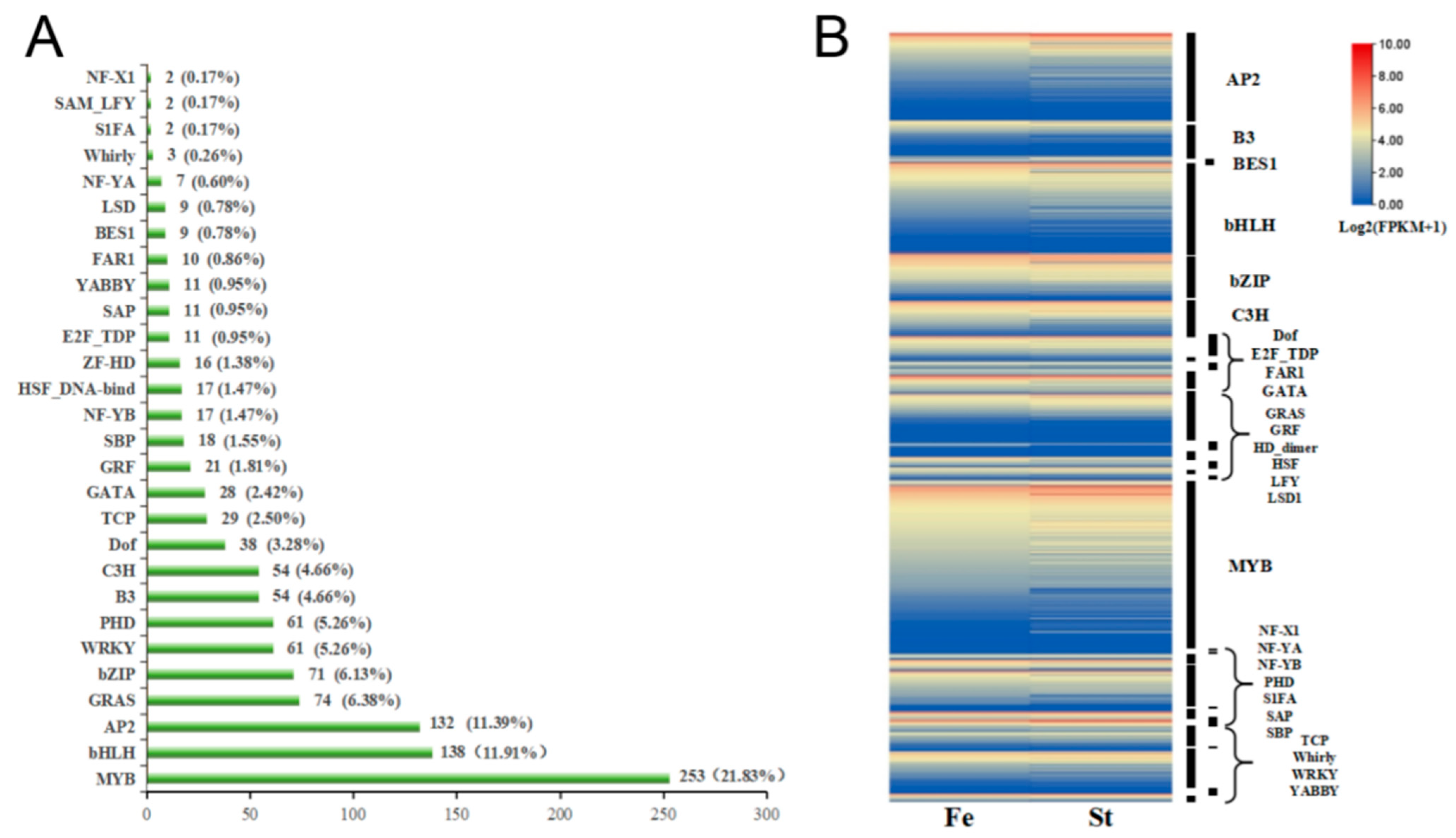
3.5. Transcription Factor Analysis of DEGs in Celery Anthers

A total of 1159 differential TFs were classified into 28 transcription factor families (Figure 6A). The top four transcription factor families, based on the number of DEGs, are as follows: the MYB transcription factor family with 253 genes, the bHLH transcription factor family with 138 genes, the AP2 transcription factor family with 132 genes, and the GRAS transcription factor family with 74 genes. A heatmap drawn from the gene expression levels of each transcription factor family member (Figure 6B) indicated that the expression levels of some transcription families, such as MYB, bHLH, and AP2, exhibited a decreasing trend in sterile lines. This suggests that the occurrence of male sterility in celery may be associated with the abnormal expression of genes from MYB, bHLH, AP2, GRAS, and other families.

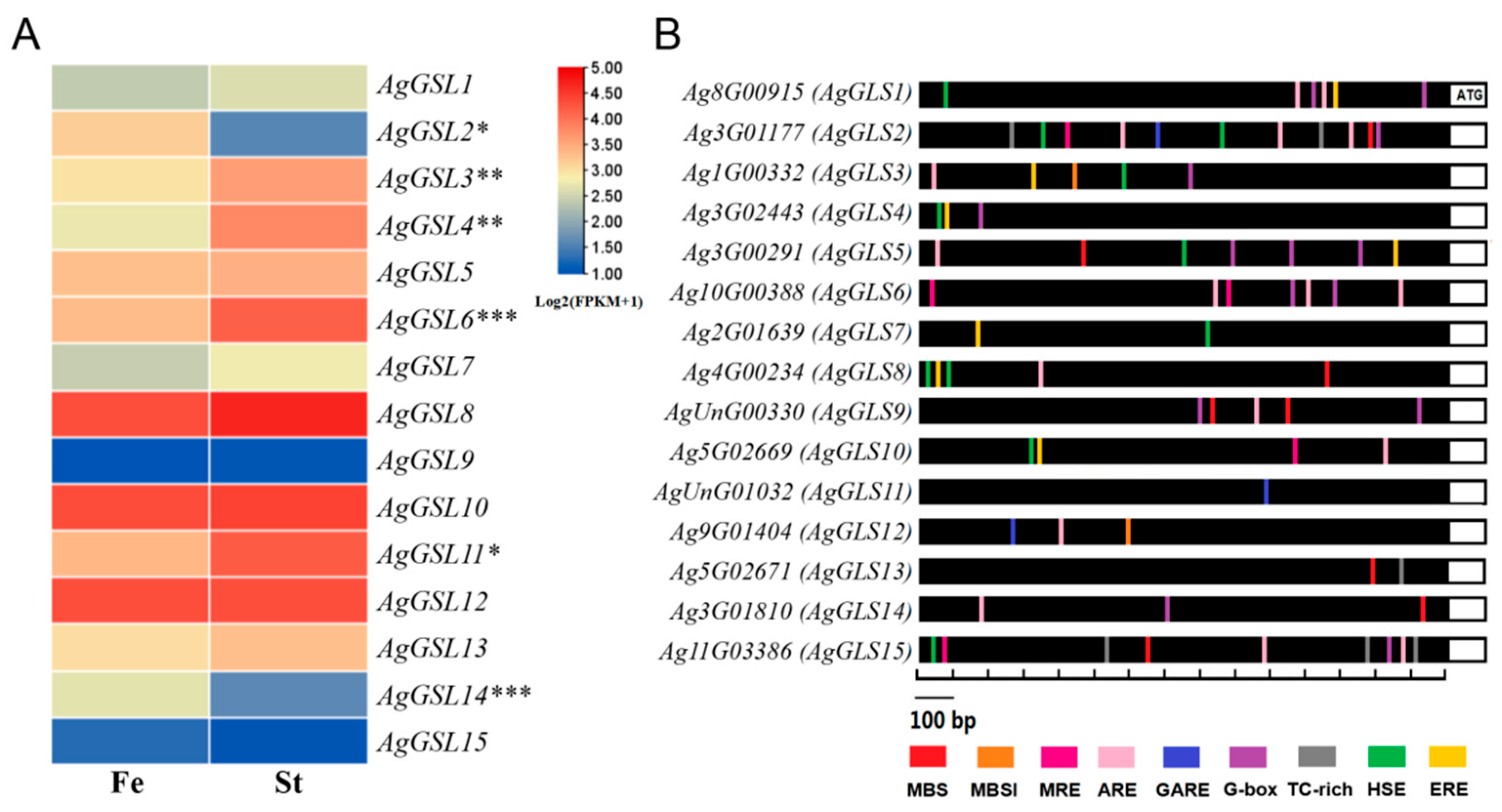
3.6. Identification and Analysis of the AgGSL Family in Celery

Based on celery genome-wide data, a total of 15 celery GSL genes were identified and named AgGSL1~15 (Table S4). The AgGSLs encoded amino acid numbers ranging from 924 (AgGSL9) to 2058 aa (AgGSL4), molecular weights ranging from 106,429.89 (AgGSL9) to 237,514.45 Da (AgGSL4), and equipoint values ranging from 6.71 (AgGSL11) to 9.34 (AgGSL6). The expression patterns of the GSL gene family were analyzed based on celery fertile and sterile transcriptome data (Figure 7A). The results revealed that relevant expression levels were detected for all 15 genes of the AgGSL gene family. Among them, the expression of all genes except AgGSL2, 14, and 15 was higher in sterile than in fertile lines. In contrast, the expression of AgGSL2, 3, 4, 6, 11, and 14 genes exhibited significant differences between the two types of lines. Additionally, this study examined the cis-regulatory elements within the 1500 bp promoter region upstream of the translation initiation site. As illustrated in Figure 7B, the most frequently identified elements in the AgGSL gene family were the anaerobic-inducible element (ARE; 17 times), MYB-binding site (containing MBS (drought-inducible), MBS1 (flavonoid biosynthesis), and MRE (light-responsive), a total of 15 times), and G-box (light-responsive element, 14 times). With the exception of AgGSL1, 4, 7, and 11, the promoters of the remaining 11 genes comprised different types of MYB binding sites, suggesting that AgGSL genes have crucial roles in environmental adaptation, pigment accumulation, and morphogenesis of floral organs. Furthermore, heat stress response element (11 times), TC-rich (defense and stress response element, 6 times), ethylene response element (7 times), and gibberellin response element (3 times) were commonly found in promoters of AgGSL family genes.

3.7. qRT-PCR Analysis of DEGs

Based on GO and KEGG analyses of DEGs, transcription factor analysis, and the identification of the obtained GSL gene family, 23 DEGs were screened for qRT-PCR quantitative validation.
As displayed in Figure 8, Ag5G02676 expression in the starch and sucrose metabolism pathway decreased significantly, with an 87.2-fold reduction in sterile lines. Similarly, the expression of Ag8G02302 in the phenylpropanoid biosynthesis pathway exhibited a 67.2-fold decrease. Conversely, the expressions of Ag6G00623 exhibited non-significant differences between fertile and sterile flowers. Notably, Ag5G00541 was upregulated 1.46-fold in sterile lines.
Furthermore, eight genes from four transcription factor families (MYB, bHLH, AP2, and GRAS) were compared in the transcriptome screen. The expression of MYB family genes differed significantly between fertile and sterile flowers. Specifically, Ag4G00837 decreased 7.4-fold in sterile lines, while Ag4G00777 was upregulated by 4.4-fold. The bHLH family of genes Ag5G01241 declined by 19-fold in sterile lines, while Ag2G03016 exhibited non-significant changes in expression. AP2 family genes Ag10G00310 and Ag10G00311 displayed divergent expression patterns, with inverse regulation trends. In contrast, Ag6G00652 of the GRAS family decreased by 2.8-fold in sterile lines, and Ag8G00010 was also downregulated in sterile lines.
Among the seven GSL genes tested, AgGSL3, 4, 5, 6, 10, and 11 were highly expressed in the sterile lines, whereas the expression level of AgGSL2 was significantly reduced, with a 4.6-fold reduction in the sterile lines. The low level of expression of this gene might be responsible for the abnormal synthesis of callose. The results revealed that the expression pattern of qRT-PCR was basically consistent with the transcriptome sequencing results, thereby supporting the reliability of the RNA-seq data obtained in this study.

4. Discussion

The mechanism of male sterility in plants originates from abnormalities in specific developmental processes during the transition from microspores to the release of mature microspores. This involves various cellular activities. Research has demonstrated that low deposition, premature degradation, or delayed degradation of callose leads to abnormal pollen development, ultimately affecting pollen fertilization and resulting in sterility [37]. Pu et al. conducted microscopic observations on paraffin sections of Chinese cabbage (Brassica rapa L. ssp. pekinensis), which revealed that the four anther locules of sterile plants successively contracted and atrophied during the late stage of pollen development [38]. They further indicated that delayed degradation of callose might be the primary cause of microspore abortion in Chinese cabbage. In this study, the morphological and cytological characteristics of celery fertile and sterile anthers at maturity were comparatively analyzed. The findings revealed distinct developmental abnormalities in sterile anthers compared to fertile anthers. Morphologically, the stamens atrophied, and the anthers were small and shriveled. The preparation of paraffin sections and observation indicated that the anther compartments were significantly atrophied, the microspores were not isolated and released, and no mature pollen grains were produced. In summary, the analysis in this study indicated that the callose developmental abnormality of ‘QCBU-001’ may be a significant factor contributing to stamen atrophy and anther abnormalities.
Anther development involves a complex biological process involving multiple metabolic pathways and regulatory factors. Mutations in fertility-critical genes can lead to male sterility. Sugar metabolism-related genes regulate pollen inner wall formation, starch deposition, and maturation, and their metabolic abnormalities may trigger male sterility in plants [39]. It was hypothesized that the disruption of starch and sucrose metabolism is a significant cause of male sterility in the wheat nuclear male sterility mutant NWMS1 [40]. Mo et al. proposed that the downregulation of β-GLU in tobacco male sterile lines impairs cellulose decomposition. This impairment results in reduced glucose content and disrupts intracellular energy metabolism, ultimately leading to male sterility [41]. In this study, the analysis of starch and sucrose metabolic pathways revealed downregulated expression of β-glucosidase (Ag5G02676, Ag11G04024, Ag11G04012, and Ag11G04029) and endoglucanase (Ag6G01234, AgUnG01208, Ag9G02274, Ag3G01891, and others) genes. This expression pattern was also observed in tobacco male sterile lines, where low expression of these enzyme genes impacts the energy metabolism during anther development, a key factor contributing to male sterility. Additionally, phenylpropanoid metabolism is one of the most important secondary metabolic pathways in plants. It produces various metabolites, such as flavonoids, lignans, and cinnamic acid amides, and is closely related to plant fertility [42]. In this study, it was found that the 4CL genes exhibited differential expression in sterile lines with decreased expression of Ag8G02302 and increased expression of Ag9G01212 compared to fertile lines. Previous research has indicated that the downregulation of 4CL reduces the synthesis of the intermediate feruloyl-CoA, thereby impairing phenolic polymer biosynthesis. This disruption hinders sporopollenin formation, inhibits pollen wall development, and ultimately results in male sterility [43]. Sun et al. found that the expression level of 4CL in all male sterile lines of rice was lower than that of fertile anthers, suggesting that the regulation of 4CL plays a significant role in promoting flavonoid metabolism and pollen wall formation [39]. This expression pattern in rice is similar to that observed in celery, indicating that the downregulation of this gene disrupts normal pollen wall and pollen development, leading to male sterility.
Transcription factor analysis revealed that numerous DEGs were enriched in the MYB, bHLH, AP2, and GRAS families, and these genes were significantly downregulated in sterile lines. The regulatory role of these transcription factors in anther development has been confirmed in various studies. For instance, the loss of function of TaMYB305 may regulate the expression of genes related to jasmonic acid synthesis in male sterile wheat, leading to abnormal pollen development and reduced fertility [44]. The bHLH transcription factors in the anthers of the model plants Arabidopsis thaliana and rice have crucial roles in the control of tapetum development, pollen wall formation, and anther development [45]. The downregulation of the AtAP2-like gene OsAP2-1 in rice RNAi lines resulted in reduced pollen viability and germination activity [46]. GRAS family member DELLA plays an important regulatory role in flower development in the model plant Arabidopsis thaliana [47]. The regulatory roles of MYB, bHLH, and AP2 transcription factors in our dataset may reflect broader integration between hormonal signaling and developmental regulation. Similar master regulatory functions have been reported in BABA-induced resistance, where such TFs coordinate hormone biosynthesis and signaling [48]. Consequently, it is hypothesized that these four classes of transcription factors may be key regulators of anther development in celery. However, their regulatory mechanisms require further investigation.
The formation of callose during anther development is a crucial process in meiosis for pollen mother cells, significantly affecting plant fertility. Research on callose has revealed that its main component is β-1,3-glucan, which is ubiquitously found in higher plants. It is synthesized under the catalytic action of GSL and degraded by endo-1,3-β-glucosidase, playing an important regulatory role in plant growth and development. Previously, 12 GSL genes (AtGSL1-AtGSL12) have been identified in Arabidopsis thaliana involved in callose synthesis [49]. Among them, AtGSL2 is a key gene regulating callose synthesis during pollen development. Loss of function in AtGSL2 disrupts normal callose deposition during pollen development and pollen tube germination, impeding callose wall formation and ultimately causing pollen wall structural collapse [50]. In this study, 15 GSL genes were identified in the celery genome based on transcriptomic and genomic data. Transcriptomic analysis and qRT-PCR validation revealed that the expression level of AgGSL2 was significantly downregulated in sterile anthers, which may be an important factor leading to callose deficiency and pollen sterility. Additionally, AgGSL3, 4, 6, and 11 were expressed at high levels in fertile anthers, indicating they may play important roles in flower development. Studies have indicated that MYB transcription factors indirectly influence anther cell differentiation and development through MBS in secondary metabolic pathways (phenylpropanoid metabolisms) related to anther development, such as lignin and anthocyanin synthesis [51]. ARE may be involved in the expression and regulation of the sucrose transporter gene family in wheat and Arabidopsis thaliana during cellular development or under environmental stress conditions [52]. In this study, we analyzed the cis-acting elements in the AgGSL promoter sequence and found that these elements were enriched in categories related to plant growth and development, abiotic stress, and phytohormone responsiveness, such as ARE and the MYB-binding site MBS. This suggests that the AgGSL may be regulated by the MYB transcription factors and could be involved in the sucrose metabolism pathway, affecting celery fertility.
This study can provide a reference framework for male sterility in Apiaceae crops (carrots, fennel, coriander, etc.). In this study, numerous DEGs associated with fertile and sterile lines of celery anthers were identified by transcriptome sequencing technology, and a regulatory model for male sterility was constructed through functional annotation and expression analysis (Figure 9). GO functional annotation showed that DEGs were mainly enriched in cellular processes, metabolic processes, catalytic activities, and binding functions, reflecting changes in basic life activities and molecular functions during anther fertility transition. KEGG pathway analysis indicated that DEGs were significantly enriched in pathways including starch and sucrose metabolism, plant–pathogen interaction, cell cycle, phytohormone signal transduction, carbon metabolism, and phenylpropanoid biosynthesis. These pathways synergistically regulate anther development, and their dysregulation may disrupt fertility balance. Meanwhile, these processes are accompanied by the synergistic action of multiple genes: differential expression of transcription factor families (MYB, bHLH, AP2, GRAS, etc.) may regulate downstream target genes to initiate the abortion program. The key callose synthesis gene AgGSL2, significantly downregulated, is a critical factor inducing anther structural abnormalities and abortion. Collectively, these factors form the molecular regulatory network underlying male sterility in celery. This study conducted transcriptome sequencing of the male sterile celery line ‘QCBU-001’ and fertile line ‘Jinnan Shiqin’, providing candidate gene resources for future genetic engineering validation of DEGs. The findings also establish a theoretical foundation and reference framework for elucidating the molecular mechanisms underlying male sterility in celery.

5. Conclusions

In this study, we observed the floral organs of the male sterile celery line ‘QCBU-001’ and the fertile line ‘Jinnan Shiqin’. Our findings revealed a significant atrophy of anther cells in the sterile line at maturity. This was accompanied by callose deficiency, aberrant microspore development, and a complete absence of mature pollen grains. Transcriptome sequencing of flowers at anthesis in both lines identified 3246 DEGs, with subsequent functional annotation for breeding-related traits and preliminary screening for potential sterility genes. Male sterility in celery is associated with abnormalities in starch and sucrose metabolism, phenylpropanoid biosynthesis pathways, and the expression of callose synthase genes. Furthermore, the expression of transcription factor family (MYB, bHLH, AP2, GRAS, and others) differed significantly between sterile and fertile lines. These results provide a foundational reference for further exploration of the molecular mechanisms underlying male sterility in celery.

Supplementary Materials

The following supporting information can be downloaded at: https://www.mdpi.com/article/10.3390/horticulturae11080901/s1, Table S1: transcriptome data for male sterility and fertility in celery; Table S2: top 20 metabolic pathways annotated with the most DEGs; Table S3: primer sequences in fluorescence quantification; Table S4: sequence analysis of callose synthase genes; Table S5: DEGs obtained from sterile and fertile lines. Figure S1: sample PCA principal component analysis and box plots of inter-sample distances.

Author Contributions

Conceptualization, M.L., G.T., and A.X.; methodology, Y.G., Z.Y., and H.L.; software, H.L. and A.X.; validation, K.L. and C.W.; formal analysis, C.W.; investigation, Y.G. and K.L.; resources, M.L. and G.T.; data curation, Y.G.; writing—original draft preparation, Y.G., Z.Y., H.L., K.L., and C.W.; writing—review and editing, M.L., G.T., A.X., and Y.Z.; visualization, Z.Y.; supervision, G.T. and Y.Z.; project administration, M.L.; funding acquisition, M.L. and G.T. All authors have read and agreed to the published version of the manuscript.

Funding

This work was supported by the earmarked fund for Sichuan Innovation Team Program of CARS (SCCXTD-2024-22), the National Natural Science Foundation of Sichuan Province (2022NSFSC1674), and the QiankeheJichu-ZK [2024] (General 543).

Institutional Review Board Statement

Not applicable.

Informed Consent Statement

Not applicable.

Data Availability Statement

Data are contained within the article and supplementary materials.

Conflicts of Interest

The authors declare no conflicts of interest.

References

  1. Smith, A.R.; Zhao, D. Sterility caused by floral organ degeneration and abiotic stresses in Arabidopsis and cereal grains. Front. Plant Sci. 2016, 7, 1503. [Google Scholar] [CrossRef]
  2. Tester, M.; Langridge, P. Breeding technologies to increase crop production in a changing world. Science 2010, 327, 818–822. [Google Scholar] [CrossRef]
  3. Cheng, Z.; Song, W.; Zhang, X. Genic male and female sterility in vegetable crops. Hortic. Res. 2022, 10, uhac232. [Google Scholar] [CrossRef]
  4. Zhou, Q.; Yuan, R.; Zhang, W.; Gu, J.; Liu, L.; Zhang, H.; Wang, Z.; Yang, J. Grain yield, nitrogen use efficiency and physiological performance of indica/japonica hybrid rice in response to various nitrogen rates. J. Integr. Agric. 2023, 22, 63–79. [Google Scholar] [CrossRef]
  5. Liu, J.; Wang, S.; Wang, H.; Luo, B.; Cai, Y.; Li, X.; Zhang, Y.; Wang, X. Rapid generation of tomato male-sterile lines with a marker use for hybrid seed production by CRISPR/Cas9 system. Mol. Breed. 2021, 41, 25. [Google Scholar] [CrossRef]
  6. Xue, M.; Li, J.; Liao, R.; Xu, J.; Zhou, M.; Yao, R.; Liu, Z.; Feng, H.; Huang, S. Fine mapping and expression characteristics analysis of male-sterile gene BrRNR1 in Chinese cabbage (Brassica rapa L. ssp. pekinensis). BMC Plant Biol. 2025, 25, 49. [Google Scholar] [CrossRef] [PubMed]
  7. Zhang, Z.; An, D.; Cao, Y.; Yu, H.; Zhu, Y.; Mei, Y.; Zhang, B.; Wang, L. Development and application of KASP markers associated with Restorer-of-fertility gene in Capsicum annuum L. Physiol. Mol. Biol. Plants 2021, 27, 2757–2765. [Google Scholar] [CrossRef]
  8. Duan, N.; Bai, J.; Xie, K.; Wang, J.; Wang, X. De novo and comparative transcriptome analysis of genetic male sterile and fertile lines in radish (Raphanus sativus). J. Hortic. Sci. Biotech. 2020, 95, 32–43. [Google Scholar] [CrossRef]
  9. He, Y.; Wang, J.; Hu, J.; Zheng, J.; Guo, Z.; Yu, Q.; Shi, Q.; Zhang, Y. Knockdown of SlEMS1 causes male sterility in tomato. Hortic. Plant J. 2025, 11, 939–942. [Google Scholar] [CrossRef]
  10. Dong, J.; Hu, F.; Guan, W.; Yuan, F.; Lai, Z.; Zhong, J.; Liu, J.; Wu, Z.; Cheng, J.; Hu, K. A 163-bp insertion in the Capana10g000198 encoding a MYB transcription factor causes male sterility in pepper (Capsicum annuum L.). Plant J. 2023, 113, 521–535. [Google Scholar] [CrossRef]
  11. Li, C.; Zhao, Z.; Liu, Y.; Liang, B.; Guan, S.; Lan, H.; Wang, J.; Lu, Y.; Cao, M. Comparative transcriptome analysis of isonuclear-alloplasmic lines unmask key transcription factor genes and metabolic pathways involved in sterility of maize CMS-C. PeerJ 2017, 5, e3408. [Google Scholar] [CrossRef]
  12. Shan, S.; Tang, P.; Wang, R.; Ren, Y.; Wu, B.; Yan, N.; Zhang, G.; Niu, N.; Song, Y. The characteristic analysis of TaTDF1 reveals its function related to male sterility in wheat (Triticum aestivum L.). BMC Plant Biol. 2024, 24, 746. [Google Scholar] [CrossRef]
  13. Bian, S.; Tian, T.; Ding, Y.; Yan, N.; Wang, C.; Fang, N.; Liu, Y.; Zhang, Z.; Zhang, H. bHLH Transcription factor NtMYC2a regulates carbohydrate metabolism during the pollen development of tobacco (Nicotiana tabacum L. cv. TN90). Plants 2022, 11, 17. [Google Scholar] [CrossRef]
  14. Krogan, N.T.; Hogan, K.; Long, J.A. APETALA2 negatively regulates multiple floral organ identity genes in Arabidopsis by recruiting the co-repressor TOPLESS and the histone deacetylase HDA19. Development 2012, 139, 4180–4190. [Google Scholar] [CrossRef] [PubMed]
  15. Ge, S.; Ding, F.; Daniel, B.; Wu, C.; Ran, M.; Ma, C.; Xue, Y.; Zhao, D.; Liu, Y.; Zhu, Z.; et al. Carbohydrate metabolism and cytology of S-type cytoplasmic male sterility in wheat. Front. Plant Sci. 2023, 14, 1255670. [Google Scholar] [CrossRef] [PubMed]
  16. Duan, Y.; Yang, L.; Han, R.; Gu, L.; Guo, J.; Sun, H.; Gong, H. Tomato sucrose synthase SUS3 is involved in flower and seed development. Plant Physiol. Biochem. 2025, 222, 109715. [Google Scholar] [CrossRef] [PubMed]
  17. Liu, Z.; Niu, F.; Yuan, S.; Feng, S.; Li, Y.; Lu, F.; Zhang, T.; Bai, J.; Zhao, C.; Zhang, L. Comparative transcriptome analysis reveals key insights into fertility conversion in the thermo-sensitive cytoplasmic male sterile wheat. Int. J. Mol. Sci. 2022, 23, 14354. [Google Scholar] [CrossRef]
  18. Guseman, J.M.; Lee, J.S.; Bogenschutz, N.L.; Peterson, K.M.; Virata, R.E.; Xie, B.; Kanaoka, M.M.; Hong, Z.; Torii, K.U. Dysregulation of cell-to-cell connectivity and stomatal patterning by loss-of-function mutation in Arabidopsis chorus (glucan synthase-like 8). Development 2010, 137, 1731–1741. [Google Scholar] [CrossRef]
  19. De Storme, N.; De Schrijver, J.; Van Criekinge, W.; Wewer, V.; Dörmann, P.; Geelen, D. GLUCAN SYNTHASE-LIKE8 and STEROL METHYLTRANSFERASE2 are required for ploidy consistency of the sexual reproduction system in Arabidopsis. Plant Cell 2013, 25, 387–403. [Google Scholar] [CrossRef] [PubMed]
  20. Töller, A.; Brownfield, L.; Neu, C.; Twell, D.; Schulze-Lefert, P. Dual function of Arabidopsis glucan synthase-like genes GSL8 and GSL10 in male gametophyte development and plant growth. Plant J. 2008, 54, 911–923. [Google Scholar] [CrossRef]
  21. Shi, X.; Sun, X.; Zhang, Z.; Feng, D.; Zhang, Q.; Han, L.; Wu, J.; Lu, T. GLUCAN SYNTHASE-LIKE 5 (GSL5) plays an essential role in male fertility by regulating callose metabolism during microsporogenesis in rice. Plant Cell Physiol. 2015, 56, 497–509. [Google Scholar] [CrossRef]
  22. Liu, Z.; Liu, Y.; Sun, Y.; Yang, A.; Li, F. Comparative transcriptome analysis reveals the potential mechanism of abortion in tobacco sua-cytoplasmic male sterility. Int. J. Mol. Sci. 2020, 21, 2445. [Google Scholar] [CrossRef] [PubMed]
  23. Sun, Y.; Xiong, X.; Wang, Q.; Zhu, L.; Wang, L.; He, Y.; Zeng, H. Integrated analysis of small RNA, transcriptome, and degradome sequencing reveals the MiR156, MiR5488 and MiR399 are involved in the regulation of male sterility in PTGMS rice. Int. J. Mol. Sci. 2021, 22, 2260. [Google Scholar] [CrossRef]
  24. Nie, Z.; Chen, J.; Song, Y.; Fu, H.; Wang, H.; Niu, Q.; Zhu, W. Comparative transcriptome analysis of the anthers from the cytoplasmic male-sterile pepper line HZ1A and its maintainer line HZ1B. Horticulturae 2021, 7, 580. [Google Scholar] [CrossRef]
  25. Wang, X.; Luo, Q.; Li, T.; Meng, P.; Pu, Y.; Liu, J.; Zhang, J.; Liu, H.; Tan, G.; Xiong, A. Origin, evolution, breeding, and omics of Apiaceae: A family of vegetables and medicinal plants. Hortic. Res. 2022, 9, uhac076. [Google Scholar] [CrossRef]
  26. Quiros, C.F.; Rugama, A.; Dong, Y.Y.; Orton, T.J. Cytological and genetic-studies of a male sterile celery. Euphytica 1986, 35, 867–875. [Google Scholar] [CrossRef]
  27. Jin, L.; Gao, G.; Zhu, X.; Lu, Z. Key points of hybrid seed production technology of male sterile dual-purpose lines in celery. Pei Fang Yuan I 2011, 8, 62. (In Chinese) [Google Scholar]
  28. Cheng, Q.; Wang, P.; Li, T.; Liu, J.; Zhang, Y.; Wang, Y.; Sun, L.; Shen, H. Complete mitochondrial genome sequence and identification of a candidate gene responsible for cytoplasmic male sterility in celery (Apium graveolens L.). Int. J. Mol. Sci. 2021, 22, 8584. [Google Scholar] [CrossRef]
  29. Tan, G.; Li, M.; Luo, Q.; Zhao, Q.; Zhong, X.; Meng, P.; Xiong, A. Creation of a male sterility line and identification of its candidate mitochondrial male sterility gene in celery. J. Plant Genet. Resour. 2022, 23, 1807–1815. (In Chinese) [Google Scholar]
  30. Li, M.; Tan, S.; Tan, G.; Luo, Y.; Sun, B.; Zhang, Y.; Chen, Q.; Wang, Y.; Zhang, F.; Zhang, Y.; et al. Transcriptome analysis reveals important transcription factor families and reproductive biological processes of flower development in celery (Apium graveolens L.). Agronomy 2020, 10, 653. [Google Scholar] [CrossRef]
  31. Li, H. Celery variety-Jinnan Shiqin. Sci. Technol. Tianjin Agric. For. 2004, 4, 21. (In Chinese) [Google Scholar]
  32. Jia, X.; Wang, G.; Xiong, F.; Yu, X.; Xu, Z.; Wang, F.; Xiong, A. De novo assembly, transcriptome characterization, lignin accumulation, and anatomic characteristics: Novel insights into lignin biosynthesis during celery leaf development. Sci. Rep. 2015, 5, 8259. [Google Scholar] [CrossRef]
  33. Love, M.I.; Huber, W.; Anders, S. Moderated estimation of fold change and dispersion for RNA-seq data with DESeq2. Genome Biol. 2014, 15, 550. [Google Scholar] [CrossRef] [PubMed]
  34. Jin, J.; He, K.; Tang, X.; Li, Z.; Lv, L.; Zhao, Y.; Luo, J.; Gao, G. An Arabidopsis transcriptional regulatory map reveals distinct functional and evolutionary features of novel transcription factors. Mol. Boil. Evol. 2015, 32, 1767–1773. [Google Scholar] [CrossRef]
  35. Li, M.; Feng, K.; Hou, X.; Jiang, Q.; Xu, Z.; Wang, G.; Liu, J.; Wang, F.; Xiong, A. The genome sequence of celery (Apium graveolens L.), an important leaf vegetable crop rich in apigenin in the Apiaceae family. Hortic Res. 2020, 7, 9. [Google Scholar] [CrossRef]
  36. Pei, Q.; Li, N.; Bai, Y.; Wu, T.; Yang, Q.; Yu, T.; Wang, Z.; Liu, Z.; Li, Q.; Lin, H.; et al. Comparative analysis of the TCP gene family in celery, coriander and carrot (family Apiaceae). Veg. Res. 2021, 1, 1–12. [Google Scholar] [CrossRef]
  37. Thiele, K.; Wanner, G.; Kindzierski, V.; Jürgens, G.; Mayer, U.; Pachl, F.; Assaad, F.F. The timely deposition of callose is essential for cytokinesis in Arabidopsis. Plant J. 2009, 58, 13–26. [Google Scholar] [CrossRef]
  38. Pu, Y.; Hou, L.; Guo, Y.; Ullah, I.; Yang, Y.; Yue, Y. Genome-wide analysis of the callose enzyme families of fertile and sterile flower buds of the Chinese cabbage (Brassica rapa L. ssp. pekinensis). FEBS Open Bio. 2019, 9, 1432–1449. [Google Scholar] [CrossRef]
  39. Sun, Y.; Fu, M.; Ang, Y.; Zhu, L.; Wei, L.; He, Y.; Zeng, H. Combined analysis of transcriptome and metabolome reveals that sugar, lipid, and phenylpropane metabolism are essential for male fertility in temperature-induced male sterile rice. Front. Plant Sci. 2022, 13, 945105. [Google Scholar] [CrossRef]
  40. Li, J.; Zhang, J.; Li, H.; Niu, H.; Xu, Q.; Jiao, Z.; An, J.; Jiang, Y.; Li, Q.; Niu, J. The major factors causing the microspore abortion of genic male sterile mutant NWMS1 in wheat (Triticum aestivum L.). Int. J. Mol. Sci. 2019, 20, 6252. [Google Scholar] [CrossRef] [PubMed]
  41. Mo, Z.; Ke, Y.; Huang, Y.; Duan, L.; Wang, P.; Luo, W.; Que, Y.; Pi, K.; Zeng, S.; Liu, R. Integrated transcriptome and proteome analysis provides insights into the mechanism of cytoplasmic male sterility (CMS) in tobacco (Nicotiana tabacum L.). J. Proteom. 2023, 275, 104825. [Google Scholar] [CrossRef] [PubMed]
  42. Dong, N.; Lin, H. Contribution of phenylpropanoid metabolism to plant development and plant-environment interactions. J. Integr. Plant Biol. 2021, 63, 180–209. [Google Scholar] [CrossRef]
  43. Yang, X.; Ye, J.; Zhang, L.; Song, X. Blocked synthesis of sporopollenin and jasmonic acid leads to pollen wall defects and anther indehiscence in genic male sterile wheat line 4110S at high temperatures. Funct. Integr. Genom. 2020, 20, 383–396. [Google Scholar] [CrossRef] [PubMed]
  44. Wang, X.; Wang, J.; Liu, Z.; Yang, X.; Chen, X.; Zhang, L.; Song, X. The R2R3 MYB gene TaMYB305 positively regulates anther and pollen development in thermo-sensitive male-sterility wheat with Aegilops kotschyi cytoplasm. Planta 2024, 259, 64. [Google Scholar] [CrossRef]
  45. Ortolan, F.; Trenz, T.S.; Delaix, C.L.; Lazzarotto, F.; Margis-Pinheiro, M. bHLH-regulated routes in anther development in rice and Arabidopsis. Genet. Mol. Biol. 2024, 46, e20230171. [Google Scholar] [CrossRef]
  46. Zhao, L.; Xu, S.; Chai, T.; Wang, T. OsAP2-1, an AP2-like gene from Oryza sativa, is required for flower development and male fertility. Sex. Plant Reprod. 2006, 19, 197–206. [Google Scholar] [CrossRef]
  47. Zentella, R.; Zhang, Z.; Park, M.; Thomas, S.G.; Endo, A.; Murase, K.; Fleet, C.M.; Jikumaru, Y.; Nambara, E.; Kamiya, Y.; et al. Global analysis of della direct targets in early gibberellin signaling in Arabidopsis. Plant Cell 2007, 19, 3037–3057. [Google Scholar] [CrossRef]
  48. Virág, E.; Nagy, Á.; Tóth, B.B.; Kutasy, B.; Pallos, J.P.; Szigeti, Z.M.; Máthé, C.; Kardos, G.; Hegedűs, G. Master regulatory transcription factors in β-Aminobutyric acid-induced resistance (BABA-IR): A perspective on phytohormone biosynthesis and signaling in Arabidopsis thaliana and Hordeum vulgare. Int. J. Mol. Sci. 2024, 25, 9179. [Google Scholar] [CrossRef]
  49. Hong, Z.; Delauney, A.J.; Verma, D.P. A cell plate-specific callose synthase and its interaction with phragmoplastin. Plant Cell 2001, 13, 755–768. [Google Scholar] [CrossRef] [PubMed]
  50. Dong, X.; Hong, Z.; Sivaramakrishnan, M.; Mahfouz, M.; Verma, D.P. Callose synthase (CalS5) is required for exine formation during microgametogenesis and for pollen viability in Arabidopsis. Plant J. 2005, 42, 315–328. [Google Scholar] [CrossRef]
  51. Shin, D.H.; Choi, M.; Kim, K.; Bang, G.; Cho, M.; Choi, S.B.; Choi, G.; Park, Y.I. HY5 regulates anthocyanin biosynthesis by inducing the transcriptional activation of the MYB75/PAP1 transcription factor in Arabidopsis. FEBS Lett. 2013, 587, 1543–1547. [Google Scholar] [CrossRef] [PubMed]
  52. Himani, S.; Sonia, S.; Sneh, N.; Rekha, M.; Indu, S.; Ravish, C. Computational analysis of cis-acting regulatory elements in 5′ regulatory regions of sucrose transporter gene families in wheat and Arabidopsis. Res. J. Biotechnol. 2014, 9, 75–81. [Google Scholar]
Figure 1. Fully open state of normal fertile and male sterile flowers in celery. (A,B) Fertile anthers; (C,D) sterile anthers. Scale bars: 1 mm (A,C), 500 µm (B,D).
Horticulturae 11 00901 g001
Figure 2. Microscopic observation of anther cross-section at the maturity stage of celery. (A,B) Fertile anthers; (C,D) sterile anthers. E: Epidermis; En: Endothecium; ML: Middle Layer; T: Tapetum; V: Vascular Region; PG: Pollen Grains. Scale bars: 200 µm (A,C), 100 µm (B,D).
Horticulturae 11 00901 g002
Figure 3. Gene expression profile and clustering analysis based on RNA sequencing analysis. Fe: Fertile line; St: Sterile line. (A) Volcano map of DEGs. (B) Cluster analysis of DEGs in heat map. The color represents the transcriptional abundance of the DEGs. Red and green represent high and low expression levels, respectively. (C) K-means clustering of gene expression trends. The gray line represents the expression pattern of genes in each cluster, and the blue line represents the average expression of all genes in the cluster.
Horticulturae 11 00901 g003
Figure 4. Functionally annotated loop diagram of the anther differential gene GO and analysis of KEGG metabolic pathway. (A) GO loop diagram: The green color represents a biological process, the red color signifies cellular component, and the blue color indicates molecular function. Purple -Log10Qvalue indicates the enrichment level; the larger the value, the more significant the enrichment. Only the top 30 entries annotated to the highest number of genes under this GO were selected for plotting. (B) KEGG pathway enrichment map: The vertical axis indicates the pathway annotation information, and the horizontal axis indicates the corresponding Rich factor of the pathway. The size of the q-value is represented by the color of the dots. The smaller the q-value is, the closer the color is to red. The number of differentiated genes contained under each pathway is indicated by the size of the dots. Only the top 30 KEGGs with the highest enrichment were selected for plotting.
Horticulturae 11 00901 g004
Figure 5. Metabolite transformation and gene expression changes in the starch and sucrose metabolism and phenylpropanoid biosynthesis pathways of fertile and sterile lines. (A) Differential gene expression for the starch and sucrose metabolism pathway. (B) Differential gene expression in the phenylpropanoid biosynthesis pathway. Trends in gene expression are indicated by the red (toned up) and blue (toned down) colors of the heatmap. Log-scale and row-scale converted FPKM values were used to create heatmap data.
Horticulturae 11 00901 g005
Figure 6. Transcription factor analysis of DEGs. (A) Names of transcription factor families and the number of transcription factors due. (B) Specific expression of transcription factors, color from blue to red indicates low to high expression, and expression was calculated using log2 (FPKM+1).
Horticulturae 11 00901 g006
Figure 7. Expression profiling and promoter analysis of AgGSL. (A) Expression of celery AgGSL family genes. (B) Analysis of cis-acting elements in the promoters of celery AgGSL family genes. Heatmap colors from blue to red indicate low to high expression, and expression was calculated using log2 (FPKM+1). * represents a significant difference (p < 0.05), ** represents a highly significant difference (p < 0.01), and *** represents an extremely significant difference (p < 0.001).
Horticulturae 11 00901 g007
Figure 8. qRT-PCR validation of DEGs. (A) Relative expression level of genes in the starch and sucrose metabolism and phenylpropanoid biosynthesis pathways. (B) Relative expression levels of genes from transcription factor families. (C) Relative expression level of the callose synthase gene. ns means the difference is non-significant (p > 0.05), * means the difference is significant (p < 0.05), ** means the difference is highly significant (p < 0.01), and *** means the difference is extremely significant (p < 0.001).
Horticulturae 11 00901 g008
Figure 9. Schematic model of the molecular mechanism underlying male sterility in celery.
Horticulturae 11 00901 g009
Disclaimer/Publisher’s Note: The statements, opinions and data contained in all publications are solely those of the individual author(s) and contributor(s) and not of MDPI and/or the editor(s). MDPI and/or the editor(s) disclaim responsibility for any injury to people or property resulting from any ideas, methods, instructions or products referred to in the content.

