Abstract
Calcium (Ca2+) plays a fundamental role in metabolic processes, and it is involved in several structural functions at the cell level, such as vacuole osmotic regulation, cell wall strengthening, and plasma membrane stability, as well as acting as a secondary messenger for several different signals. The role of Ca2+ in signal transduction and cell wall organization is crucial for stress responses, cell activity, and plant tissue development. In addition, Ca2+ is essential in modulating enzymatic activities, hormonal control, water, and ion transport across the plasma membrane. Although calcium’s role in fruit trees is well studied, many of its specific functions in kiwifruit remain unclear, including the optimal amount of Ca2+ in fruit and its distribution in fruit cells for the best pre- and post-harvest fruit quality. Calcium transport to the fruit is mainly regulated by the xylem sap flow; however, the contribution of fruit transpiration and the requirements of fruit cells are not clear. Understanding the kinetics of Ca2+ accumulation in fruit under different environmental conditions can help establish correct nutrient management. This review addresses the current knowledge on Ca2+ involvement in plant physiology, metabolic processes, structural functions, and fruit growth, quality, and storage, with particular emphasis on Actinidia chinensis. In addition, the different analytical techniques used for the quantification and definition of Ca2+ in different plant organs, including stain technology, X-rays, and advanced imaging methods, are here explored.
1. Introduction
Calcium (Ca2+) is an essential nutrient for plants and plays a pivotal role in the development and physiology of fruits, serving as a critical element in various cellular processes. Calcium stands out as a regulator of cell division, cell wall formation, and cellular integrity. Despite its leading role, the mechanisms of action of Ca2+ in fruit growth pre- and post-harvest are not fully understood and remain controversial. Ca deficiencies are well known to compromise cell wall stability [1] and promote membrane breakdown [2], both resulting in fruit disorders [3]. In particular, there is a large body of literature dealing with the symptoms of Ca deficiency [4], unbalanced K:Ca and N:Ca ratios [5,6], and negative interactions between Ca2+ and Mg [7], which are responsible for a number of disorders in pome fruit [4], as well as in other species. These issues are countered in different ways, including through the post-harvest application of edible Ca-based coatings [8].
These symptoms are common in sandy, low-pH soils, like those in North and South America or Central and Northern Europe, where applications of Ca2+ are necessary to prevent deficiency symptoms (Figure 1). In these environmental conditions, kiwifruit can also benefit from Ca2+ treatments, which represent a secure and eco-friendly approach to efficiently postpone fruit ripening, uphold fruit quality, and decrease both weight loss and decay incidence during storage, increasing shelf-life [9].
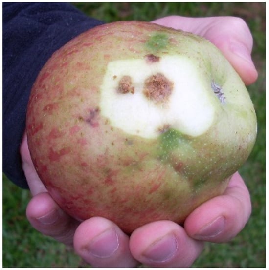
Figure 1.
Bitter pit Ca-deficiency symptoms are recurrent in pome fruits cultivated in non-calcareous soil, and can appear not only during storage, but also pre-harvest, as in this picture from Rio Grande do Sul (Brazil).
However, excess Ca2+ can also have negative effects, as experimentally observed in potted vines of Actinidia chinensis grown in Ca-depleted soil, and subsequently fertilized with different rates of Ca in the form of CaCl2 (Larocca, unpublished); leaf CO2 fixation increased in response to an application rate of 30 g Ca vine−1. However, it decreased with the application of 90 g Ca vine−1 to the same level as the unfertilized control (Table 1), showing the negative effect of a possible excess of nutrients.

Table 1.
Effect of application of Ca on leaf carbon assimilation of 2-year-old bearing potted vine of Zespri Gold 3 (A. chinensis var. chinensis).
In lime soils, like those of the Mediterranean area, symptoms of Ca deficiency are not common and appear in specific situations only, such as in sandy, non-calcareous soils, or in association with excess K availability or copper toxicity as a result of the frequent sprays of Cu-based fungicides used in disease control in humid climates (Figure 2). Nevertheless, the use of Ca-based foliar sprays is common for most fruit trees worldwide regardless of knowledge of soil Ca2+ availability, with the risk of reaching a toxicity threshold that can negatively affect crop yield and fruit quality. Correct Ca2+ management should be based on the knowledge of Ca2+ partitioning within fruit districts and its effect on fruit quality.
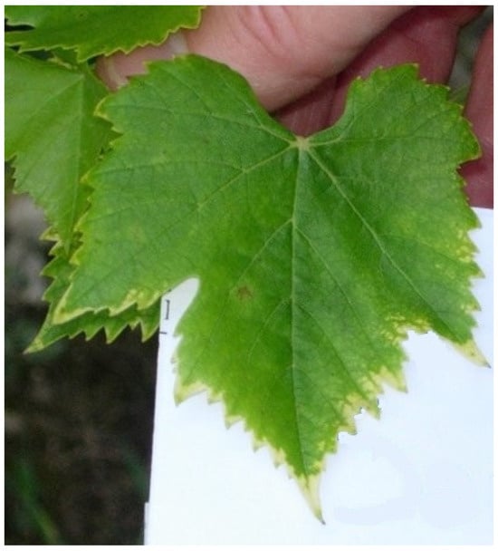
Figure 2.
Symptoms of Ca deficiency in response to Cu toxicity, induced by a concentration of 400 mg Cu kg−1 of sandy soil.
The role of Ca2+ in cell wall is crucial for fruit tissue organization in response to both abiotic and biotic stress, such as (1) pathogen infections, where fruit epidermal cells, rich in pectin, are the first defense against disease [10], and (2) fruit maturation, where the hormonal control of fruit development involves cytoplasmic Ca2+ as a secondary signal messenger [11]. Due to its low mobility in the phloem, Ca2+ accumulation within fruit depends mainly on xylem transport, which in turn is regulated by transpiration water flow. Despite this, Ca2+ affects the movement of water and other ions within plants more than any other nutrient, through the control of aquaporin activity, cell plasma membrane properties, and cell wall modification.
2. Calcium and Plant Physiology
Calcium has several functions in plant physiology [10], including (1) signaling activity; (2) organization of the plasma membrane (aquaporins and ion channels); and (3) contributing to cell wall structure.
2.1. Signaling Activity
Calcium plays a role in hormone signaling, interacting with endogenous auxins (indole-3-acetic acid—IAA), gibberellin (gibberellic acid—GA), and abscisic acid (ABA) for the regulation of cell division and expansion from fruit set to fruit softening. The close relationship between Ca2+ and IAA was earlier discussed in kiwifruit [12], where the IAA (ng g−1 FW) and Ca2+ (mg g−1 FW) concentrations were linearly correlated; hence, it can be hypothesized that IAA stimulates Ca2+ accumulation, which in turn delays ethylene synthesis and maturation and extends fruit storability. In this context, IAA acts as a fruit anti-senescent capable of delaying the ripening process by inhibiting ethylene [13]. This hypothesis was also confirmed in tomato, where IAA stimulates Ca2+ uptake and distribution in fruit [10].
The correlation between IAA and Ca2+ in fruit can be explained by the fact that IAA positively affects the induction and formation of vascular tissues, which are needed for Ca2+ upward transport. In particular, in climacteric apple fruit, Ca2+ content was found to be negatively correlated with both respiratory intensity and the rate of ethylene production during storage [14,15]. In addition, Ca2+ can enhance the binding of IAA at the membrane level through the modification of binding sites [16]. As a result, fruits with symptoms of disorders usually have lower Ca2+ concentrations than disorder-free ones [11].
In contrast, physiologically active GA has been shown to inhibit Ca2+ translocation into tomato fruits [17]. High levels of GAs during periods of intense growth could therefore be responsible for the decline in Ca2+ uptake during fruit development. As a rule, applications of GAs with the aim of increasing fruit size and crunchiness have the potential to inhibit Ca2+ accumulation in fruits, with negative effects on their susceptibility to pre- and post-harvest disorders. On the other hand, factors that reduce the biosynthesis of GA, suppress its action, or promote the endogenous antagonists of GAs, are also able to promote the translocation of Ca2+ into the fruit [18].
Abscisic acid is recognized as a major regulator capable of interaction with Ca2+ signaling, governing the opening of leaf stomata in response to drought stress [19,20]. In the context of apple bitter pit prevention, the application of ABA was proven to be effective in facilitating a preferential partitioning of Ca2+ in the fruit. In this regard, one of the mechanisms described involves competition between leaves and fruits; the foliar application of ABA in apple was found to force stomata closure, with a consequent reduction in leaf transpiration and a decrease in leaf water potential in comparison with fruit [19], which impaired the movement of Ca2+ to the leaves in favor of fruits. Transcriptomic analyses have revealed that the expression of Ca-responsive genes is significantly reliant on ABA transactivation elements [21]. This relationship is facilitated by Ca2+ sensor major intrinsic proteins (MIPs): Ca-dependent protein kinases (CDPKs), interacting protein kinases (CIPKs), calcineurin B-like (CBL) protein, protein kinases (CPKs), and ABA regulators like protein phosphatase 2C (PP2C) and Ca-independent kinase sucrose non-fermenting (SnRK2) [22]. These all control membrane water channels. The expression of CDPK and CBL/CIPK influences plant ABA sensitivity, thereby impacting the overall ABA response [22].
Calcium is also involved in the signaling of nutrient availability variations: the literature refers to potassium, nitrate- and ammonium-N, iron, boron, and magnesium as potentially being controlled by Ca2+ signals [23]. As an example, in Arabidopsis roots, the proteins responsible for potassium uptake and transport are regulated by Ca2+ decoding complexes (CIPKs). A similar control was observed for nitrate-N, whose transporter is regulated by the same CIPK, that under low-nitrate conditions turns the protein into a high-affinity transporter [24].
2.2. How Calcium Helps Organize the Plasma Membrane
2.2.1. Ion Channels
Ion channels are specialized proteins in the plasma membrane that allow charged ions to cross the plasma membrane down their electrochemical gradient. This movements usually generates an electrical current. Ion channels are selective and permit the plasma membrane crossing of a single type of ion.
Many ion channels have been studied, including slow anion channel-associated (SLAC1), gated outwardly rectifying K (GORK), open stomata (OST1), and heat shock protein (HSP1), all of them located in the guard cells where they can modulate stomata opening [25]. Other water channels are force-gated (FG) channels and mechanosensitive (MS) channels capable of instantly converting membrane forces into Ca2+ movements and electrical signals [26].
Plasma membrane FG ion channels are important in translating physical cues into chemical signals, like triggering Ca-dependent protein phosphorylation via channel opening, while MS channels convert mechanical stimuli into a flow of ions.
The activation of Ca channels allows transmembrane fluxes of Ca2+, resulting in variation in the cytosolic Ca2+ concentration from ‘resting’ levels of approximately 100 nM to ‘excitatory’ levels of 300 nM–400 nM and vice versa. In fact, there are two general classes of Ca2+ channels: the first, located in the plasma membrane, allows Ca2+ influx from the cell wall space into the cytosol; the second, a Ca2+ release channel located in the membrane of intracellular organelles, allows the release of stored Ca2+. Both channels can be found in the same cell type [27]. According to Schroeder and Thuleau [27], the plasma membrane influx in Ca channels can be divided into two types.
The first type is voltage-dependent, as observed in Mimosa pudica, which shows a rapid touch-sensitive response and stretch-activated channels that operate in response to a mechanical perturbation, like membrane tension under osmotic pressure, observed during cell wall loosening to increase membrane area. These channels, including mammalian Piezo channels and plant OSCA channels, instantly transduce force into electrical and ion signals, influencing Ca2+ concentration. The diverse OSCA family’s conductance and activation kinetics offer possibilities for generating Ca2+ signatures in response to membrane tension changes. This conversion of mechanical signals into biological ones underscores the role of Ca2+ as an intracellular messenger, rapidly altering cytosolic Ca2+ concentration upon channel opening due to the strong electrochemical gradient [26].
The second type of influx Ca channel is ABA-activated ones, which regulate ion fluxes across the vacuole membrane for the control of stomata opening. This mechanism also includes channels responsible for the release of vacuolar K+ in response to increases in ABA concentration as a consequence of drought stress [28]. These channels are down-regulated at low turgor and up-regulated at high turgor.
Guard cell osmoregulation seems, at least in Arabidopsis, to be activated by inward water flow to equilibrate different osmotic concentrations and promote an osmoregulation between the cytoplasm and vacuole, with the involvement of an aquaporin acting as an ion Ca-activated channel [28] responsible for the release of vacuolar K+ in response to ABA. This mechanism probably includes other ions (i.e., Na+) in different cell physiological responses. Ca2+ release channels are found in the membrane of intracellular organelles and regulate the storage of Ca2+. The concentration can be 104 times higher than that in the cytosol; therefore, the opening of these channels allows a rapid increase in Ca2+ concentration in the cytosol [27].
Above all, Ca2+ channels are involved in several signal transduction processes in higher plant cells, including bud formation, polar growth, gas exchange, secretion, movement, and light-regulated and hormone-regulated growth and development [29].
Cell elongation and plant growth are regulated by the interplay between osmotic pressure and cell extensibility. While the role of turgor pressure is acknowledged, the plasma membrane’s involvement in mechanical properties is often overlooked.
2.2.2. Aquaporins
The composition of the plasma membrane and its involvement in translating mechanical into electrical signals contribute significantly to the development and growth of cells and tissues. As previously mentioned, plasma membrane contains MIPs, which are water channel aquaporins (AQP) [30]. The mechanisms of their opening and closure involve a Ca-dependent CPK that is stimulated by the external environment, such as drought, anoxia, salt stress, disease, etc.
Calcium is responsible for the activation of AQP at the membrane level of both roots [31] and stomata guard cells [32]; consequently, it is involved in water movement through the whole plant, since it can regulate both apoplastic and symplastic cell-to-cell water flow from the roots to the leaves as a response to biotic and abiotic stress.
The different mechanisms that control AQP include plasma membrane phosphorylation and dephosphorylation, cytoplasm acidification, and Ca2+ modulation [31]. While it is known that root anoxia has a negative effect on plasma membrane water flow, as a consequence of a reduction in cytoplasmatic pH, the explanation of the similar negative effect observed with high cytoplasmic Ca2+ concentrations is still not clear. It seems that cytoplasmic Ca2+ concentration increases the activation energy with a reduction in outflow water movement through the plasma membrane pores.
Roots of sugar beet show a biphasic Ca2+ sensitivity that appears with cytosolic nanomolar or micromolar Ca2+ concentrations as a result of two kinds of AQPs with binding sites with high and low sensitivity to cytosolic Ca2+ [31]. The inhibition effect at high concentrations was estimated to be between 100 and 200 micro-M of Ca2+ in both sugar beet [31] and Arabidopsis [33]. Since the normal cytosolic concentration is approximately 100 nano-M, a mechanism of AQP turning on and off via changes in cytosolic Ca2+ may be hypothesized [31].
The presence of Ca2+ seems pivotal in regulating water transport in plants under salt stress, as its concentration within the nutrient solution can dictate the recovery of root hydraulic conductivity through the regulation of aquaporin functionality [34,35]. Unlike root cells, in which water channels are controlled by cytosolic Ca2+, at the stomata level, AQPs seem to be controlled by external Ca2+, as observed in broad bean [32]. The mechanism involves low extracellular Ca2+, which activates CDPK, CaM, and CBL proteins, inducing an increase in cytosolic Ca2+ with an effect on stomata closure.
In broad bean leaves, extracellular Ca2+ at high concentrations (i.e., >200 mM) was found to block water channel and reduce water flow into the guard cells, with stomatal closure and a consequent reduction in CO2 assimilation [32]. The experimental data shown in Table 1 seem to confirm this response in kiwifruit. With excess soil Ca2+ availability, the concentration in the xylem sap may rise above the toxicity threshold, with a negative impact on leaf C fixation. This hypothesis needs further evidence.
2.3. Cell Wall Structure
One of the main functions of Ca2+ in the plant is the organization of the cell wall and cell functionality, which are directly related to the stability of plant tissues and affect fruit integrity both pre- and post-harvest.
The space between two fruit cells includes primary and secondary cell walls and a middle lamella, along with empty space. These structures are made of pectin (35%), proteins (10%), cellulose (25%), and hemicellulose (20%). Pectins are chemical compounds made of polysaccharides such as homogalacturonan, rhamnogalacturonan-I, rhamnogalacturonan-II, and xylogalacturonan that combine together to form five types of pectin: S-type, A-type, B-type, C-type, P-type [36].
In the fruit flesh middle lamella, negatively charged carboxylic groups of pectins bind with Ca2+ to form structural bridges in the typical organization of ‘egg boxes’; an example of Ca-pectate is pectin bound to two molecules of polygalacturonic acid, belonging to two adjacent cell walls (egg box) to confer structural firmness to the fruit. By cross-linking negatively charged regions in pectin, mainly found in short stretches of demethylated homogalacturonans, Ca2+ imparts rigidity to the cell walls and regulates the cell’s ability to extend. When Ca2+ concentrations are too low, the wall is weakened and may even break, whereas at high Ca2+ levels, pectin chains become cross-linked and aggregated, resulting in maximal wall rigidity [37].
Calcium deficiency promotes physiological disorders caused by the structural collapse of the cell wall because of the absence of a specialized egg box. This mechanism is typical of pollen tubes and root hairs, where a decrease in middle lamella Ca2+ and cell rigidity is necessary during tip growth [38]. At the same time, salinity stress can bring about an excessive replacement of Ca2+ with Na+, disturbing the cell wall organization with anomalies in root growth [23].
During fruit ripening, various chemical transformations take place within the cell walls, including alterations in pectin side chains, the breakdown of pectin through depolymerization, and the degradation of hemicellulose xyloglucan [10]. When combined with other ripening-related activities, such as solute accumulation, these transformations contribute to distinct physical and textural alterations in fruits [36]. The majority of pectin populates the middle lamella in the cell junction, with little amounts in the primary cell wall. Cellular turgor pressure promotes the separation of cells and the formation of extra-cellular spaces, affecting the structural organization of fruit flesh during maturation [10]. Fruit softening is the result of a transformation of cellular wall composition, particularly due to pectin de-esterification and decreases in Ca2+-polygalacturonic acid cross-linking, which alter the physical attributes of the cell wall, such as strength, elasticity, loosening, and swelling [10]. Pectin de-esterification is promoted by pectin methyl esterase (PME), revealing carboxyl residues that can be cross-linked by Ca2+. The level of PME activity and the presence of Ca2+ within the apoplasm have a direct impact on the strength and expansion of the cell wall [39] and affect cell wall properties, such as porosity and ionic status [40].
When compared with other species, the cell wall of kiwifruit fruit contains a larger amount of polysaccharide. As previously reported [41] the cell wall of green-flesh kiwifruit (cv. Hayward) at harvest consists mainly of heterogeneous pectin galactan mixtures (40–50%) and hemicellulose (15–25%). The hemicellulose mainly consists of xyloglucans [42]. The concentrations of these polysaccharides vary at different stages of maturity because of the physiological changes that occur mostly during the maturation process. These changes could lead to significant differences in the functional properties of the polysaccharides of kiwifruit.
The characterization of these structural polymers during all stages of growth and maturation of the kiwifruit fruit contributes to defining their functions throughout the entire fruit growth process. The role of Ca2+ in cell wall organization and biotic stress responses, such as those induced by pathogen infections, is still a matter of discussion [10,43]. Calcium signals are an early response of cells to the presence of pathogenic and symbiotic microorganisms; it has been shown that microbe-associated molecules increase cytosolic Ca2+ by mobilizing it from both extracellular (apoplast) and intracellular (vacuole/endoplasmic reticulum) stores, with an alteration in nuclear Ca2+ [43]. This increase in cytosolic Ca2+ concentration leads to the activation of mitogen, salicylic acid, and wound-activated protein kinases, which are active against microbial infection. Some pathogens secrete pectinases (i.e., PME) that degrade pectin, breaking down its structure [44]. As for fruit maturation, Ca2+ can contrast the action of these enzymes, as was shown in grape, where Ca2+ inhibited enzyme activity, although the concentration required was different for each of the isolates of Botrytis cinerea tested [45]; consequently, the Ca2+ concentration of grape skin cell walls was positively correlated with Botrytis enzymatic digestion tolerance [46]. In kiwifruit, Botrytis cinerea and Alternaria alternata were found to stimulate the synthesis of defensive enzymes, such as peroxidase and phenylalanine ammonia-lyase, in response to PMEs produced by the pathogens [44,47]
On the other hand, pectin plays an active role in mediating immune responses during interactions between plants and pathogens; this role was discussed in a recent review by Riseh et al. [44].
The authors underlined that the bactericidal activity of pectin is mainly due to its free undissociated forms of some organic acids, such as sorbic acid, dehydroacetic acid, salicylic acid, and benzoic acid [44]. In addition, when plants are exposed to stress, they release molecules derived from the cell wall, such as pectin fragments or oligogalacturonides (OGAs), which have been implicated in pathogen defense signaling activation [44]. It is likely that functional OGAs may be prevalent in fruit and affect ripening, as ripening fruit has a high pectin content and is attractive to a variety of pathogens.
Many factors influence the defense-response-eliciting capacity and specificity of OGAs, including Ca2+ availability and the length of the OGA [10]. Complex interactions between Ca2+ nutrition and the diversity of pectin profiles influence susceptibility to fungal pathogens in different ways, according to genotypes, tissues, and organs.
The mitigation of oxidative stress induced by metalloid arsenic (As) on Vicia faba was observed as a consequence of the combination of the signaling molecules melatonin (a similar hormone to endogenous IAA) and Ca2+ [48]. Alone or in combination, they reduced electrolyte leakage, malondialdehyde synthesis, reactive oxygen species (ROS), hydrogen peroxide, and superoxide anions, as well as their related enzymes: NADPH oxidase and glycolate oxidase, stimulated by the presence of As in the growing medium [48].
The upregulation of enzymatic components of the ascorbate–glutathione pathway seems responsible for this mitigation.
3. Mechanisms and Challenges of Calcium Transport in Fruit Trees
Calcium long-distance transport is mainly carried out through the apoplastic pathway, involving intercellular space and xylem vessels; on the other hand, Ca2+ short-distance transport, such as from cell organelles to the cytoplasm or from cell to cell (i.e., stomata aperture regulation), or eventually from leaf to fruit via the phloematic pathway, follows a symplastic movement that could take advantage of cell permeability and requires energy to activate the Ca2+- or H+-ATPase pump [32]. From this picture, since Ca2+ can regulate both symplastic aquaporins and ion channels, as well as the apoplastic cell wall structure and xylem vessels, it seems that Ca2+ has the potential to modify its own delivery [10,49].
Calcium long-distance delivery and distribution into tree canopies is affected by (1) the rate of water mass flow in the xylem vessels; (2) the competition with other cations for binding charges in the xylem vessel walls and pit membrane; (3) and the possibility for Ca2+ to be chelated by low-weight organic molecules, which increases its water solubility and mobility. Calcium short-distance pathways depend on the abundance of mechanisms that increase cellular permeability to water and ions, such as water and ion channels [10].
Once entered into the root apoplast, Ca2+ is forced by the Casparian band to cross the plasma membrane and enter into the cytosol; therefore, the movement of Ca2+ into the xylem must be through a sympalstic pathway, so that the flux involves both protein-mediated channels and Ca-chelates [49].
It is clear that in most fruit trees, long-distance Ca2+ transport in tissues with a low transpiration rate—such fruits at maturation, characterized by a reduction in lenticel gas exchange rate, which is necessary to maintain fruit cell turgor—is reduced compared to those with a higher transpiration rate, such as leaves. Therefore, leaf gas exchange is more affected by atmospheric variation than fruit; for example, the increase in atmospheric CO2 related to climate change and increases in drought stress, both of which limit stomata aperture, have the potential to reduce water transpiration rate and Ca2+ movement [49].
While it is universally accepted that transpiration and water flow affect Ca2+ long-distance movement, the interdependence between water and Ca2+ transport is not completely clear [49]. In fact, Ca2+ transport to the leaves is not only affected by mass flow; it is affected by extracellular factors such as the binding of Ca2+ to the xylem wall, the intensity of which increases with plant growth [49], and hydraulic resistance, which eventually develops during the season and may reduce Ca2+ delivery [10]. Compartmentation is one of the main causes for increased hydraulic resistance; it occurs, for example, in Casparian strip formation at the root level or during the separation of the leaf xylem from the leaf apoplast by bundle sheath cells [10].
These morphological modifications can also include physical barriers, like decreases in xylem vessel diameter or the formation of pectin gel within the xylem sieve, which usually happens late in the season [10]. This development is common to many fruit species and involves the switch from xylem to phloem Ca2+ delivery; it can be considered an adaptation mechanism of plants that removes fruit hydraulic independence and ensures that fruit water status depends on the whole plant’s requirements [50].
4. Calcium Accumulation in Kiwifruit Fruit
Since Ca2+ mobility through the phloem is negligible, Ca2+ accumulation in aerial sink organs depends on xylem flow, and considering that fruit transpiration rate declines significantly during development [51], Ca2+ delivery seasonal trends reflect the formation and functionality of xylem conduits. The reduction in Ca2+ import during ripening is correlated with a drop in xylem hydraulic conductance. The gradual modification of the xylem system is visible in kiwifruit fruits [52]. It is known that the functionality of the xylem bundle undergoes progressive deterioration, leading to total and permanent dysfunction 8 weeks after full bloom. This dysfunction is attributed to the rupture of xylem vessels due to fruit elongation and expansion [53]. The alteration in the xylem bundle, in conjunction with certain environmental factors, such as increases in daily evapotranspiration rate, results in reduced fruit transpiration rates and finally a decrease in Ca2+ accumulation [54,55]. This was experimentally observed in the green flesh of Hayward kiwifruit, where the limitation of fruit transpiration, induced by wrapping them in plastic bags, significantly reduced fruit Ca2+ partitioning during the first 80 days after flowering [56]. However, once the plastic bag was removed, the fruit recovered the accumulation of Ca2+ to the level of the unbagged control fruit (Table 2). These results show the ability of kiwi to accumulate Ca2+ in the fruit until harvest, unlike the results of previous reports. This late-season fruit capacity to recover from Ca2+ deficiency can be related to the recognized ability of kiwifruit to maintain positive water fluxes from both the phloem and xylem [10], or it can be explained by the high level of soluble Ca in the calcareous soil of the investigation area (Table 3). Finally, a more intriguing explanation is the ability of Ca2+ to autoregulate itself by moving to deficient plant districts.
Table 2.
Fruit calcium content (mg Ca fruit−1) during the season in bagged and not-bagged fruits [56].
Table 3.
Calcium and potassium water solubility in soil and in irrigation water, as determined in 2022 in a kiwifruit orchard in the south-eastern side of the Italian Po valley (Toselli, unpublished).
Several external factors can influence fruit transpiration, such as light and atmospheric humidity. Increased radiation availability appears to prolong fruit transpiration activity, likely extending the functionality of fruit conducting systems [57]. A relationship between increased vascular development and higher Ca2+ concentration in kiwifruit fruits exposed to light has been demonstrated [58].
Fruits exposed to light had approximately 40% more xylem vascular bundles in the carpel section and double Ca2+ concentration compared to shaded fruits [55].
As already mentioned, different Ca2+ concentrations can be explained by a regulatory mechanism governed by IAA; in fact, exposure to full sunlight leads to a 30% higher concentration of hydroxycinnamic acids (HCAs), protectors of IAA, compared to shaded fruits. Therefore, it is suggested that light, by inducing the biosynthesis of these phenols, indirectly reduces IAA degradation and increases Ca2+ accumulation [57].
A recent study on yellow kiwifruits demonstrated that, in Northern Greece, the application of Ca2+ post-harvest significantly delays ripening and softening by reducing ethylene production, as confirmed by the change in the expression level of the genes and the synthesis of proteins involved in ethylene signaling and cell wall structure deterioration [59]. Calcium treatment has a lasting impact on the transcriptome and proteome of kiwifruit, influencing ripening-related changes [59]. The total Ca2+ content in fruit can be separated into various fractions with different solubilities, leading to different physiological activities [60]. Water-soluble Ca2+, which is associated with components like organic acids, nitrates, and chlorides, as well as exchangeable Ca2+ bound to pectin and proteins, is considered physiologically active in the plant. This contrasts with the more tightly bound forms, such as the phosphate and oxalate fractions [18]. Another factor influencing Ca-cross-linked pectates is the stress imposed on wall binding sites by turgor pressure. Under this physical tension (e.g., 0.2 MPa), the bonds elongate, weaken, and reduce their affinity for Ca2+ [61].
The primary roles of Ca-oxalate (CaOx) crystal formation in plants involve the regulation of Ca2+ at high concentrations and protection against herbivores. These crystals are formed from oxalic acid synthesized within the plant and Ca2+ taken up by the roots. Each plant species produces only specific types or subsets of crystal morphologies, including prismatic crystals, crystal sand, raphide crystals, and druse crystals. CaOx crystals can be deposited into the vacuoles or associated with the cell wall.
Cells that have vacuolar crystals are often specialized for this purpose and are termed crystal idioblasts. In some plants, crystals are found in the vacuoles of regular cell types, such as storage parenchyma, bundle sheath cells, epidermal cells, or chlorenchyma. Considering the variety in crystal shapes and sizes, along with their prevalence and spatial distribution, many theories have been proposed about their functions in plants. These functions include Ca2+ regulation, plant defense, detoxification, ion balance, structural support and rigidity, and even the gathering and reflection of light [62,63].
Earlier research has indicated that green kiwifruit contains moderate amounts of total oxalates [64]—up to 18–45 mg oxalate 100 g−1 FW—mostly in the form of insoluble Ca-oxalate raphide crystals [65]. These insoluble oxalate crystals are responsible for the irritant factor or “catch”, which describes the irritation of the mouth’s mucous membranes [66]. In addition, soluble oxalates may be of interest for kidney stone patients who are trying to decrease their urinary oxalate excretion by avoiding the consumption of oxalate-rich foods [67].
The pulp of golden kiwifruit has a lower total oxalate content (15.7 mg 100 g−1 FW) compared to the green variety, but it contains higher levels of soluble oxalates (8.5 mg 100 g−1 FW) than the green cultivar (7.6 mg 100 g−1 FW) [67].
The fruit skin of green kiwifruit cultivars has lower levels of insoluble oxalates (36.9 mg 100 g−1 FW) compared to yellow varieties (43.6 mg 100 g−1 FW), whereas the seeds of the green-flesh cultivars have higher levels of insoluble oxalates (107 mg 100 g−1 FW) than the yellow ones (85 mg 100 g−1 FW) [67].
Among the various cultivars of Actinidia, not only does the oxalate content differ, but there can also be variations in its form [68]. Kiwifruit raphide calcium-oxalate crystals show a long and needle-like shape in green-fleshed cultivars, but a short shape in yellow-fleshed ones. The raphide crystals in A. arguta and A. rufa are similarly short.
5. Methodologies for the Study of Calcium in Plants
The evaluation of the concentration of different fractions of Ca2+ in various plant organs is essential for a better understanding of its kinetics and for obtaining a clear picture of its functions and roles in various metabolic processes of plants. The most commonly used methodologies are listed below (Table 4).
Table 4.
Summary of the technology for studying calcium in plants.
5.1. Microscope Technique
The use of a microscope can provide spatial Ca2+ imaging across kiwifruit sections, as shown by the real-time Ca2+ imaging described by Polychroniadu [59] to visualize Ca2+ signals. The cell-permeable fluorescent indicator Fluo-3 was used before the analysis with a stereomicroscope equipped with a digital camera. Hand-cut and razor-cut fruit slices were incubated for 2 h at 4 °C with 5 μM Fluo-3 AM in MES buffer (10 mM, pH 6.5). After washing with MES, samples were transferred to glass slides and observed under a UV source [59].
5.2. X-Ray Fluorescence Microscopy (XFM)
The X-ray fluorescence technique detects elements based on their characteristic fluorescent X-rays. These fluorescent X-rays are emitted when the specimen is irradiated with a focused beam of high-energy X-rays (XFM). This beam excites a range of elements, which are then detected and quantified by a detector to ascertain their concentrations in the specimen. By scanning the specimen through the incident beam in an x-y pattern, a raster map is generated where each point corresponds to a pixel containing data on element concentrations or relative intensities. High-energy (greater than 15 keV) X-rays can penetrate deeply, making it possible to analyze both sectioned and potentially whole, intact plant tissues.
Generally, the X-ray beam does not damage the sample, so the method is considered non-destructive. However, since X-rays comprise ionizing radiation, the energy and exposure duration can cause damage by producing free radicals, which are highly reactive and can harm the examined tissue [69].
The validity of the XRF technique was demonstrated in comparison with traditional laboratory analyses for determining Ca2+ concentration in apple tissues [70]. This novel technique offers several advantages, particularly its non-destructive nature, allowing for time-resolved, in vivo analyses of changes in elemental distribution without compromising the integrity of the sample. Its ability to penetrate deep into plant tissues, both sectioned and intact, enables a detailed examination of nutrient movement, molecular biology functions, and contaminant behavior at the microscopic level.
With this technique, it is possible to understand the spatial variability of Ca2+ concentrations in plant tissues.
5.3. Calcium Imaging Techniques
Calcium imaging is a microscopy method used to visually assess the Ca2+ levels in isolated cells or tissues. This technique utilizes Ca2+ indicators, which are fluorescent molecules that change their fluorescence properties upon binding with Ca2+ ions. Calcium imaging has facilitated the study of Ca2+ signaling in numerous cell types, with confocal laser scanning microscopy (CLSM) employed for detailed analysis. Calcium imaging is also used to measure the concentration of Ca2+ in cells or tissues using fluorescent dyes.
Measurement techniques can be classified into ratiometric and non-ratiometric, each with distinct properties and applications. Non-ratiometric dyes, also known as single-wavelength probes, such as Fluo-4, Rhod-2, and Ca2+ Green-1, emit fluorescence at a specific wavelength when excited. These dyes change their fluorescence intensity in response to Ca2+ binding, allowing for the detection of Ca2+ concentration in cells. However, these measurements can be influenced by various factors, including light intensity, photobleaching, and sample geometry. As a result, additional corrections may be necessary to ensure accuracy. These dyes are particularly useful for their simplicity and ease of use, but are more sensitive to experimental variations. Non-ratiometric dyes measure Ca2+ concentration by monitoring changes in fluorescence intensity.
Ratiometric dyes, such as Fura-2 and Indo-1, emit fluorescence at two or more wavelengths, and the ratio of these emissions correlates with the Ca2+ concentration. This ratiometric approach provides more reliable and accurate measurements, as it compensates for variables like light intensity, photobleaching, and uneven dye loading.
The ratio of fluorescence intensities at different wavelengths provides a direct measure of Ca2+ concentration, reducing the influence of experimental artifacts [71]. Calcium imaging with fluorescent dyes offers significant advantages for studying Ca2+ signaling in living cells and tissues. One of the key benefits is its high sensitivity and specificity in detecting Ca2+, allowing researchers to observe dynamic changes in Ca2+ levels in real time.
The use of advanced microscopy techniques such as CLSM also allows for high spatial resolution, making it possible to examine the distribution of Ca2+ at the cellular level.
Ratiometric dyes, in particular, provide more reliable and accurate data by compensating for experimental variations such as changes in light intensity or photobleaching, thus enhancing measurement precision. However, there are several challenges associated with these techniques.
Calibration is necessary, particularly with non-ratiometric dyes, as fluorescence intensity may vary due to factors like dye loading or sample thickness. Both types of dyes are also susceptible to photobleaching, where prolonged light exposure reduces their fluorescence, potentially influencing the results.
Furthermore, in thicker tissues, the penetration of fluorescent dyes can be limited, which may hinder the effectiveness of Ca2+ imaging in larger or more complex samples. Ratiometric dyes, while offering more accurate measurements, also require more complex data analysis, involving calculating ratios of fluorescence at different wavelengths.
This can be computationally demanding and may require specialized software, making the process more difficult.
5.4. Stain Technology
Stain technologies are methodologies used for the coloration and marking of specific cellular components, such as Ca2+, in biological tissues. These techniques utilize chemical dyes that selectively bind to certain ions or cellular structures, allowing the visualization of their distribution and accumulation within cells and tissues.
Alizarin Red S is a versatile stain that is widely used in plant histochemistry to localize and visualize Ca2+ in plant tissues. This red-color synthetic dye reacts with Ca2+ ions, forming a red complex. Although traditionally used for staining calcified animal tissues, its application in plant research is gaining attention, particularly in studying plant cells [72].
Alizarin Red S is particularly effective for highlighting Ca2+ associated with cell walls, but it can also stain protoplasmic Ca2+ when used in combination with other reagents.
The dye is commonly applied in an aqueous solution, with the pH adjusted to 4.2 to optimize its interaction with Ca2+ compounds [73].
The technique is valuable for investigating the role of Ca2+ in plant development. The method has been employed in various plants, including ferns (Vittaria graminifolia and Onoclea sensibilis), where it revealed Ca2+ localization in both the protoplasm and cell walls [75].
The application of the dye is relatively straightforward, and it is an inexpensive option, making it accessible for many research laboratories. Additionally, the technique is well established and has been employed in various plant species, making it versatile in research on plant development and physiology. However, this technique has some limitations, including the possibility that Alizarin Red S binds to other ions with similar properties of Ca2+, which may interfere with the interpretation of the results. Sample preparation is critical, as variations in fixation and sectioning protocols can affect the quality of staining and reduce the consistency of results. Moreover, although the technique is useful for visualizing Ca2+ distribution, it does not allow precise quantitative measurements, offering instead a qualitative observation of the stained areas. Finally, staining may not be uniform across all tissues if the pH or dye concentration is not properly optimized.
5.5. Size-Exclusion Chromatography
Size-exclusion chromatography (SEC) can be used to quantify de-polymerized pectins previously separated according to their size by filtration through a gel that usually consists of spherical beads containing pores of a specific size distribution. It can be used in ripening fruits, where the de-esterification and depolymerization of pectins to shorter sub-units occurs, and their quantification indirectly measures the amount of cross-linked Ca2+ [10]. The employment of a chelator agent such as CDTA [10], followed by SEC, is a means to characterize the time-course of ionically bound pectins induced by treatments, particularly with regard to soil Ca2+ availability.
5.6. Nuclear Magnetic Resonance
Nuclear Magnetic Resonance (NMR) proton relaxometry can be used in the analysis of biochemical changes in fruits [37] because plant cellular tissue contains water compartmentalized into subcellular structures, such as the cytoplasm, vacuoles, cell walls, granules, and extracellular space, ensuring proton exchange between these structures. This exchange can be measured by an NMR acquisition time, since these compartments are characterized by different proton relaxation rates. Biochemical changes in plant tissues are accompanied by changes in water content, distribution, and the integrity of subcellular compartments, leading to variations in relaxation times that can be used to investigate ripening, internal decay, and disease-related changes in fruits [37].
6. Conclusions
Although Ca2+ is generally recognized as one of the crucial nutrients for fruit quality pre- and post-harvest, its role in kiwifruit fruit is not as clear as it is in pome fruits. The movement of Ca2+ is influenced by various factors, such as transpiration rates, hydraulic resistance, the formation of physical barriers within the xylem, etc., that often limit the mobility of Ca2+; in addition, the efficiency of Ca2+ delivery depends on vascular development, which is itself correlated with Ca2+ concentration. However, in most of the Mediterranean area, the soils are calcareous and present high levels of water-soluble Ca2+, which may in part counter the limitations of mobility of this ion in kiwifruit. Understanding the complex interplay of Ca2+ transport mechanisms and its accumulation in different plant organs is essential for improving agricultural practices, especially under changing environmental conditions. Future research should focus on mechanisms that regulate Ca2+ transport and accumulation in kiwifruit fruit, including its distribution in fruit cells, the optimal amount for good fruit quality pre- and post-harvest, its interactions with other micronutrients, and the molecular pathways regulating its transport and storage.
Funding
This research was funded by Zespri contract number 2454 (27/03/2024).
Acknowledgments
We thank ZESPRI INTERNATIONAL LTD (400 Maunganui Road, Mount Maunganui 3116, New Zealand) for their financial support.
Conflicts of Interest
The authors declare no conflict of interest.
References
- Demarty, M.; Morvan, C.; Thellier, M. Calcium and cell wall. Plant Cell Environ. 2006, 7, 441–448. [Google Scholar] [CrossRef]
- Balantič, K.; Weiss, U.; Allmaier, G.; Kramar, P. Calcium ion effect on phospholipid bilayers as cell membrane analogues. Bioelectrochemistry 2022, 143, 107988. [Google Scholar] [CrossRef] [PubMed]
- De Freitas, S.T.; Handa, A.K.; Wu, Q.; Park, S.; Mitcham, E.J. Role of pectin methylesterases in cellular calcium distribution and blossom-end rot development in tomato fruit. Plant J. 2012, 71, 824–835. [Google Scholar] [CrossRef] [PubMed]
- Ferguson, I.B.; Watkins, C.B. Bitter pit in apple fruit. Hort. Rev. 1989, 11, 289–355. [Google Scholar]
- Bramlage, W.J.; Drake, M.; Lord, W.J. The influence of mineral nutrition on the quality and storage performance of pome fruits grown in North America. In Proceedings of the Symposium on Mineral Nutrition and Fruit Quality of Temperate Zone Fruit Trees, Canterbury, UK, 1–7 April 1979; Volume 92, pp. 29–40. [Google Scholar]
- Kirkby, E.A.; Pilbeam, D.J. Calcium as a plant nutrient. Plant Cell Environ. 1984, 7, 397–405. [Google Scholar] [CrossRef]
- Retamales, J.; Valdes, C.; Dilley, D.; León, L.; Lepe, V.P. Bitter pit prediction in apples through Mg infiltration. Acta Hort. 2000, 512, 169–179. [Google Scholar] [CrossRef]
- Zhang, Y.; Kong, Q.; Niu, B.; Liu, R.; Chen, H.; Xiao, S.; Wu, W.; Zhang, W.; Gao, H. The dual function of calcium ion in fruit edible coating: Regulating polymer internal crosslinking state and improving fruit postharvest quality. Food Chem. 2024, 447, 138952. [Google Scholar] [CrossRef]
- Shiri, M.A.; Ghasemnezhad, M.; Moghadam, J.F.; Ebrahimi, R. Efficiency of CaCl2 spray at different fruit development stages on the fruit mineral nutrient accumulation in cv. hayward kiwifruit. J. Elem. 2016, 21, 195–209. [Google Scholar] [CrossRef]
- Hocking, B.; Tyerman, S.D.; Burton, R.A.; Gilliham, M. Fruit calcium: Transport and physiology. Front. Plant Sci. 2016, 7, 569. [Google Scholar] [CrossRef]
- Gao, Q.; Xiong, T.; Li, X.; Chen, W.; Zhu, X. Calcium and calcium sensors in fruit development and ripening. Sci. Hort. 2019, 253, 412–421. [Google Scholar] [CrossRef]
- Sorce, C.; Lombardi, L.; Montanaro, G. Occurrence of natural auxin and accumulation of calcium during early fruit development in kiwifruit. Aust. J. Crop Sci. 2011, 5, 895–898. [Google Scholar]
- Bregoli, A.M.; Fabbroni, C.; Costa, F.; Raimondi, V.; Costa, G. Auxin and ethylene interaction during fruit growth and ripening of Actinidia deliciosa. Adv. Plant Ethyl. Res. 2007, 2, 105–107. [Google Scholar]
- Poovaiah, B.W.; Glenn, G.M.; Reddy, A.S.N. Calcium and fruit softening: Physiology and biochemistry. Hortic. Rev. 1988, 10, 107–152. [Google Scholar]
- Sharma, R.R.; Pal, R.K.; Singh, D.; Singh, J.; Dhiman, M.R.; Rana, M.R. Relationships between storage disorders and fruit calcium contents, lipoxygenase activity, and rates of ethylene evolution and respiration in ‘Royal Delicious’ apple (Malus × domestica Borkh.). J. Pomol. Hortic. Sci. 2012, 87, 367–373. [Google Scholar] [CrossRef]
- Ferguson, I.B. Calcium in plant senescence and fruit ripening. Plant Cell Environ. 1984, 7, 477–489. [Google Scholar] [CrossRef]
- De Freitas, S.T.; Jiang, C.Z.; Mitcham, E.J. Mechanisms involved in calcium deficiency development in tomato fruit in response to gibberellins. J. Plant Growth Regul. 2012, 31, 221–234. [Google Scholar] [CrossRef]
- Saure, M.C. Calcium translocation to fleshy fruit: Its mechanism and endogenous control. Sci. Hort. 2005, 105, 65–89. [Google Scholar] [CrossRef]
- Falchi, R.; D’Agostin, E.; Mattiello, A.; Coronica, L.; Spinelli, F.; Costa, G.; Vizzotto, G. ABA regulation of calcium-related genes and bitter pit in apple. Postharvest Biol. Technol. 2017, 132, 1–6. [Google Scholar] [CrossRef]
- Xiong, T.; Tan, Q.; Li, S.; Mazars, C.; Galaud, J.P.; Zhu, X. Interactions between calcium and ABA signaling pathways in the regulation of fruit ripening. J. Plant Physiol. 2021, 256, 153309. [Google Scholar] [CrossRef] [PubMed]
- Kaplan, B.; Davydov, O.; Knight, H.; Galon, Y.; Knight, M.R.; Fluhr, R.; Fromm, H. Rapid transcriptome changes induced by cytosolic Ca2+ transients reveal ABRE-related sequences as Ca2+- responsive cis elements in Arabidopsis. Plant Cell 2006, 18, 2733–2748. [Google Scholar] [CrossRef]
- Edel, K.H.; Kudla, J. Integration of calcium and ABA signaling. Curr. Opin. Plant Biol. 2016, 33, 83–91. [Google Scholar] [CrossRef]
- Thor, K. Calcium—Nutrient and Messenger. Front. Plant Sci. 2019, 10, 440. [Google Scholar] [CrossRef]
- Ho, C.H.; Lin, S.H.; Hu, H.C.; Tsay, Y.F. CHL1 functions as a nitrate sensor in plants. Cell 2009, 138, 1184–1194. [Google Scholar] [CrossRef] [PubMed]
- Wang, X.; Lv, S.; Han, X.; Guan, X.; Shi, X.; Kang, J.; Zhang, L.; Cao, B.; Li, C.; Zhang, W.; et al. The calcium-dependent protein kinase CPK33 mediates strigolactone-induced stomatal closure in Arabidopsis thaliana. Front. Plant Sci. 2019, 10, 1630. [Google Scholar] [CrossRef] [PubMed]
- Frachisse, J.M.; Thomine, S.; Allain, J.M. Calcium and plasma membrane force-gated ion channels behind development. Curr. Opin. Plant Biol. 2020, 53, 57–64. [Google Scholar] [CrossRef]
- Schroeder, J.I.; Thuleau, P. Ca2+ Channels in Higher Plant Cells. Plant Cell. 1991, 3, 555–559. [Google Scholar] [CrossRef]
- MacRobbie, E. Control of Volume and Turgor in Stomatal Guard Cells. J. Membr. Biol. 2006, 210, 131–142. [Google Scholar] [CrossRef]
- White, P.J. Calcium channels in higher plants. Biochim. Biophys Acta 2000, 1465, 171–189. [Google Scholar] [CrossRef] [PubMed]
- Németh-Cahalan, K.L.; Hall, J.E. pH and calcium regulate the water permeability of aquaporin. J Biol. Chem. 2000, 275, 6777–6782. [Google Scholar] [CrossRef]
- Alleva, K.; Niemietz, C.M.; Sutka, M.; Maurel, C.; Parisi, M.; Tyerman, S.D.; Amodeo, G. Plasma membrane of Beta vulgaris storage root shows high water channel activity regulated by cytoplasmic pH and a dual range of calcium concentrations. J. Exp. Bot. 2006, 57, 609–621. [Google Scholar] [CrossRef]
- Yang, H.M.; Zhang, X.Y.; Tang, Q.L.; Wang, G.X. Extracellular calcium is involved in stomatal movement through the regulation of water channels in broad bean. Plant Growth Regul. 2006, 50, 79–83. [Google Scholar] [CrossRef]
- Gerbeau, P.; Amodeo, G.; Henzler, T.; Santoni, V.; Ripoche, P.; Maurel, C. The water permeability of Arabidopsis plasma membrane is regulated by divalent cations and pH. Plant J. 2002, 30, 71–81. [Google Scholar] [CrossRef]
- Cabañero, F.J.; Martínez-Ballesta, M.C.; Teruel, J.A.; Carvajal, M. New evidence about the relationship between water channel activity and calcium in salinity-stressed pepper plants. Plant Cell Physiol. 2006, 47, 224–233. [Google Scholar] [CrossRef] [PubMed]
- Martínez-Ballesta, M.C.; Cabañero, F.; Olmos, E.; Periago, P.M.; Maurel, C.; Carvajal, M. Two different effects of calcium on aquaporins in salinity-stressed pepper plants. Planta 2008, 228, 15–25. [Google Scholar] [CrossRef] [PubMed]
- Prasanna, V.; Prabha, T.N.; Tharanathan, R.N. Fruit ripening phenomena-an overview. Crit. Rev. Food Sci. Nutr. 2007, 47, 1–19. [Google Scholar] [CrossRef]
- Kirtil, E.; Oztop, M.-H.; Sirijariyawat, A.; Ngamchuachit, P.; Barrett, D.M.; McCarthy, M.J. Effect of pectin methyl esterase (PME) and CaCl2 infusion on the cell integrity of fresh-cut and frozen-thawed mangoes: An NMR relaxometry study. Food Res. Inter. 2014, 66, 409–416. [Google Scholar] [CrossRef]
- Bascom, C.S.; Hepler, P.K.; Bezanilla, M. Interplay between ions, the cytoskeleton, and cell wall properties during tip growth. Plant Physiol. 2018, 176, 28–40. [Google Scholar] [CrossRef]
- Conn, S.J.; Gilliham, M.; Athman, A.; Schreiber, A.W.; Baumann, U.; Moller, I.; Cheng, N.H.; Stancombe, M.A.; Hirschi, K.D.; Webb, A.A.R.; et al. Cell-specific vacuolar calcium storage mediated by CAX1 regulates apoplastic calcium concentration, gas exchange, and plant productivity in Arabidopsis. Plant Cell 2011, 23, 240–257. [Google Scholar] [CrossRef]
- Kohli, P.; Kalia, M.; Gupta, R. Pectin Methylesterases: A Review. J. Bioprocess. Biotech. 2015, 5, 1000227. [Google Scholar] [CrossRef]
- Redgwell, R.J.; Melton, L.D.; Bkasch, D.J. Cell-wall polysaccharides of kiwifruit (Actinidia deliciosa): Chemical features in different tissue zones of the fruit at harvest. Carbohydr. Res. 1988, 2, 241–258. [Google Scholar] [CrossRef]
- Yuliarti, O.; Matia-Merino, L.; Goh, K.K.T.; Mawson, J.; Williams, M.A.K.; Brennan, C. Characterization of gold kiwifruit pectin from fruit of different maturities and extraction methods. Food Chem. 2015, 166, 479–485. [Google Scholar] [CrossRef]
- Dodd, A.N.; Kudla, J.; Sanders, D. The language of calcium signaling. Annu. Rev. Plant Biol. 2010, 61, 593–620. [Google Scholar] [CrossRef]
- Riseh, R.S.; Vazvani, M.G.; Taheri, A.; Kennedy, J.F. Pectin-associated immune responses in plant-microbe interactions: A review. Inter. J. Biol. Macromol. 2024, 273, 132790. [Google Scholar]
- Chardonnet, C.O.; Sams, C.E.; Trigiano, R.N.; Conway, W.S. Variability of three isolates of Botrytis cinerea affects the inhibitory effects o fcalcium on this fungus. Phytopathology 2000, 90, 769–774. [Google Scholar] [CrossRef] [PubMed]
- Chardonnet, C.; Doneche, B. Relation between calcium content and resistance to enzymatic digestion of the skin during grape ripening. Vitis 1995, 34, 95–98. [Google Scholar]
- Gao, Z.; Zhang, R.; Xiong, B. Management of postharvest diseases of kiwifruit with a combination of the biocontrol yeast Candida oleophila and an oligogalacturonide. Biol. Control 2021, 156, 104549. [Google Scholar] [CrossRef]
- Siddiqui, M.H.; Alamri, S.; Khan, M.N.; Corpas, F.J.; Al-Amri, A.A.; Alsubaie, Q.D.; Alia, H.M.; Kalajid, H.M.; Ahmad, P. Melatonin and calcium function synergistically to promote the resilience through ROS metabolism under arsenic-induced stress. J. Hazard. Mater. 2020, 398, 122882. [Google Scholar] [CrossRef]
- Gilliham, M.; Dayod, M.; Hocking, B.J.; Xu, B.; Conn, S.J.; Kaiser, B.N.; Leigh, R.A.; Tyerman, S.D. Calcium delivery and storage in plant leaves: Exploring the link with water flow. J. Exp. Bot. 2011, 62, 2233–2250. [Google Scholar] [CrossRef]
- Greenspan, M.D.; Shackel, K.A.; Matthews, M.A. Developmental changes in the diurnal water budget of the grape berry exposed to water deficits. Plant Cell Environ. 1994, 17, 811–820. [Google Scholar] [CrossRef]
- Montanaro, G.; Treutter, D.; Xiloyannis, C. Phenolic compounds in young developing kiwifruit in relation to light exposure: Implications for fruit calcium accumulation. J. Plant Interact. 2007, 2, 63–69. [Google Scholar] [CrossRef]
- Mazzeo, M.; Dichio, B.; Clearwater, M.J.; Montanaro, G.; Xiloyannis, C. Hydraulic resistance of developing Actinidia fruit. Ann. Bot. 2013, 112, 197–205. [Google Scholar] [CrossRef] [PubMed]
- Dichio, B.; Remorini, D.; Lang, S. Developmental changes in xylem functionality in kiwifruit fruit: Implications for fruit calcium accumulation. Acta Hortic. 2003, 610, 191–195. [Google Scholar] [CrossRef]
- Xiloyannis, C.; Celano, G.; Montanaro, G.; Dichio, B.; Sebastiani, L.; Minnocci, A. Water relations, calcium and potassium concentration in fruits and leaves during annual growth in mature kiwifruit plants. Acta Hort. 2000, 564, 129–134. [Google Scholar] [CrossRef]
- Xiloyannis, C.; Celano, G.; Montanaro, G.; Dichio, B. Calcium absorption and distribution in mature kiwifruit. Acta Hort. 2003, 610, 331–334. [Google Scholar] [CrossRef]
- Montanaro, G.; Dichio, B.; Xiloyannis, C.; Celano, G. Light influences transpiration and calcium accumulation in fruit of kiwifruit plants (Actinidia deliciosa var. deliciosa). Plant Sci. 2006, 170, 520–527. [Google Scholar] [CrossRef]
- Baldi, E.; Toselli, M.; Bonora, A.; Boini, A.; Quartieri, M.; Germani, M.; Polidori, G.; Corelli Grappadelli, L. Agronomic strategies to manipulate kiwifruit calcium content to understand its role in fruit physiology. Horticulturae 2025, 11, 237. [Google Scholar] [CrossRef]
- Biasi, R.; Altamura, M.M. Light enhances differentiation of the vascular system in the fruit of Actinidia deliciosa. Physiol. Plantarum 1996, 98, 28–35. [Google Scholar] [CrossRef]
- Polychroniadou, C.; Michailidis, M.; Samiotaki, M.; Adamakis, I.D.S.; Giannoutsou, E.; Skodra, C.; Karagiannis, E.; Bazakos, C.; Molassiotis, A.; Tanou, G. Understanding the effect of calcium in kiwifruit ripening and establishment of early and late response mechanisms through a cross-omics approach. Postharvest Biol. Technol. 2024, 211, 112803. [Google Scholar] [CrossRef]
- Mostafa, M.A.E.; Ulrich, A. Absorption, distribution, and form of Ca in relation to Ca deficiency (Tip Burn) of sugarbeets. Crop Sci. 1976, 16, 27–30. [Google Scholar] [CrossRef]
- Hepler, P.K.; Winship, L.J. Calcium at the cell wall-cytoplast interface. J. Integr. Plant Biol. 2010, 52, 147–160. [Google Scholar] [CrossRef]
- Franceschi, V.R.; Horner, H.T. Calcium oxalate crystals in plants. Bot. Rev. 1980, 46, 361–427. [Google Scholar] [CrossRef]
- Franceschi, V.R.; Nakata, P.A. Calcium oxalate in plants: Formation and function. Annual Rev. Plant Biol. 2005, 56, 41–71. [Google Scholar] [CrossRef]
- Nguyễn, H.V.; Savage, G.P. The effects of temperature and pH on the extraction of oxalate and pectin from green kiwifruit (Actinidia deliciosa L.), golden kiwifruit (Actinidia chinensis L.), kiwiberry (Actinidia arguta) and persimmon (Diospyros kaki). Int. J. Food Sci. Technol. 2013, 48, 794–800. [Google Scholar] [CrossRef]
- Rassam, M.; Bulley, S.M.; Laing, W.A. Oxalate and Ascorbate in Actinidia Fruit and Leaves. Acta Hort. 2007, 753, 479–485. [Google Scholar] [CrossRef]
- Perera, C.O.; Halett, I.; Nguyen, T.T.; Charles, J.C. Calcium Oxalate Crystals: The Irritant Factor in Kiwifruit. J. Food Sci. 1990, 55, 1066–1069. [Google Scholar] [CrossRef]
- Nguyễn, H.V.H.; Savage, G.P. Total, soluble and insoluble oxalate contents of ripe green and golden kiwifruit. Foods 2013, 2, 76–82. [Google Scholar] [CrossRef] [PubMed]
- Watanabe, K.; Takahashi, B. Determination of soluble and insoluble oxalate contents in kiwifruit (Actinidia deliciosa) and related species. J. Jpn. Soc. Hortic. Sci. 1998, 67, 299–305. [Google Scholar] [CrossRef]
- Kopittke, P.M.; Lombi, E.; van der Ent, A.; Wang, P.; Laird, J.S.; Moore, K.L.; Persson, D.P.; Husted, S. Methods to visualize elements in plants. Plant Physiol. 2020, 182, 1869–1882. [Google Scholar] [CrossRef]
- Kalcsits, L.A. Non-destructive measurement of calcium and potassium in apple and pear using handheld X-ray fluorescence. Front. Plant Sci. 2016, 7, 442. [Google Scholar] [CrossRef]
- Kanchiswamy, C.N.; Malnoy, M.; Occhipinti, A.; Maffei, M.E. Calcium imaging perspectives in plants. Inter. J. Mol. Sci. 2014, 15, 3842–3859. [Google Scholar] [CrossRef]
- Otulak, K.; Garbaczewska, G. Cellular localisation of calcium ions during potato hypersensitive response to Potato virus Y. Micron 2011, 42, 381–391. [Google Scholar] [CrossRef] [PubMed]
- McGee-Russell, S.M. Histochemical methods for calcium. J. Histochem. Cytochem. 1958, 6, 22–42. [Google Scholar] [CrossRef] [PubMed]
- Belton, P.; Capozzi, F. Magnetic resonance in food science—Meeting the challenge. Magn. Reson. Chem. 2011, 49, S1. [Google Scholar] [CrossRef] [PubMed]
- Miller, J.H.; Kotenko, J.L. The use of alizarin red S to detect and localize calcium in gametophyte cells of ferns. Stain Technol. 1987, 62, 237–245. [Google Scholar] [CrossRef] [PubMed]
Disclaimer/Publisher’s Note: The statements, opinions and data contained in all publications are solely those of the individual author(s) and contributor(s) and not of MDPI and/or the editor(s). MDPI and/or the editor(s) disclaim responsibility for any injury to people or property resulting from any ideas, methods, instructions or products referred to in the content. |
© 2025 by the authors. Licensee MDPI, Basel, Switzerland. This article is an open access article distributed under the terms and conditions of the Creative Commons Attribution (CC BY) license (https://creativecommons.org/licenses/by/4.0/).