Article

Analysis of the Structures of Mating-Type A and B Loci in Stropharia rugosoannulata Based on Genomic Data and Development of SNP Molecular Markers

1
Key Laboratory of Vegetable Germplasm Innovation and Utilization of Hebei, College of Horticulture, Hebei Agricultural University, Baoding 071001, China
2
College of life Sciences, Hebei Agricultural University, Baoding 071001, China
*
Authors to whom correspondence should be addressed.
These authors contributed equally to this work.
Horticulturae 2025, 11(11), 1325; https://doi.org/10.3390/horticulturae11111325
Submission received: 12 September 2025 / Revised: 11 October 2025 / Accepted: 20 October 2025 / Published: 3 November 2025
(This article belongs to the Section Medicinals, Herbs, and Specialty Crops)

Abstract

Stropharia rugosoannulata is a widely cultivated edible fungus with high economic and nutritional value. It is a tetrapolar heterothallic basidiomycete. The development of single nucleotide polymorphism (SNP) markers for mating-type identification holds considerable promise for enhancing breeding efficiency. In our study, one group of test crosses and three-round mating experiments from one parental strain were conducted in order to ascertain the mating type in this species. Segregation distortion in mating types was observed after single-spore isolation, which was deviated from Mendelian inheritance. The monokaryotic strain Q25 was derived from the dikaryotic mycelium S1 of S. rugosoannulata. The genome map of strain Q25 with 48.27 Mb and 14 chromosomes was constructed using genomic, transcriptomic, and high-throughput chromosome conformation capture (Hi-C) sequencing technologies. The locations of mating-type loci were identified using genomic annotation. The mating-type A locus is located in chromosome 1, with the gene sequence of β-fg, HD2, HD1, and MIP. The mating-type B locus is located in chromosome 12. It contains five pheromone receptors and five pheromone precursor genes. Two pairs of highly specific and stable primers were designed based on SNP loci in A and B mating types. A1, A2, B1, and B2 alleles were precisely distinguished with these primers, which were subsequently applied in cultivation experiments. This study lays a foundation for the precise breeding of S. rugosoannulata.

1. Introduction

Stropharia rugosoannulata Farl. ex Murrill is a rare edible and medicinal fungus (Agaricales, Strophariaceae) [1,2]. It has a bright color [3] and a rich aroma, as well as abundant nutritional components, including proteins, mineral elements [4], polysaccharides [5], steroids [6], phenols [7], sterols [8], etc. These components exhibit properties such as immunomodulatory effects [9], antioxidant activity [10], lectin [11], and anti-tumor activity [12]. It is a fungus responsible for its strong bioremediation and lignin-degrading abilities [13] and is also recognized as one of the edible fungus species recommended for cultivation by the Food and Agriculture Organization (FAO) of the United Nations [14].
The sustainable development of the S. rugosoannulata cultivation industry relies on breeding programs to select and develop high-quality varieties [15]. Traditional breeding methods for S. rugosoannulata mainly include wild-strain domestication, systematic selection, cross mutation, and protoplast fusion breeding [16]. These methods have been primarily used for new fine-strain selection; however, these methods are inevitably associated with heavy workloads and long growth cycles. Molecular breeding, which integrates molecular biology techniques with traditional breeding methods, can effectively shorten the breeding cycle and enable precision breeding [17,18]. In edible fungus breeding, marker-assisted selection (MAS) utilizes DNA molecular markers closely linked to target genes to accurately identify the genotypes of different individuals in hybrid progenies and rapidly distinguish edible fungus strains [19]. Currently, molecular markers (including ISSR, ITS, MNP, RAPD, SRAP, SSR, and SNP) have been employed for germplasm identification, genetic relationship assessment, and genetic diversity analysis of S. rugosoannulata [20,21,22].
Stropharia rugosoannulata is a tetrapolar heterothallic basidiomycete [15,16]. The mating system consists of two mating-type loci, A and B [23]. The unlinked mating-type loci generate four distinct (A1B1, A2B2, A1B2, A2B1) basidiospore mating types following karyogamy [24]. The A locus regulates nuclear pairing and clamp-connection formation [25]. The B locus controls nuclear migration and clamp-connection fusion [26]. Dikaryotic mycelia of both A and B mating-type loci are essential in fruiting body differentiation and development [27]; since there are no obvious morphological distinguishing markers between monokaryotic and dikaryotic mycelia, the hybridization result can only be judged by the presence of clamp connections. Therefore, research on mating types is particularly crucial for the breeding of S. rugosoannulata.
Mating types can be used for species discrimination, strain identification, variety protection, and parental traceability to improve breeding efficiency [15]. Currently, the conventional methods for identifying the mating types of edible fungi involve protoplast monokaryotization, three-round hybridization of single-spore strains, and nuclear migration experiments [28,29]. S. rugosoannulata is a tetrapolar heterothallic basidiomycete [16]. For S. rugosoannulata, these methods are characterized by heavy workloads, long time consumption, and high false detection rates, which can exert significant impacts on production. Through modern molecular biology and genomics approaches, the mating-type systems of several edible fungus species have been accurately elucidated, including Agrocybe salicacola [30], F. filiformis [31], L. edodes [32], and Grifola frondosa [29]. Shang et al. [15] analyzed the mating system of S. rugosoannulata at the genomic level using next-generation sequencing (NGS) technology and found that A and B are two unlinked mating-type loci in its mating system, which confirms at the molecular genetic level that it is a tetrapolar heterothallic basidiomycete. Moreover, it is clear that the A locus is composed of HD1, HD2, and MIP, but the specific location of β-fg has not been identified; the B locus consists of five pheromone receptor genes and three pheromone precursor genes.
In the breeding process, the development of mating-type molecular markers facilitates more precise breeding of S. rugosoannulata. Single nucleotide polymorphism (SNP) refers to a base variation in a single DNA nucleotide at a specific position within the genome [33]. SNPs are the most abundant source of genetic variability in eukaryotes and represent a codominant and the most commonly used marker system in genetic research [34,35]. SNP molecular marker technology can not only accurately and efficiently distinguish haploids and diploids, but it also has strong recognizability for differences between parents and offspring and is not affected by the environment [36,37]. Meanwhile, due to the continuous development and low cost of high-throughput sequencing technology, combining sequencing technology on the basis of whole-genome analysis can achieve more efficient and accurate identification of edible fungus strains [38,39]. In the field of edible fungi, the application of SNP molecular marker technology has not yet been discovered.
In this study, monokaryotic strains of S. rugosoannulata were obtained via protoplast-based monokaryon isolation [28] and single-spore culture [29]. Mating types were confirmed through three-round hybridization and nuclear migration assays, followed by genome, transcriptome, and Hi-C sequencing technology. Gene annotation and homology comparison were performed to characterize the genetic architecture of mating-type A and B loci. Based on these findings, SNP molecular markers targeting mating-type genes were developed and validated, providing a theoretical foundation for hybrid breeding and precision molecular breeding of S. rugosoannulata.

2. Materials and Methods

2.1. Fungal Strains

The S. rugosoannulata dikaryotic strain S1 [40] (the strains are preserved in the Edible Fungus Laboratory of the College of Life Sciences, Northwest A&F University), which could develop mature fruiting bodies with basidiospores, was provided by the Edible Fungus Laboratory, Hebei Agricultural University (Baoding, China). Strains were preserved on potato dextrose agar medium (PDA: 200 g potato infusion; 20 g glucose; 20 g agar, and 1 L distilled water) at 4 °C.

2.2. Methods for Acquisition of Monokaryotic Strains

A total of 120 monokaryotic strains (Y1-Y120) were isolated from the dikaryotic strain S1 via protoplast monokaryotization, following a previously described method [41]. Mycelium of the S1 strain was cultured in liquid medium (24 g PDB powder, 2 g tryptone, 3 g KH2PO4, 1.5 g MgSO4·7H2O, 0.1 g vitamin B1, and 1 L distilled water) on a shaking table at 25 °C for 3 d, followed by washing with sterile water. After drying the excess water on mycelial surface with sterile filter paper, then mycelia were transferred into a 10 mL centrifuge tube containing 2% fungal lysozyme (Institute of Microbiology, Guangdong Academy of Sciences, Guangzhou, China). The centrifuge tube was placed in a 30 °C water bath for enzymatic hydrolysis for 3 h, with gentle shaking once every one hour to ensure full contact between the mycelia and enzyme solution. A sterile syringe equipped with a cotton wool column filter drew out the residual mycelia after enzymatic hydrolysis in a sterile environment. The filtrate was transferred to a 10 mL centrifuge tube and centrifuged, then supernatant was discarded, and the precipitate was washed twice with 0.6 mol/L mannitol (Beijing Solarbio Science & Technology Co., Ltd., Beijing, China) for protoplast purification. Dilute the protoplasts with a stable osmotic solution (using 0.6 mol/L mannitol as the osmotic stabilizer). Accurately draw 0.1 mL of the protoplast suspension and evenly spread it on the surface of the regeneration medium. Finally, the protoplasts were spread on regeneration medium (39 g PDA powder, 2 g tryptone, 2 g yeast extract powder, 1.5 g MgSO4·7H2O, 1.5 g K2HPO4, 1.5 g KH2PO4, 0.1 g vitamin B1, 0.1 g vitamin B6, 109 g mannitol, and 1 L deionized water) and cultured at 25 °C for 7 d. Transfer newly germinated protoplasts to PDA slants for maintenance culture.
Suspend fully mature 3 × 3 cm cap blocks in 100 mL sterile Erlenmeyer flasks for 24–48 h. Add about 5 mL of sterile water to the bottle to suspend the spores in the liquid. Take 100 µL aliquots of 1 × 10⁵ spores/mL suspension of basidiospores and evenly spread them on a 90 mm culture dish containing PDA. Incubate at 25 °C for about 7 days until the spores germinate. The monokaryotic strain obtained from the basidiospore suspension is designated as strain Q.

2.3. Chromosome Number Determination and Morphological Observation

The modified germ-tube burst microscopy (GTBM) method [42] was employed. Five fungal plugs (3 mm diameter) were placed in a 100 mL Erlenmeyer flask containing potato dextrose broth (PDB) medium and statically incubated at 25 °C for 1 day. The flasks were then transferred to a shaker (150 rpm, 25 °C) for 2 days, followed by the addition of TBZ and further incubation for 1 day to enrich metaphase chromosome division. Next, 10 mL of the mycelium-containing medium was transferred to a centrifuge tube and centrifuged at 5000 rpm for 3 min. The supernatant was discarded, and the pellet was fixed with a fixative solution (methanol–glacial acetic acid = 17:3) overnight. The following day, the mycelia were re-fixed sequentially with absolute ethanol and 70% ethanol, each for 2 h. After fixation, the mycelia were pelleted by centrifugation, and the final suspension was adjusted to 1 mL of mycelium–ethanol mixture.
A total of 80 μL mycelium–ethanol mixture was transferred onto a clean glass slide. The slide was immediately heat-fixed over an alcohol lamp to evenly adhere the mycelia to the glass surface and air-dried for subsequent use. Next, 1 mol/L hydrochloric acid (HCl) was added dropwise to the slide, which was then incubated in a 60 °C water bath for 15 min to digest the mycelial cell walls. Subsequently, pre-cooled 1× phosphate-buffered saline (PBS) was applied dropwise, and the slide was kept at low temperature under reduced pressure for 20 min to allow cellular hydration and expansion. After removing excess PBS, a drop of pre-cooled fixative was added, and the slide was quickly heat-fixed again over the alcohol lamp. This step aimed to induce thermal shock to the expanded cells, causing them to burst and disperse the condensed chromosomes onto the slide. Following air-drying, the chromosomes were stained using the Giemsa inverted-hook method. Specifically, the slide was immersed in Giemsa stain (www.shyuanye.com, accessed on 10 February 2024) for 20 min, rinsed with neutral pH sterile water, air-dried in the dark, and finally examined under a 100× oil-immersion objective lens.

2.4. Mating Experiments and Identification of Mating Types

The monokaryotic strains used in the mating experiments were obtained via the aforementioned protoplast monokaryotization method and single-spore isolation. Two strains of monokaryotic hypha were inoculated with PDA in 90 mm Petri dishes 1 mm apart from the center and grown at 25 °C in darkness for 2 weeks. The mononuclear nature of the two strains was confirmed with self-crossing experiments and by checking the mycelia from the interacting regions of the two monokaryons for clamp connections using a visible-light microscope. The presence of multiple clamp connections was taken as confirmation of successful mating. Incompatibility reactions and the absence of clamp connections signaled unsuccessful mating.
To investigate the protoplast monokaryotization process and sexual reproduction mechanism of the dikaryotic strain S1, this study detected the mating types of monokaryotic strains and conducted three rounds of hybridization experiments. From the 120 protoplast-derived monokaryotic strains (designated Y1–Y120), monokaryotic strain Y25 was randomly selected as the tester strain T1 (its mating type was tentatively designated as A1B1). In the detection experiment, a total of 106 monokaryotic strains obtained via single-spore isolation (designated Q1–Q106) were confirmed by microscopic observation. The hyphal morphology was observed under a microscope after strain Y25 was hybridized with the remaining 106 protoplast-derived monokaryotic strains; any strain that successfully mated with the test strain could be identified as a monokaryotic strain carrying the opposite mating-type T2 (A2B2).
The mating types of the single-spore isolated monokaryotic strains were identified through three rounds of hybridization and nuclear migration assays. A chi-square test (χ2) was performed in this study to analyze the proportional distribution of the four mating types among the 106 monokaryotic strains. Detailed information on the intra-strain mating grid is provided in Supplementary Tables S1–Table S3.

2.5. DNA/RNA Preparation

Inoculate the mononuclear strain Q25 of the S. rugosoannulata strain S1 into the liquid culture medium and incubate in a 25 °C artificial climate chamber for 1 day. The next day, place the liquid medium on a shaker for cultivation at 25 °C with a rotation speed of 150 rpm. After 10 days of cultivation, the mycelia were filtered and dried, followed by genomic DNA extraction using a modified cetyltrimethyl ammonium bromide (CTAB) method [43]. For transcriptome analysis, total RNA was isolated using the EZNA kit (purchased from Omega, Stamford, CT, USA).

2.6. Genome Sequencing and Assembly

The genome of a randomly selected S. rugosoannulata strain, Q25, was sequenced using the Pacific Sequel II method (sequencing depth of 100×) by Biomarker Technologies Co., Ltd. (Beijing, China). First, 39.53 Gb of sub-reads was obtained using PacBio’s circular consensus sequencing (CCS) program (https://github.com/PacificBiosciences/ccs, accessed on 10 February 2025). To obtain more accurate sequencing data, the sub-reads were processed by means of Smartlink’s CCS (https://www.pacb.com/products-and-services/analytical-software/smrt-analysis, accessed on 10 February 2025). Hifiasm [44] (https://github.com/chhylp123/hifiasm/, accessed on 10 February 2025) and Pilon [45] (http://github.com/broadinstitute/pilon/releases/, accessed on 10 February 2025) software were used to assemble the CCS reads and correct the assembled genome, respectively. After assembling the genome, its integrity was evaluated from the second-generation data-return ratio and BUSCO (benchmarking universal single-copy orthologues) [46]. Transcript RNAs were predicted using an RNA Nano2000 assay kit (Thermo Scientific, Waltham, MA, USA) and the Qubit® RNA assay kit on the Qubit® 2.0 fluorometer (Life Technologies, Carlsbad, CA, USA). An RNA sample of S. rugosoannulata, Y25, was sequenced with the Illumina NovaSeq by Biomarker Technologies, Ltd. (Beijing, China). A total of 6 Gb of clean data was used for genome-assisted assembly.

2.7. Chromosome-Level Genome Assembly

DNA isolation, library construction, sequencing and assembly were carried out by Biomarker Technologies, Ltd. (Beijing, China). The chromatin was digested with HindIII and ligated in situ after cross-linking with methyl aldehyde. DNA fragments were tagged with biotin, and those containing interactions were cyclized. Lastly, the DNA fragments were captured and purified using streptavidin-coated magnetic beads and subjected to Illumina HiSeq sequencing. Hi-C sequencing libraries were constructed, and the concentration and insert size were determined using Qubit (2.0) (Thermo) (Waltham, MA, USA), and Agilent (2100) (Agilent) software (Agilent, Santa Clara, CA, USA). In addition, the effective concentration of the library was quantitated via q-PCR to ensure quality. High-quality samples were sequenced using Illumina Hiseq with read lengths of PE150. Clean reads were mapped to the S. rugosoannulata genome using BWA [47]. Data on the division, order and direction of scaffolds and contigs were used in Lachesis (http://shendurelab.github.io/LACHESIS/, accessed on 10 February 2025) to assemble them into super scaffolds [48].

2.8. Mating-Type A and B Loci of Analyzed S. rugosoannulata Strain

Based on the genome functional annotation results of the single-spore strain of S. rugosoannulata and homologous sequence alignment, the gene sequences and structures of the mating-type A and mating-type B loci were determined. The protein domain of the HD gene at the mating-type A locus was analyzed using InterproScan (http://www.ebi.ac.uk/interpro/search/sequence_search, accessed on 10 February 2025), and the nuclear localization signal of the HD protein was analyzed using PSORT II Prediction. SOPMA (https://npsa-prabi.ibcp.fr/cgi-bin/npsa_automat.pl?page=/NPSA/npsa_sopma.html, accessed on 10 February 2025) was used to analyze the secondary structure of the HD protein. The structure of the HD protein at the A mating-type locus was further investigated. Using the TMHMM Server software (http://www.cbs.dtu.dk/services/TMHMM-2.0/, accessed on 10 February 2025), a transmembrane structural analysis of the pheromone receptors at the mating-type B locus was carried out. After identifying the pheromone receptor gene, an open reading frame (ORF) search was conducted within 5 kb upstream and downstream of the gene. Conserved motifs such as CAAX, AF, and ER motifs, with amino acid lengths ranging from 20 to 100, as well as the 3′ end region, were searched for.

2.9. SNP Primer Design and Validation

The hyphae of the mononuclear strains A1B1 (Y25), A2B2 (Q6, Q48), A2B1 (Q27, Q25), and A1B2 (Q106, Q19) of the large-cap mushroom strain S1 were sent to Beijing Baimaike Biotechnology Co., Ltd. (Beijing, China) for resequencing. Through resequencing the A1B1 and A2B2 strains, the sequence information of the mating-type loci A1 and A2 can be obtained. Based on the sequence alignment results, non-homologous segments of the A1 and A2 sequences were selected to design primers. Specifically, primers capable of specifically amplifying the A1 locus and those for the A2 locus were designed [49,50]. During primer design, it is crucial to ensure absolute sequence specificity at the 3′ end of the primer. Similarly [51], based on the sequence alignment results of the A1B1 and A2B2 mating-type B loci, primers were designed to specifically amplify the B1 locus and the B2 locus. SNP primers for the A and B loci were designed using Primer 5 software [51]. Four unfruited strains and four fruited strains were randomly selected for SNP primer validation.

3. Results

3.1. Protoplasts and Basidiospores of Monokaryotic Cultures

The gill of the mature fruiting body of S. rugosoannulata is black (Figure 1A), and its basidiospores can be observed (Figure 1B). Protoplasts were obtained after the treatment of young mycelia (Figure 1C). A total of 120 monokaryotic strains (designated Y1~Y120) were isolated from protoplasts, and 106 monokaryotic strains (designated Q1~Q106) were obtained via single-spore isolation.

3.2. Observation of Chromosome Number and Morphology

The haploid chromosome number (n = 14) of S. rugosoannulata strain S1 exhibits good dispersion, enabling clear visualization of centromere positions in several chromosomes despite their small size relative to plant chromosomes (Figure 1E and Figure 2).

3.3. Mating Experiments and Mating-Type Identification

In the test-cross experiments, successful hybridization was determined by observing clamp connections and the monokaryotic/dikaryotic status of mycelia (Figure 1D). After microscopic examination, only three protoplast-derived monokaryotic strains (Y25, Y30, and Y49) were identified; pairwise crosses confirmed these strains belonged to the same mating type (A1B1), and Y25 was selected as the tester strain. Only one mating type was obtained, possibly due to deviations in protoplast regeneration. In three rounds of crossing experiments, mating-type identification of 106 monosporous isolates (obtained via single-spore isolation) showed that the ratio of the four mating types was 31:22:42:12. Calculations yielded a χ2 value of 17.38, indicating that the ratio did not conform to Mendel’s laws and that segregation distortion occurred [29].

3.4. Fine Genomic Map of S. rugosoannulata Strain Q25 from S1

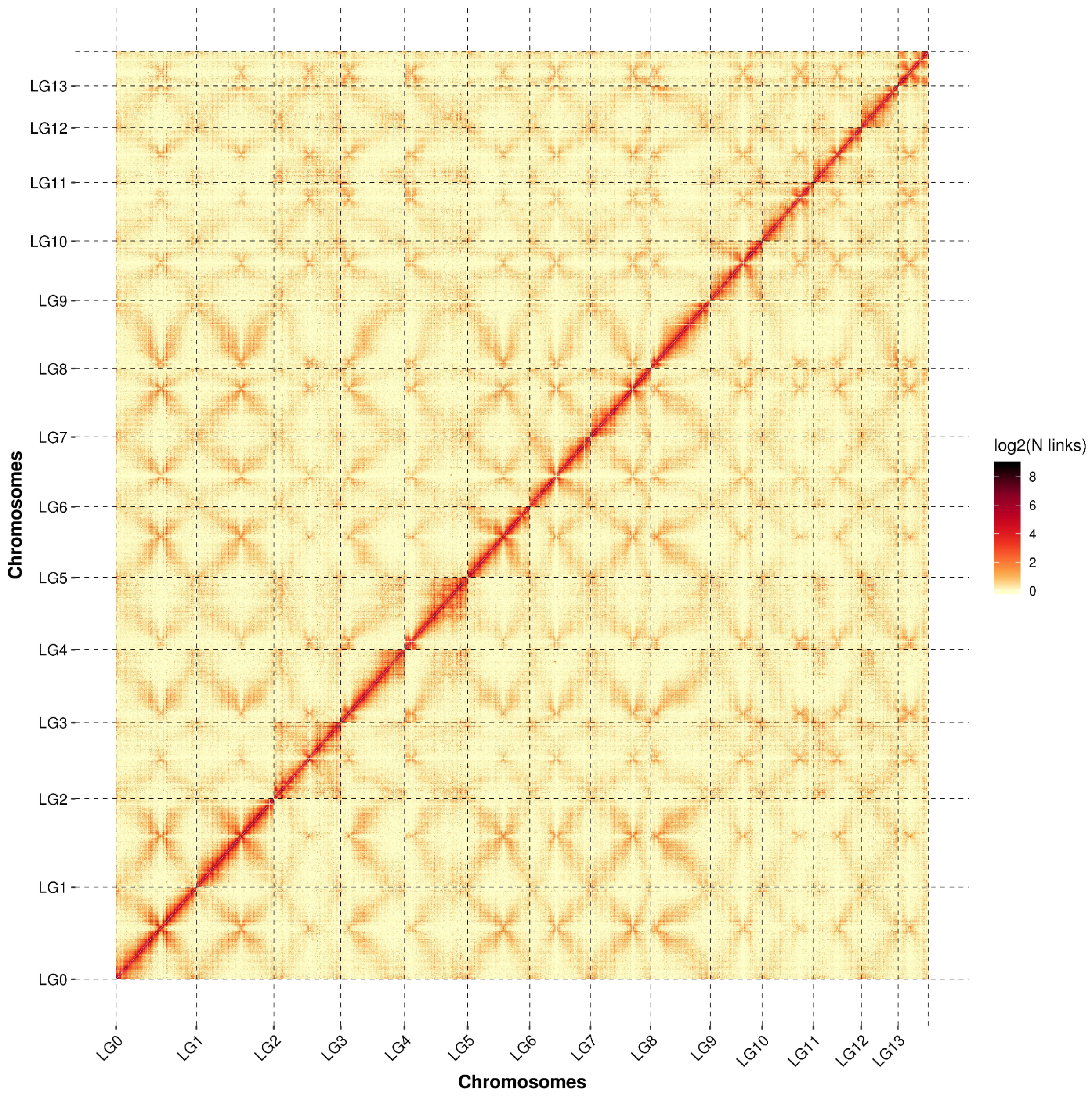
We constructed the genome map of the monokaryotic strain Q25 and studied its mating-type loci and mating types. The total genome length of the S. rugosoannulata monokaryotic strain Q25 is 48.27 Mb, with an N50 length of 2.71 Mb and a sequencing depth of 45X. The Hi-C assembly chromosome interaction heatmap (Figure 2) clearly shows 14 chromosomes. The two mating-type loci A and B are located on chromosome 1 and chromosome 12, respectively. To our knowledge, this is the first chromosome-level genome assembly of S. rugosoannulata.

3.5. Mating-Type Loci in S. rugosoannulata

3.5.1. Mating-Type Locus A

Through resequencing-based gene function annotation, the HD2 gene and MIP gene at locus A were identified. The position of HD1 in the S. rugosoannulata genome was obtained through homology alignment with the known HD1 sequence of Coprinopsis cinerea. Based on the amino acid sequence of β-fg from Coprinopsis cinerea, homologous alignment with other basidiomycetes was performed on NCBI to obtain the gene function annotation information of the β-fg gene. Using this information, a search was conducted in the local genome database, and the location of the β-fg gene was finally determined. The β-fg gene is located upstream of HD2, with a physical distance of 228 kb from HD2. The gene order at locus A is β-fg, HD2, HD1, and MIP, with physical intervals of 228 kb, 384, and 569, respectively. The analysis of homologous domains of HD protein sequences showed that the domain names of HD1 and HD2 proteins are both HOX_1 (IPR001356). Nuclear localization signal (NLS) analysis of HD1 and HD2 protein sequences showed that HD1 harbors four NLS motifs, whereas HD2 contains two NLS motifs. The gene order within the mating-type A locus is β-fg, HD2, HD1, and MIP, as illustrated in Figure 3.

3.5.2. Mating-Type Locus B

Analysis of conserved domains and corresponding protein transmembrane structures indicated that the mating-type B locus, which contains five pheromone receptor genes, is located on chromosome 12. The five identified pheromone precursor genes were named PP1, PP2, PP3, PP4, and PP5. All these five pheromone precursors contain CaaX motifs, which are CLVS, CRAS, CRAA, CAAA, and CRAS, respectively (Figure 4). Among them, CRAS and CRAA are different from the typical amino acid CaaX motif, suggesting that base mutations may have occurred in the pheromone precursor genes during the evolutionary process. The detailed gene structure of the B locus is shown in Figure 5 and Supplementary Table S4. The pheromone precursor genes are presented in Figure 4 and Supplementary Table S5, while the receptor genes are provided in Supplementary Table S6 and Supplementary Figure S1.

3.6. SNP Validation and Application

Suitable SNP loci were selected from the conserved regions of the mating-type A and B loci, and 32 and 44 primer pairs were designed, respectively, using Primer 5. The usable rate of primers targeting the A locus was 96% and that of primers targeting the B locus was 95%. From these primers, four pairs were selected for their high stability and specificity (Table 1, Supplementary Tables S7 and S8). It is known that the mating types of the monokaryotic strains Y25, 6, 27, and 106 are A1B1, A2B2, A2B1, and A1B2, respectively. The primer amplification profiles are shown in Figure 6, indicating that these four primer pairs can be used as primers for SNP molecular markers to distinguish the mating types of monokaryotic strains. The amplified bands of primers A1 and A2 are approximately 600 bp and 750 bp, respectively; the amplified bands of primers B1 and B2 are approximately 250 bp and 300 bp, respectively.
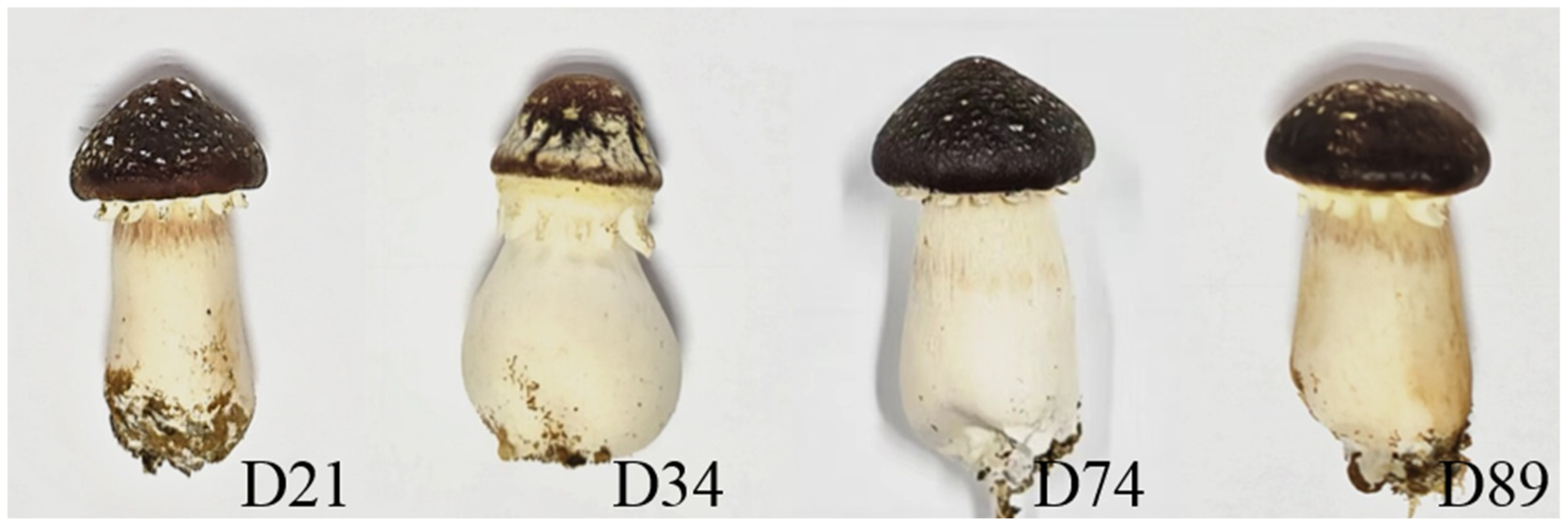
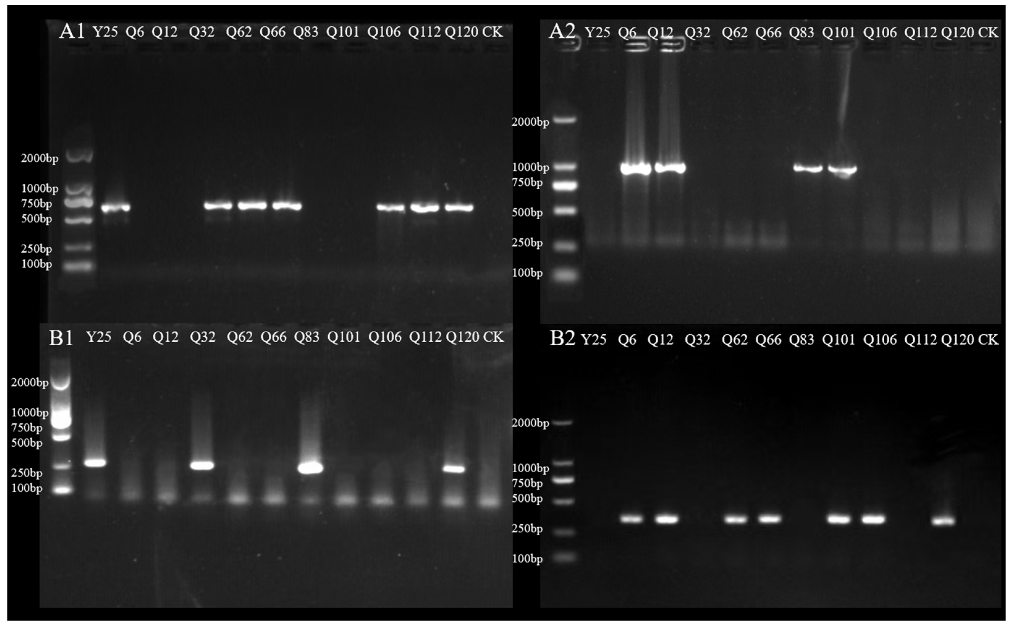
The mating types of 106 spore-derived monokaryotic strains were verified using the SNP primers, and the ratio of the four mating types (A1B1, A2B2, A2B1, and A1B2) was found to be 33:16:19:38. Eight strains were randomly selected from 97 selfed strains to test their fruiting performance (Table 2, Figure 7 and Figure 8, Supplementary Table S9 and S10). In the actual cultivation process, the fruiting results were consistent with the primer validation results; hybrid strains with incomplete parental mating types failed to fruit.

4. Discussion

Edible fungus breeding has transitioned from traditional to precise as a development of modern biology and the agricultural industry [16]. Currently, whole-genome sequencing has become a crucial approach in basic research. The application of whole-genome sequencing has enabled a deeper understanding of the molecular genetic structure of mating-type loci [52]. The mating-type systems of common edible basidiomycetes are highly complex and exhibit numerous unique biological characteristics [18]. Genome analysis has elucidated the mating types of F. filiformis [31], L. edodes [26], Agaricus bisporus [53], and other species. Additionally, the application of genome-sequencing technology provides more comprehensive theoretical support for the design of molecular markers.
In this study, different mating types were identified through nuclear migration assay. It was observed that the protoplast monokaryotic strains possessed the same mating-type gene, with segregation distortion occurring. A total of 106 monokaryotic strains were isolated from spores, and the ratio of the four obtained mating types was 31:22:42:12. The chi-square test value was 17.38, which did not conform to Mendel’s segregation rules, indicating the occurrence of segregation distortion. This may be attributed to the difference in regeneration capacity among individual monokaryons, which is caused by their distinct genotypes [54,55]. Numerous studies have reported this phenomenon in various edible fungi, including A. auricula [56], F. filiformis [57], et al. The mating types of 106 spore-derived monokaryotic strains were validated using SNP primers, yielding mating-type proportions of A1B1:A2B2:A2B1:A1B2 = 33:16:19:38. The results are inconsistent with the mating-type identification outcomes from the test-cross nuclear migration assays. However, in our actual cultivation process, the fruiting results were consistent with the primer verification data: none of the hybrid strains with incomplete parental mating types produced mushrooms. This phenomenon may be attributed to the occurrence of false clamp connections during hybridization, which has also been reported in Ganoderma lucidum [58], G. frondosa [29], and other species. It is hypothesized that S. rugosoannulata may possess multiple alleles, such as A3 and B3. This study not only significantly reduces the identification time for monokaryotic and dikaryotic strains and lowers breeding costs, but it also enables the combination of strain phenotypic analysis to identify key agronomic trait genes, rapidly screen for desired target strains, and accelerate breeding progress
Complete genomes and a high-resolution gene map were obtained using transcriptome, resequencing, and Hi-C sequencing technologies, which allowed the confirmation of the locations and structures of the mating-type A and B loci. In this study, both Hi-C sequencing and the modified germ-tube burst chromosome preparation method revealed that S. rugosoannulata contains 14 chromosomes with significant differences in chromosome size. However, a study by Liu [59] reported that S. rugosoannulata has 13 chromosomes.
Research on numerous basidiomycete species has shown that the mating-type A locus consists of one or several pairs of homeodomain protein genes [60]. While the number of A mating-type genes varies across species, significant structural conservation exists. The gene sequence of the HD region in mating-type A is highly conserved, flanked by MIP and β-fg, with their distance from the A locus typically within 1 kb [61]. In this study, β-fg was not closely linked to HD2, with a physical distance of up to 228 kb. The distance and position of β-fg and HD vary significantly among different mushroom species, such as L. edodes [62,63] and Schizophyllum commune [64]. In L. edodes [62], the distance between the β-fg gene and HD genes is 7.6 kb, while in S. commune [64], this distance reaches 838 kb. These findings indicate that chromosomal rearrangements have occurred at the A mating-type locus during species evolution; the relatively long intergenic distances around the locus may permit sequence translocations and gene duplications [63]. Stropharia rugosoannulata shows a similar pattern in above-mentioned genes; further research is still needed to confirm this. In Ganoderma, the β-fg gene has been translocated to other locations in the genome via gene rearrangement; specifically, it has moved to the region where the B mating-type locus is located; the significant changes in gene localization near the mating-type locus may be associated with a unique evolutionary and adaptive processes of this fungus (adaptation to specific ecological environments or the evolution of distinct mating strategies) [65]. Additionally, the β-fg and MIP genes were located on the same side of the A locus. The gene order of the A mating type is β-fg, HD2, HD1, and MIP, and the HD genes exhibit a typical “head-to-head” arrangement, consistent with Shang et al.’s [15] study.
The B mating-type genes of Basidiomycota encode pheromone receptors and pheromone precursors [66]. In basidiomycetes, these genes often occur in pairs and are genetically linked, forming a B mating-type locus or sub-locus [67]. Studies have shown that fungal pheromone receptors typically possess a seven-transmembrane domain structure [25], but multiple atypical receptors exist in edible fungi [68]. In this study, all five pheromone precursors contained CaaX motifs, which is consistent with previous research. However, their C-terminal sequences are CLVS, CRAS, CRAA, CAAA, and CRAS, respectively. The amino acid compositions of the CRAS and CRAA motifs differ from those of typical CaaX motifs, suggesting that base mutations occurred in the pheromone precursor genes during evolution, leading to changes in their sequence characteristics. A similar mutation pattern was observed in the study of Pleurotus [69]. Among them, the terminal conserved motifs of PP1, PP2, PP3, and PP5 are ER motifs, while that of PP4 is an AF motif. This differs from the findings of Shang et al.’s study; the direction of PP3 is also different. Previous studies identified three pheromone receptor genes: PP1 with a conserved CaaX box, PP2 containing both FF and ER motifs, and PP3 containing both AF and ER motifs. Atypical pheromone receptor phenomena have also been reported in Pleurotus pulmonarius [70]. In the pheromone precursors of the B mating-type locus, atypical motifs (CRAS, CRAA) may alter receptor–ligand interactions by disrupting pheromone structure, interfering with pheromone processing, or modifying the receptor-binding interface [71]. The study reveals that minimal sequence changes, including single-amino acid substitutions in either pheromones or receptors, can significantly redirect their ligand-receptor recognition profile. These changes can directly reshape mating compatibility and may contribute to prezygotic reproductive isolation between populations by impeding mate recognition. To clarify the specific effects of CRAS and CRAA motifs on receptor activation and mating, further functional studies are required [66].
Our research on the mating-type system and SNP molecular markers of S. rugosoannulata provides a theoretical foundation for its hybrid breeding and precision breeding. During the study, it was discovered that certain molecular markers closely linked to the mating type are located in regions where genes involved in lignin and cellulose degradation are also present. This discovery provides genomic evidence for elucidating the role of substances in ecosystem cycling and their substrate utilization mechanisms during cultivation [28]. The genes that were linked to specific mating types will serve as important molecular markers for breeding. As a result, it promotes the in-depth exploration of S. rugosoannulata, transitioning from basic genetic studies to functional genomics.

5. Conclusions

This study clearly identified the mating-type A and B loci of S. rugosoannulata and designed four pairs of SNP primers, providing an accurate, rapid, convenient, and efficient method for distinguishing monokaryotic and dikaryotic strains. In the future, the function of mating-type genes among different strains could be analyzed by integrating the mycelial growth rate and vigor [55]. This will facilitate the full exploration of mating-type genes associated with key traits, thereby improving breeding efficiency and enabling the prediction of a strain’s potential in future breeding programs. Furthermore, the findings of this study can serve as a reference for phylogenetic analysis and trait-linkage analysis of current S. rugosoannulata strains, enriching research methodologies. It will also provide strong support for the robust development of subsequent studies, including marker-assisted breeding, functional gene discovery, and mechanism exploration.

Supplementary Materials

The following supporting information can be downloaded at https://www.mdpi.com/article/10.3390/horticulturae11111325/s1, Figure S1: Transmembrane structure of the pheromone receptor, Table S1: Conduct the first-round mating experiment using Y25, which is randomly selected as the tester strain and isolated from the S. rugosoannulata strain S1, Table S2: Conduct the second-round mating experiment using Q25, which is randomly selected as the tester strain and isolated from the S. rugosoannulata strain S1, Table S3: Conduct the third-round mating experiment using Q19, which is randomly selected as the tester strain and isolated from the S. rugosoannulata strain S1, Table S4: Genetic structure of mating-type B locus, Table S5: Pheromone precursor gene structure, Table S6: Transmembrane helical structure of pheromone receptor, Table S7: SNP primers designed based on the mating-type A locus, Table S8: SNP primers designed based on the mating-type B locus, Table S9: Selfing combinations, Table S10: Mating type of monokaryotic strain.

Author Contributions

Conceptualization, M.L. and S.L.; methodology, S.L.; software, J.S.; validation, J.S., P.Z. (Peng Zhu) and P.Z. (Panpan Zhang); formal analysis, S.L., G.L., X.L. and A.M.; investigation, J.S., Y.S., H.L. and S.X.; resources, S.L.; data curation, J.S.; writing—original draft preparation, P.Z. (Panpan Zhang); writing—review and editing, S.L., G.L., X.L. and A.M.; visualization, J.S.; supervision, J.W. and J.T.; project administration, S.L. and M.L.; funding acquisition, S.L. and M.L. All authors have read and agreed to the published version of the manuscript.

Funding

This research was funded by “Edible Fungus Species Cultivation Position (No. CARS-20-4)”, the “Talent Introduction Scientific Research Special Project of Hebei Agricultural University (YJ201849)”, the “Key Research and Development Planning Project in Science and Technology of Hebei Province (21326315D)”, the “Innovation Team of Edible Fungi of Hebei Modern Agricultural Industrial Technology System (HBCT2018050205)”, and the “Analysis of the Mating Type System and Application of Molecular Marker-Assisted Precision Breeding in Stropharia rugosoannulata (C2025204257)”.

Institutional Review Board Statement

Not applicable.

Informed Consent Statement

Not applicable.

Data Availability Statement

The whole genome sequence data reported in this paper have been deposited in the Genome Warehouse of the National Genomics Data Center, Beijing Institute of Genomics, Chinese Academy of Sciences/China National Center for Bioinformation, under accession number GWHGWFB01000000, which is publicly accessible at https://ngdc.cncb.ac.cn/gwh, accessed on 10 February 2025.

Conflicts of Interest

The authors declare no conflicts of interest.

Abbreviations

The following abbreviations are used in this manuscript:
ISSRInter-Simple Sequence Repeat
ITSInternal Transcribed Spacer
MNPMulti-Nucleotide Polymorphism
RAPDRandom Amplified Polymorphic DNA
SNPSingle-Nucleotide Polymorphism
SRAPSequence-Related Amplified Polymorphism
SSRSimple Sequence Repeat

References

  1. Huang, N. Classification and characterization of Strapharia rugoso-annulata. Edible Fungi 1995, 6, 11. [Google Scholar]
  2. Liu, D.; Chen, Y.Q.; Xiao, X.W.; Zhong, R.T.; Yang, C.F.; Liu, B.; Zhao, C. Nutrient properties and nuclear magnetic resonance-based metabonomic analysis of macrofungi. Foods 2019, 8, 397. [Google Scholar] [CrossRef] [PubMed]
  3. Shen, M.; Yan, J.; Wang, R.S.; Quan, X.H.; Yuan, Y.; Yao, X.T.; Jin, Q.L. Molecular mechanisms and horticultural implications of temperature-induced color variation in Stropharia rugosoannulata mushrooms. Food Chem. Mol. Sci. 2025, 11, 100276. [Google Scholar] [CrossRef]
  4. Hu, S.; Feng, X.; Huang, W.; Ibrahim, S.A.; Liu, Y. Effects of drying methods on non-volatile taste components of Stropharia rugoso-annulata mushrooms. LWT 2020, 127, 109428. [Google Scholar] [CrossRef]
  5. Wang, Q.; Zhao, Y.L.; Feng, X.; Ibrahim, S.A.; Huang, W.; Liu, Y. Effects of drying on the structural characteristics and antioxidant activities of polysaccharides from Stropharia rugosoannulata. J. Food Sci. Technol. 2021, 58, 3622–3631. [Google Scholar] [CrossRef]
  6. Wu, J.; Kobori, H.; Kawaide, M.; Suzuki, T.; Choi, J.H.; Yasuda, N.; Noguchi, K.; Matsumoto, T.; Hirai, H.; Kawagishi, H. Isolation of bioactive steroids from the Stropharia rugosoannulata mushroom and absolute configuration of strophasterol B. Biosci. Biotechnol. Biochem. 2013, 77, 1779–1781. [Google Scholar] [CrossRef]
  7. Zhou, B.; Jia, L.; Meng, F.Y.; Song, Z.; Liu, X.N.; Deng, P.; Fan, K.M. Statistical optimization of cultivation conditions for exopolysacchride production and mycelia growth by Stropharia rugosoannulata. Ann. Microbiol. 2010, 60, 89–96. [Google Scholar] [CrossRef]
  8. Wu, J.; Suzuki, T.; Choi, J.H.; Yasuda, N.; Noguchi, K.; Hirai, H.; Kawagishi, H. An unusual sterol from the mushroom Stropharia rugosoannulata. Tetrahedron Lett. 2013, 54, 4900–4902. [Google Scholar] [CrossRef]
  9. Otakar, R.; Jiri, M.; Tunde, J. Beta-glucans in higher fungi and their health effects. Nutr. Rev. 2009, 67, 624–631. [Google Scholar] [CrossRef]
  10. Liu, Y.; Hu, C.F.; Feng, X.; Cheng, L.; A Ibrahim, S.; Wang, C.T.; Huang, W. Isolation, characterization and antioxidant of polysaccharides from Stropharia rugosoannulata. Int. J. Biol. Macromol. 2020, 155, 883–889. [Google Scholar] [CrossRef]
  11. Zhang, W.W.; Tian, G.T.; Geng, X.R.; Zhao, Y.C.; Ng, T.; Zhao, L.Y.; Wang, H.X. Isolation and characterization of a novel lectin from the edible mushroom Stropharia rugosoannulata. Molecules 2014, 19, 19880–19891. [Google Scholar] [CrossRef]
  12. Hyun, J.W.; Kim, C.K.; Park, S.H.; Yoon, J.Y.; Shim, M.J.; Kang, C.Y.; Choi, E.C.; Kim, B.K.l. Antitumor components of Agrocybe cylindracea. Arch. Pharmacal Res. 1996, 19, 207–212. [Google Scholar] [CrossRef]
  13. Yang, Y.; Meng, G.L.; Ni, S.J.; Zhang, H.F.; Dong, C.H. Genomic analysis of Stropharia rugosoannulata reveals its nutritional strategy and application potential in bioremediation. J. Fungi 2022, 8, 162. [Google Scholar] [CrossRef]
  14. Liers, C.; Arnstadt, T.; Ullrich, R.; Hofrichter, M. Patterns of lignin degradation and oxidative enzyme secretion by different wood-and litter-colonizing basidiomycetes and ascomycetes grown on beech-wood. FEMS Microbiol. Ecol. 2011, 78, 91–102. [Google Scholar] [CrossRef] [PubMed]
  15. Shang, J.J.; Hou, D.; Li, Y.; Zhou, C.L.; Guo, T.; Tang, L.H.; Mao, W.J.; Chen, Q.; Bao, D.P.; Yang, R.H. Analyses of mating systems in Stropharia rugosoannulata based on genomic data. Mycosystema 2020, 39, 1152–1161. [Google Scholar] [CrossRef]
  16. Huang, L.; Si, C.; Shi, H.Y.; Huang, F.Q.; Duan, J.; He, C.M. Research progress on genetic breeding of Stropharia rugosoannulata. North. Hortic. 2024, 48, 118–125. (In Chinese) [Google Scholar] [CrossRef]
  17. Kumar, R.; Das, S.P.; Choudhury, B.U.; Kumar, A.; Prakash, N.R.; Verma, R.; Chakraborti, M.; Devi, A.G.; Bhattacharjee, B.; Das, R.; et al. Advances in genomic tools for plant breeding: Harnessing DNA molecular markers, genomic selection, and genome editing. Biol. Res. 2024, 57, 80. [Google Scholar] [CrossRef]
  18. Bao, D.P. Scientific Problems in Crossbreeding of Edible Fungi. Acta Edulis Fungi 2020, 27, 1–24. [Google Scholar] [CrossRef]
  19. Devi, E.L.; Devi, C.P.; Kumar, S.; Sharma, S.K.; Beemrote, A.; Chongtham, S.K.; Singh, C.H.; Tania, C.; Singh, T.B.; Ningombam, A.; et al. Marker assisted selection (MAS) towards generating stress tolerant crop plants. Plant Gene 2017, 11, 205–218. [Google Scholar] [CrossRef]
  20. Ke, L.N. Molecular Polymorphism in Germplasm Resource of Tricholoma giganteum Massee, Stropharia rugoso-annulata Farlow, Hercium erinaceus, Agrocybe cylindracea. Master’s Thesis, Fujian Agriculture and Forestry University, Fuzhou, China, 2008. [Google Scholar]
  21. Liu, F.; Cao, B.; Dai, H.M.; Li, G.J.; Li, S.M.; Gao, W.; Zhao, R.L. High-resolution core gene-associated multiple nucleotide polymorphism (cgMNP) markers for strain identification in the wine cap mushroom Stropharia rugosoannulata. Microorganisms 2025, 13, 1685. [Google Scholar] [CrossRef]
  22. Yan, P.S.; Jiang, J.H.; Cui, W.S. Characterization of protoplasts prepared from the edible fungus, Stropharia rugoso-annulata. World J. Microbiol. Biotechnol. 2004, 20, 173–177. [Google Scholar] [CrossRef]
  23. Kües, U. From two to many: Multiple mating types in Basidiomycetes. Fungal Biol. Rev. 2015, 29, 126–166. [Google Scholar] [CrossRef]
  24. Raper John, R. Genetics of sexuality in higher fungi. Bioscience 1966, 17, 846. [Google Scholar]
  25. Kothe, E. Tetrapolar fungal mating types: Sexes by the thousands. FEMS Microbiol. Rev. 1996, 18, 65–87. [Google Scholar] [CrossRef]
  26. Ha, B.; Moon, Y.J.; Song, Y.; Kim, S.; Kim, M.; Yoon, C.W.; Ro, H.S. Molecular analysis of B mating type diversity in Lentinula edodes. Sci. Hortic. 2019, 243, 55–63. [Google Scholar] [CrossRef]
  27. Casselton, L.A.; Olesnicky, N.S. Molecular genetics of mating recognition in basidiomycete fungi. Microbiol. Mol. Biol. Rev. 1998, 62, 55–70. [Google Scholar] [CrossRef] [PubMed]
  28. Li, Y.Q.; Yang, Y.; Huang, X.H.; Huang, J.; Dong, C.H. Molecular and genetic evidence for a tetrapolar mating system in Sparassis latifolia. Fungal Biol. 2020, 124, 1004–1012. [Google Scholar] [CrossRef] [PubMed]
  29. Zhang, S.S.; Li, X.; Li, G.J.; Huang, Q.; Tian, J.H.; Wang, J.L.; Li, M.; Li, S.M. Genetic and molecular evidence of a tetrapolar mating system in the edible mushroom Grifola frondosa. J. Fungi 2023, 9, 959. [Google Scholar] [CrossRef] [PubMed]
  30. Chen, W.; Chai, H.; Yang, W.; Zhang, X.; Chen, Y.; Zhao, Y. Characterization of non-coding regions in B mating loci of Agrocybe salicacola groups: Target sites for B mating type identification. Curr. Microbiol. 2017, 74, 772–778. [Google Scholar] [CrossRef]
  31. Wang, W.; Lian, L.D.; Xu, P.; Chou, T.S.; Irum, M.; Aron, O.; Muhammad, W.; Chen, B.Z.; Liu, X.R.; Liu, F.; et al. Advances in understanding mating type gene organization in the mushroom-forming fungus Flammulina velutipes. G3 Genes Genomes Genet. 2016, 6, 3635–3645. [Google Scholar] [CrossRef]
  32. Ha, B.; Kim, S.; Kim, M.; Moon, Y.J.; Song, Y.; Ryu, J.S.; Ryu, H.; Ro, H.S. Diversity of A mating type in Lentinula edodes and mating type preference in the cultivated strains. J. Microbiol. 2018, 56, 416–425. [Google Scholar] [CrossRef] [PubMed]
  33. Zhang, J.; Yang, J.J.; Zhang, L.K.; Luo, J.; Zhao, H.; Zhang, J.N.; Wen, C.L. A new SNP genotyping technology Target SNP-seq and its application in genetic analysis of cucumber varieties. Sci. Rep. 2020, 10, 5623. [Google Scholar] [CrossRef] [PubMed]
  34. Onda, Y.; Takahagi, K.; Shimizu, M.; Inoue, K.; Mochida, K. Multiplex PCR targeted amplicon sequencing (MTA-Seq): Simple, flexible, and versatile SNP genotyping by highly multiplexed PCR amplicon sequencing. Front. Plant Sci. 2018, 9, 201. [Google Scholar] [CrossRef] [PubMed]
  35. Chen, K.; Zhou, Y.X.; Li, K.; Qi, L.X.; Zhang, Q.F.; Wang, M.C.; Xiao, J.H. A novel three-round multiplex PCR for SNP genotyping with next generation sequencing. Anal. Bioanal. Chem. 2016, 408, 4371–4377. [Google Scholar] [CrossRef]
  36. Meuwissen, T.H.; Hayes, B.J.; Goddard, M.E. Prediction of total genetic value using genome-wide dense marker maps. Genetics 2001, 157, 1819–1829. [Google Scholar] [CrossRef]
  37. Zhang, M.Y.; Song, C.Y.; Yu, H.L.; Tan, Q.; Xu, Z.; Wang, R.J.; Zhang, L.J.; Shang, X.D. AS-PCR-based identification of mating types in Lentinula edodes using SNP genotyping. Acta Edulis Fungi 2019, 26, 1–9. (In Chinese) [Google Scholar] [CrossRef]
  38. Campbell, N.R.; Harmon, S.A.; Narum, S.R. Genotyping-in-Thousands by sequencing (GT-seq): A cost effective SNP genotyping method based on custom amplicon sequencing. Mol. Ecol. Resour. 2014, 15, 855–867. [Google Scholar] [CrossRef]
  39. Song, H.Y.; Zhao, P. Research progress on molecular biological detection of edible mushroom strains. China Cucurbits Veg. 2024, 37, 9–17. [Google Scholar] [CrossRef]
  40. Gao, Y. Study on Key Technologies and Screening Strains of Stropharia rugosoannulata. Master’s Thesis, Northwest A&F University, Xianyang, China, 2014; p. 63. (In Chinese). [Google Scholar]
  41. Zhou, F.; Yang, H.L.; Zhao, Y.; Jiang, S.X.; Chen, M.J.; Li, Z.P.; Cha, L.; Li, Y. Characteristics of mononuclear properties and the protoplast monokaryogenesis of different varieties of Stropharia rugosoannulata. Acta Agric. Shanghai 2023, 39, 33–37. (In Chinese) [Google Scholar] [CrossRef]
  42. Guo, B.F. Karyotype Analysis and FISH Analysis of Infection Related Genes in Metarhizium Acridum. Master’s Thesis, Chongqing University of Technology, Chongqing, China, 2014; p. 63. (In Chinese). [Google Scholar]
  43. Du, X.H.; Zhao, Q.; Xia, E.H.; Gao, L.Z.; Richard, F.; Yang, Z.L. Mixed-reproductive strategies, competitive mating-type distribution and life cycle of fourteen black morel species. Sci. Rep. 2017, 7, 1493. [Google Scholar] [CrossRef]
  44. Cheng, H.; Concepcion, G.T.; Feng, X.; Zhang, H.; Li, H. Haplotype-resolved de novo assembly using phased assembly graphs with hifiasm. Nat. Methods 2021, 18, 170–175. [Google Scholar] [CrossRef]
  45. Walker, B.J.; Abeel, T.; Shea, T.; Priest, M.; Abouelliel, A.; Sakthikumar, S.; Cuomo, C.A.; Zeng, Q.; Wortman, J.; Young, S.K.; et al. An integrated tool for comprehensive microbial variant detection and genome assembly improvement. PLoS ONE 2014, 9, e112963. [Google Scholar] [CrossRef]
  46. Simão, F.A.; Waterhouse, R.M.; Ioannidis, P.; Kriventseva, E.V.; Zdobnov, E.M. BUSCO: Assessing genome assembly and annotation completeness with single-copy orthologs. Bioinformatics 2015, 31, 3210. [Google Scholar] [CrossRef]
  47. Li, H. Aligning sequence reads, clone sequences and assembly contigs with BWA-MEM. arXiv 2013, arXiv:1303.3997. [Google Scholar] [CrossRef]
  48. Burton, J.N.; Adey, A.; Patwardhan, R.P.; Qiu, R.; Kitzman, J.O.; Shendure, J. Chromosome-scale scaffolding of de novo genome assemblies based on chromatin interactions. Nat. Biotechnol. 2013, 31, 1119–1125. [Google Scholar] [CrossRef] [PubMed]
  49. Stanke, M.; Waack, S. Gene prediction with a hidden markov model and a new intron submodel. Bioinformatics 2003, 19, 1453–1454. [Google Scholar] [CrossRef] [PubMed]
  50. Majoros, W.H.; Pertea, M.; Salzberg, S.L. Tigrscan and glimmer HMM: Two open source ab initio eukaryotic gene-finders. Bioinformatics 2004, 20, 2878–2879. [Google Scholar] [CrossRef] [PubMed]
  51. Koressaar, T.; Lepamets, M.; Kaplinski, L.; Raime, K.; Andreson, R.; Remm, M. Primer3_masker: Integrating masking of template sequence with primer design software. Bioinformatics 2018, 34, 1937–1938. [Google Scholar] [CrossRef]
  52. Lu, M.; Li, J.; Sun, X.; Zhao, D.Q.; Zong, H.Z.; Tang, C.; Li, K.; Zhou, Y.X.; Xiao, J.H. Genotyping single nucleotide polymorphisms in homologous regions using multiplex kb level amplicon capture sequencing. Mol. Genet. Genom. 2024, 299, 99. [Google Scholar] [CrossRef]
  53. Choi, Y.J.; Jung, S.; Eom, H.; Hoang, T.; Han, H.G.; Kim, S.; Ro, H.S. Structural analysis of the A mating type locus and development of the mating type marker of Agaricus bisporus var. bisporus. J. Fungi 2023, 9, 284. [Google Scholar] [CrossRef]
  54. Fen, P.; Sun, Y.; Hao, L.B.; An, J.L.; Gai, Z.P.; Wang, Y.X.; Zheng, S.Y.; Wang, C.X. Study on isolation and mating type determination of monokaryotic spores of Pleurotus ostreatus ‘Jiping 16’. Edible Med. Mushrooms 2024, 32, 386–392. [Google Scholar]
  55. Larraya, L.M.; Pérez, G.; Iribarren, I.; Blanco, J.A.; Alfonso, M.; Pisabarro, A.G.; Ramírez, L. Relationship between monokaryotic growth rate and mating type in the edible basidiomycete Pleurotus ostreatus. Appl. Environ. Microbiol. 2001, 67, 3385–3390. [Google Scholar] [CrossRef] [PubMed]
  56. Zhang, H.; Cao, H.; Pan, Y.J.; Cao, J.Y. Study on mating system of Auricularia auricula. Mycosystema 2002, 21, 559–564. (In Chinese) [Google Scholar]
  57. Wang, Y.; Xiong, H.Y.; Yang, R.H.; Wu, Y.Y.; Liu, X.; Bao, D.P. Nuclear partial segregation occurred in protoplast monokaryotization and asexual spore monosporous isolation of wild strains of Flammulina filiformis. Mycosystema 2025, 44, 75. (In Chinese) [Google Scholar] [CrossRef]
  58. Ke, B.R.; Lu, Z.H.; Wu, X.P.; Guo, L.X.; Lan, Q.X. Determination and diversity analysis of mating types of Ganoderma lucidum basidispores. Chin. J. Trop. Crops 2018, 39, 145–150. [Google Scholar]
  59. Liu, X.Y. Preliminary Research on the Breeding of Excellent Hybrids of Stropharia rugosoannulata in this Mushroom and Rice Rotation. Master’s Thesis, Huazhong Agricultural University, Wuhan, China, 2023; p. 99. [Google Scholar] [CrossRef]
  60. Casselton, L.A. Fungal sex genes e searching for the ancestors. Bioassay 2008, 30, 711–714. [Google Scholar] [CrossRef]
  61. Raudaskoski, M.; Kothe, E. Basidiomycetes mating type gens and pheromone signaling. Eukaryot. Cell 2010, 9, 847–859. [Google Scholar] [CrossRef]
  62. Chen, L.; Gong, Y.; Cai, Y.; Liu, W.; Zhou, Y.; Xiao, Y.; Xu, Z.; Liu, Y.; Lei, X.; Wang, G.Z.; et al. Genome sequence of the edible cultivated mushroom Lentinula edodes (shiitake) reveals insights into lignocellulose degradation. PLoS ONE 2016, 11, e0160336. [Google Scholar] [CrossRef]
  63. Au, C.H.; Wong, M.C.; Bao, D.P.; Zhang, M.Y.; Song, C.Y.; Song, W.H.; Law, P.T.W.; Kües, U.; Kwan, H.S. The genetic structure of the A mating-type locus of Lentinula edodes. Gene 2014, 535, 184–190. [Google Scholar] [CrossRef]
  64. Ohm, R.A.; De Jong, J.F.; Lugones, L.G.; Aerts, A.; Kothe, E.; Stajich, J.E.; de Vries, R.P.; Record, E.; Levasseur, A.; Baker, S.E.; et al. Genome sequence of the model mushroom Schizophyllum commune. Nat. Biotechnol. 2010, 28, 957–963. [Google Scholar] [CrossRef]
  65. Nishimura, A.; Linder, M.E. Identification of a novel prenyl and palmitoyl modification at the CaaX Motif of Cdc42 that regulates RhoGDI binding. Mol. Cell. Biol. 2013, 33, 1417–1429. [Google Scholar] [CrossRef]
  66. Fowler, T.J.; Mitton, M.F.; Vaillancourt, L.J. Carlene a raper, changes in mate recognition through alterations of pheromones and receptors in the multisexual mushroom fungus Schizophyllum commune. Genetics 2001, 158, 1491–1503. [Google Scholar] [CrossRef] [PubMed]
  67. Meritxell, R.; Michael, P.C.; Lorna, A.C.; Andrew, J.B. The origin of multiple B mating specificities in Coprinus cinereus. Genetics 2005, 170, 1105–1119. [Google Scholar] [CrossRef] [PubMed]
  68. James, T.Y.; Kües, U.; Rehner, S.A.; Vilgalys, R. Evolution of the gene encoding mitochondrial intermediate peptidase and its cosegregation with the A mating-type locus of mushroom fungi. Fungal Genet. Biol. 2004, 41, 381–390. [Google Scholar] [CrossRef]
  69. Chen, B.S.; Fu, J.S.; Long, Y.; Zhang, L.; Zhang, X.Y.; Zhang, X.; Hu, F.L.; Jiang, Y.J.; Xie, B.G. Sexual life cycle of Volvariella volvacea revealed by investigation of the random migration of tetrad. Mycosystema 2017, 36, 466–472. [Google Scholar]
  70. Zhao, Y.N. Study on Mating Type of Pleurotus. Master’s Thesis, Anhui Agriclitural University, Hefei, China, 2023; p. 70. (In Chinese). [Google Scholar]
  71. Chen, B.Z.; Qiu, M.M.; Ye, L.Y.; Chen, T.C.; Lu, X.; Jiang, Y.; Wu, X.P. Genomic data reveal unique structures of the mating loci of the progeny of Pleurotus pulmonarius wild-type strain X1. Mycosystema 2019, 38, 2205–2213. (In Chinese) [Google Scholar] [CrossRef]
Figure 1. Mature fruiting bodies, basidiospores, protoplasts, and clamp connections of S. rugosoannulata. Note: (A): fruiting body of S. rugosoannulata dikaryotic strain S1, (B): basidiospores, (C): protoplasts, (D): clamp connections, (E): chromosomes (1 n, haploid).
Horticulturae 11 01325 g001
Figure 2. Hi-C assembly chromosome interaction heatmap.
Horticulturae 11 01325 g002
Figure 3. Schematic diagram of the gene structure of the mating-type A locus.
Horticulturae 11 01325 g003
Figure 4. Sequence alignment of pheromone precursor proteins. Note: ✳ indicates that amino acid bases at this site/column are identical.
Horticulturae 11 01325 g004
Figure 5. Schematic diagram of the gene structure of the mating-type B locus.
Horticulturae 11 01325 g005
Figure 6. SNP primer validation profiles for four loci. Note: (A): result of A1 gene amplification-gel image; (B): result of B1 gene amplification-gel image; (C): result of A2 gene amplification-gel image; (D): result of B2 gene amplification-gel image.
Horticulturae 11 01325 g006
Figure 7. Fruiting bodies of S. rugosoannulata produced via selfing.
Horticulturae 11 01325 g007
Figure 8. Validation of selected selfed strains using SNP molecular markers. Note: (A1): Image of A1 primer band; (A2): Image of A2 primer band; (B1): Image of B1 primer band; (B2): Image of B2 primer band.
Horticulturae 11 01325 g008
Table 1. Primer sequence.
Primer NameUpstream Prime (5′-3′)Primer NameDownstream Primer (5′-3′)Annealing Temperature
A1F8GCTCTTCAAACCCTCAATAATAGTCA1R8-1TATTCTGGAATGAGGATCTCAGAGC55 °C
A2F1CATTGTTGCCAGCGAGGACA2R1-4ATGATGGTCCCAATACCCGA58 °C
B1F9TGTTGCGTAGGATCAGGGGAGTB1R9-2CCATCTCTTCGCTCACCGATGT57 °C
B2F9TTGCGTAGGATCAGGGGAGGB2F9-2ATCCACCTCTGGCAAGTTTAGGA58 °C
Table 2. Verification of the fruiting performance of partial selfed strains.
Selfed StrainsHybrid CombinationsMating TypesFruiting Performance
D19Q6*Q62A2B2*A1B2
D21Q101*Y25A2B2*A1B1+
D34Q83*Q66A2B1*A1B2+
D41Q12*Q120A2B2*A1B2
D67Q83*Q32A2B1*A1B1
D72Q101*Q112A2B2*A1B1+
D87Q83*Q106A2B1*A1B2+
D90Q101*Q62A2B2*A1B2
Note: +: Fruiting occurred; −: No fruiting occurred.
Disclaimer/Publisher’s Note: The statements, opinions and data contained in all publications are solely those of the individual author(s) and contributor(s) and not of MDPI and/or the editor(s). MDPI and/or the editor(s) disclaim responsibility for any injury to people or property resulting from any ideas, methods, instructions or products referred to in the content.