Article

Optimizing Target Metabolites Production in Coleus blumei Indoor Cultivation: Combined Effects of LED Light and Salinity Stress

1
Department of Agro-Food Sciences and Technologies (DISTAL), University of Bologna, 40127 Bologna, Italy
2
Department of Pharmacy and Biotechnology (FaBiT), University of Bologna, 40126 Bologna, Italy
*
Author to whom correspondence should be addressed.
Horticulturae 2025, 11(10), 1205; https://doi.org/10.3390/horticulturae11101205
Submission received: 4 September 2025 / Revised: 24 September 2025 / Accepted: 1 October 2025 / Published: 6 October 2025

Abstract

Light quality is a recognized driver of plant growth and secondary metabolism in Coleus blumei, a valuable source of rosmarinic acid (RA) and quercetin (QU), whereas its combination with salinity stress represents a potential strategy that still requires further investigation. We evaluated four LED spectra, red–blue (RB) (6:1, control), blue (B), red (R), and RB + Far-Red, under both control (0 mM NaCl) and moderate salt stress (120 mM NaCl), measuring biomass (dry weight) and RA/QU in leaves and roots after three (T1) and five weeks (T2). Blue light produced the greatest root biomass, while the leaf dry weight under B did not differ significantly from RB or RBfr. RA peaked at T2 under B in leaves and under R in roots; QU was maximal under B in leaves and under RB in roots. Extending exposure from T1 to T2 markedly increased both metabolites’ yield. Salinity had little effect on biomass, increased the total QU yield, and did not enhance the total RA yield. These results indicate that targeted LED regimes and longer exposure can raise the yields of bioactive compounds, and that combining specific spectra with moderate salinity is an effective strategy for selectively increasing quercetin accumulation in indoor-grown C. blumei.

1. Introduction

Since antiquity, Coleus blumei (Lamiaceae family) has not been valued as an ornamental plant but has also been chosen for its curative effects in the treatment of several diseases [1]. C. blumei is currently being evaluated for its psychoactive substance contents [2], mainly for its known capacity of accumulating rosmarinic acid [3,4]. Additionally, this plant’s flavonoid content has been investigated in relation to the therapeutic properties of quercetin-3-glucoside, quercitrin, quercetin 3-(6″-acetylglucoside), and quercetin 3-O-acetyl-rhamnoside [5]. The increasing interest in extracting rosmarinic acid and quercetin underlines their numerous applications in the pharmaceutical [6,7] and food industries [8]. In this context, indoor agriculture enables precise control of the light spectrum and intensity, temperature, humidity, and nutrient supply [9,10], allowing for the optimization of abiotic factors for growth and metabolites accumulation. Applying targeted stress treatments can reshape biosynthetic pathways and thus modulate the concentration of therapeutic compounds. Among these, high soil salinity stands out as one of the major abiotic stresses impacting cultivated plants. Its effects are mediated through increased osmotic stress, limiting water uptake and ion toxicity, which disrupts cellular functions, with consequences on photosynthesis and the overall metabolism [11].
With a value of 4 dS m−1 of electrical conductivity (EC), equivalent to 40 mM NaCl and an osmotic pressure of about 0.2 MPa, soil is considered saline [12]. Researchers already confirmed that salt stress alters plant growth, leaf water potential, and relative water content in five Coleus species [13]. The tolerance mechanism to salt stress is generally recognized as a multigenic response involving numerous salt-induced genes [14]. The response of secondary metabolism to combined environmental stressors is not widely explored, limiting its application for optimizing the accumulation of bioactive compounds in plant tissues. For instance, the synergistic effects of light and salinity treatments have been studied in Portulaca oleracea L. microgreens. Results showed that plants grown under red–blue (RB) light exhibited the highest total phenolic content, whereas salinity significantly reduced the total flavonoid content (TFC), highlighting that growing purslane in moderate salinity and under RB or RB + Far-Red (Fr) light is the optimal condition for improving yield and quality, reducing the content of anti-nutritional compounds [15]. Similarly, an experiment on Ginkgo biloba demonstrated that TFC, particularly levels of quercetin and isorhamnetin, was significantly higher in plants treated with 100 mmol L−1 NaCl compared to the control group (no salt) and plants exposed to other salt concentrations [16].
In this context, this experiment evaluates Coleus blumei’s response to LED treatments combined with salt stress, focusing on biomass, rosmarinic acid, and quercetin production (contents and yields) in plant roots and leaves, observing their trend over time. A previous preliminary study focused on the effect of different light spectra on metabolite production in C. blumei, revealing that RB light was more effective than white light in enhancing the accumulation of bioactive compounds [17]. Based on this result, RB treatment was selected as the control condition in the present experiment. Additionally, we aimed to understand whether the coexistence of more than one stressor could represent a viable strategy to enhance the accumulation of target metabolites in plant organs, as already demonstrated in other species. To this end, the present experiment integrates the variability of salt stress into the light treatment framework to investigate possible combined or antagonistic effects on the biosynthesis of rosmarinic acid and quercetin in C. blumei.

2. Materials and Methods

The experiment was conducted at the Department of Agriculture and Food Science (Alma Mater Studiorum, University of Bologna), Bologna, Italy.

2.1. Plants Material, Light Treatment, and Indoor Setting

For the experiment, seedlings of Coleus blumei, variety “Coleus Sun Ruby Heart”, at the four-leaf stage were used. Each plant was transplanted into individual plastic pots (D 10 × 10 cm; H 17 cm. Bamaplast; Margine Coperta-traversagna, Italy) containing topsoil (pH 5.5–6.5, EC 0.25–0.35 dS m−1, bulk density 280 kg m−3, and total porosity 90%; VIV COCCO TIRRENO 2. Virgorplant, Lodi, Italy). Plants acclimatization was performed in a growth chamber with white LEDs. After 18 days of pre-treatment, plants were split under four LED spectra: red–blue (RB 6:1) as control, blue (B), red (R), and an RB + Far-Red (R:B 7:3 + 3.7%Fr) (C-LED S.r.l, Imola, Italy). Plant density was fixed to avoid shading. Plants were rotated daily, reducing buffer effects and increasing light absorbance homogeneity. A light irradiance of 150 ± 10 μmol m−1s−1 and a photoperiod of 12 h dark/light (8.00–20.00) were constantly maintained during the entire trial (measured with LP471/PAR quantum radiometric sensor; Delta Ohm S.r.l., Padua, Italy). The distance of lamps from the upper leaves was adjusted according to plants’ growth. Temperature was set at 20 °C.

2.2. Irrigation

During pre-treatment, plants were irrigated with tap water, with pH corrected at 6.0 using 38% nitric acid, according to plant needs. When split under the four different light spectra, plants under each light treatment were divided between two irrigation schemes: half were irrigated with tap water (control), with pH corrected at 6.0 using 38% nitric acid, and half were exposed to salinity stress, simulated using 120 mM NaCl. NaCl was added to the same corrected tap water. Irrigated soil EC values were measured using Combo Meter Plus (Bluelab) instrument. Plants were irrigated manually: to maintain uniformity, each plant was provided with 240 mL of water. Split irrigation was carried out 4 times in total during the treatment.

2.3. Sampling Scheme

To assess the temporal dynamics of the measured variables, an initial sampling time (T0) was carried out at the end of the pre-treatment phase, which lasted 18 days. This sampling occurred before plants were assigned to the four light spectra that constituted the actual experimental treatment to establish a baseline for comparison prior to light exposure. Subsequently, two additional sampling times were conducted: the first at 20 days (T1) and the second at 33 days (T2) after the beginning of the treatment phase (i.e., from T0). For each treatment group, sampled plants were randomly selected.

2.4. Biomass and Bioactive Compounds Quantification

Biomass corresponds to the weight of leaves and roots at two harvesting times and is expressed as g per plant. Afterwards, rosmarinic acid (RA) and quercetin (QU) content were analyzed. Compound concentrations (mg g−1) were measured with HPLC-MS/MS analysis, carried out using a Waters Alliance e2695 chromatographic system (Waters; Milford, MA, USA) equipped with an autosampler, coupled to a Waters Micromass Quattro Micro triple-quadrupole mass spectrometer (Milford, MA, USA) featuring an electrospray ion source operating in negative ionization mode (ESI-). Separation was achieved on an Agilent Zorbax SB-Aq C18 (Agilent Technologies; Santa Clara, CA, USA) Narrow Bore column (2.1 × 100 mm, 3.5 µm) at room temperature. The mobile phase comprised two components: water with 5 mM ammonium formate + 0.1% formic acid (A) and a mixture of acetonitrile/water (95/5, V/V) containing 5 mM ammonium formate + 0.1% formic acid (B) and flowing at a constant rate of 0.3 mL/min under a composition gradient.
For the HPLC-MS/MS analysis, optimized multiple reaction monitoring (MRM) transitions were employed. The parameters included ion source voltage (3.2 kV), ion source temperature (155 °C), desolvation temperature (355 °C), and desolvation gas flow (580 L/h), with N2 serving as the desolvation gas and Ar as the collision gas. Cone voltage and collision energy ranged from 20 to 40 V and 15 to 35 eV, respectively. The optimized precursor/product MRM ion transitions were 359.0 → 197.0 for rosmarinic acid and 301.0 → 272.9 for quercetin. Stable deuterium-labeled analogs of the target analytes were used as internal standards.
Samples were dried at 70 °C until a constant mass was achieved and then ground. A 0.20 g extract of powdered material was mixed with 2.0 mL of 70% ethanol and stirred continuously for 6 h at room temperature. The process was repeated thrice; then, the pooled extract was filtered using nylon syringe filters and solvent evaporation at 40 °C in order to obtain crude extracts. An appropriate amount was dissolved in methanol (1 mg/mL concentration) and subjected to microextraction by packed sorbent (MEPS) as the clean-up procedure. The MEPS procedure was based on the use of a SGE Analytical Science device, including a 100 μL HPLC syringe fitted with a barrel insert and needle assembly (BIN). MEPS BIN contains a solid-phase extraction functionalized silica sorbent built into the syringe needle. The sorbent was activated with methanol and equilibrated with ultrapure water before loading the sample extract. After washing and elution steps, the eluate was dried under vacuum, redissolved with a mixture of components A and B, and injected into the HPLC-MS/MS system for analysis.

2.5. Experimental Design and Statistical Analysis

A factorial design (4 × 2 × 2) was implemented to investigate the effects of three experimental factors on various traits of C. blumei: light spectrum (four levels: “R”, “B”, “RB”, and “RBFr”), salt stress (two levels: “YES”, “NO”), and sampling time (“Time points -T-” for samples; two levels: “T1”, “T2”). This design produced eight treatments, further subdivided by sampling time, each with four replicates. Replicates were randomly placed on separate shelves within the controlled growth chamber, and each treatment was randomly allocated to them. Although preliminary uniformity tests confirmed that environmental conditions were homogeneous (with no significant differences) across the growth chamber shelving units, these conditions were also carefully monitored and adjusted throughout the experiment to ensure consistency and homogeneity across the replicates to which the treatments were allocated.
The traits examined included the following:
  • Leaf and root DW;
  • Leaf and root concentrations of RA and QU;
  • Leaf, root, and total yields of RA and QU.
To calculate the yields and total yields of RA and QU, the following formulas were applied:
L e a f   y i e l d s   ( µ g   p l a n t 1 ) = D W L e a v e s × S M   L e a v e s
R o o t   y i e l d s   ( µ g   p l a n t 1 ) = D W R o o t s × S M   R o o t s
T o t a l   y i e l d s   ( µ g   p l a n t 1 ) = L e a f   y i e l d s + R o o t   y i e l d s
where SM refers to secondary metabolites (RA and QU). Data at “T0” (before treatment application) were analyzed by calculating the arithmetic mean of yields, concentrations, and DWs from five sampled plants. To facilitate the visual interpretation of the results for the transformed variables, specifically to enhance the understanding of their temporal trends, the arithmetic means at T0 were incorporated both into the bar charts representing the main effect of time on these variables and into those representing the interaction between light and time.
The calculation procedure applied for the contribution analysis of biomass (DW) and metabolites concentration (C) to yield variation in time (from T1 to T2) is reported in Supplementary Materials (Section S1.1).
The data analysis for this study was conducted using R Studio version 4.3.3 [18]. Linear models (lm) were used to assess the effects of LED light spectrum, salt stress, and sampling time, together with their interactions on the previously described traits, considered as response variables in the models. Normality and homogeneity of variances were checked using quantile–quantile and residual plots, respectively. Variables not meeting these assumptions were transformed: logarithmic transformation for roots QU yield and total QU yield; Box–Cox family transformations were performed for root RA yield, total RA yield, and leaf QU yield using the ‘make.tran’ function from the ‘emmeans’ package [19]. p-values were obtained using the Anova function (Type III) from the car package [20]. Results were analyzed with the emmeans function from the emmeans package [19] to calculate estimated marginal means (EMMs). When necessary, EMMs were back-transformed to their original scale for interpretation using the type = “response” argument.
For multiple comparisons (more than two means), the cld function from the multcomp package was employed to generate compact letter displays (CLDs) of EMMs [21], applying the Tukey adjustment. In contrast, for simple comparisons (between two means), t-tests were conducted using the same cld function on the EMMs to generate CLDs. All statistical analyses were performed at a significance level of 0.05. Data visualization was performed with the ggplot function from the ggplot2 package [22], and results are presented as means ± SE.

3. Results

In this study, the combined effect of salinity stress and LED treatment was investigated. This section shows the results of factor interactions (when significant) and the main factor effects on plants’ dry weight and target compounds accumulation (concentration and yield). To better understand the accumulation trend, yield results are first presented separately for leaves and roots and then scaled to the whole plant. Firstly, ANOVA results are presented in Table 1, then rosmarinic acid and quercetin values are discussed. For simplification, only the effect of time as a main factor is reported in Table 2, as it showed a high level of significance (p ≤ 0.001) across all the observed variables; other results are presented graphically. Results of the contribution analysis of biomass (DW) and metabolites concentrations (C) on yield variation in time (from T1 to T2) are reported in Supplementary Materials (Table S1).

3.1. ANOVA Results

The ANOVA results (Table 1) highlight the central role of time (p ≤ 0.001) in modulating plant responses throughout the experimental period. Dry weight (DW) increased significantly over time in all tissues (leaves, roots, and total plant), indicating a consistent progression of biomass accumulation. In leaves, a significant light/salt stress (L:S) interaction (p ≤ 0.05) suggests that the effect of light quality on biomass is modulated by the presence of salt stress, although salt stress alone did not exert a significant effect (p > 0.1). No significant interactions involving time (salt stress/time, S:T or light/salt stress/time, L:S:T) were detected for DW. Light quality alone also significantly affected the root (p ≤ 0.05) and total plant (p ≤ 0.05) DW.
As for secondary metabolite responses, both rosmarinic acid and quercetin were strongly influenced by time (p ≤ 0.001) across all measured parameters: concentrations and yields in both leaves and roots. In leaves, the RA concentration was significantly modulated by light (p ≤ 0.05), showed a trend for salt stress (0.05 < p ≤ 0.1), and was weakly influenced by the three-way interaction L:S:T (0.05 < p ≤ 0.1). The RA yield was affected by light and time (p ≤ 0.05 and p ≤ 0.001, respectively) but not by salt stress or any factor interactions. The QU concentration responded strongly to light (p ≤ 0.001), exhibited a trend for salt stress (0.05 < p ≤ 0.1), and showed a significant L:S interaction (p ≤ 0.01). Similarly, the QU yield was influenced by light (p ≤ 0.001), time (p ≤ 0.001), and their interaction (p ≤ 0.05). In roots, the RA concentration was affected by light (p ≤ 0.05), time (p ≤ 0.001), and their interaction (p ≤ 0.05), while the RA yield depended only on time (p ≤ 0.001). The QU concentration in roots responded to light (p ≤ 0.01) and time (p ≤ 0.001), whereas the QU yield was influenced only by time (p ≤ 0.001).
At the whole-plant level, total RA and QU yields were both significantly modulated by time (p ≤ 0.001). Light affected the dry weight (p ≤ 0.05), while salt stress significantly influenced only the QU yield (p ≤ 0.05), indicating that salt stress may influence specific metabolic traits more strongly than overall plant growth.

3.2. Dry Weight

As shown in Table 1, time significantly modulated plant dry weight accumulation: leaves biomass showed an increase of 86.6% from T1 to T2, reaching values of 2.65 ± 0.14 g at the final harvest (T2) (Table 2). Additionally, Figure 1A shows that, while no statistically significant differences emerge, an interesting trend can be observed: under salt stress conditions, plants exposed to B and R separately tend to show a slight increase in the leaves’ DW compared to their respective non-saline conditions. However, when B and R are combined (RB and RBFr treatments), salt stress has a negative impact on DW, if compared to the non-saline counterpart, then the combination of B and R spectra does not confer the same positive effect observed when these wavelengths are applied independently. Similarly, the roots’ DW increase by 115% from T1 to T2, with a mean value of 17.88 ± 0.75 g at T2 (Table 2). This time, observing light effects (Figure 1B), the best result is obtained by the B light with a value of 15.3 ± 1.06 g, 1.2 times higher than the control light (RB).
Total plant values confirmed that time (Table 2) and B light have an effect on biomass accumulation. B light registered an average value of 17.5 ± 0.75 g DW, higher than other treatments (in Supplementary Materials, Figure S1).

3.3. Rosmarinic Acid

3.3.1. Leaves

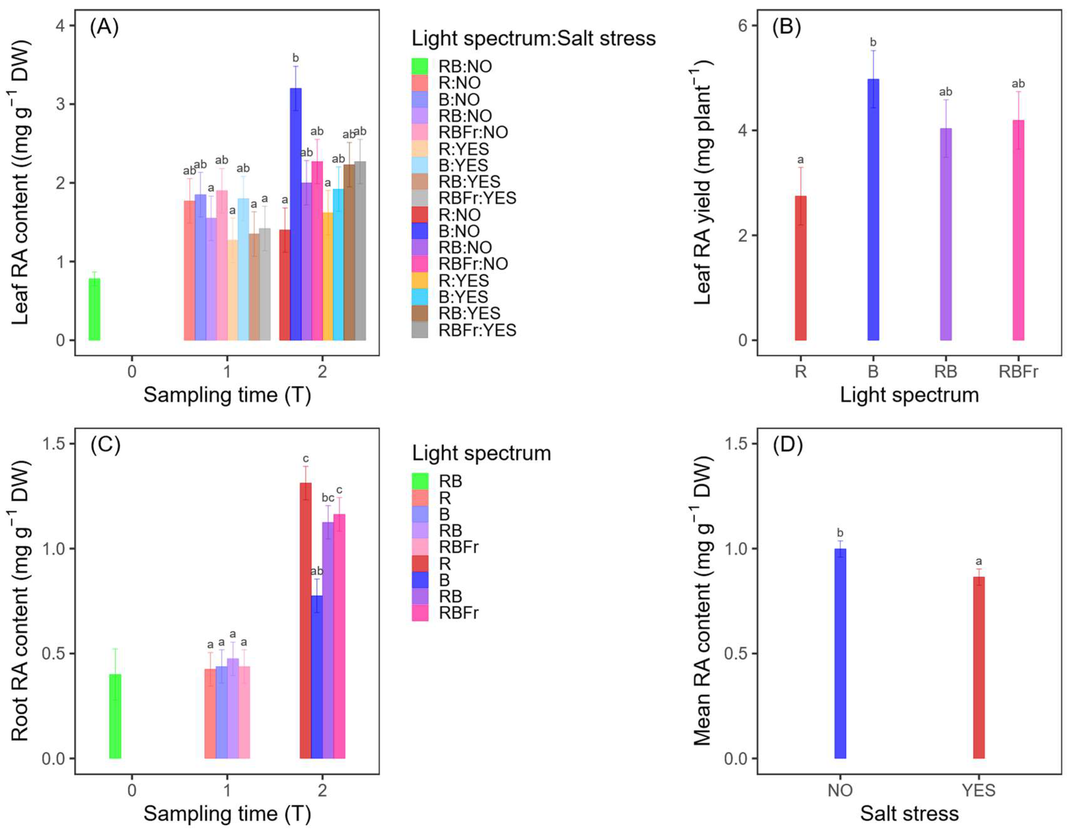
Figure 2A shows that an RA concentration at T0 is notably lower than at later time points, indicating a minimal initial accumulation. By T1, the RA concentration increases across all treatments, with no significant differences among light and salt stress conditions, suggesting that at T1, neither the light spectrum nor salt stress significantly impact RA accumulation. At T2, a more pronounced variability emerges: the B:NO treatment shows the highest RA content, higher than the results under R light, indicating that blue light under non-saline conditions may enhance RA synthesis. Other treatments, particularly those exposed to R light and combined light treatments (RB, RBFr), exhibit moderate RA concentrations without significant differences among them. Salt stress does not seem to induce a clear pattern of RA accumulation at T2, as RA concentrations under saline and non-saline conditions remain within similar ranges (in Supplementary Materials, Figure S1). However, some non-saline conditions (RB:NO and R:NO) appear to support higher RA concentrations compared to their corresponding saline treatments, suggesting that salt stress might not be a strong inducer of RA biosynthesis under these spectra. To summarize, the light spectrum plays a role in RA biosynthesis, with blue light (B:NO) showing the strongest effect at T2 (4.98 mg). Since the three-way interaction shows only a marginal significance, it may be interesting to examine the effects of the individual factors: in this case, the highest RA concentration values are observed at T2 (Table 2) under blue light and in non-saline conditions (data shown in Supplementary Materials, Figure S1), supporting the result of the three-way interaction.
In RA yield analysis, an increase around 139% from T1 to T2 was obtained (Table 2). Moreover, the blue spectrum confirmed what was observed in the DW and concentration graphs (Figure 2B): a higher RA final yield under blue light may be partially driven by an increased availability of leaf tissue, supporting an overall greater content of secondary metabolites.

3.3.2. Roots

Figure 2C illustrates the RA concentration across different sampling time and light spectrum conditions. By T1, the RA concentration shows comparable results for all light treatments with no significant differences between them. At T2, a more pronounced differentiation among treatments is observed: R and RBFr treatments exhibit the highest root RA concentrations, significantly differing from the other treatments. The B treatment obtained the lowest RA concentration among T2 treatments, although still significantly higher than values observed at earlier time points. These results indicate that the sampling time has a strong effect on the root RA concentration, with a marked increase from T0 to T2. Unlike in leaves, roots’ RA biosynthesis is driven by the R spectrum, regardless of the presence of salt stress. Only time as single factor statistically affected the RA yield, with an increase of 410% from T1 to T2 and a mean value of 19.36 ± 1.20 mg plant−1 at T2 (Table 2).

3.3.3. Total Plant

Considering the total plant RA yield, at T2 we measured a yield of 24.98 ± 1.34 mg plant−1, 4.1 times higher than at T1 (Table 2). In this case, only time as a main factor had a significant effect on the RA yield. In this case, it could be interesting to highlight the different responses under the salt treatments (Figure 2D): on average, plants under non-stressed conditions showed a higher RA accumulation.

3.4. Quercetin

3.4.1. Leaves

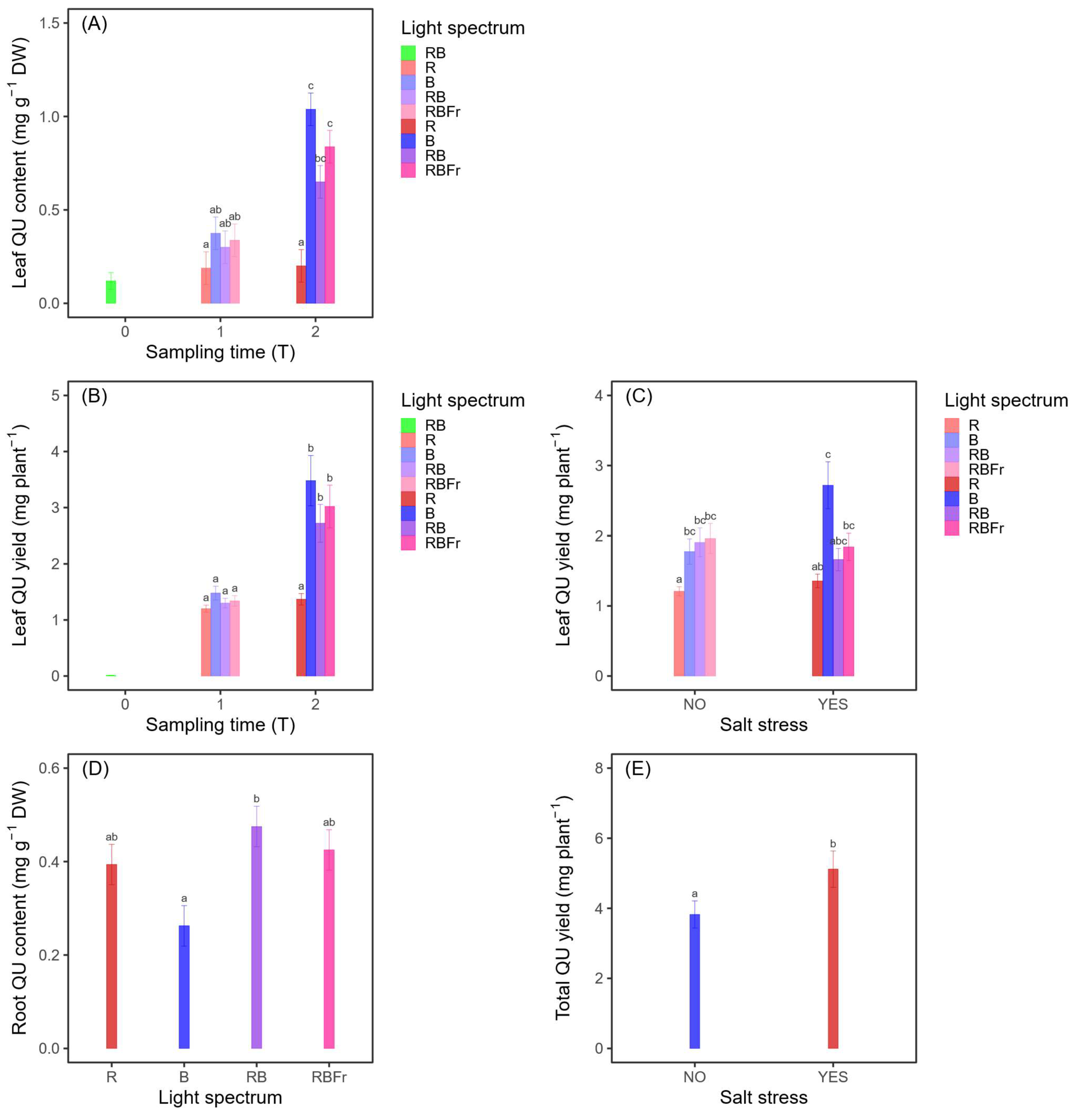
The quercetin concentration in leaves was statistically affected by the L:S interaction, with a strong result of B light (1.04 mg g−1 DW) at T2, not statistically different from the RBfr and RB treatments (Figure 3A). These results suggest that quercetin accumulation in leaves is strongly time-dependent, with a substantial increase from T0 to T2. Additionally, light played a key role in quercetin biosynthesis, with blue light-based treatments obtaining higher values. Otherwise, the R treatment was less effective in promoting quercetin accumulation (Figure 3A). Considering the L:T effect on the QU yield, Figure 3B shows that B light at T2 maximized the flavonoid content, obtaining a value of 3.48 ± 0.44 mg plant−1. Exploring the L:S interaction, there is a general increase in the QU yield under stressed conditions: B spectrum maximizes the yield under stress conditions due to its role in stimulating both biomass production and flavonoid biosynthesis. R treatment, despite its lower QU accumulation in control conditions, experiences a moderate increase in yield under salt stress but remained among the lowest values overall. The RB and RBFr treatments, which have intermediate yields under NO stress, maintain a similar ranking under salt stress: the combine light spectra support stable QU production regardless of stress conditions (Figure 3C).

3.4.2. Roots

No statistically relevant effect was observed by the interaction of factors on the roots’ QU concentration. A 141% increase in the flavonoid content was obtained from T1 to T2 (Table 2). According to Figure 3D, light plays a role in the modulation of QU content in roots: RB is the most effective spectrum, significantly outperforming blue light. Overall, data highlight that combined light spectra enhance root quercetin biosynthesis, while blue light alone is less effective in promoting its accumulation. The roots’ QU yield registered a significant increase from T1 to T2 (+449%), with a mean value of 8.22 ± 0.96 mg plant−1 at final harvest (33 days of treatment) (Table 2).

3.4.3. Total Plant

Considering the yield of quercetin in the whole plant, a significant positive effect was observed in salt-treated plants, suggesting a stronger activation of quercetin biosynthesis in plants exposed to stress (Figure 3E).

4. Discussion

4.1. Effects of Salt Stress and Light on Biomass Accumulation

Previous studies have investigated abiotic stressors [23] or integrated abiotic and biotic treatments [24] as a strategy to boost plant yield and secondary metabolite production under controlled conditions. Yet the specific interaction between salt stress and light quality remains underexplored. In crop plants, salinity imposes immediate osmotic stress by reducing soil water potential and inducing specific ion toxicities, which disrupt K+/Na+ homeostasis and nutrient uptake. Indirectly, salinity provokes oxidative stress via excess reactive oxygen species, altered hormonal regulation, photosynthetic inhibition, and membrane destabilization, ultimately compromising growth, reproduction and yield [25,26]. In our study, however, Coleus blumei displayed a degree of salt tolerance, as biomass accumulation was determined primarily by the time of exposure and light quality rather than salinity. No significant three-way interaction among light, time, and salinity was observed; blue light promoted the highest biomass accumulation, though values were statistically comparable to RB and RBfr treatments. This aligns with recent evidence that LED lighting can enhance both biomass yield and antioxidant capacity under saline conditions [15]. In general, it is widely reported that plant photoreceptors are highly responsive to red and blue light. Given that plant photoreceptors are particularly responsive to red and blue light, their combination represents an optimal strategy to support growth [27,28].
Salt stress induces a range of physiological and biochemical adaptations [29], including profound shifts in carbohydrate metabolism, particularly in raffinose oligosaccharide (RFO) pathways, which contribute to osmoprotection. In C. blumei, exposure to 60 mM NaCl reduced the starch accumulation in leaves, accompanied by a transient decline and subsequent recovery in photosynthetic activity. Salinity also promoted the synthesis of novel carbohydrates, including high-molecular-weight RFOs (DP 5–8) and O-methylated inositol, both linked to osmotic adjustments [30]. Another study on Coleus species reported that increasing NaCl concentrations (100, 200, and 300 mM) reduce the relative water content and leaf water potential and also increase the electrolyte leakage and water uptake capacity. The most salt-tolerant species, Coleus aromaticus and Coleus amboinicus, exhibited higher carbohydrate accumulation, again demonstrating an effective osmoprotectant mechanism [13]. These findings indicate that Coleus responds to salt stress by adjusting carbon partitioning, favoring the accumulation of osmoprotective sugars over starch storage to stabilize cellular functions and maintain metabolic homeostasis.
In our study, light treatments yielded comparable outcomes regardless of salinity. Except for a significant blue–red difference in root growth, light quality did not affect root or leaf biomass production.

4.2. Effect of Salt Stress and Light on Secondary Metabolites

Beyond the primary metabolism, salinity and light elicit the modulation of specialized metabolite biosynthesis, enhancing antioxidant capacity [31]. Whereas many metabolomic studies delineate gene- and metabolite-level activations to single abiotic stimuli, here we evaluate the combined action of salinity and light on the accumulation of quercetin and rosmarinic acid. Without direct gene expression or metabolite profiling, our interpretation should be considered as a hypothesis-driven reconstruction of the underlying metabolic mechanism supported by the quantitative patterns we report. In mechanistic terms, we propose that salinity and light-derived reactive oxygen species (ROS) co-activate the shikimate-derived phenylpropanoid pathway at its entry point, the phenylalanine ammonia-lyase (PAL) and rosmarinic acid branch that links phenylalanine and tyrosine metabolism [32,33]. Indeed, under salinity, excess Na+ and Cl accumulation displaces essential nutrients and imposes osmotic stress, lowering stomatal conductance and photosynthetic enzyme activity and thereby increasing ROS, activating enzymatic and non-enzymatic antioxidant mechanisms [34]. First-line detoxification is provided by superoxide dismutase (SOD), catalase (CAT) and the ascorbate–glutathione (AsA–GSH) cycle, with auxiliary antioxidant enzymes extending the ROS-scavenging network [35]. Beyond detoxification, low-molecular-weight antioxidants, such as AsA, GSH, and the AsA–GSH redox state, act as regulators: AsA serves as a peroxidase co-substrate and influences the expression of PAL and other entry enzymes of phenylpropanoid metabolism (PAL, C4H, and 4CL), upregulating the flavonoid branch (CHS, CHI, F3H/F3′H, and FLS) and ultimately increasing the flavonol output [36,37]. In parallel, blue light, perceived through cryptochromes, can elevate redox signaling and promote AsA accumulation [38], thereby amplifying phenylpropanoid activation and the induction of PAL and downstream flavonoid genes [39,40]. The convergence of salinity and blue light-mediated redox signaling coherently explains the time-dependent rise in quercetin in our plants, indicating a positive combined effect of salt stress and blue-enriched lighting on QU leaves accumulation [41]. Consistently, multiple studies report comparable responses: in Ginkgo biloba, moderate salt stress regulates key transcription factors that turn on stress-responsive genes and increase flavonoid biosynthesis [16]. In Phaseolus vulgaris, salinity upregulates phenylpropanoid and flavonoid pathway genes, leading to higher flavonoid accumulation [28], supporting our observation that mild salinity can elicit quercetin accumulation with limited impact on growth.
In contrast to quercetin, foliar rosmarinic acid increased, primarily under blue light, but showed no clear positive correlation with salinity. This pattern likely reflects the more constitutive role of RA in Lamiaceae, making it less additively inducible by NaCl than flavonols [42,43]. Moreover, RA biosynthesis relies on a dual-route architecture: the phenylalanine and tyrosine arms joined by rosmarinic acid synthase [43]. Experimental evidence shows that metabolic engineering can markedly boost RA in S. miltiorrhiza hairy roots by reinforcing key nodes, especially the tyrosine-arm enzymes TAT and HPPR (alone or together) alongside PAL/C4H/4CL, although aspects of the regulatory control remain unresolved [44]. In our experiment, without gene-expression profiling, we therefore hypothesize that, given that RA draws on two precursor routes, elevating PAL alone is insufficient: if the salt/light interaction either fails to co-induce downstream RA steps or engages stress-responsive transcriptional repression, its content will not show an additive salt × light gain. In parallel, RA and quercetin compete for p-coumaroyl-CoA [45]; by strongly activating the flavonol pathway, blue light likely diverted precursors toward quercetin, so the robust salt × blue effect on quercetin may have obscured any additive response in RA. Consistently, cultivar-dependent patterns in basil support this dissociation: NaCl increased quercetin-rutinoside and RA in the ‘Sweet Broadleaf’ but reduced RA in ‘Siam Queen’ leaves, underscoring compound- and genotype-specific regulation under salinity [46].
In roots, red-containing spectra consistently promoted phenolic accumulation: RA peaked under monochromatic red, while quercetin was numerically highest under RB, though not significantly different from RBFr or R. A mixed LED spectra often yield synergistic effects by co-activating multiple photoreceptor pathways, thereby enhancing both growth and the secondary metabolism [47]; notably, RB with a higher red fraction (e.g., 3B:7R) has been reported to simultaneously promote growth and phenolic acids and to upregulate TAT and PAL in Salvia miltiorrhiza, indicating the coordinated activation of both tyrosine- and phenylalanine-derived RA precursors [48]. According to our observation, RA responds strongly to red-enriched lighting, while adding blue preferentially boosts the flavonol branch. These results confirm what we observed in our earlier Coleus analysis [17]. These results reinforce that spectral composition effects are highly organ- and species-specific, in line with the complex picture emerging from the literature [49].
Beyond spectral quality and the combination between salt and light stressors, the time and severity of salinity application is a second, decisive driver of polyphenols accumulation. For example, prolonged exposure can lead to plant adaptation with a consequent stabilization or even decline of flavonoid level over time [50]. In addition, a study on Dracocephalus kotschyi showed that moderate salinity (75 mM) maximizes phenolic and flavonoid accumulation, whereas higher levels (100 mM) cause a drop in these compounds [51]. In our case, both rosmarinic acid and quercetin exhibited an increasing trend from 20 (T1) to 33 (T2) days of treatment, including salt-treated plants, indicating that Coleus’ adaptive response had not yet reached a plateau or begun to decline over the period examined. However, the effect we observed could result from the salinity level applied (120 mM NaCl), which could be lower than the threshold necessary to elicit a more pronounced metabolic response in Coleus.
Taken together, these outcomes indicate that, in Coleus blumei, light quality exerts a stronger control than salinity over the accumulation of the profiled bioactive compounds, with clear, organ-specific responses. Practically, this experiment optimized rosmarinic acid (RA) content under blue-enriched LEDs and non-saline conditions, since salinity did not confer an additional gain. By contrast, for quercetin, combining blue/RB lighting with mild salinity acted as a selective elicitor and increased the QU content.

4.3. Strategic Optimization of Rosmarinic Acid and Quercetin Yields: Implications and Future Perspectives

The specific contributions of biomass and concentration to yield variation in time (from T1 to T2) are reported in the Supplementary Materials (Table S1).
In leaves, the RA yield increased primarily due to biomass accumulation (~69% DW), with a secondary contribution from concentration (~31% C). This pattern supports that the strong time effect (T***) on the RA yield was largely biomass-driven, with light providing an additional but minor modulation of RA concentration (L*). In contrast, the leaf QU yield was predominantly explained by concentration (~56% C) rather than biomass (~44% DW), consistent with the strong light effect on the QU concentration (L***). The significant L:T interaction (**) further indicates that blue light maximized the QU accumulation specifically at T2, while biomass supplied an additive background effect. Notably, salt showed a trend-level effect on the QU concentration (S·), which was also reflected in the yield (L:S·), suggesting that salinity amplifies the effect of light, primarily at the concentration level.
In roots, the RA yield at T2 resulted from joint increases in the biomass and concentration, with concentration explaining a larger share (~54% C) than biomass (~46% DW). Thus, the overall time effect (T***) on the RA yield reflects the parallel rise in both factors. Similarly, the root QU yield increased through parallel contributions, again with concentration prevailing (~54% C) over biomass (~46% DW), pointing to a time-driven maturation effect (T***). Despite significant light effects on the QU concentration (L**), these changes did not translate into yield (L ns).
At the whole-plant level, both RA and QU yields increased with plant maturation, reflecting the combined action of biomass expansion and concentration changes. For total RA, biomass accounted for a modest majority of the gain (~56% DW vs. ~44% C), whereas for total QU, the opposite pattern was observed (~54% C vs. ~46% DW). The total QU yield reflected not only the strong maturation effect (T***) but also a positive salt effect (S*), consistent with the trend-level stimulation of the QU concentration observed in leaves and its light–salt interplay at the organ level. Overall, these patterns indicate that total QU production benefits from mild salinity, primarily through the concentration-level modulation in photosynthetic tissues, whereas the total RA yield largely follows biomass accumulation with time.
Since the leaf RA yield peaks at five weeks (T2) under blue light, when both biomass and the RA concentration are at their highest, and the root RA is driven almost entirely by harvest timing, the optimal strategy to maximize total RA in Coleus blumei is to use blue-enriched LED regimes, delaying harvesting to five weeks while avoiding salinity. Similarly, since foliar QU accumulation peaks at five weeks (T2) under blue or red–blue spectra, while the root QU yield is determined solely by harvest timing, and moderate salt stress can further boost whole-plant QU, the recommended strategy to maximize the total QU in Coleus blumei is to apply blue-enriched LED strategies, delay harvesting to five weeks, and, when whole-plant yield is the primary goal, introduce moderate salinity.
However, prolonging the duration of light treatments entails a trade-off, as LEDs often represent around 60–70% of total electricity use in vertical farms [52,53]. Therefore, truly cost-effective protocols must balance the percentage increase in the metabolite yield achieved per additional week of lighting against the corresponding rise in energy consumption. Moreover, to unlock the full potential of this system, in-depth transcriptomic and metabolomic profiling of Coleus species will be essential to identify and regulate the key genes controlling carbohydrate metabolism and flavonoid biosynthesis.

5. Conclusions

Our findings provide insights into strategies for optimizing rosmarinic acid and quercetin production in Coleus blumei in LED-based indoor environments. Time consistently emerged as the primary driver across variables, underscoring the central role of the developmental stage in metabolite accumulation. This study also confirms that the impact of light quality on secondary-metabolite accumulation is organ-dependent and that blue and red spectra modulate metabolic pathways in distinct ways.
The highest rosmarinic acid yield in leaves was observed under blue monochromatic light; in roots, red light optimized the RA content
Similarly, quercetin yield in leaves reached its peak under the blue spectrum; differently, in roots, the RB light resulted in the highest quercetin concentration
On average, the total plant RA content was reduced by salt application, while the total plant QU yield was higher in salt-treated plants
Prolonged treatment, from 20 to 33 days, increased plants’ biomass, RA, and QU content.
Values of 24.98 ± 1.34 mg of RA and 10.09 ± 1.02 mg of QU per plant were measured at T2.
Taken together, these results support extending the cultivation to five weeks to achieve maximal yields of both compounds. Moderate salinity selectively amplifies QU without enhancing RA, highlighting spectral-salinity modulation as a promising strategy for targeted metabolite accumulation in Coleus blumei indoor systems.

Supplementary Materials

The following supporting information can be downloaded at: https://www.mdpi.com/article/10.3390/horticulturae11101205/s1. Section S1 (S1.1: Contribution analysis of biomass and concentration to yield. Section S2 (Table S1: Contribution of biomass accumulation (DW) and metabolite concentration (C) to yield increase (ΔY) from T1 to T2, computed with mean values reported in Table 2. Figure S1: Additional main factors (light and salt stress) effects on Coleus blumei total dry weight and rosmarinic acid content). Supplementary reference: Shorrocks, A.F. Decomposition Procedures for Distributional Analysis: A Unified Framework Based on the Shapley Value. J. Econ. Inequal. 2013, 11, 99–126. https://doi.org/10.1007/s10888-011-9214-z [54].

Author Contributions

Conceptualization, B.S., A.Q., M.T., and A.B.; methodology, B.S., A.Q., M.T., L.M., M.P., and A.B.; validation, A.B.; formal analysis, A.Q.; investigation, B.S., A.Q., M.T., L.M., and M.P.; resources, P.T., D.T., L.M., M.P., and A.B.; data curation, B.S.; writing—original draft preparation, B.S., A.Q., L.M., and M.P.; visualization, A.Q.; supervision, P.T., D.T., and A.B.; project administration, P.T. and D.T. All authors have read and agreed to the published version of the manuscript.

Funding

This research received no external funding.

Data Availability Statement

The original contributions presented in this study are included in the article/supplementary material. Further inquiries can be directed to the corresponding author.

Acknowledgments

We would like to thank C-Led s.r.l. Imola (IT) for the collaboration in designing and making LED lamps for the specific needs of this research.

Conflicts of Interest

The authors declare no conflicts of interest.

References

  1. Andrade-Cetto, A. Ethnobotanical Study of the Medicinal Plants from Tlanchinol, Hidalgo, México. J. Ethnopharmacol. 2009, 122, 163–171. [Google Scholar] [CrossRef]
  2. Jakobina, M.; Łyczko, J.; Szumny, A.; Galek, R. The Influence of Cultivation Conditions on the Formation of Psychoactive Salvinorin A, Salvinorin B, Rosmarinic Acid and Caffeic Acid in Coleus Scutellarioides. Sci. Rep. 2024, 14, 6693. [Google Scholar] [CrossRef] [PubMed]
  3. Razzaque, A.; Ellis, B.E. Rosmarinic Acid Production in Coleus Cell Cultures. Planta 1997, 137, 287–291. [Google Scholar] [CrossRef] [PubMed]
  4. Bauer, N.; Vuković, R.; Likić, S.; Jelaska, S. Potential of Different Coleus Blumei Tissues for Rosmarinic Acid Production. Food Technol. Biotechnol. 2015, 53, 3–10. [Google Scholar] [CrossRef] [PubMed]
  5. Bismelah, N.A.; Ahmad, R.; Mohamed Kassim, Z.H.; Ismail, N.H.; Rasol, N.E. The Antibacterial Effect of Plectranthus scutellarioides (L.) R.Br. Leaves Extract against Bacteria Associated with Peri-Implantitis. J. Tradit. Complement. Med. 2022, 12, 556–566. [Google Scholar] [CrossRef]
  6. Mirza, M.A.; Mahmood, S.; Hilles, A.R.; Ali, A.; Khan, M.Z.; Zaidi, S.A.A.; Iqbal, Z.; Ge, Y. Quercetin as a Therapeutic Product: Evaluation of Its Pharmacological Action and Clinical Applications—A Review. Pharmaceuticals 2023, 16, 1631. [Google Scholar] [CrossRef]
  7. Guan, H.; Luo, W.; Bao, B.; Cao, Y.; Cheng, F.; Yu, S.; Fan, Q.; Zhang, L.; Wu, Q.; Shan, M. A Comprehensive Review of Rosmarinic Acid: From Phytochemistry to Pharmacology and Its New Insight. Molecules 2022, 27, 3292. [Google Scholar] [CrossRef]
  8. Marchev, A.S.; Vasileva, L.V.; Amirova, K.M.; Savova, M.S.; Koycheva, I.K.; Balcheva-Sivenova, Z.P.; Vasileva, S.M.; Georgiev, M.I. Rosmarinic acid—From bench to valuable applications in food industry. Trends Food Sci. Technol. 2021, 117, 182–193. [Google Scholar] [CrossRef]
  9. Hazrati, S.; Pignata, G.; Casale, M.; Hosseini, S.J.; Nicola, S. Influence of Nutrient Solutions in an NGS® Soilless System on the Yield, Quality and Shelf Life of Fresh-Cut Commercial Mint at Different Harvest Times. Agronomy 2024, 14, 610. [Google Scholar] [CrossRef]
  10. Quadri, A.; Barbaresi, A.; Tassinari, P.; Bertaccini, A.; Contaldo, N.; Mercolini, L.; Protti, M.; Montalbetti, R.; Laurita, R.; Torreggiani, D. Use of LED Light and Plasma Activated Water (PAW) to Stimulate Pharmaceutical Compound Production in Catharanthus roseus Plants. In Proceedings of the X International Symposium on Light in Horticulture, Seoul, Republic of Korea, 19–22 May 2024; pp. 255–268. [Google Scholar] [CrossRef]
  11. Mohammadi, H.; Khoshi, N.; Hazrati, S.; Aghaee, A.; Falakian, M.; Ghorbanpour, M. Interaction of NaCl Salinity and Light Intensity Affect Growth, Physiological Traits and Essential Oil Constituents in Artemisia dracunculus L. (Tarragon). Biochem. Syst. Ecol. 2023, 107, 104626. [Google Scholar] [CrossRef]
  12. Acosta-Motos, J.R.; Ortuño, M.F.; Bernal-Vicente, A.; Diaz-Vivancos, P.; Sanchez-Blanco, M.J.; Hernandez, J.A. Plant Responses to Salt Stress: Adaptive Mechanisms. Agronomy 2017, 7, 18. [Google Scholar] [CrossRef]
  13. Kotagiri, D.; Kolluru, V.C. Effect of Salinity Stress on the Morphology & Physiology of Five Different Coleus Species. Biomed. Pharmacol. J. 2017, 10, 1639–1649. [Google Scholar] [CrossRef]
  14. Rao, K.V.M.; Raghavendra, A.S.; Reddy, K.J. Physiology and Molecular Biology of Stress Tolerance in Plants; Springer: Dordrecht, The Netherlands, 2006. [Google Scholar]
  15. Giménez, A.; Martínez-Ballesta, M.D.C.; Egea-Gilabert, C.; Gómez, P.A.; Artés-Hernández, F.; Pennisi, G.; Orsini, F.; Crepaldi, A.; Fernández, J.A. Combined Effect of Salinity and Led Lights on the Yield and Quality of Purslane (Portulaca oleracea L.) Microgreens. Horticulturae 2021, 7, 180. [Google Scholar] [CrossRef]
  16. Xu, N.; Liu, S.; Lu, Z.; Pang, S.; Wang, L.; Wang, L.; Li, W. Gene Expression Profiles and Flavonoid Accumulation during Salt Stress in Ginkgo biloba Seedlings. Plants 2020, 9, 1162. [Google Scholar] [CrossRef] [PubMed]
  17. Sambuco, B.; Quadri, A.; Trenta, M.; Tassinari, P.; Torreggiani, D.; Barbaresi, A.; Mercolini, L.; Protti, M. Enhancing Secondary Metabolites Accumulation in Coleus blumei through LED Light Application. In Proceedings of the X International Symposium on Light in Horticulture, Seoul, Republic of Korea, 19–22 May 2024; pp. 243–254. [Google Scholar] [CrossRef]
  18. Posit team. RStudio: Integrated Development Environment for R. Posit Software, Version 2025.05.01; PBC: Boston, MA, USA, 2025. Available online: http://www.posit.co/ (accessed on 15 June 2025).
  19. Lenth, R. emmeans: Estimated Marginal Means, aka Least-Squares Means. R Package, Version 1.8.4-1. 2023. Available online: https://CRAN.R-project.org/package=emmeans (accessed on 15 June 2025).
  20. Fox, J.; Weisberg, S. An R Companion to Applied Regression, 3rd ed.; Sage: Thousand Oaks, CA, USA, 2019. [Google Scholar]
  21. Hothorn, T.; Bretz, F.; Westfall, P. Simultaneous Inference in General Parametric Models. Biom. J. 2008, 50, 346–363. [Google Scholar] [CrossRef] [PubMed]
  22. Wickham, H. ggplot2: Elegant Graphics for Data Analysis; Springer: New York, NY, USA, 2016; ISBN 978-3-319-24277-4. [Google Scholar]
  23. Wang, J.; Han, Z.; He, J.; Kang, H.; Li, Q.; Chen, H.; Zhang, X.; Miao, W.; Shang, X.; Chen, W.; et al. Exploring the Effects of Light–Water Interaction in Plant Factory to Improve the Yield and Quality of Panax notoginseng (Burkill) F. H. Chen. Agronomy 2025, 15, 368. [Google Scholar] [CrossRef]
  24. Quadri, A.; Sambuco, B.; Trenta, M.; Tassinari, P.; Torreggiani, D.; Mercolini, L.; Protti, M.; Zambonelli, A.; Barbaresi, A. Bioengineered Indoor Farming Approaches: LED Light Spectra and Bi-2 Ostimulants for Enhancing Vindoline and Catharanthine Production in 3 Catharanthus roseus. Horticulturae 2025, 11, 828. [Google Scholar] [CrossRef]
  25. Stavridou, E.; Webster, R.J.; Robson, P.R.H. The Effects of Moderate and Severe Salinity on Composition and Physiology in the Biomass Crop Miscanthus × Giganteus. Plants 2020, 9, 1266. [Google Scholar] [CrossRef]
  26. Atta, K.; Mondal, S.; Gorai, S.; Singh, A.P.; Kumari, A.; Ghosh, T.; Roy, A.; Hembram, S.; Gaikwad, D.J.; Mondal, S.; et al. Impacts of Salinity Stress on Crop Plants: Improving Salt Tolerance through Genetic and Molecular Dissection. Front. Plant Sci. 2023, 14, 1241736. [Google Scholar] [CrossRef]
  27. Luo, S.; Zou, J.; Shi, M.; Lin, S.; Wang, D.; Liu, W.; Shen, Y.; Ding, X.; Jiang, Y. Effects of Red-Blue Light Spectrum on Growth, Yield, and Photosynthetic Efficiency of Lettuce in a Uniformly Illumination Environment. Plant Soil Environ. 2024, 70, 305–316. [Google Scholar] [CrossRef]
  28. Zhang, S.; Zhang, L.; Zou, H.; Qiu, L.; Zheng, Y.; Yang, D.; Wang, Y. Effects of Light on Secondary Metabolite Biosynthesis in Medicinal Plants. Front. Plant Sci. 2021, 12, 781236. [Google Scholar] [CrossRef] [PubMed]
  29. Zhao, H.; Liang, H.; Chu, Y.; Sun, C.; Wei, N.; Yang, M.; Zheng, C. Effects of Salt Stress on Chlorophyll Fluorescence and the Antioxidant System in Ginkgo biloba L. Seedlings. HortScience 2019, 54, 2125–2133. [Google Scholar] [CrossRef]
  30. Gilbert, G.A.; Wilson, C.; Madore, M.A. Root-Zone Salinity Alters Raffinose Oligosaccharide Metabolism and Transport in Coleus. Plant Physiol. 1997, 115, 1267–1276. [Google Scholar] [CrossRef] [PubMed]
  31. Dixon, R.A.; Paiva, N.L. Stress-Induced Phenylpropanoid Metabolism. Plant Cell. 1995, 7, 1085–1097. [Google Scholar] [CrossRef]
  32. Trócsányi, E.; György, Z.; Zámboriné-Németh, É. New Insights into Rosmarinic Acid Biosynthesis Based on Molecular Studies. Curr. Plant Biol. 2020, 23, 100162. [Google Scholar] [CrossRef]
  33. Falcone Ferreyra, M.L.; Rius, S.P.; Casati, P. Flavonoids: Biosynthesis, Biological Functions, and Biotechnological Applications. Front. Plant Sci. 2012, 3, 222. [Google Scholar] [CrossRef]
  34. Hossain, M.S.; Dietz, K.J. Tuning of Redox Regulatory Mechanisms, Reactive Oxygen Species and Redox Homeostasis under Salinity Stress. Front. Plant Sci. 2016, 7, 548. [Google Scholar] [CrossRef]
  35. Hasanuzzaman, M.; Raihan, M.R.H.; Masud, A.A.C.; Rahman, K.; Nowroz, F.; Rahman, M.; Nahar, K.; Fujita, M. Regulation of Reactive Oxygen Species and Antioxidant Defense in Plants under Salinity. Int. J. Mol. Sci. 2021, 22, 9326. [Google Scholar] [CrossRef]
  36. Ma, D.; Guo, Y.; Ali, I.; Lin, J.; Xu, Y.; Yang, M. Accumulation Characteristics of Plant Flavonoids and Effects of Cultivation Measures on Their Biosynthesis: A Review. Plant Physiol. Biochem. 2024, 215, 108960. [Google Scholar] [CrossRef]
  37. Foyer, C.H.; Kunert, K. The Ascorbate-Glutathione Cycle Coming of Age. J. Exp. Bot. 2024, 75, 2682–2699. [Google Scholar] [CrossRef]
  38. Chibani, K.; Gherli, H.; Fan, M. The Role of Blue Light in Plant Stress Responses: Modulation through Photoreceptors and Antioxidant Mechanisms. Front. Plant Sci. 2025, 16, 1554281. [Google Scholar] [CrossRef]
  39. Stracke, R.; Ishihara, H.; Huep, G.; Barsch, A.; Mehrtens, F.; Niehaus, K.; Weisshaar, B. Differential Regulation of Closely Related R2R3-MYB Transcription Factors Controls Flavonol Accumulation in Different Parts of the Arabidopsis Thaliana Seedling. Plant J. 2007, 50, 660–677. [Google Scholar] [CrossRef] [PubMed]
  40. Daryanavard, H.; Postiglione, A.E.; Mühlemann, J.K.; Muday, G.K. Flavonols Modulate Plant Development, Signaling, and Stress Responses. Curr. Opin. Plant Biol. 2023, 72, 102350. [Google Scholar] [CrossRef] [PubMed]
  41. Ko, E.Y.; Nile, S.H.; Sharma, K.; Li, G.H.; Park, S.W. Effect of Different Exposed Lights on Quercetin and Quercetin Glucoside Content in Onion (Allium cepa L.). Saudi J. Biol. Sci. 2015, 22, 398–403. [Google Scholar] [CrossRef] [PubMed]
  42. Petersen, M.; Simmonds, M.S. Rosmarinic Acid. Phytochemistry 2003, 62, 121–125. [Google Scholar] [CrossRef]
  43. Ru, M.; Wang, K.; Bai, Z.; Peng, L.; He, S.; Wang, Y.; Liang, Z. A Tyrosine Aminotransferase Involved in Rosmarinic Acid Biosynthesis in Prunella vulgaris L. Sci. Rep. 2017, 7, 4892. [Google Scholar] [CrossRef]
  44. Kim, G.D.; Park, Y.S.; Jin, Y.H.; Park, C.S. Production and Applications of Rosmarinic Acid and Structurally Related Compounds. Appl. Microbiol. Biotechnol. 2015, 99, 2083–2092. [Google Scholar] [CrossRef]
  45. Chen, I.G.J.; Lee, M.S.; Lin, M.K.; Ko, C.Y.; Chang, W. The Blue Light Decreases Tanshinone IIA Content in Salvia Miltiorrhiza Hairy Roots via Genes Regulation. J. Photochem. Photobiol. B 2018, 183, 164–171. [Google Scholar] [CrossRef]
  46. Scagel, C.F.; Lee, J.; Mitchell, J.N. Salinity from NaCl Changes the Nutrient and Polyphenolic Composition of Basil Leaves. Ind. Crops Prod. 2019, 127, 119–128. [Google Scholar] [CrossRef]
  47. Cioć, M.; Szopa, A.; Prokopiuk, B.; Pawłowska, B.; Łopusiewicz, Ł. Influence of LED Light Spectra on Morphogenesis, Secondary Metabolite Production and Antioxidant Potential in Eucomis autumnalis Cultured In Vitro. Agronomy 2025, 15, 2197. [Google Scholar] [CrossRef]
  48. Zhang, S.; Ma, J.; Zou, H.; Zhang, L.; Li, S.; Wang, Y. The Combination of Blue and Red LED Light Improves Growth and Phenolic Acid Contents in Salvia miltiorrhiza Bunge. Ind. Crops Prod. 2020, 158, 112959. [Google Scholar] [CrossRef]
  49. Thoma, F.; Somborn-Schulz, A.; Schlehuber, D.; Keuter, V.; Deerberg, G. Effects of Light on Secondary Metabolites in Selected Leafy Greens: A Review. Front. Plant Sci. 2020, 11, 497. [Google Scholar] [CrossRef]
  50. Du, Y.; Zhao, Q.; Chen, L.; Yao, X.; Zhang, H.; Wu, J.; Xie, F. Effect of Drought Stress during Soybean R2–R6 Growth Stages on Sucrose Metabolism in Leaf and Seed. Int. J. Mol. Sci. 2020, 21, 618. [Google Scholar] [CrossRef]
  51. Vafadar, F.; Amooaghaie, R.; Ehsanzadeh, P.; Ghanadian, M.; Talebi, M.; Ghanati, F. Melatonin and Calcium Modulate the Production of Rosmarinic Acid, Luteolin, and Apigenin in Dracocephalum kotschyi under Salinity Stress. Phytochemistry 2020, 177, 112422. [Google Scholar] [CrossRef]
  52. Pennisi, G.; Sanyé-Mengual, E.; Orsini, F.; Crepaldi, A.; Nicola, S.; Ochoa, J.; Fernandez, J.A.; Gianquinto, G. Modelling Environmental Burdens of Indoor-Grown Vegetables and Herbs as Affected by Red and Blue LED Lighting. Sustainability 2019, 11, 4063. [Google Scholar] [CrossRef]
  53. Avgoustaki, D.D.; Xydis, G. Plant Factories in the Water-Food-Energy Nexus Era: A Systematic Bibliographical Review. Food Secur. 2020, 12, 253–268. [Google Scholar] [CrossRef]
  54. Shorrocks, A.F. Decomposition Procedures for Distributional Analysis: A Unified Framework Based on the Shapley Value. Econ. Inequal. 2013, 11, 99–126. [Google Scholar] [CrossRef]
Figure 1. Dry weight (g) in Coleus blumei leaves and roots. (A) S:L interaction on leaves DW (g plant−1): bars are grouped by salinity level (NO–YES; 0 mM-120 mM NaCl) to highlight the combined effect of the two factors. (B) Light treatment effect on roots’ DW: colors represent different light spectra. Bars indicate SE (n = 4). Means with different letters are significantly different at the 5% level by Tukey’s adjustment.
Horticulturae 11 01205 g001
Figure 2. Leaves, roots, and total plant rosmarinic acid concentration and yield across different time sampling and treatment conditions (light and salt stress). (A) S:L:T interaction effect on leaves’ RA concentration (mg g−1 DW): bars are grouped by sampling time to show the compound accumulation trend; colors represent S:L combinations. (B) Light effect on leaves’ RA yield (mg plant−1); colors represent different light spectra. (C) S:L:T interaction effect on roots’ RA conc. (mg g−1 DW). (D) Salt stress effect on total plant mean RA conc. (mg g−1 DW); colors represent the two salinity levels (0 mM–120 mM NaCl). Bars indicate SE (n = 4). Means with different letters are significantly different at the 5% level by Tukey’s test.
Horticulturae 11 01205 g002
Figure 3. Leaves, roots, and total plant quercetin concentrations and yields across different time sampling and treatment conditions (light and salt stress). (A) L:T interaction effect on leaves’ QU concentration (mg g−1 DW): bars are grouped by sampling time to show the compound accumulation trend; colors represent different treatment combinations. (B) L:T interaction effect on leaves’ QU yield (mg plant−1). (C) S:L interaction effect on leaves’ QU yield: bars are grouped by salinity level (NO–YES; 0 mM-120 mM NaCl) to highlight the synergistic effect of the two factors. (D) Light effect on roots’ QU concentration (mg g−1 DW); colors represent different light treatments. (E) Salt stress effect on total plants’ mean QU yield; different colors represent the two salinity levels (0 mM–120 mM NaCl). Bars indicate SE (n = 4). Means with different letters are significantly different at the 5% level by Tukey’s test.
Horticulturae 11 01205 g003
Table 1. ANOVA results of dry weight (g), RA-QU concentration (mg g−1 DW), and yield (mg plant−1).
Main FactorFactors Combination
LeavesLightSalt StressTimeL:SL:TS:TL:S:T
Dry weightnsns***·nsnsns
RA concentration*·***nsnsns·
RA yield*ns***nsnsnsns
QU concentration***·***ns**nsns
QU yield***ns***·*nsns
RootsLightSalt stressTimeL:SL:TS:TL:S:T
Dry weight*ns***nsnsnsns
RA concentration*ns***ns*nsns
RA yieldnsns***nsnsnsns
QU concentration**ns***nsnsnsns
QU yieldnsns***nsnsnsns
Total plantLightSalt stressTimeL:SL:TS:TL:S:T
Dry weight*ns***nsnsnsns
RA yieldnsns***nsnsnsns
QU yieldns****nsnsnsns
Asterisks indicate levels of significance: * p ≤ 0.05, ** p ≤ 0.01, and *** p ≤ 0.001; “·” indicates a trend towards significance (0.05 < p ≤ 0.1), while “ns” denotes non-significant differences (p > 0.1).
Table 2. Time main factor effect on leaves, roots, and total plant dry weight (g), rosmarinic acid (RA) and quercetin (QU) concentration (mg g−1 DW), and yield (mg plant−1) at 20 (T1) and 33 (T2) days of treatments.
Time Levels
LeavesT0T1T2
Dry weight0.04 ± 0.011.42 ± 0.14 a2.65 ± 0.14 b
RA concentration0.78 ± 0.081.62 ± 0.09 a2.12 ± 0.09 b
RA yield0.03 ± 0.0032.35 ± 0.38 a5.62 ± 0.38 b
QU concentration0.12 ± 0.0440.30 ± 0.04 a0.68 ± 0.04 b
QU yield0.002 ± 0.001.32 ± 0.04 a2.42 ± 0.14 b
RootsT0T1T2
Dry weight0.19 ± 0.048.29 ± 0.75 a17.88 ± 0.75 b
RA concentration0.26 ± 0.020.44 ± 0.03 a1.09 ± 0.03 b
RA yield0.05 ± 0.0184.34 ± 0.41 a19.36 ± 1.20 b
QU concentration0.24 ± 0.040.22 ± 0.03 a0.55 ± 0.03 b
QU yield0.15 ± 0.061.50 ± 0.17 a8.22 ± 0.96 b
Total plantT0T1T2
Dry weight0.22 ± 0.059.71 ± 0.79 a20.53 ± 0.79 b
RA yield0.09 ± 0.016.61 ± 0.48 a24.98 ± 1.34 b
QU yield0.05 ± 0.011.94 ± 0.19 a10.09 ± 1.02 b
Values are means ± SE (n = 4). Means with different letters are significantly different at the 5% level by Tukey’s adjustment.
Disclaimer/Publisher’s Note: The statements, opinions and data contained in all publications are solely those of the individual author(s) and contributor(s) and not of MDPI and/or the editor(s). MDPI and/or the editor(s) disclaim responsibility for any injury to people or property resulting from any ideas, methods, instructions or products referred to in the content.