Article

Effects of Temperature Regulation on the Physiological Characteristics and Platycodin Synthesis of Platycodon grandiflorum

Cultivation Base of State Key Laboratory for Ecological Restoration and Ecosystem Management of Jilin Province and Ministry of Science and Technology, College of Chinese Medicinal Materials, Jilin Agricultural University, Changchun 130118, China
*
Authors to whom correspondence should be addressed.
Horticulturae 2024, 10(8), 848; https://doi.org/10.3390/horticulturae10080848
Submission received: 4 July 2024 / Revised: 6 August 2024 / Accepted: 8 August 2024 / Published: 9 August 2024
(This article belongs to the Section Medicinals, Herbs, and Specialty Crops)

Abstract

:
Platycodon grandiflorum, a dual-purpose herb for food and medicine, is widely distributed in Asia. Although P. grandiflorum has relatively low requirements for its growing environment, temperature remains an important ecological factor affecting its growth, development, and quality formation. In order to explore the effect of different temperatures on P. grandiflorum during their growth period, the diversity in growth physiology, platycodin contents, and gene expression of key enzymes were investigated under constant (8 °C, 18 °C, and 28 °C) and variable (8–18 °C, 8–28 °C, and 18–28 °C) temperature conditions at each of the three levels. The results suggested that both constant and variable temperatures at high levels significantly increased the aboveground fresh weight of P. grandiflorum. However, the low–variable temperature was beneficial for the accumulation of dry and fresh weight in the roots. Regardless of whether temperatures were constant or variable at low levels, this increased the content of soluble sugars, proline, and peroxidase in P. grandiflorum, while upregulating the expression levels of key enzyme genes involved in platycodin synthesis. Meanwhile, a low–constant temperature inhibited the photosynthetic rate of P. grandiflorum. Furthermore, medium–constant and large-scale variable temperatures were conducive to the accumulation of platycodins in the roots. This research provides a theoretical basis and data support for the influence of temperature variations on P. grandiflorum quality formation.

1. Introduction

An appropriate temperature is an essential condition for the normal growth and development of medicinal plants, as well as an important ecological factor affecting the quality formation of their medicinal materials [1]. Temperature can affect the rhythm of the growth and development of medicinal plants at different stages, e.g., the aboveground biomass accumulation of cannabis and the flowering time of safflower [2,3]. Certain alterations in plants can be caused by physiological factors, especially photosynthetic and resistance physiological factors [4]. Photosynthesis, as the main pathway for carbohydrate synthesis and energy metabolism in plants, is highly sensitive to temperature [5,6]. Generally, low temperatures can cause changes in the photochemical quenching coefficient (qP) and non-photochemical quenching (NPQ) of plants, while inhibiting the photosystem, and affecting the maximum photochemical efficiency of PSII (Fv/Fm) and actual photochemical efficiency (φPSII) [7]. In the meantime, alterations in stomatal conductance (Gs) and stomatal limitation values (Ls) can occur under low-temperature conditions, resulting in a decrease in the net photosynthetic rate (Pn), photosynthesis function, and water use efficiency (WUE), ultimately affecting the morphological and physiological characteristics of the plants [8]. On the flip side, an elevated temperature can increase photorespiration and mitochondrial respiration [9,10], which induces the reduction of chlorophyll content and the occurrence of photoinhibition [11], thereby decreasing plant photosynthesis [12]. Moreover, plants possess specific mechanisms to adapt to the environment through physiological and physiochemical changes. For instance, soluble sugars (SS), soluble proteins (SP), and proline (PRO) can protect enzymes, proteins, and biofilm systems by regulating the osmotic potential of the cytoplasm [13]. Some protective enzymes in plants, e.g., superoxide dismutase (SOD) and peroxidase (POD), maintain the homeostasis of reactive oxygen species (ROS), playing a role in ensuring the integrity and stability of the cell membrane structure [14]. Furthermore, the degree of damage to the plant cell membranes can be reflected by malondialdehyde (MDA), which is a critical index for membrane peroxidation [15]. In brief, synergistic effects between various physiological characteristics sustain the internal balance of plants in response to fluctuations in temperature [4]. Considering the intuitive ecological relationship between medicinal plants and temperature, exploration of their reactions to temperature regulation is of great significance for the cultivation strategies used for medicinal plants and the quality formation of medicinal materials.
Platycodon grandiflorum (Jacq.) A. DC, a perennial herbaceous plant, is widely distributed in Asian countries such as China, Japan, and South Korea [16]. The vast geographical span of distribution areas for P. grandiflorum leads to an obvious contrast in ecological factors, especially temperature [17]. P. grandiflorum has been utilized in the medical field since ancient times, and modern pharmacological research has evidenced that it has multiple effects, including cough and phlegm relief, cardiovascular protection, cancer prevention, and immune regulation [16]. The main active ingredients of P. grandiflorum are oleanane-type triterpenoid saponins with double sugar chains, including platycoside E (PE), platycodin D3 (PD3), deapioplatycodin D (DPD), and platycodin D (PD) [18]. These secondary metabolites of P. grandiflorum are mainly synthesized via the mevalonate pathway [19,20], by means of the various key enzymes involved in platycodin synthesis, which go through three stages [21,22]. Temperature shifts, as external stimuli, have a considerable effect on the accumulation of secondary metabolites in medicinal plants [23]. Taking triterpenoid saponins as an example, reports have demonstrated that short-term low-temperature stimulation can promote the accumulation of ginsenosides in Panax ginseng and platycodin D in P. grandiflorum [24,25,26].
Hitherto, the mechanisms underlying the physiological- and molecular-level responses of P. grandiflorum under different temperature regulations are still unclear. We supposed that the yield and quality of P. grandiflorum could be obviously affected by temperature regulation, regardless of constant or variable temperatures, during the growth period. Therefore, this approach, which simulates the growth period of P. grandiflorum through a single environmental factor, temperature, established three constant temperature regulation modes: 8 °C (low–constant temperature), 18 °C (medium–constant temperature), and 28 °C (high–constant temperature), as well as three variable temperature regulation modes: 8–18 °C (low–variable temperature), 8–28 °C (large-scale variable temperature), and 18–28 °C (high–variable temperature). Hence, the response mechanisms of P. grandiflorum to temperature regulations were investigated to determine the appropriate temperature for its growth and the accumulation of secondary metabolites, while ensuring the yield of medicinal parts and the content of effective ingredients during the cultivation process.

2. Materials and Methods

2.1. Test Plants

The tested plants were cultivated in the medicinal botanical garden of Jilin Agricultural University (Changchun, Jilin, China, 43°48′ N, 125°25′ E). In total, 130 P. grandiflorum plants were transplanted into plastic tubes with a diameter of 7.5 cm and a height of 40 cm, which were filled with an equal amount of soil matrix. Each pipe was buried deeply in the ground, leaving the upper edge of every pipe slightly higher than the surrounding ground. Regular field treatments, consisting of weeding and watering, were carried out to ensure the plants had good growth conditions.

2.2. Chemicals and Reagents

Chromatographic-grade acetonitrile and methanol were obtained from Fisher Scientific (Fair Lawn, NJ, USA), and chromatographic-grade formic acid was acquired from Merck (Darmstadt, Germany). Water was purified using a Milli-Q water purification system (Millipore, Billerica, MA, USA). Four authentic standards, including platycoside E (PE), platycodin D3 (PD3), deapioplatycodin D (DPD), and platycodin D (PD), were obtained from Push Bio-Technology (Chengdu, Sichuan, China). As determined using a high-performance liquid chromatography–evaporative light scattering detector (HPLC-ELSD) approach, the purities of all the standards were greater than 97%.

2.3. Temperature Regulation Scheme

Temperature regulation trials began in June 2021. The biennial P. grandiflorum plants were randomly divided into 6 groups, with 20 tubes for each group placed into 6 artificial climate incubators (Nuoji instrument, Changzhou, Jiangsu, China). The 6 incubators were set to either a constant temperature of 8 °C, 18 °C, or 28 °C, or a variable temperature of 8–18 °C, 8–28 °C, or 18–28 °C. The lighting time was set to be from 6:00 a.m. to 6:00 p.m. (12 h/12 h), and the illuminance intensity was set to 40,000 lx. The plants were rotated and adjusted every 3 days to ensure uniform illumination exposure on their leaves. Photosynthesis and chlorophyll fluorescence parameters were measured between 9:00 a.m. and 10:00 a.m. on days 10 and 20 after temperature management, and sampling was conducted between 10:00 a.m. and 12:00 a.m. for subsequent analysis.

2.4. Physiological Index Measurement

A portable photosynthesis system (LCpro+, ADC BioScientific, Herts, UK) was utilized for measuring photosynthesis parameters. The largest leaves of five randomly selected P. grandiflorum plants were chosen in each of the six regulatory groups (30 leaves in total) for measuring and calculating the Gs, Pn, Ls, and WUE values between 9:00 a.m. and 10:00 a.m.
Chlorophyll fluorescence parameters were measured by a chlorophyll fluorometer (Opti-Sciences, Hudson, NH, USA) using the same leaves as were used for photosynthesis detection, as abovementioned. In this part, the leaves were clamped with blade clamps and then measured for four parameters, including qP, NPQ, Fv/Fm, and φPSII, after 30 min of dark adaptation.
Ten uniformly growing P. grandiflorum plants were screened from each temperature treatment group, and half of them (five plants in each group) were measured for root fresh weight (RFW), root dry weight (RDW), and aboveground fresh weight (AFW). In detail, the roots and aboveground parts were washed with distilled water, the water was wiped off, and they were weighed separately. Then, the roots were dried in the shade to a constant weight, weighed in sequence, and ground into powder through a 60-mesh sieve. The other half of fresh P. grandiflorum roots were cut into pieces and fully mixed, frozen by liquid nitrogen, and stored in a −80 °C freezer for further analysis.
Six types of commercial kits (Jiancheng Bioengineering Institute, Nanjing, Jiangsu, China), including SS, SP, PRO, MDA, SOD (EC 1.15.1.1), and POD (EC 1.11.1.7), were utilized to evaluate the contents of the osmoregulatory substances SS, SP, PRO, and MDA, as well as the levels of the antioxidant enzymes SOD and POD. Frozen root tissue (0.5 g) in each group was ground into a homogenate in 5 mL of phosphate-buffered saline (0.05 mol/L, pH 7.8) under ice-bath conditions. The samples were transferred to 10 mL falcon tubes and centrifuged at 4 °C at 10,000 rpm for 10 min. The supernatant of each sample was collected and processed according to the manufacturer’s instructions. The absorbance values of SS, SP, PRO, MDA, SOD, and POD in the homogenate of P. grandiflorum were measured using a microplate reader (Molecular Devices, San Jose, CA, USA) at wavelengths of 595 nm, 520 nm, 620 nm, 530 nm, 550 nm, and 405 nm, respectively. Each treatment group was subjected to three repeated experiments, and the six types of permeation-regulating substance activities were calculated according to the formulas in the assay kits.

2.5. Quantitative Analysis of Platycodins

Five batches of ground samples (1.0 g) in each group were accurately weighed and individually placed into conical flasks, vortexed with 20 mL of 80% (v/v) methanol, and sonicated (40 kHz, 250 W) at 60 °C for one hour. After the sample was cooled naturally to an ambient temperature, the extracted sample solution was weighed again, and 80% methanol was added to compensate for the lost volume. The supernatant was filtered and evaporated to dryness by an Eyela rotary evaporator (CCA-1112A, Tokyo, Japan) at 60 °C. Then, the residue was dissolved with 5 mL of methanol in a brown volumetric flask. The reconstituted solutions were filtered using a 0.22 μm PTFE membrane (Agilent Technologies, Santa Clara, CA, USA) to remove the solid particles for HPLC-ELSD analysis.
The chromatogram analysis was conducted using an Agilent 1260 HPLC system equipped with a quaternary pump, an automatic sampler, and a 1260 Infinity II evaporative light scattering detector. The tested samples were separated on a ZORBAX SB C18 chromatography column (4.6 × 250 mm, 5 µm, Agilent) at a column temperature of 30 °C. The binary gradient elution system consists of 0.2% formic acid (A) and acetonitrile (B), with a gradient separation program: 0–15 min, 20% B; 15–45 min, 20–22% B; 45–50 min, 22–26% B; 50–55 min, 26–20% B; and 55–60 min, 20% B. The flow rate was set to 1.0 mL/min, and the injection volume was 20 μL. The drift tube temperature was 35 °C, and the carrier gas (N2) flow rate was 1.7 L/min. The HPLC-ELSD chromatograms were acquired and processed by ChemStation software (Agilent, version C.01.05).
Stock solutions of four reference standards were prepared separately in methanol (4.00 mg/mL). Subsequently, working standard solutions containing a mixture of the four analytes were prepared and diluted with methanol to acquire a series of concentrations. The calibration curve was obtained by plotting the peak area of each authentic reference against the corresponding concentration using linear regression.

2.6. Key Genes Involved in the Platycodin Synthesis Pathway

Total RNA of P. grandiflorum roots was extracted with a Plant RNApure Kit (Zoman Biotechnology, Beijing, China) and evaluated by agarose electrophoresis, then the A260/A280 values of total RNA were measured with a P330 nanophotometer (Thermo Fisher Scientific, Waltham, MA, USA) to ensure the quality of the total RNA. Complementary DNA was then synthesized from total RNA by reverse transcription-polymerase chain reaction (PCR), as previously described [25]. Using PgGAPDH as an internal reference, the relative gene expression levels of PgAACT, PgPMK, PgMVK, PgMVD, PgSS, PgSE, Pgβ-AS, PgUGT2, and PgUGT4 were measured (the primer information is listed in Table 1) and evaluated by an Mx3000P quantitative real-time PCR system (Agilent). The qPCR reaction conditions contained in each 20.0 μL PCR reaction system were as follows: 10.0 μL of SYBR Premix Ex Taq, 1.0 μL of each primer, 1.0 μL of cDNA, and 7.0 μL of ddH2O. The reaction sequence was as follows: 30 s at 94 °C for pre-denaturation, 5 s at 94 °C for denaturation, 30 s at 55 °C for annealing, and 20 s at 72 °C for extension. This thermal cycle was repeated 45 times.

2.7. Statistical Analysis

The original data was processed in SPSS 21 (IBM, Armonk, NY, USA). Either a student’s t test or a two-way analysis of variance (ANOVA) was used for determining significant differences. The correlation analysis between variables was visualized by utilizing Pearson correlation coefficients on the online data analysis platform OmicShare (https://www.omicshare.com/tools, accessed on 4 July 2024). GraphPad Prism 9.0 (GraphPad Software, San Diego, CA, USA) was used to draw the graphics.

3. Results

3.1. Effects of Temperature Regulation on Growth Characteristics and Physiological Indexes

All of these three parameters could be affected by both temperature variation and regulation duration simultaneously. The growth characteristics of P. grandiflorum under different temperature regulations for 10 and 20 days are shown in Figure 1. The levels of root fresh weight (RFW) and root dry weight (RDW) in test plants reached their peaks at a constant temperature of 18 °C (Figure 1a,b) and decreased with rising temperatures under variable temperature conditions (Figure 1d,e). Regardless of constant or variable temperatures, the aboveground fresh weight (AFW) values of P. grandiflorum significantly increased with rising temperatures after 20 days of temperature management (Figure 1c,f). To sum up, higher temperatures were more conducive to the aboveground growth of P. grandiflorum but less conducive to the accumulation of dry matter in their roots.
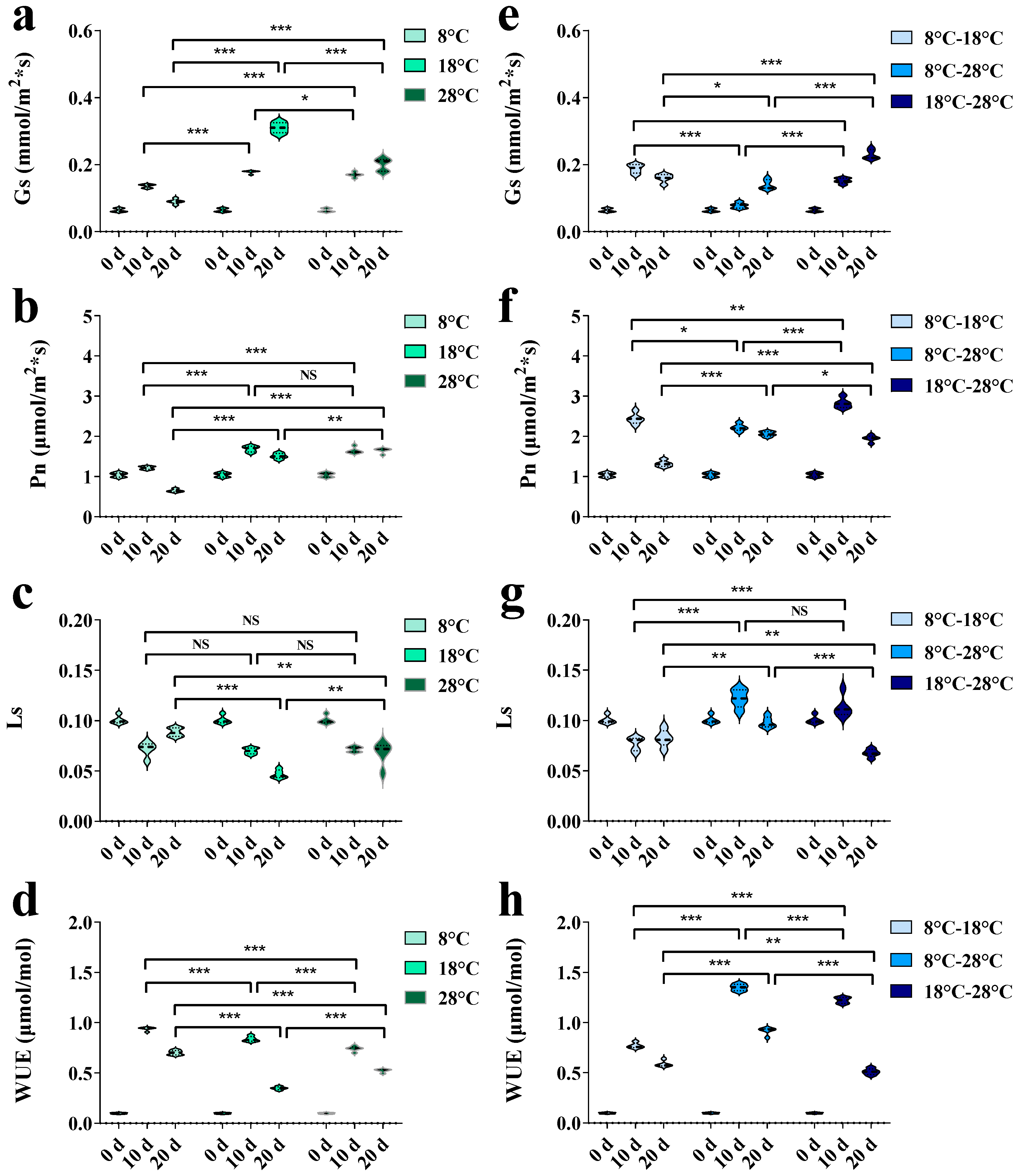
As represented in Figure 2, temperature regulation affected the photosynthetic indexes of P. grandiflorum. The values of stomatal conductance (Gs) significantly increased at a constant temperature of 18 °C and a variable temperature of 18–28 °C, reaching their high points after 20 days of regulation (p < 0.01) (Figure 2a,e). Compared to the unprocessed state, the net photosynthetic rate (Pn) values in each group first increased after 10 days of regulation and then decreased after 20 days. Among them, the low–constant temperature of 8 °C and the low–variable temperature of 8–18 °C were particularly significant (Figure 2b,f). Both the stomatal limitation (Ls) and water use efficiency (WUE) values were at relatively lower levels under constant temperature regulations of 18 °C and 28 °C (Figure 2c,d), indicating that P. grandiflorum can control water loss by reducing its stomatal opening in a high-temperature environment. For variable temperature regulation, the fluctuation trends of Ls and WUE values were also similar, reaching their lowest points when controlled at 18–28 °C for 20 days (Figure 2g,h). This indicates that temperature regulation might alter the original photosynthetic mode and physiological function of P. grandiflorum by affecting its photosynthetic rate, stomatal limitation value, and water use efficiency.
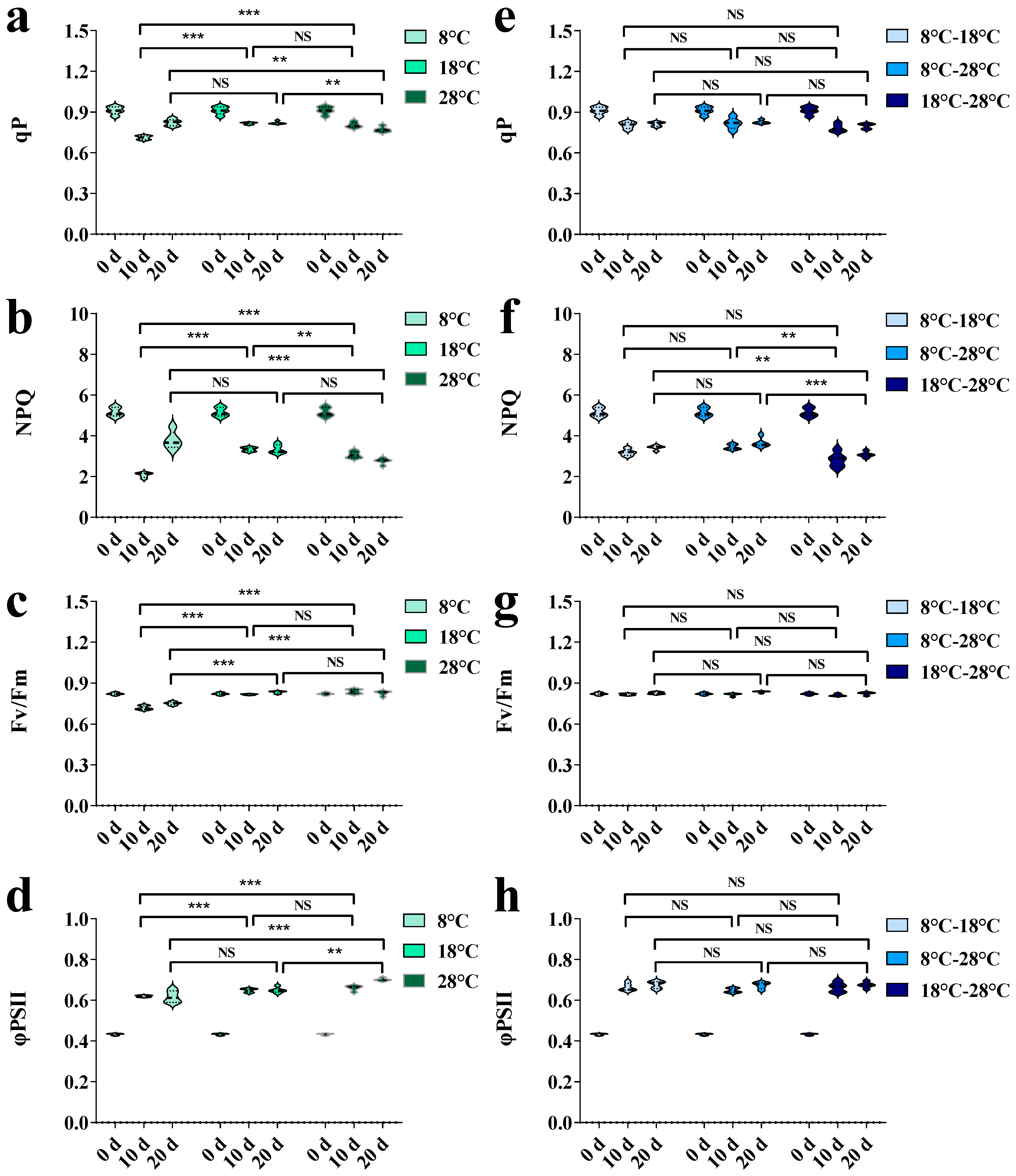
The chlorophyll fluorescence parameters of P. grandiflorum leaves under different temperature regulations for 10 and 20 days are exhibited in Figure 3. When the temperatures were regulated for 10 days, the values of the photochemical quenching coefficient (qP), non-photochemical quenching (NPQ), maximum photochemical efficiency of PSII (Fv/Fm), and actual photochemical efficiency (φPSII) in the 8 °C treatment group were obviously lower than those in the other constant treatment groups (Figure 3a–d). Gradually, all parameters increased with time, while the qP and NPQ reached their high points after 20 days of regulation (Figure 3a,b). Among the variable temperature regulations, three parameters varied little (Figure 3e,g,h), with only a slight fluctuation in the NPQ (Figure 3f). It can be inferred that low–constant temperatures had a negative impact on the chlorophyll fluorescence parameters of P. grandiflorum leaves, causing a certain degree of stress on the plants.
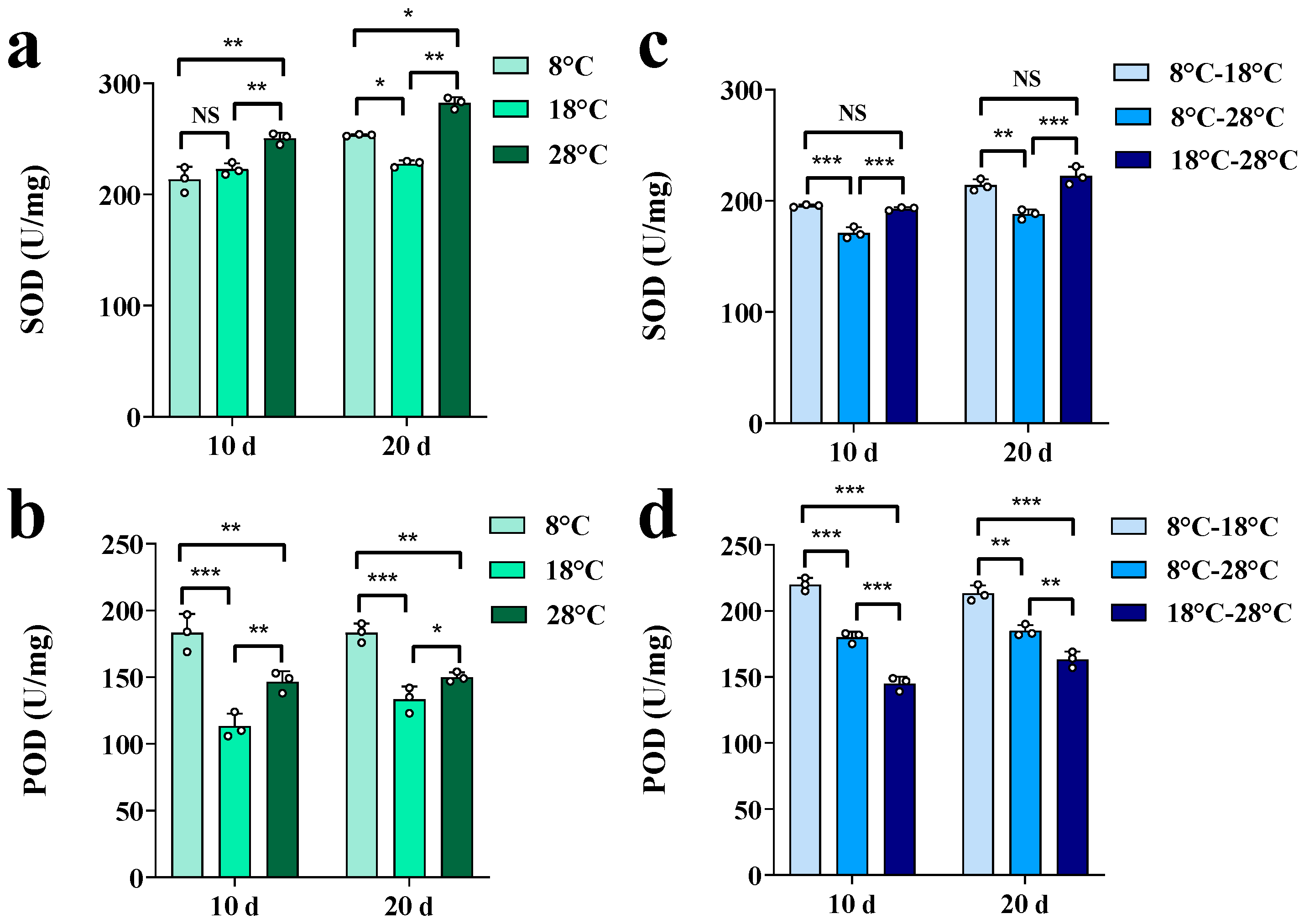
The influences of temperature regulation on osmotic substances and protective enzymes in P. grandiflorum roots over 10 and 20 days are depicted in Figure 4 and Figure 5. Under constant temperature conditions, the levels of soluble sugars (SS), soluble proteins (SP), proline (PRO), malondialdehyde (MDA), and peroxidase (POD) were significantly higher in the 8 °C treatment than in others with the same regulatory duration (Figure 4a–d and Figure 5b), while only the superoxide dismutase (SOD) value reached a high point at a constant temperature of 18 °C (Figure 5a).
Under variable temperature conditions, the levels of SS, SP, PRO, MDA, and POD were at the highest point within the same management duration in the regulation of 8–18 °C (Figure 4e–h and Figure 5d), and only the difference in SOD values between the treatment groups was not obvious (Figure 5c). In summary, low temperatures had significant impacts on the indexes of physiological resistance in P. grandiflorum roots, indicating that P. grandiflorum can resist the cell oxidative damage caused by lower temperatures by regulating osmotic substances and protecting enzyme systems.
The correlations between growth indexes (RFW, RDW, and AFW) and physiological stress indexes (photosynthesis characteristics, chlorophyll fluorescence parameters, osmoregulatory substances, and antioxidant enzymes) of P. grandiflorum are displayed in Figure 6.
The growth indexes of P. grandiflorum were strongly correlated with several indicators, e.g., chlorophyll fluorescence parameters and osmoregulatory substances. Specifically, RFW was strongly positively correlated with Fv/Fm (correlation coefficient r > 0.7), while RDW was highly positively associated with SP (r > 0.7). AFW had a close positive correlation with Pn and φPSII (r > 0.7), an extremely strong negative correlation with SS (r > 0.9, p < 0.05), and a high negative correlation with qP, NPQ, PRO, and MDA (r > 0.6). There were close positive correlations between Ls and WUE, qP and NPQ, and MDA and PRO (r > 0.9, p < 0.01), and close negative correlations between Ls and Gs, as well as MDA and Pn (r > 0.9, p < 0.01). These associations indicated that multiple physiological stress indexes might be involved in the accumulation of substances and energy, synergistically affecting the growth characteristics of P. grandiflorum.

3.2. Effects of Temperature Regulation on Key Enzyme Gene Expressions and Platycodin Contents

The expression levels of key enzyme genes involved in the platycodin synthesis pathway under different temperature regulations are represented in Figure 7. Compared to 10-day management, the expression levels of PgAACT, Pgβ-AS, and PgUGT4 were significantly upregulated after 20 days of constant temperature interventions, while the expression levels of PgMVK, PgPMK, PgMVD, PgSE, PgSS, and PgUGT2 were significantly downregulated. Similarly, compared to 10-day management, the expression level of PgUGT4 was notably upregulated after 20 days of variable temperature interventions, while the expression levels of PgMVK, PgPMK, PgMVD, PgSE, PgSS, and Pgβ-AS were obviously downregulated. However, the expression levels of PgAACT and PgUGT2 varied, but the fluctuation trends were inapparent. Overall, the biosynthesis processes of platycodins were greatly influenced by temperature regulation at the transcriptional level.
The HPLC chromatograms of the standard mixture and P. grandiflorum extract were profiled in Figure S1, and the calibration curves of four platycodins are listed in Table S1. Thereby, the contents of four platycodins after 10- and 20-day regulations were calculated precisely and displayed separately in Figure 8. The Pharmacopoeia of People’s Republic of China (2020 edition) stipulates that the content of platycodin D (PD) in P. grandiflorum roots shall not be less than 0.10% [27].
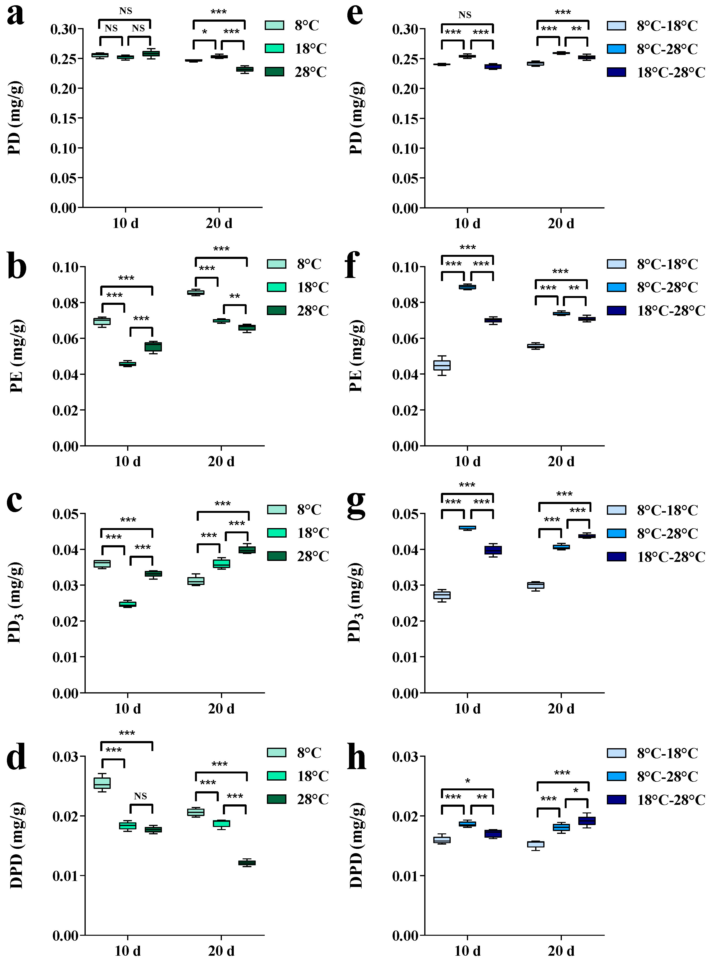
Although regulated by different temperatures, the PD contents in the tested samples varied little and were consistently above this criterion. It is noteworthy that the peaks of the PD contents were reached after the 20-day regulation at 18 °C and 8–18 °C (Figure 8a,e). In comparison to the PD, temperature regulation had a more apparent impact on the other monomeric platycodins, including platycoside E (PE), platycodin D3 (PD3), and deapioplatycodin D (DPD). Both the PE and PD3 contents were significantly increased at 18 °C and 28 °C, after 20-day constant temperature regulation, compared to 10-day management (Figure 8b,c). The DPD content reached a high point among the treatment groups at 8 °C (Figure 8d). Under variable temperature regulations, the contents of PD and PE were significantly higher than others in the 8–28 °C condition (Figure 8e,f), and the PD3 and DPD contents were also at high levels in the same condition (Figure 8g,h). Therefore, we inferred that when P. grandiflorum is kept at a low–constant temperature for an extended period, it has a negative impact on its growth, thereby generating more secondary metabolites for defense. On the other hand, the appropriate temperatures were also beneficial for the accumulation of secondary metabolites in P. grandiflorum, which might be due to the regulation of different temperatures affecting the accumulation of platycodins with respect to both the type and degree of structural modifications occurring during their synthesis process.
The correlations seen between the contents of platycodins and the expression levels of key enzyme genes involved in the platycodin biosynthesis pathway are characterized in Figure 9. Among them, there was an extremely strong negative association between PE and PgUGT4 (correlation coefficient r > 0.8). PD3 is highly negatively correlated with PgPMK, PgMVD, and PgSE (r > 0.6), while DPD is highly positively associated with PgMVK and PgMVD (r > 0.6). PD had a moderately positive correlation with PgMVK (r > 0.4) and a moderately negative association with Pgβ-AS and PgUGT2 (r > 0.4). There were strong positive correlations between PgAACT and PgMVD, PgAACT and PgSE, and PgPMK and PgSE (r > 0.9, p < 0.05), as well as an extremely strong negative association between PgMVD and PgSE (r > 0.8, p < 0.05). These relationships indicated that key enzyme genes are likely to participate in the biosynthesis of platycodins at the transcriptional level and ultimately affect the accumulation of platycodins.

4. Discussion

In general, temperature plays a decisive role in the phenological development and metabolic accumulation of medicinal plants without water and nutrient limitations [28]. Although certain plants have their own preferences for temperature, relatively higher temperatures will accelerate the growth of most plants within a normal range [29,30]. RFW and RDW are two important yield indicators for P. grandiflorum, while AFW can reflect the growth status of plants themselves. In this research, P. grandiflorum plants reached the end of their growth period after 20 days of regulation, while the aboveground parts grew and developed rapidly. The AFW levels within the temperature regulation range increased with the rising temperature, and reached their high points at a high–constant temperature of 28 °C and a high–variable temperature of 18–28 °C. However, there was an evident difference in the response of the roots and aboveground parts of P. grandiflorum to temperature regulation. The levels of RFW and RDW decreased with rising temperatures, reaching their peak values at a medium–constant temperature of 18 °C and a low–variable temperature of 8–18 °C. The results of this part corresponded to a previous study that found that the root biomass of P. grandiflorum reached its maximum at 15 °C, and the shoot biomass reached its maximum at 25 °C [29]. It was speculated that the physiological and ecological indexes of temperature-mediated photosynthesis affect the distribution of energy and substances in the plant organs [31,32]. Among the photosynthetic physiology indexes, Pn and Gs were always at high levels except at a low–constant temperature of 8 °C and a low–variable temperature of 8–18 °C after 20 days of management. This indicated that P. grandiflorum has a strong photosynthetic function at relatively high temperatures, which is conducive to biomass accumulation. Compared to 10-day management, qP and NPQ first decreased and then increased at a low–constant temperature of 8 °C after 20-day regulation. The reason for this was deduced to be that prolonged low temperatures lead to a decrease in the demand for adenosine 5′-triphosphate (ATP) and nicotinamide adenine dinucleotide phosphate (NADPH) in chloroplasts undergoing photosynthesis, resulting in excess light energy and NPQ dissipating the excess energy as heat [33,34]. Low–constant temperatures caused mild stress on P. grandiflorum in the early stages of regulation, but as the regulation period was extended, the plants exhibited some sort of adaptability. As a general rule, plants can use osmoregulatory substances to adapt to external changes [35], while responding to oxidative damage in the photosystem caused by increased ROS in the antioxidant enzyme system [36,37]. In this section, the levels of PRO, MDA, and POD were markedly higher in the low–constant and low–variable temperatures than in other regulation groups. However, there were various fluctuation trends for different physiological indexes of resistance, which could be due to the different peak times of each index [4]. Under low–temperature conditions, plant photosynthesis is usually inhibited, and osmoregulatory substances will increase to accommodate the low-temperature environment. Therefore, AFW had strong negative correlations with photosynthesis characteristics and osmoregulatory substances. Thus, different parts of P. grandiflorum plants had different responses to temperatures and to the mutual influences of physiological indicators. These synergistic effects maintained the balance in P. grandiflorum plants and increased the accumulation of the substances in each organ.
The bioactive components of medicinal plants are mostly secondary metabolites, which play a crucial role in responding to different external stimuli [1]. Numerous studies have demonstrated that temperature has a significant impact on the composition and content of secondary metabolites in medicinal plants [38,39]. Platycodins are the main active ingredients in P. grandiflorum roots and are also the key substances that determine the quality of its medicinal material [16]. The high points of PD contents came from the medium–constant temperature of 18 °C and the large-scale variable temperature of 8–28 °C. The contents of PE, PD3, and DPD were higher at a constant temperature of 8 °C and a variable temperature of 8–28 °C. Therefore, during the growth period, P. grandiflorum was more conducive to the accumulation of platycodins under medium–constant temperatures and large-scale variable temperatures, which have an average temperature of 18 °C. Variations in the secondary metabolites affected by temperature are achieved by upregulating or downregulating the expression levels of the corresponding genes in medicinal plants [40,41]. In the present work, temperature degree and regulation duration notably affected the key enzyme genes involved in the platycodin synthesis pathway, thereby regulating the platycodin contents. However, the alterations in platycodins did not completely match the changes in expression of key enzyme genes, which might be due to the limitation of substrate content for platycodin synthesis and the influence of platycodin transportation between different organs of P. grandiflorum [42]. Based on the association values observed between key enzyme genes and platycodins, it was found that PgPMK, PgMVD, and PgUGT-4 were highly correlated with platycodins. They are located at different positions in the biosynthetic pathway of platycodins and synergistically participate in platycodin synthesis at the transcriptional level, ultimately affecting the accumulation of platycodins. In light of the comprehensive physiological characteristics of plant growth and variations in platycodin content, we proposed that a medium–constant temperature of 18 °C and a large-scale variable temperature of 8–28 °C are more favorable for the growth period of P. grandiflorum; that is, a suitable temperature for the growth period of P. grandiflorum under natural conditions is an average temperature of 18 °C. The root growth and platycodin contents are at a high level, while not being subjected to temperature stress under these conditions.

5. Conclusions

In this research, the effects of temperature on plant growth and medicinal material quality were investigated by revealing the physiological indexes of P. grandiflorum and the responses of platycodin accumulation under different temperature regulations. This investigation proposed that constant or variable temperature regulations could significantly affect the yield and quality of P. grandiflorum during the growth period. The aboveground biomass accumulation of P. grandiflorum during the growth period significantly increased under high–constant and high–variable temperature regulations. The root fresh weight and root dry weight of P. grandiflorum were at their peak values when the plants grew at a low–variable temperature. Low temperatures can affect the photosynthetic physiological indexes, osmoregulatory substances, and antioxidant enzyme activities of P. grandiflorum, ultimately affecting its growth. The expressions of key enzyme genes were remarkably influenced by both the temperature degree and regulation duration. Under a medium–constant temperature of 18 °C and a large-scale variable temperature of 8–28 °C, i.e., an average temperature of 18 °C in natural conditions, the contents of the platycodins in the P. grandiflorum roots were at high points. The systematic evaluations of this work will be helpful to elucidate the physiological ecology of P. grandiflorum and the mechanisms of platycodin accumulation under different temperature regulations. This study has provided a theoretical basis and data support for the determination of suitable cultivation temperatures and the selection of high-quality production areas for P. grandiflorum, as well as guidelines for the quality formation of their medicinal materials under temperature regulation. To further improve the quality of P. grandiflorum, the combination of temperature regulation with corresponding nutrient intake could be explored as a future study direction.

Supplementary Materials

The following supporting information can be downloaded at: https://www.mdpi.com/article/10.3390/horticulturae10080848/s1, Figure S1: HPLC-ELSD chromatograms of the standard mixture (a) and P. grandiflorum extract (b); Figure S2: Field planting picture of P. grandiflorum before the management of temperature regulations; Table S1: Regression equations and linear ranges of the four platycodins; Table S2: Effect of temperature regulation on the differentially expressed genes of key enzymes in P. grandiflorum.

Author Contributions

Conceptualization, M.H. and L.Y.; Formal analysis, Z.W.; Funding acquisition, M.H. and L.Y.; Investigation, Z.W. and Y.Y.; Visualization, Z.W.; Writing—original draft, Z.W.; Writing—review and editing, M.H. All authors have read and agreed to the published version of the manuscript.

Funding

This research was funded by the Modern Agricultural Industrial Technology System Project (CARS-21).

Data Availability Statement

The datasets for this study are available in this manuscript. Further inquiries can be directed to the corresponding authors.

Acknowledgments

We would like to thank Meng Zhang and Ping Di for assisting in operating the experimental instruments.

Conflicts of Interest

The authors declare no conflicts of interest.

References

  1. Li, Y.; Kong, D.; Fu, Y.; Sussman, M.R.; Wu, H. The effect of developmental and environmental factors on secondary metabolites in medicinal plants. Plant Physiol. Biochem. 2020, 148, 80–89. [Google Scholar] [CrossRef] [PubMed]
  2. Luca, D.; Omid, A.; Giles, E.; John, H.; Graham, O. The cannabinoid profile and growth of hemp (Cannabis sativa L.) is influenced by tropical daylengths and temperatures, genotype and nitrogen nutrition. Ind. Crop Prod. 2022, 178, 114605. [Google Scholar]
  3. Torabi, B.; Adibnya, M.; Rahimi, A.; Azari, A. Modeling flowering response to temperature and photoperiod in safflower. Ind. Crop Prod. 2020, 151, 112474. [Google Scholar] [CrossRef]
  4. Zhang, T.; Gao, Y.; Han, M.; Yang, L. Changes in the physiological characteristics of Panax ginseng embryogenic calli and molecular mechanism of ginsenoside biosynthesis under cold stress. Planta 2021, 253, 79. [Google Scholar] [CrossRef]
  5. Allakhverdiev, S.; Kreslavski, V.; Klimov, V.; Los, D.; Carpentier, R.; Mohanty, P. Heat stress: An overview of molecular responses in photosynthesis. Photosynth. Res. 2008, 98, 541–550. [Google Scholar] [CrossRef]
  6. Gifford, R. Whole plant respiration and photosynthesis of wheat under increased CO2 concentration and temperature long-term vs short-term distinctions for modelling. Glob. Chang. Biol. 1995, 1, 385–396. [Google Scholar] [CrossRef]
  7. Ensminger, I.; Busch, F.; Huner, N. Photostasis and cold acclimation: Sensing low temperature through photosynthesis. Physiol. Plant. 2006, 126, 28–44. [Google Scholar] [CrossRef]
  8. Liu, Z.; Sun, W.; Zhao, Y.; Li, X.; Fang, Y.; Wu, J.; Zeng, X.; Yang, N.; Wang, Y.; He, L. Effects of low nocturnal temperature on photosynthetic characteristics and chloroplast ultrastructure of winter rapeseed. Russ. J. Plant Physiol. 2016, 4, 451–460. [Google Scholar] [CrossRef]
  9. Wise, R.; Olson, A.; Schrader, S.; Sharkey, T. Electron transport is the functional limitation of photosynthesis in field grown Pima cotton plants at high temperature. Plant Cell Environ. 2004, 27, 717–724. [Google Scholar] [CrossRef]
  10. Li, Y.; Liu, M.; Li, Y.; Liu, P.; Zhao, S.; Gao, H.; Zhang, Z. Photoprotection by mitochondrial alternative pathway is enhanced at heat but disabled at chilling. Plant J. 2020, 104, 403–415. [Google Scholar] [CrossRef]
  11. Mathur, S.; Agrawal, D.; Jajoo, A. Photosynthesis: Response to high temperature stress. J. Photochem. Photobiol. B Biol. 2014, 137, 116–126. [Google Scholar] [CrossRef]
  12. Salvucci, S.; Crafts, B. Inhibition of photosynthesis by heat stress: The activation state of Rubisco as a limiting factor in photosynthesis. Plant Physiol. 2004, 120, 179–186. [Google Scholar] [CrossRef] [PubMed]
  13. Blum, A. Osmotic adjustment is a prime drought stress adaptive engine in support of plant production. Plant Cell Environ. 2017, 40, 4–10. [Google Scholar] [CrossRef] [PubMed]
  14. Jebara, S.; Jebara, M.; Limam, F.; Aouani, M. Changes in ascorbate peroxidase, catalase, guaiacol peroxidase and superoxide dismutase activities in common bean (Phaseolus vulgaris) nodules under salt stress. J. Plant Physiol. 2005, 162, 929–936. [Google Scholar] [CrossRef]
  15. Blokhina, O.; Virolainen, E.; Fagerstedt, K.V. Antioxidants, oxidative damage and oxygen deprivation stress: A review. Ann. Bot. 2003, 91, 179–194. [Google Scholar] [CrossRef] [PubMed]
  16. Du, X.; Li, H.; Zhai, R.; Xu, K.; Fu, X. Phytochemical and pharmacological studies of natural saponins from Platycodon grandiflorum. Rec. Nat. Prod. 2023, 17, 963–985. [Google Scholar]
  17. Zhang, Y.; Sun, M.; He, Y.; Gao, W.; Wang, Y.; Yang, B.; Sun, Y.; Kuang, H. Polysaccharides from Platycodon grandiflorum: A review of their extraction, structures, modifications, and bioactivities. Int. J. Biol. Macromol. 2024, 271, 132617. [Google Scholar] [CrossRef]
  18. Zhang, S.; Chai, X.; Hou, G.; Zhao, F.; Meng, Q. Platycodon grandiflorum (Jacq.) A. DC. A review of phytochemistry, pharmacology, toxicology and traditional use. Phytomedicine 2022, 106, 154422. [Google Scholar] [CrossRef]
  19. Lichtenthaler, H. The 1-deoxy-D-xylulose-5-phosphate pathway of isoprenoid biosynthesis in plants. Annu. Rev. Plant Biol. 1999, 50, 47–65. [Google Scholar] [CrossRef]
  20. Mahmoud, S.; Croteau, R. Strategies for transgenic manipulation of monoterpene biosynthesis in plants. Trends Plant Sci. 2002, 7, 366–373. [Google Scholar] [CrossRef]
  21. Lange, B.; Rujan, T.; Martin, W.; Croteau, R. Isoprenoid biosynthesis: The evolution of two ancient and distinct pathways across genomes. Proc. Natl. Acad. Sci. USA 2000, 97, 13172–13177. [Google Scholar] [CrossRef] [PubMed]
  22. Lee, M.; Jeong, J.; Seo, J.; Shin, C.; Kim, Y.; In, J.; Yang, D.; Yi, J.; Choi, Y. Enhanced triterpene and phytosterol biosynthesis in Panax ginseng overexpressing squalene synthase gene. Plant Cell Physiol. 2004, 45, 976–984. [Google Scholar] [CrossRef] [PubMed]
  23. Vinocur, B.; Altman, A. Recent advances in engineering plant tolerance to abiotic stress: Achievements and limitations. Curr. Opin. Biotechnol. 2005, 16, 123–132. [Google Scholar] [CrossRef] [PubMed]
  24. Wang, S.; Liang, W.; Yao, L.; Wang, J.; Gao, W. Effect of temperature on morphology, ginsenosides biosynthesis, functional genes, and transcriptional factors expression in Panax ginseng adventitious roots. J. Food Biochem. 2019, 43, e12794. [Google Scholar] [CrossRef] [PubMed]
  25. Zhang, T.; Han, M.; Yang, L.; Han, Z.; Cheng, L.; Sun, Z.; Yang, L. The effects of environmental factors on ginsenoside biosynthetic enzyme gene expression and platycodin abundance. Molecules 2018, 24, 14. [Google Scholar] [CrossRef] [PubMed]
  26. Yu, H.; Li, J.; Chang, X.; Dong, N.; Chen, B.; Wang, J.; Zha, L.; Gui, S. Genome-wide identification and expression profiling of the WRKY gene family reveals abiotic stress response mechanisms in Platycodon grandiflorus. Int. J. Biol. Macromol. 2024, 257, 128617. [Google Scholar] [CrossRef] [PubMed]
  27. The State Commission of Pharmacopoeia. Pharmacopoeia of People’s Republic of China; Part I; The Medicine Science and Technology Press: Beijing, China, 2020; p. 289.
  28. Shi, Y.; Ding, Y.; Yang, S. Cold signal transduction and its interplay with phytohormones during cold acclimation. Plant Cell Physiol. 2015, 56, 7–15. [Google Scholar] [CrossRef] [PubMed]
  29. Kwon, S.; Kim, H.; Roy, S.; Boo, H.; Woo, S.; Kim, H. Effects of temperature, light intensity and DIF on growth characteristics in Platycodon grandiflorum. J. Crop Sci. Biotechnol. 2019, 22, 379–386. [Google Scholar] [CrossRef]
  30. Nguyen, T.; Lee, J.; Lee, G.; Cho, K.; Cho, D.; Son, K. Optimization of cultivation type and temperature for the production of balloon flower (Platycodon grandiflorum A. DC) sprouts in a plant factory with artificial lighting. Horticulturae 2022, 8, 315. [Google Scholar] [CrossRef]
  31. Schwenkert, S.; Fernie, A.; Geigenberger, P.; Leister, D.; Moehlmann, T.; Naranjo, B.; Neuhaus, H. Chloroplasts are key players to cope with light and temperature stress. Trends Plant Sci. 2022, 27, 577–587. [Google Scholar] [CrossRef]
  32. Kitashova, A.; Adler, S.; Richter, A.; Eberlein, S.; Dziubek, D.; Klipp, E.; Nägele, T. Limitation of sucrose biosynthesis shapes carbon partitioning during plant cold acclimation. Plant Cell Environ. 2022, 46, 464–478. [Google Scholar] [CrossRef] [PubMed]
  33. Ploschuk, E.; Bado, L.; Salinas, M.; Wassner, D.; Windauer, L.; Insausti, P. Photosynthesis and fluorescence responses of Jatropha curcas to chilling and freezing stress during early vegetative stage. Environ. Exp. Bot. 2014, 102, 18–26. [Google Scholar] [CrossRef]
  34. Ashrostaghi, T.; Aliniaeifard, S.; Shomali, A.; Azizinia, S.; Koohpalekani, J.; Moosavi-Nezhad, M.; Gruda, N. Light intensity: The role player in cucumber response to cold stress. Agronomy 2022, 12, 201. [Google Scholar] [CrossRef]
  35. Wu, S.; Zhang, J.; Ke, J.; Zhou, K.; Wang, J.; Dai, B.; Xue, S.; Zhou, Y.; Xie, Y.; Wang, Y. Evaluation of low-temperature adaptability of different of Lily varieties. Appl. Ecol. Environ. Res. 2022, 2101, 665679. [Google Scholar] [CrossRef]
  36. Meng, A.; Wen, D.; Zhang, C. Maize seed germination under low-temperature stress impacts seedling growth under normal temperature by modulating photosynthesis and antioxidant metabolism. Front. Plant Sci. 2022, 13, 843033. [Google Scholar] [CrossRef] [PubMed]
  37. Nadarajah, K. ROS homeostasis in abiotic stress tolerance in plants. Int. J. Mol. Sci. 2020, 21, 5208. [Google Scholar] [CrossRef] [PubMed]
  38. Goh, H.; Khairudin, K.; Sukiran, N.; Normah, M.; Baharum, S. Metabolite profiling reveals temperature effects on the VOCs and flavonoids of different plant populations. Plant Biol. 2020, 95, 197–203. [Google Scholar] [CrossRef]
  39. Palaniyandi, K.; Wu, J. Low temperature enhanced the podophyllotoxin accumulation vis-a-vis its biosynthetic pathway gene(s) expression in Dysosma versipellis (Hance) M. Cheng-A pharmaceutically important medicinal plant. Process Biochem. 2016, 18, 130–139. [Google Scholar] [CrossRef]
  40. Vashisth, D.; Kumar, R.; Rastogi, S.; Patel, V.; Kalra, A.; Gupta, M.; Gupta, A.; Shasany, A. Transcriptome changes induced by abiotic stresses in Artemisia annua. Sci. Rep. 2018, 8, 3423. [Google Scholar] [CrossRef]
  41. He, J.; Yao, L.; Pecoraro, L.; Liu, C.; Wang, J.; Huang, L.; Gao, W. Cold stress regulates accumulation of flavonoids and terpenoids in plants by phytohormone, transcription process, functional enzyme, and epigenetics. Crit. Rev. Biotechnol. 2022, 43, 680–697. [Google Scholar] [CrossRef]
  42. Di, P.; Sun, Z.; Cheng, L.; Han, M.; Yang, L.; Yang, L. LED light irradiations differentially affect the physiological characteristics, ginsenoside content, and expressions of ginsenoside biosynthetic pathway genes in Panax ginseng. Agriculture 2023, 13, 807. [Google Scholar] [CrossRef]
Figure 1. Effects of temperature regulation on the growth characteristics of P. grandiflorum. (ac) Histograms of root fresh weight (RFW), root dry weight (RDW), and aboveground fresh weight (AFW) values in three-level constant temperature regulations. (df) Histograms of RFW, RDW, and AFW values in three-level variable temperature regulations. ** p < 0.01, *** p < 0.001. NS, no statistical significance.
Horticulturae 10 00848 g001
Figure 2. Effects of temperature regulation on photosynthetic characteristics of P. grandiflorum leaves. (ad) Violin plots of stomatal conductance (Gs), net photosynthetic rate (Pn), stomatal limitation values (Ls), and water use efficiency (WUE) values in three-level constant temperature regulations. (eh) Violin plots of Gs, Pn, Ls, and WUE values in three-level variable temperature regulations. * p < 0.05, ** p < 0.01, *** p < 0.001. NS, no statistical significance.
Horticulturae 10 00848 g002
Figure 3. Effects of temperature regulation on chlorophyll fluorescence parameters of P. grandiflorum. (ad) Violin plots of photochemical quenching coefficient (qP), non-photochemical quenching (NPQ), maximum photochemical efficiency of PSII (Fv/Fm), and actual photochemical efficiency (φPSII) values in three-level constant temperature regulations. (eh) Violin plots of qP, NPQ, Fv/Fm, and φPSII values in three-level variable temperature regulations. ** p < 0.01, *** p < 0.001. NS, no statistical significance.
Horticulturae 10 00848 g003
Figure 4. Effects of temperature regulation on antioxidant enzymes and osmoregulatory substances of P. grandiflorum roots. (ad) Histograms of soluble sugars (SS), soluble proteins (SP), proline (PRO), and malondialdehyde (MDA) values in three-level constant temperature regulations. (eh) Histograms of SS, SP, PRO, and MDA values in three-level variable temperature regulations. * p < 0.05, ** p < 0.01, *** p < 0.001. NS, no statistical significance.
Horticulturae 10 00848 g004
Figure 5. Effects of temperature regulation on antioxidant enzymes and osmoregulatory substances of P. grandiflorum roots. (a,b) Histograms of superoxide dismutase (SOD), and peroxidase (POD) values in three-level constant temperature regulations. (c,d) Histograms of SOD and POD values in three-level variable temperature regulations. * p < 0.05, ** p < 0.01, *** p < 0.001. NS, no statistical significance.
Horticulturae 10 00848 g005
Figure 6. Correlation analysis between growth indexes and physiological stress indexes of P. grandiflorum. * p < 0.05, ** p < 0.01.
Horticulturae 10 00848 g006
Figure 7. Heat map of differentially expressed genes of key enzymes under constant and variable temperature regulations in the platycodin synthesis pathway of P. grandiflorum. In the data comparison between 10 d and 20 d, the significance symbols were marked on elements of 10 d. * p < 0.05, ** p < 0.01, *** p < 0.001.
Horticulturae 10 00848 g007
Figure 8. Effects of temperature regulation on platycodin content in P. grandiflorum (ad). Content of platycoside E (PE), platycodin D3 (PD3), deapioplatycodin D (DPD), and platycodin D (PD) under three-level constant temperature regulations (eh). Content of PE, PD3, DPD, and PD under three-level variable temperature regulations. * p < 0.05, ** p < 0.01, *** p < 0.001. NS, no statistical significance.
Horticulturae 10 00848 g008
Figure 9. Correlation analysis between platycodins and key enzyme genes involved in the platycodin synthesis pathway. * p < 0.05.
Horticulturae 10 00848 g009
Table 1. Primers sequences used in this research.
GenePrimer Sequence (5′−3′)
PgGAPDHF: CAGGGAGGCTTTTAGTTCAGGT
R: ATCACATCTACACCCCTCCAGC
PgAACTF: CCTCAATACCCCCAAGAGTGTC
R: AATGAAGCCTTTGCTGTCGTC
PgPMKF: ATCGTTGGCAGCCCTTCC
R: CCAGTCTTTGCTACTTCAGGCTT
PgMVKF: CCTTTAGCATCATCATTTCGCA
R: CCATACTCTAAAACTGTTGTTCGCTA
PgMVDF: ACATCTCCTTTGGATTTCTGCG
R: CTTCAGAGGCTGCTTTTTCACTT
PgSSF: CGGATGATTTCTACCCGTTGTT
R: CTGTTGAATAACGAGGGCGAAG
PgSEF: CACCACGACTTCTATCAACGGA
R: GAGATAGCCGCCTGGTTGTAG
Pgβ-ASF: GTTGGTCGTCTCCCACAATCAC
R: CCAGCAGTGACTCCCTAAACCA
PgUGT2F: AGAGCGTGTGGTGTGGGGT
R: CACCGTTCTGAAATCCCTCCTAT
PgUGT4F: CCCACAATGAGTCACGAATCC
R: TTCATTTGGGTAATAAGGAAAGTGA
Disclaimer/Publisher’s Note: The statements, opinions and data contained in all publications are solely those of the individual author(s) and contributor(s) and not of MDPI and/or the editor(s). MDPI and/or the editor(s) disclaim responsibility for any injury to people or property resulting from any ideas, methods, instructions or products referred to in the content.