Article

Unlocking the Potential of In Vitro Photoautotrophy for Eryngium foetidum: Biomass, Morphophysiology, and Acclimatization

by
Darlyara Reis Silva
1,
Marion Nayon Braga Soares
1,
Maria Cristina Rocha Silva
1,
Mariana Costa Lima
1,
Vitória Karla de Oliveira Silva-Moraes
2,
Givago Lopes Alves
2,
Anyela Marcela Ríos-Ríos
3,
Aldilene da Silva Lima
2,
Marcos Vinícius Marques Pinheiro
4,
Thais Roseli Corrêa
2,5,
Diego Silva Batista
5,6,
Fábio Afonso Mazzei Moura de Assis Figueiredo
5,
Fabrício de Oliveira Reis
2,
Tiago Massi Ferraz
2,5 and
Sérgio Heitor Sousa Felipe
5,*
1
Centro de Ciências Agrárias, Universidade Estadual do Maranhão, São Luís 65055-310, Brazil
2
Programa de Pós-Graduação em Agroecologia, Universidade Estadual do Maranhão, São Luís 65055-310, Brazil
3
Programa de Pós-Graduação em Biodiversidade e Biotecnologia da Amazônia Legal–BIONORTE, Universidade Estadual do Maranhão, São Luís 65055-310, Brazil
4
Universidade Regional Integrada do Alto Uruguai e das Missões, Campus Frederico Westphalen (URI-FW), Frederico Westphalen 98400-000, Brazil
5
Programa de Pós-Graduação em Agricultura e Ambiente, Universidade Estadual do Maranhão, São Luís 65055-310, Brazil
6
Departamento de Agricultura, Universidade Federal da Paraíba, Campus III, Bananeiras 58220-000, Brazil
*
Author to whom correspondence should be addressed.
Horticulturae 2024, 10(1), 107; https://doi.org/10.3390/horticulturae10010107
Submission received: 26 December 2023 / Revised: 17 January 2024 / Accepted: 17 January 2024 / Published: 22 January 2024
(This article belongs to the Special Issue Innovative Micropropagation of Horticultural and Medicinal Plants)

Abstract

:
Eryngium foetidum L., a biennial herb with diverse applications in food and traditional medicine, holds economic and pharmacological significance. Given its growing commercial interest, implementing biotechnological approaches like plant tissue culture is vital for sustainable propagation and metabolite production. In this study, we investigated the in vitro photoautotrophic potential of Eryngium foetidum, examining growth, chlorophyll a fluorescence, photosynthetic pigments, and anatomical features under sucrose concentrations (0 and 30 g L−1) and gas exchange rate (14 and 25 μL L−1 s−1 CO2). Acclimatization and survival rates of plants after ex vitro transfer were also assessed. Eryngium foetidum exhibited robust growth in both photoautotrophic and photomixotrophic conditions, with natural ventilation significantly enhancing plant development. Chlorophyll a fluorescence and photosynthetic performance were influenced by sucrose and gas exchange, highlighting the importance of these factors in plant micropropagation. Moreover, the species demonstrated remarkable plasticity during acclimatization, with high survival rates and rapid inflorescence development. The research provides valuable insights into optimizing in vitro cultivation conditions for Eryngium foetidum, emphasizing the potential for large-scale clonal propagation and exploring secondary metabolites. The observed phenotypic plasticity underscores the adaptability of the species to diverse environments. These biotechnological strategies open avenues for future studies, including the application of elicitors for enhanced secondary metabolite production.

Graphical Abstract

1. Introduction

Eryngium foetidum L. (Apiaceae family), commonly known as culantro, is a biennial herb with an erect growth habit, featuring a rosette of long spatulate spiny-toothed leaves and reaching a height of 10–50 cm after inflorescence development. It is commonly found in tropical and subtropical regions [1,2]. This species serves a dual purpose, functioning both as a food source (unconventional seasoning vegetable) and a traditional medicine in Latin America, the Central and Pacific Islands, Africa, and Asia [3]. The leaves of Eryngium foetidum are notably rich in phosphorus, potassium, and dietary micronutrients, making them suitable for potential use in food fortification [4,5,6]. In the realm of medicine, Eryngium foetidum is recognized for its high biological activity as an antioxidant. It is utilized in the treatment of diseases related to the gastrointestinal tract and exhibits antibacterial, analgesic, anti-inflammatory, anthelmintic, anticonvulsant, and anticancer properties [7,8,9].
The pharmacological importance attributed to Eryngium foetidum is particularly notable due to the various classes of compounds produced in its secondary metabolism, including aldehydes, carotenoids, phenols, and anthraquinones [7]. Furthermore, it is important to note that essential oils are found in the leaves, including molecules such as (2E)-2-dodecenal, trans-2-tetradecenal, 1-dodecanal, 1-decanal, tetradecanal, γ-terpinene, mesitylene, ρ-cymene, α-pinene, 1-undecanal, carotol, and 1-tetradecene [10]. Collectively, this phytochemical profile adds economic value to international trade and in the cosmetic, pharmaceutical, and food industries [7,8,9,10,11].
Due to the increasing commercial interest in Eryngium foetidum, it becomes essential to implement biotechnological approaches, such as plant tissue culture, for the rational propagation of the species. This is crucial to mitigate the risk of genetic erosion resulting from the disorderly exploitation of the population [12]. Furthermore, apart from being a biotechnological strategy, plant tissue culture offers the potential for controlled production of metabolites of interest, in contrast to field conditions. It also enables the exploration of complex biosynthetic pathways that have not been achievable artificially until now [13,14].
Plant tissue culture serves as a platform for plant cloning under aseptic conditions and environmental control in the laboratory [15]. Traditionally, in vitro multiplication relies on three nutritional types of cultures: heterotrophic, where there are no photosynthetically active organs, and the carbon source is solely the carbohydrate supplied in the medium; photomixotrophic, where photosynthetically active organs use the carbohydrate in the medium, along with the consumption of CO2 in the flask headspace; and photoautotrophic (or sugar-free medium), where photosynthetically active organs rely on CO2 in the flask headspace as the carbon source [16,17]. The latter two cultivation systems, photoautotrophic and photomixotrophic, promote in vitro plant growth compared to the first (heterotrophic), which is more suitable for callus cultivation [16].
While photoautotrophic cultivation requires the use of gas-permeable films to enhance gas exchange between the flask headspace and the external environment, these films can also be employed in photomixotrophic (sugar-provided medium) cultivation to improve the ventilation rate of the culture flask [17,18,19]. Investigating the impact of natural ventilation on both photoautotrophy and photomixotrophy in the in vitro cultivation of Eryngium foetidum plants may offer valuable insights into the morphophysiological characteristics influencing the growth and development of plants in these systems. This research can contribute new knowledge for implementing strategies applicable to clonal propagation on a large scale and in the in vitro germplasm banks.
Considering the information presented above, our objective was to assess the in vitro photoautotrophic potential of Eryngium foetidum. In this study, we initially characterized the growth and development of Eryngium foetidum plants under in vitro photomixotrophic conditions (without and with natural ventilation) and photoautotrophic systems. Subsequently, we examined the acclimatization of these plants. Our research provides novel insights into how Eryngium foetidum responds to photomixotrophy and photoautotrophy, demonstrating its adaptability to different carbon sources.

2. Materials and Methods

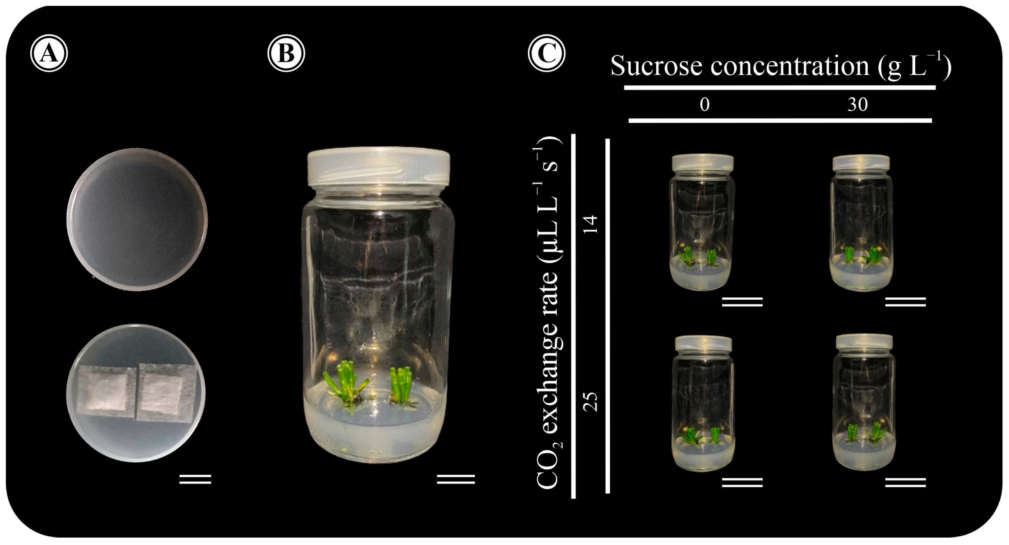
2.1. Plant Material and Experimental Design

The Eryngium foetidum plants from a natural population utilized in the experiments were sourced from the Germplasm Bank at the Tissue Culture Laboratory (LCT), State University of Maranhão, MA, Brazil (2°34′00″ S and 44°12′00″ W). These plants are regularly maintained in 350 mL transparent glass flasks, each containing 60 mL of Murashige and Skoog (MS) medium with vitamins [20] (PhytoTechnology®, Lenexa, KS, USA), 3% sucrose (w/v; Dinâmica® Química Contemporânea Ltd.a, São Paulo, SP, Brazil), and solidified with 5.5 g L−1 agar (Dinâmica® Química Contemporânea Ltd.a, São Paulo, SP, Brazil). The maintenance conditions include a temperature of 25 ± 2 °C, an irradiance of 85 μmol m−2 s−1 (provided by four tubular white LED lamps, T8, 9 W, Avant, São Paulo, SP, Brazil), and a 16 h photoperiod.
Explants (approximately 2.0–2.5 cm) were inoculated into 350 mL transparent glass flasks containing 60 mL of MS culture medium [20] (PhytoTechnology®, Lenexa, KS, USA), following the conditions employed in vitro cultivation in the Germplasm Bank, with sucrose concentrations of 0 and 30 g L−1. Two explants were inoculated per flask. We employed two flask-sealing systems, resulting in different gas exchange rates [21]: polypropylene lids without membranes (14 μL L−1 s−1 CO2 exchange rate) and polypropylene lids with two 10 mm holes covered by microporous tape membranes, as proposed by Saldanha et al. [18] (25 μL L−1 s−1 CO2 exchange rate) (Figure 1A).
The flasks were maintained for 45 days in a growth room under an irradiance of 85 μmol m−2 s−1, a 16 h photoperiod, and a temperature of 25 ± 2 °C (Figure 1B). The design was completely randomized in a 2 × 2 factorial [sucrose concentrations (0 and 30 g L−1) and gas exchange rate (14 and 25 μL L−1 s−1 CO2)], consisting of twelve replications for each treatment (Figure 1C). The experiments were repeated twice.

2.2. Chlorophyll a Fluorescence

Chlorophyll fluorescence measurements were conducted on the third fully expanded leaf below the apex. The following parameters: initial fluorescence (F0), maximum fluorescence (Fm), variable fluorescence (Fv), PSII maximum quantum yield (Fv/Fm), total number of active reaction centers per absorption (RC/ABS), maximum primary efficiency of PSII (Fv/F0), and performance index (PI) were assessed using a portable nonmodulated fluorimeter (Pocket PEA, Hansatech Instrument Ltd., King’s Lynn, Norfolk, UK). The instrument was calibrated to the leaf under dark conditions for 30 min using a leaf-clip system (Hansatech Instrument Ltd., King’s Lynn, Norfolk, UK). This calibration ensured the complete opening of reaction centers with minimal heat loss and the complete oxidation of QA [22].

2.3. Determination of Photosynthetic Pigments

Pigments were extracted from leaf discs (5 mm in diameter), which were obtained from the third fully expanded leaf below the apex. The leaf discs were immersed in 5 mL of dimethyl sulfoxide (DMSO; Isofar® Ltd.a, Duque de Caxias, RJ, Brazil) and left in the dark for 48 h [23]. Absorbance measurements of the samples at 665 nm, 649 nm, and 480 nm were taken using a UV-visible spectrophotometer (mono-beam) (model UV-M51; BEL Engineering Company, Monza, Italy) in a 10 mm quartz cuvette. The calculation of chlorophyll a, b, and carotenoids followed the method outlined by Wellburn [24].

2.4. Quantification of Photosynthetic Performance

Gas exchange and quantification of the in vitro photosynthetic rate were conducted following the procedures outlined by Castro et al. [25]. A CO2 gas analyzer (Model SBA-5; PP Systems International, Amesbury, MA, USA) and LoggerLite 1.9.3 software (Vernier Software & Technology Caliper, Beaverton, OR, USA) were used for these measurements. Gas exchange was determined by calculating the difference between the reference CO2 and the CO2 levels of plants exposed to atmospheric air. This calculation was based on the leaf area per plant (cm2).

2.5. Growth Analyzes

To analyze the growth and development of plants grown in vitro for 45 d, various parameters were assessed, including shoot length (cm), root length (cm), base diameter of the rosette (mm), leaf area (cm2), number of leaves, shoot dry mass (g), root dry mass (g), and shoot-to-root dry mass ratio. The leaf area was determined following the method outlined by Silva et al. [26]. To determine the dry mass, the samples were subjected to 60 °C in an oven with forced air circulation (AmbiKontrol, Arujá, SP, Brazil) until a constant weight was reached.

2.6. Leaf Anatomical Analysis

For an anatomical structural characterization of plants grown in vitro for 45 days, samples of the third pair of expanded leaves of the median region were fixed in FAA solution (formalin: acetic acid: 50% ethyl alcohol in 1:1:18 ratio) [27]. After fixation, the samples were dehydrated in increasing ethanolic series (10, 20, 30, 40, 50, 60, 70, 80, 85, 90, and 95%) and included in acrylic resin (Historesin; Leica Instruments, Jena, Germany). Cross-sections of 6 μm thickness were obtained on a semi-automated rotary microtome (model MRP2015, Lupetec Tecnologia Aplicada, São Carlos, SP, Brazil) and stained with toluidine blue (pH 3.2) [28]. Slides were mounted and images were captured using a light microscope (model B20T; Bioptika, Colombo, PR, Brazil) with the U-photo system, coupled to a digital photographic camera (model CMOS-5.0; Bioptika, Colombo, PR, Brazil) and computer with Capture V2.1 software.

2.7. Ex Vitro Acclimatization

The culture medium was removed from the root systems of 45-day-old Eryngium foetidum plants grown in vitro by gently rinsing them under running water. The plants of each in vitro treatment were then transplanted into 350 mL polystyrene pots (model KP-350 TR; Cristalcopo Descartaveis S/A, Içara, SC, Brazil) containing commercial substrate (Carolina soil®, Santa Cruz do Sul, RS, Brazil). The plants were grown in a greenhouse with an average temperature of 28.5 ± 2 °C; humidity relative of 80 ± 5%; natural light conditions with an intensity of ~1050 µmol photons m−2 s−1, and a 12 h photoperiod.

2.8. Statistical Analyzes

The data were statistically analyzed by two-way analysis of variance (ANOVA), with the means compared by Tukey’s test. All statistical analyses were performed with Genes software, version 1990.2018.39 [29].

3. Results

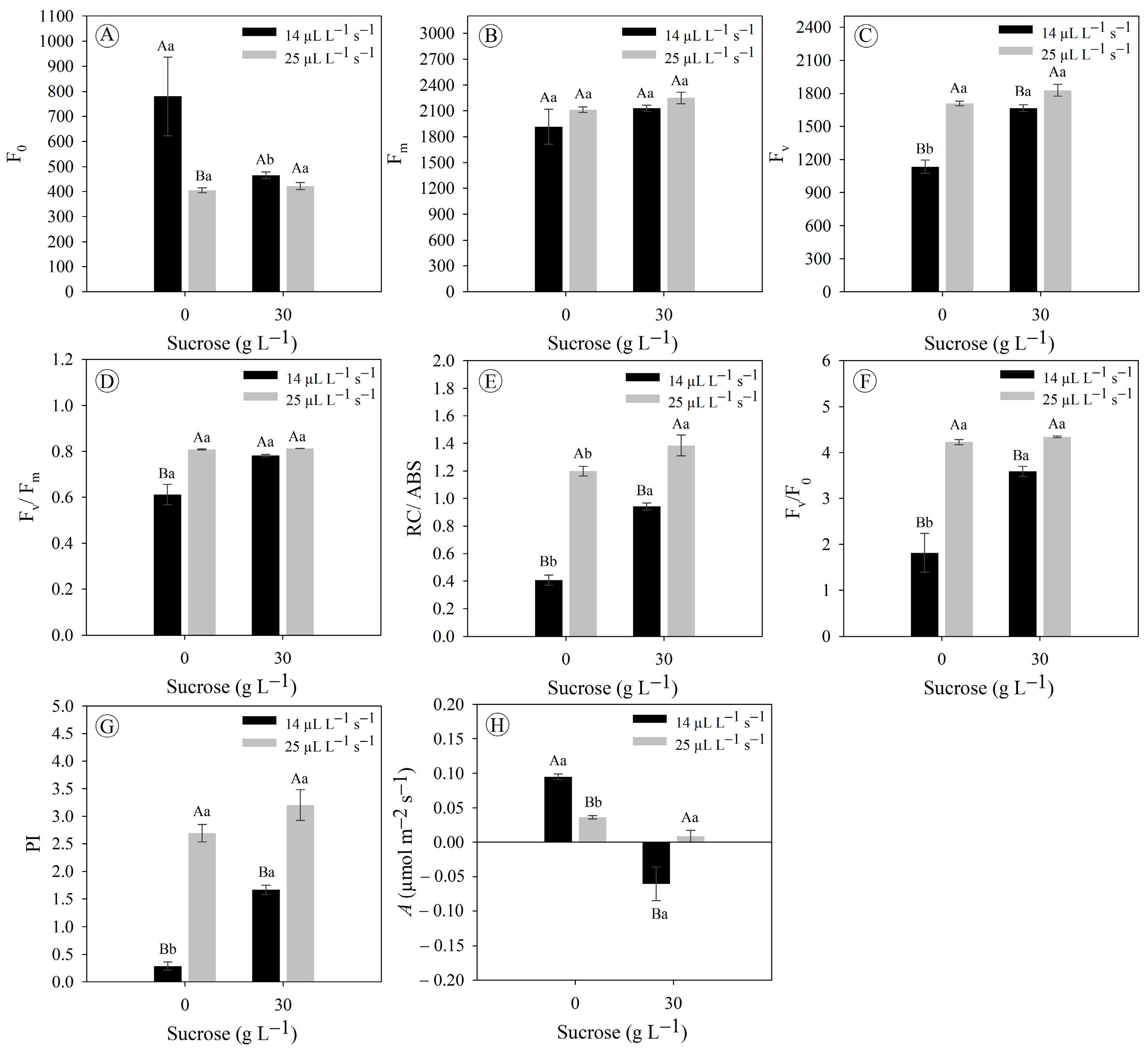
3.1. Gas Exchange Rates Improve Chlorophyll a Fluorescence and Photosynthetic Rate in Eryngium foetidum Grown In Vitro

Our results indicate that Eryngium foetidum plants exhibit phenotypic traits indicative of robust growth and development under photoautotrophic conditions (0 g L−1 sucrose and 25 μL L−1 s−1 CO2) and photomixotrophic conditions (30 g L−1 sucrose) both without (14 μL L−1 s−1 CO2; lower gas exchange rate) and with (25 μL L−1 s−1 CO2; higher gas exchange rate) natural ventilation. However, plants under 0 g L−1 sucrose and 14 μL L−1 s−1 CO2 were negatively impacted, likely due to the limiting condition of carbon availability—considered as the control treatment—(Figure 2).
Eryngium foetidum plants grown under 0 g L−1 sucrose and 14 μL L−1 s−1 CO2 displayed a significantly higher initial fluorescence compared to the other treatments (45% higher) (Figure 3A). In parallel, there were no significant differences in the maximum fluorescence for all treatments (ranged from 2112 to 2250) (Figure 3B). Plants under a higher gas exchange rate (25 μL L−1 s−1 CO2), regardless of sucrose concentration, displayed better results for variable fluorescence, total number of active reaction centers per absorption, maximum primary efficiency of PSII, and performance index (Figure 3C,E–G). Lastly, only plants under the limiting condition of in vitro cultivation (0 g L−1 sucrose and 14 μL L−1 s−1 CO2) exhibited an average of 0.61 for PS II maximum quantum yield (Figure 3D), which indicates that they are facing stress.
The photosynthetic rate showed that Eryngium foetidum plants in the absence of sucrose exhibited better performance compared to treatments with the presence of this carbohydrate in the culture medium (3–8 times in sucrose-free with 14 and 25 μL L−1 s−1 CO2) (Figure 3H). Notably, plants under cultivation with sucrose and low gas exchange (30 g L−1 sucrose and 25 μL L−1 s−1 CO2) did not show photosynthetic carbon assimilation when their gas exchange was analyzed (Figure 3H). Collectively, the data indicate that sucrose negatively affects photosynthetic performance, but is mitigated when using a higher gas exchange rate (25 μL L−1 s−1 CO2).

3.2. Sucrose Concentrations and Gas Exchange Rates Do Not Modulate Pigment Concentration in Eryngium foetidum Grown In Vitro

Eryngium foetidum plants grown under photoautotrophic (0 g L−1 sucrose and 25 μL L−1 s−1 CO2), photomixotrophic (0 g L−1 sucrose and 14 μL L−1 s−1 CO2), and photomixotrophic with ventilation natural (0 g L−1 sucrose and 25 μL L−1 s−1 CO2) did not differ statistically from each other for the chlorophyll a (ranged from 37 to 49 µg cm−2), total chlorophyll (ranged from 45 to 57 µg cm−2), carotenoids (ranged from 10 to 12 µg cm−2), and total chlorophyll/carotenoid ratio (~5 µg cm−2); but these three cultivation systems differed significantly for cultivation under 0 g L−1 sucrose and 14 μL L−1 s−1 CO2 (Figure 4A,D–F). Chlorophyll b concentrations and the chlorophyll a/b ratio did not show significant differences between all treatments, with averages of 8 and 6 µg cm−2, respectively (Figure 4B,C).

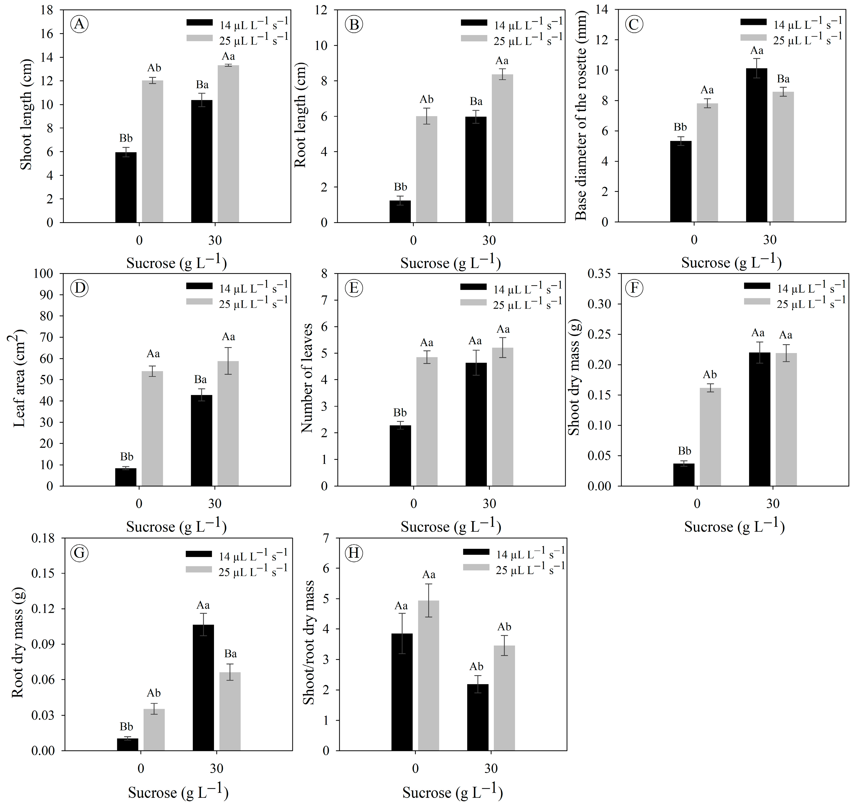
3.3. Sucrose Concentrations and Gas Exchange Rates Affect the In Vitro Growth and Development of Eryngium foetidum

Eryngium foetidum plants grown under a higher gas exchange rate (25 μL L−1 s−1 CO2) displayed an increase in shoot size of 74% and 11% under concentrations of 0 and 30 g L−1 sucrose, respectively, compared to the lower gas exchange rate (14 μL L−1 s−1 CO2) (Figure 5A). Furthermore, in this same condition of greater gas exchange, there was a significant increase in root length by 382% and 39% at concentrations of 0 and 30 g L−1 sucrose, respectively (Figure 5B). Plants under a lower gas exchange rate (14 μL L−1 s−1 CO2) displayed a larger mean rosette base diameter compared to the treatment with a higher gas exchange rate (25 μL L−1 s−1 CO2) under the concentration of 30 g L−1 sucrose (10% larger diameter); while at the concentration of 0 g L−1 sucrose, the opposite occurred, an expected result since this treatment (0 g L−1 and 14 μL L−1 s−1 CO2) is an extremely limiting cultivation condition for growth and plant development (Figure 5C).
Interestingly, plants grown in a photoautotrophic system (0 g L−1 sucrose and 25 μL L−1 s−1 CO2) displayed as good an increase in leaf area as the photomixotrophic with natural ventilation (30 g L−1 sucrose and 25 μL L−1 s−1 CO2; it ranged from 43 to 59 cm2), both treatments being significantly superior to the system with the lowest gas exchange rate (14 μL L−1 s−1 CO2) (Figure 5D). However, these differences in leaf area did not follow for the variable number of leaves, in which the plants under 0 g L−1 sucrose and 14 μL L−1 s−1 CO2 and the plants under 30 g L−1 sucrose and 14 or 25 μL L−1 s−1 CO2 displayed number of leaves with close values (~5 leaves) (Figure 5E).
Plants grown with sucrose supplementation displayed greater accumulation of dry mass in the shoot and root, independent of the gas exchange rate (Figure 5F,G). Concerning the shoot-to-root dry mass ratio, it was observed that in plants under 0 g L−1 sucrose, there was a greater carbon allocation in the shoot, while plants under 30 g L−1 sucrose displayed a more balanced carbon allocation between the shoot and root (Figure 5H).

3.4. Sucrose Supplementation and Gas Exchange Rates Affect Leaf Anatomy

Eryngium foetidum plants displayed the same leaf histological organization in all in vitro cultivation conditions accessed. The anatomy of the midrib cross-sections consisted of a uniseriate and amphistomatic epidermis, subepidermal collenchyma on the adaxial and abaxial surfaces; secretory canal and idioblasts distributed throughout the fundamental parenchyma; vascular bundles and fundamental parenchyma distributed throughout the central cavity (Figure 6). However, natural ventilation affected the histological differentiation of leaves compared to treatments with less gas exchange, leading to the formation of thicker and larger leaves (Figure 6).

3.5. Ex Vitro Acclimatization of Eryngium foetidum Grown under Sucrose Concentrations and Gas Exchange Rates

Plants showed interesting plasticity to face the acclimatization phase (Figure 7A). Eryngium foetidum plants grown under photoautotrophic (0 g L−1 sucrose and 25 μL L−1 s−1 CO2), photomixotrophic (30 g L−1 sucrose) without (14 μL L−1 s−1 CO2) and with (25 μL L−1 s−1 CO2) natural ventilation displayed 100% survival, while only the limiting (0 g L−1 sucrose and 14 μL L−1 s−1 CO2) displayed a low survival (~33%) (Figure 7B). An interesting fact was the development of inflorescences in plants grown under these photoautotrophy and photomixotrophy treatments (Figure 7C).

4. Discussion

Plants coordinate their growth and development with carbon availability and source in vitro [30,31,32]. Understanding how sucrose levels (carbon source provided in the cultivation medium) and natural ventilation (carbon source provided in the flask headspace) can affect plant micropropagation offers new perspectives for producing plants with improved morphophysiological characteristics [19]. Our results here demonstrate that Eryngium foetidum plants exhibit in vitro photoautotrophic potential. However, the addition of sucrose in the photomixotrophic cultivation system also increased the accumulation of plant dry mass.
It is worth highlighting that the Eryngium foetidum plants under limiting cultivation conditions (14 μL L−1 s−1 CO2 and 0 g L−1 sucrose) were drastically affected in their physiological entirety, reducing growth and affecting plant development. This response was expected, since there is a strong limitation in the supply of carbon to plants, both in the cultivation medium due to only organic components making carbon available in small quantities (e.g., vitamins and myoinositol), and in the headspace of the flask due to the use of rigid lids making it difficult for CO2 to diffuse [16,19]. Although this cultivation condition is an obstacle, the explants did not die, which reflects the ability of Eryngium foetidum to adapt to adverse environments. In contrast, the other three in vitro culture systems exemplified by photoautotrophic and photomixotrophic without and with natural ventilation provided better morphophysiological responses.
In the photomixotrophic system with a lower gas exchange rate (or without natural ventilation), the microenvironment of the flask is the factor with the greatest influence on the growth and development, since carbon is made available through the supply of sucrose in the medium [33,34,35]. Undeniably, sealing the bottles (rigid lids or without membranes) provides high humidity inside them, greater accumulation of the phytohormone ethylene, reduced gas exchange, and low CO2 concentration [36,37,38,39]. These factors lead to the development of plants with morphophysiological disorders, such as leaf formation displaying mesophyll with large intercellular spaces, poorly developed vascular system, lack of epicuticular wax formation, and non-functional stomata [16,18,19,40]. In this system, the accumulation of ethylene is a key factor that can strongly affect the growth and development of plants [36].
Ethylene affects several processes in plant growth and development, in which its synthesis increases as the concentration of its precursor increases, suggesting that 1-aminocyclopropane-1-carboxylic acid can act as a signaling molecule in plants [41]. Depending on the plant species and type of flask sealing, there may be a high accumulation of ethylene in the flask headspace, causing a lower photosynthetic rate due to inducing leaf senescence pathways and, consequently, lower plant growth [36,42,43]. In the current study, although some symptoms of leaf chlorosis were seen in plants under systems with a lower gas exchange rate (or 14 μL L−1 s−1 CO2), there was no leaf abscission; thus, we believe that ethylene may indeed harm plants, but not to the level of being decisive for lower photosynthetic performance. Probably, the sucrose supplied in the cultivation medium is the main inhibitor of photosynthesis in these systems without natural ventilation [19,44].
Sucrose supplementation in the culture medium affects carbon assimilation by photosynthesis based on the activity reduction in the ribulose-1,5-bisphosphate carboxylase/oxygenase (RuBisCO) enzyme [44] since active carboxylation sites of Rubisco are blocked with phosphorylated sugars [45]. To face these morphophysiological disorders, there is the photoautotrophic system, which improves the environment in the flask headspace in line with the elimination of the carbon source (carbohydrate; e.g., sucrose) [16,40,42]. Despite this, we cannot consider the sucrose supplemented in the cultivation medium to be completely deleterious. In our study, sucrose supplemented in the in vitro culture medium increased the absolute values of plant dry weight.
Previous studies with Solanum tuberosum plants grown under 3% sucrose estimated that CO2 assimilation contributed about 40% of the dry mass, while the other 60% came from sucrose absorbed from the culture medium [33]. This may be the explanation for the greater biomass of Eryngium foetidum plants in a photomixotrophic without and with natural ventilation compared to photoautotrophic, as, often, these plants grown in a photoautotrophic system may not be achieving maximum photosynthesis performance due to extrinsic factors such as quantity (irradiance) and quality (spectral) of light, in addition to the low concentration of CO2 in the headspace of the bottle. Based on this, CO2 enrichment—or forced ventilation—has been applied to plant tissue culture (see [30,31,32,46]).
Considering the chlorophyll a fluorescence and photosynthesis of Eryngium foetidum plants, it appears that the response of CO2 assimilation by photosynthesis was not likely associated with photochemical limitations within cultivation systems. Instead, it suggests potential obstacles in terms of biochemical reactions to CO2 fixation. The utilization of natural ventilation in the in vitro cultivation of Eryngium foetidum plants (both photoautotrophic and photomixotrophic with natural ventilation) significantly improved fluorescence aspects in correlation with the photosynthetic rate, compared to photomixotrophic cultivation without natural ventilation.
Photoautotrophic or photomixotrophic with natural ventilation systems also provided greater development of the leaf area of Eryngium foetidum, in addition to altering the cellular differentiation of the leaves. These results corroborate findings observed in other species, for example, Vernonia condensate Baker [19], Pfaffia glomerata (Spreng.) Pedersen [40], Aechmea blanchetiana (Baker) L.B.Sm. [47], Lippia dulcis Trev. [48], and Brassavola tuberculata Hook. [49], in which natural ventilation led to the formation of thicker and larger leaves. These response patterns are important in photosynthetic performance; however, even better, natural ventilation also leads to the formation of functional stomata even in vitro cultivation, capable of better controlling gas exchange and evapotranspiration during the acclimatization phase [16,42].
Eryngium foetidum plants grown in vitro showed excellent survival during post-culture acclimatization, indicating that not only the photoautotrophic system but also photomixotrophy without and with natural ventilation are effective micropropagation systems. An interesting fact was the rapid development of inflorescences in plants during acclimatization. We do not know for sure whether the cultivation conditions caused an effective stress capable of leading to this, or whether it could be related to the vegetative propagation that favored the rapid development of inflorescences in the photoautotrophic and photomixotrophic treatments without and with natural ventilation.
Our initial findings, based on biotechnological strategies, open avenues for future studies. These studies could explore the application of elicitors to develop methods promoting the exploration of secondary metabolites in the medicinal context of the species. The considerable phenotypic plasticity we observed in the in vitro cultivation this species points to emphasizes the potential for further advancements in this field. Furthermore, it is necessary to advance knowledge through studies that can evaluate different genotypes in light of the phenotypic plasticity of the species (see [50]).

5. Conclusions

Eryngium foetidum plants demonstrate in vitro photoautotrophic potential. Moreover, when supplemented with sucrose and provided with natural ventilation, there was a significant increase in plant biomass. The implications of this research extend to the mass propagation of this species, aligning with the exploration of secondary metabolites. This is particularly relevant in the phytochemical industry, especially concerning essential oils.

Author Contributions

Conceptualization, S.H.S.F. and M.V.M.P.; methodology, S.H.S.F. and D.R.S.; formal analysis, D.R.S., M.N.B.S., M.C.R.S., M.C.L., V.K.d.O.S.-M., G.L.A., A.M.R.-R. and A.d.S.L.; investigation, D.R.S., M.N.B.S., M.C.R.S., M.C.L., V.K.d.O.S.-M., G.L.A., A.M.R.-R. and A.d.S.L.; resources, T.R.C., F.A.M.M.d.A.F., F.d.O.R. and T.M.F.; data curation, S.H.S.F., D.R.S. and G.L.A.; writing—original draft preparation, S.H.S.F., D.S.B. and D.R.S.; writing—review and editing, S.H.S.F.; D.S.B.; M.V.M.P.; T.R.C., F.A.M.M.d.A.F., F.d.O.R. and T.M.F.; supervision, S.H.S.F. All authors have read and agreed to the published version of the manuscript.

Funding

Funding by the National Council for Scientific and Technological Development (CNPq, Brasília, DF, Brazil: Grant no. PQ 304214/2022-1 to Diego S Batista).

Data Availability Statement

The original contributions presented in the study are included in the article, further inquiries can be directed to the corresponding author.

Acknowledgments

The authors thank the Universidade Estadual do Maranhão (UEMA) and the Fundação de Amparo à Pesquisa e ao Desenvolvimento Científico e Tecnológico do Maranhão (FAPEMA), MA, Brazil.

Conflicts of Interest

The authors declare no conflict of interest.

References

  1. Castro, T.F.D.; Carneiro, W.F.; Reichel, T.; Fabem, S.L.; Machado, M.R.F.; Souza, K.K.C.; Resende, L.V.; Murgas, L.D.S. The toxicological effects of Eryngium foetidum extracts on zebrafish embryos and larvae depend on the type of extract, dose, and exposure time. Toxicol. Res. 2022, 11, 891–899. [Google Scholar] [CrossRef] [PubMed]
  2. Lucas, D.B.; Cardozo, A.L. Eryngium in Flora e Funga do Brasil. Jardim Botânico do Rio de Janeiro. Available online: https://floradobrasil.jbrj.gov.br/FB15529 (accessed on 5 December 2023).
  3. POWO. Plants of the World Online. Available online: https://powo.science.kew.org/ (accessed on 30 November 2023).
  4. Singh, S.; Singh, D.R.; Banu, S.; Salim, K.M. Determination of bioactives and antioxidant activity in Eryngium foetidum L.: A traditional culinary and medicinal herb. Proc. Natl. Acad. Sci. India Sect. B Biol. Sci. 2013, 83, 453–460. [Google Scholar] [CrossRef]
  5. Anju, T.; Rai, N.K.S.; Uthirchamkavu, I.; Sreedharan, S.; Ndhlala, A.R.; Singh, P.; Kumar, A. Analysis of nutritional and antioxidant potential of three traditional leafy vegetables for food security and human wellbeing. S. Afr. J. Bot. 2022, 145, 99–110. [Google Scholar] [CrossRef]
  6. Do Socorro Teixeira Costa Leitão, D.; Pereira Barbosa-Carvalho, A.P.; Campos de Siqueira, F.; Pontes s Sousa, R.; Santos Lopes, A.; Campos Chisté, R. Extracts of Eryngium foetidum Leaves from the Amazonia Were Efficient Scavengers of ROS and RNS. Antioxidants 2023, 12, 1112. [Google Scholar] [CrossRef]
  7. Rodrigues, T.L.M.; Silva, M.E.P.; Gurgel, E.S.C.; Oliveira, M.S.; Lucas, F.C.A. Eryngium foetidum L. (Apiaceae): A literature review of traditional uses, chemical composition, and pharmacological activities. Evid. Based Complement. Alternat Med. 2022, 14, 2896895. [Google Scholar] [CrossRef] [PubMed]
  8. Paw, M.; Gogoi, R.; Sarma, N.; Saikia, S.; Chanda, S.K.; Lekhak, H.; Lal, M. Anti-microbial, anti-oxidant, anti-diabetic study of leaf essential oil of Eryngium foetidum L. along with the chemical profiling collected from North East India. J. Essent. Oil-Bear. 2022, 26, 814–829. [Google Scholar] [CrossRef]
  9. Zhang, X.; Chen, J.; Zhou, S.; Zhao, H. Ethanol extract of Eryngium foetidum leaves induces mitochondrial associated apoptosis via ROS generation in human gastric cancer cells. Nutr. Cancer 2022, 74, 2996–3006. [Google Scholar] [CrossRef]
  10. Rodrigues, T.L.M. Desempenho Fisiológico e Perfil Químico do Óleo Essencial. Orientadora: Eloisa Helena de Aguiar Andrade. Masters’s Thesis, Universidade Federal Rural da Amazônia, Museu Paraense Emílio Goeldi, Belém, Brazil, 2019; 60p. Available online: http://repositorio.ufra.edu.br/jspui/handle/123456789/812 (accessed on 10 December 2023).
  11. Devi, P.B.; Deb, P.; Singh, H.B. Promotion, utilization, and commercial cultivation of local spices with special reference to eryngo (Eryngium foetidum L.) as a measure for livelihood improvement towards achieving the goal of sustainable development in the indo-burma biodiversity hotspot: A case study from manipur, North-East India. In Climate Change Adaptation, Risk Management and Sustainable Practices in the Himalay; Sharma, S., Kuniyal, J.C., Chand, P., Singh, P., Eds.; Springer International Publishing: Cham, Switzerland, 2023; pp. 253–266. [Google Scholar] [CrossRef]
  12. Rajamohan, S. Harbouring the potential of medicinal and aromatic plants of India: Novel biotechnological approach and extraction technologies. In Medicinal and Aromatic Plants of India Vol. 1. Medicinal and Aromatic Plants of the World, Vol. 8; Máthé, Á., Khan, I.A., Eds.; Springer International Publishing: Cham, Switzerland, 2022; pp. 323–339. [Google Scholar] [CrossRef]
  13. Karuppusamy, S. A review on trends in production of secondary metabolites from higher plants by in vitro tissue, organ and cell cultures. J. Med. Plants Res. 2009, 3, 1222–1239. [Google Scholar]
  14. Fazili, M.A.; Bashir, I.; Ahmad, M.; Yaqoob, U.; Geelani, S.N. In vitro strategies for the enhancement of secondary metabolite production in plants: A review. Bull. Natl. Res. Cent. 2022, 46, 35. [Google Scholar] [CrossRef]
  15. Nhut, D.T. General information: Some aspects of plant tissue culture. In Plant Tissue Culture: New Techniques and Application in Horticultural Species of Tropical Region; Nhut, D.T., Tung, H.T., Yeung, E.C.T., Eds.; Springer: Singapore, 2022; pp. 1–23. [Google Scholar] [CrossRef]
  16. Kozai, T.; Kubota, C. Concepts, definitions, ventilation methods, advantages and disadvantages. In Photoautotrophic (Sugar–Free Medium) Micropropagation as a New Micropropagation and Transplant Production System; Kozai, T., Afreen, F., Zobayed, S., Eds.; Springer: Dordrecht, The Netherlands, 2005; pp. 19–30. [Google Scholar] [CrossRef]
  17. Ramos, J.C.M.; Ribeiro, L.M.; Nunes, G.P.; Soares, J.S.; Sorgato, J.C. In vitro and ex vitro production of Schomburgkia crispa: Effect of flask sealing systems and different light sources. Rodriguesia 2023, 74, e01062022. [Google Scholar] [CrossRef]
  18. Saldanha, C.W.; Otoni, C.G.; Azevedo, J.L.F.; Dias, L.L.C.; Rêgo, M.M.; Otoni, W.C. A low-cost alternative membrane system that promotes growth in nodal cultures of Brazilian ginseng [Pfaffia glomerata (Spreng.) Pedersen]. Plant Cell Tiss. Organ. Cult. 2012, 110, 413–422. [Google Scholar] [CrossRef]
  19. Fortini, E.A.; Batista, D.S.; Mamedes-Rodrigues, T.C.; Felipe, S.H.S.; Correia, L.N.F.; Chagas, K.; Silva, P.O.; Rocha, D.I.; Otoni, W.C. Gas exchange rates and sucrose concentrations affect plant growth and production of flavonoids in Vernonia condensata grown in vitro. Plant Cell Tiss. Organ. Cult. 2021, 144, 593–605. [Google Scholar] [CrossRef]
  20. Murashige, T.; Skoog, F. A revised medium for rapid growth and bio assays with tobacco tissue cultures. Physiol. Plant 1962, 15, 473–497. [Google Scholar] [CrossRef]
  21. Batista, D.S.; Dias, L.L.C.; Rêgo, M.M.; Saldanha, C.W.; Otoni, W.C. Flask sealing on in vitro seed germination and morphogenesis of two types of ornamental pepper explants. Ciênc. Rural. 2017, 47, e20150245. [Google Scholar] [CrossRef]
  22. Bolhar-Nordenkampf, H.R.; Long, S.P.; Baker, N.R.; Oquist, G.; Schreiber, U.L.E.G.; Lechner, E.G. Chlorophyll fluorescence as a probe of the photosynthetic competence of leaves in the field: A review of current instrumentation. Funct. Ecol. 1989, 3, 497–514. [Google Scholar] [CrossRef]
  23. Santos, R.P.; Ferreira Da Cruz, A.C.; Iarema, L.; Kuki, K.N.; Campos Otoni, W. Protocolo para extração de pigmentos foliares em porta-enxertos de videira micropropagados. Rev. Ceres 2008, 55, 356–364. [Google Scholar]
  24. Wellburn, A.R. The spectral determination of chlorophylls a and b, as well as total carotenoids, using various solvents with spectrophotometers of different resolution. J. Plant Physiol. 1994, 144, 307–313. [Google Scholar] [CrossRef]
  25. Castro, K.M.; Batista, D.S.; Fortini, E.A.; Silva, T.D.; Felipe, S.H.S.; Fernandes, A.M.; Sousa, R.M.J.; Nascimento, L.S.Q.; Campos, V.R.; Grazul, R.M.; et al. Photoperiod modulates growth, morphoanatomy, and linalool content in Lippia alba L. (Verbenaceae) cultured in vitro. Plant Cell Tiss. Organ. Cult. 2019, 139, 139–153. [Google Scholar] [CrossRef]
  26. Silva, T.D.; Batista, D.S.; Fortini, E.A.; Castro, K.M.; Felipe, S.H.S.; Fernandes, A.M.; Sousa, R.M.J.; Chagas, K.; Silva, J.V.S.; Correia, L.N.F.; et al. Blue and red light affects morphogenesis and 20-hydroxyecdisone content of in vitro Pfaffia glomerata accessions. J. Photochem. Photobiol. B Biol. 2020, 203, 111761. [Google Scholar] [CrossRef]
  27. Johansen, D.A. Plant Microtechnique; Mac Graw-Hill Book Co., Inc.: New York, NY, USA, 1940; 523p. [Google Scholar]
  28. O’Brien, T.P.; McCully, M.E. The Study of Plant Structure: Principles and Selected Methods; Termarcarphi Pty., Ltd.: Melbourne, Australia, 1981. [Google Scholar]
  29. Cruz, C.D. Genes: A software package for analysis in experimental statistics and quantitative genetics. Acta Sci. Agron. 2013, 35, 271–276. [Google Scholar] [CrossRef]
  30. Ferreira, P.R.B.; Cruz, A.C.F.; Batista, D.S.; Nery, L.A.; Andrade, I.G.; Rocha, D.I.; Felipe, S.H.S.; Koehler, A.D.; Nunes-Nesi, A. CO2 enrichment and supporting material impact the primary metabolism and 20-hydroxyecdysone levels in Brazilian ginseng grown under photoautotrophy. Plant Cell Tiss. Organ. Cult. 2019, 139, 77–89. [Google Scholar] [CrossRef]
  31. Louback, E.; Batista, D.S.; Pereira, T.A.R.; Mamedes-Rodrigues, T.C.; Silva, T.D.; Felipe, S.H.S.; Rocha, D.I.; Steinmacher, D.A.; Otoni, W.C. CO2 enrichment leads to altered cell wall composition in plants of Pfaffia glomerata (Spreng.) Pedersen (Amaranthaceae). Plant Cell Tiss. Organ. Cult. 2021, 145, 603–613. [Google Scholar] [CrossRef]
  32. Luis, S.J.; Jabín, B.J. CO2–enriched air in a temporary immersion system induces photomixotrophism during in vitro multiplication in vanilla. Plant Cell Tiss. Organ. Cult. 2023, 155, 29–39. [Google Scholar] [CrossRef]
  33. Wolf, S.; Kalman-Rotem, N.; Yakir, D.; Zrv, M. Autotrophic and heterotrophic carbon assimilation of in vitro grown potato (Solanum tuberosum L) plants. J. Plant Physiol. 1998, 153, 574–580. [Google Scholar] [CrossRef]
  34. Shin, K.S.; Park, S.Y.; Paek, K.Y. Sugar metabolism, photosynthesis, and growth of in vitro plantlets of Doritaenopsis under controlled microenvironmental conditions. In Vitro Cell Dev. Biol. Plant 2013, 49, 445–454. [Google Scholar] [CrossRef]
  35. Ribeiro, I.S.; Ribeiro, L.M.; Soares, J.S.; Ramos, J.C.M.; Sorgato, J.C. Light condition, flask sealing, and cultivation time on the germination and initial in vitro development of Dendrobium nobile Lindl. Ornam. Hortic. 2022, 28, 407–413. [Google Scholar] [CrossRef]
  36. Vinh, B.V.T.; Tung, H.T.; Mai, N.T.N.; Khai, H.D.; Luan, V.Q.; Phong, T.H.; Phong, T.N.P.; Nhut, D.T. Enhanced efficient micropropagation and reduced abnormal phenomena in Phyllanthus amarus plantlets cultured on medium containing silver nanoparticles. S. Afr. J. Bot. 2023, 163, 217–225. [Google Scholar] [CrossRef]
  37. Askari, N.; Aliniaeifard, S.; Visser, R.G.F. Low CO2 Levels are detrimental for in vitro plantlets through disturbance of photosynthetic functionality and accumulation of reactive oxygen species. Horticulturae 2022, 8, 44. [Google Scholar] [CrossRef]
  38. Silva, L.M.; Carvalho, V.S.; Generoso, A.L.; Miranda, D.P.; Costa Júnior, O.D.; Simioni, P.F.; Santana, D.B.; Cunha, M.; Oliveira, J.G.; Viana, A.P. Micropropagation of interspecific hybrids of Vitis spp. in microenvironments with different gas exchanges. Sci. Hortic. 2022, 305, 111413. [Google Scholar] [CrossRef]
  39. Carrari-Santos, R.; Vettorazzi, R.G.; Pinto, V.B.; Pinto, V.B.; Sena, E.O.A.; Oliveira, J.G.; Campostrini, E.; Silveira, V.; Santa-Catarina, C. Microporous membrane and culture medium affect in vitro seedling development of Dalbergia nigra (Vell.) Ex Benth. (Fabaceae) by modulation of the protein profile and accumulation of ethylene and CO2. Plant Cell Tiss. Organ. Cult. 2023, 153, 559–576. [Google Scholar] [CrossRef]
  40. Iarema, L.; Cruz, A.C.F.; Saldanha, C.W.; Dias, L.L.C.; Vieira, R.F.; Oliveira, E.J.; Otoni, W.C. Photoautotrophic propagation of Brazilian ginseng [Pfaffia glomerata (Spreng.) Pedersen]. Plant Cell Tiss. Organ. Cult. 2012, 110, 227–238. [Google Scholar] [CrossRef]
  41. Wang, K.L.C.; Li, H.; Ecker, J.R. Ethylene biosynthesis and signaling networks. Plant Cell 2002, 14 (Suppl. S1), S131–S151. [Google Scholar] [CrossRef] [PubMed]
  42. Kubota, C. Concepts and background of photoautotrophic micropropagation. In Molecular Breeding of Woody Plants; Morohoshi, N., Komamine, A., Eds.; Elsevier: Amsterdam, The Netherlands, 2001; pp. 325–334. [Google Scholar]
  43. Cournac, L.; Dimon, B.; Carrier, P.; Lohou, A.; Chagvardieff, P. Growth and photosynthetic characteristics of Solanum tuberosum plantlets cultivated in vitro in different conditions of aeration, sucrose supply, and CO2 enrichment. Plant Physiol. 1991, 97, 112–117. [Google Scholar] [CrossRef]
  44. Desjardins, Y.; Hdider, C.; Riek, J. Carbon nutrition in vitro—Regulation and manipulation of carbon assimilation in micropropagated systems. In Automation and Environmental Control in Plant Tissue Culture; Aitken-Christie, J., Kozai, T., Smith, M.A.L., Eds.; Springer: Dordrecht, The Netherlands, 1995; pp. 441–471. [Google Scholar] [CrossRef]
  45. Mayak, S.; Tirosh, T.; Ilan, A.; Duvdevani, A.; Khayat, E. Growth and development of pineapple (Ananas comosus L.) plantlets cultured in vitro at enriched and ambient CO2 environments. In International Symposium on Biotechnology of Tropical and Subtropical Species Part 2; ISHS: Brisbane, Australia, 1998; Volume 461, pp. 225–230. [Google Scholar] [CrossRef]
  46. Marques, I.; Fernandes, I.; Paulo, O.S.; Lidon, F.C.; DaMatta, F.M.; Ramalho, J.C.; Ribeiro-Barros, A.I. A transcriptomic approach to understanding the combined impacts of supra-optimal temperatures and CO2 revealed different responses in the polyploid Coffea arabica and its diploid progenitor C. canephora. Int. J. Mol. Sci. 2021, 22, 3125. [Google Scholar] [CrossRef] [PubMed]
  47. Martins, J.P.R.; Almeida-Rodrigues, L.C.; Santos, E.R.; Gontijo, A.B.P.L.; Falqueto, A.R. Impacts of photoautotrophic, photomixotrophic, and heterotrophic conditions on the anatomy and photosystem II of in vitro-propagated Aechmea blanchetiana (Baker) L.B. Sm. (Bromeliaceae). In Vitro Cell. Dev. Biol. Plant 2020, 56, 350–361. [Google Scholar] [CrossRef]
  48. Rocha, T.T.; Araújo, D.X.; Silva, A.M.; Oliveira, J.P.V.; Carvalho, A.A.; Gavilanes, M.L.; Bertolucci, S.K.V.; Alves, E.A.; Pinto, J.E.B.P. Morphoanatomy and changes in antioxidant defense associated with the natural ventilation system of micropropagated Lippia dulcis plantlets. Plant Cell Tiss. Organ. Cult. 2022, 151, 467–481. [Google Scholar] [CrossRef]
  49. Soares, J.S.; Ramos, J.C.M.; Sorgato, J.C.; Ribeiro, L.M.; Reis, L.C. Brassavola tuberculata Hook.: In vitro growth and ex vitro establishment as a function of the micropropagation system and sucrose. Braz. J. Biol. 2023, 83, e270892. [Google Scholar] [CrossRef]
  50. Gonçalves, V.P.; Andrade, F.L.N.; Gomes, R.F.; Santos, L.S. Genetic diversity in creole genotypes of Amazon chicory. Acta Amazon. 2022, 52, 89–95. [Google Scholar] [CrossRef]
Figure 1. Schematic representation of the experimental design. (A) Polypropylene lids without a membrane (14 μL L−1 s−1 CO2 exchange rate) and with two membranes (25 μL L−1 s−1 CO2 exchange rate). (B) Explants of Eryngium foetidum at the beginning of in vitro culture. (C) Experimental design. Further details can be found in the Materials and Methods section. Scale bars: (A) = 1 cm, (B) = 2 cm, and (C) = 5 cm.
Horticulturae 10 00107 g001
Figure 2. Forty-five-day-old Eryngium foetidum plants under sucrose concentrations (0 and 30 g L−1) and gas exchange levels (14 and 25 μL L−1 s−1 CO2 exchange rates). Scale bars: 2 cm.
Horticulturae 10 00107 g002
Figure 3. Chlorophyll fluorescence parameters and photosynthetic rate in Eryngium foetidum after 45 days of in vitro cultivation under sucrose concentrations (0 and 30 g L−1) and gas exchange levels (14 and 25 μL L−1 s−1 CO2 exchange rates). (A) initial fluorescence; (B) maximum fluorescence; (C) variable fluorescence; (D) PS II maximum quantum yield; (E) total number of active reaction centers per absorption; (F) maximum primary efficiency of PSII; (G) performance index; and (H) photosynthetic rate. Uppercase letters compare the means of plants under different CO2 exchange rates at the same sucrose concentration, while lowercase letters compare plants under the same CO2 exchange rate at different sucrose concentrations (Tukey’s test; p ≤ 0.05). The bars represent the standard error (n = 5).
Horticulturae 10 00107 g003
Figure 4. Concentration of photosynthetic pigments in Eryngium foetidum after 45 days of in vitro cultivation under sucrose concentrations (0 and 30 g L−1) and gas exchange levels (14 and 25 μL L−1 s−1 CO2 exchange rates). (A) chlorophyll a; (B) chlorophyll b; (C) ratio of chlorophyll a to chlorophyll b; (D) total chlorophyll; (E) carotenoids; and (F) ratio of total chlorophyll to carotenoids. Uppercase letters compare the means of plants under different CO2 exchange rates at the same sucrose concentration, while lowercase letters compare plants under the same CO2 exchange rate at different sucrose concentrations (Tukey’s test; p ≤ 0.05). The bars represent the standard error (n = 5).
Horticulturae 10 00107 g004
Figure 5. Growth parameters of Eryngium foetidum after 45 days of in vitro cultivation under sucrose concentrations (0 and 30 g L−1) and gas exchange levels (14 and 25 μL L−1 s−1 CO2 exchange rate). (A) shoot length; (B) root length; (C) base diameter of the rosette; (D) leaf area; (E) number of leaves; (F) shoot dry mass; (G) root dry mass; and (H) shoot-to-root dry mass ratio. Uppercase letters compare the means of plants under different CO2 exchange rates at the same sucrose concentration, while lowercase letters compare plants under the same CO2 exchange rate at different sucrose concentrations (Tukey’s test; p ≤ 0.05). The bars represent the standard error (n = 7).
Horticulturae 10 00107 g005
Figure 6. Leaf midrib cross-sections of Eryngium foetidum after 45 days of in vitro cultivation under sucrose concentrations (0 and 30 g L−1) and gas exchange levels (14 and 25 μL L−1 s−1 CO2 exchange rate). Ab: abaxial epidermis; Ad: adaxial epidermis; Cc: central cavity; Co: collenchyma; Ep: epidermis; Gp: ground parenchyma; Id: idioblast; Sc: secretory canal; and Vb: vascular bundle. Scale bars: 300 μm.
Horticulturae 10 00107 g006
Figure 7. Acclimatization of Eryngium foetidum plants after 45 days of in vitro cultivation under sucrose concentrations (0 and 30 g L−1) and gas exchange levels (14 and 25 μL L−1 s−1 CO2 exchange rate). (A) Sixty-five-day-old Eryngium foetidum plants during acclimatization; (B) Survival after 60 days of ex vitro cultivation; and (C) Inflorescence development after 60 days of ex vitro cultivation. Uppercase letters compare the means of plants under different CO2 exchange rates at the same sucrose concentration, while lowercase letters compare plants under the same CO2 exchange rate at different sucrose concentrations (Tukey’s test; p ≤ 0.05). Scale bars: (A) = 5 cm and (B,C) = the bars represent the standard error (n = 6).
Horticulturae 10 00107 g007
Disclaimer/Publisher’s Note: The statements, opinions and data contained in all publications are solely those of the individual author(s) and contributor(s) and not of MDPI and/or the editor(s). MDPI and/or the editor(s) disclaim responsibility for any injury to people or property resulting from any ideas, methods, instructions or products referred to in the content.

