Article

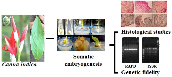
Somatic Embryogenesis and Genetic Fidelity Study of the Micropropagated Medicinal Species, Canna indica

1
Molecular Cytogenetics Laboratory, Department of Botany, University of North Bengal, Siliguri 734013, West Bengal, India
2
Bamboo Technology, Department of Biotechnology, Bodoland University, Kokrajhar 783370, Assam, India
*
Author to whom correspondence should be addressed.
Horticulturae 2015, 1(1), 3-13; https://doi.org/10.3390/horticulturae1010003
Submission received: 23 February 2015 / Revised: 28 April 2015 / Accepted: 30 April 2015 / Published: 8 May 2015

Abstract

:
Canna indica Linn. (Cannaceae) is used both as medicine and food. Traditionally, various parts of C. indica are exploited to treat blood pressure, dropsy, fever, inflammatory diseases etc. However, to date there is no reliable micropropagation protocol for C. indica. We present here a regeneration technique C. indica with banana micropropagation medium (BM). BM supplemented with 3% sucrose, 0.7% agar, and 0.17% NH4NO3 and different plant growth regulators like BAP (2 mg∙L−1) and NAA (0.5 mg∙L−1) was found to be effective in inducing callus. BM with BAP (2 mg∙L−1) was ideal for somatic embryogenesis and plantlet regeneration. After a period of 3 months, regenerated plantlets were successfully transferred to the field conditions. Appearance of somaclonal variation among the regenerated plants is a common problem which was assessed by DNA fingerprinting. To detect genetic fidelity in C. indica, RAPD and ISSR markers were employed. Ten RAPD primers produced 60 amplicons, while 7 ISSR primers generated 45 bands in both in vitro plantlets and mother plants. RAPD and ISSR analyses showed no evidence of polymorphism between parent plants and the regenerated plants as all the amplified products were found to be monomorphic.

Graphical Abstract

1. Introduction

Canna, the solitary genus of the family Cannaceae, is a well-known ornamental plant with beautiful flowers. The plant is cultivated extensively throughout the world for its decorative and widely-varying flower colors [1]. In addition, species of Canna are also edible [2] and have medicinal properties [3,4,5,6]. The common practice for Canna propagation is asexual, mainly through multiplication of rhizomes. However, this poses the problem of genetic stagnancy so genetic variation is limited. Varietal improvement of Canna lies in genetic manipulation and protoplast fusion [7]. In both the cases a preliminary requirement is a robust micropropagation protocol. Only two groups have attempted this. Sakai and Imai [8] regenerated Canna edulis Ker Gawler of callus culture but with limited success. Hosoki and Sasaki [9] produced C. edulis with the longitudinal shoot split method. However, no attempts to regenerate has been made to C. indica or any other Indian varieties through callus culture have been reported. Therefore, the aim of the present study was to develop and standardize a protocol for regeneration of C. indica by regeneration through callus culture.
One major drawback of tissue cultured-grown plants is somaclonal variation (SV) among the micropropagated plantlets. This may reduce the quality of regenerated plantlets considerably, and needs to be checked thoroughly before release. Magnitude of the arrival of SV may depend upon the robustness of the regeneration protocol. One good way of assessing somaclonal uniformity is to study the genetic fidelity. In the present study we employed various fingerprinting techniques like RAPD analysis with random primer-based PCR markers and various ISSR markers to assess genetic fidelity of regenerated plantlets.

2. Experimental Section

2.1. In vitro Germination of Seed

Immature healthy fruit were collected from two-year old Canna indica plant grown in a laboratory greenhouse for aseptic culture (accession No. 9590, deposited in the “NBU Herbarium”, Department of Botany, University of North Bengal). The fruit were washed in 1% Tween 20 for 30 min and then rinsed several times with double distilled water (DDW). They were then surface sterilized with 70% alcohol for 45 s, and rinsed several times with sterile DDW under the laminar air flow cabinet. The fruits were finally treated with 0.1% HgCl2 for 1.5 min and washed several times with sterile DDW to remove traces of HgCl2. Surface-sterilized fruit were blot dried, and seeds were removed. Single cotyledonous seeds were cut into pieces (usually 2 pieces) on a sterile glass plate and placed on MS medium [10] supplemented with 3% sucrose, 0.42% CaCl2 and 0.7% agar as a solidifying agent without any growth regulators. Cultures were incubated at 25 °C ± 2 °C with 8 h light exposure. Seeds germinated within 2–3 days. Leaves were excised from 18 ± 2 day old in vitro germinated plants and further subcultured in suitable medium for growth and multiplication.

2.2. Callus Induction and Somatic Embryo Production

Leaves of in vitro germinated plants were aseptically cultured in banana micropropagation medium (BM) containing various concentrations of growth regulators and supplemented with 1% agar, 30 g∙L−1 sucrose and 0.17% NH4NO3. The pH of the medium was adjusted to 5.6 ± 0.2 with 1N NaOH or 1N HCl prior to the addition of agar. The media was autoclaved at 121 °C at 15 psi for 20 min.
Callus induction was initiated in media containing different concentrations of 6-benzyl amino purine (BAP) as cytokinin and 1-napthaleneacetic acid (NAA) or 2,4 dichloro-phenoxy acetic acid (2,4-D) as auxins. Subculturing was done at a regular interval of 2 weeks in the same media having the same hormonal composition [11]. BAP, NAA and 2,4-D at the rates of 1, 2, 3 or 4 mg∙L−1 were supplied to observe various stages of in vitro callus induction. For somatic embryogenesis, callus tissues were treated with a series of various concentrations of BAP ranging from 1–4 mg∙L−1. Controlled cultures with no hormone were also prepared. All of the culture tubes were kept at 25 °C ± 2 °C with a photoperiod of 16 h at 2000–3000 lux light intensity of cool white fluorescent light.

2.3. Histological Observations and Plantlet Regeneration

Somatic embryos were further subjected to histological examination. Histological studies were performed on 15-day-old somatic embryos which were maintained by regular transfer into fresh medium. Transverse and longitudinal sections were dehydrated in graded ethanol (30% to 100%) and stained with safranin and light green [12]. Tissues were suitably mounted in DPX after passing through xylol.
From the embryonic calli, roots and shoots were formed without addition of any other growth regulators by around 16 and 22 days, respectively.

2.4. Hardening and Transfer of Plant to Soil

After 4 weeks of root and shoot in vitro, the whole plantlet was carefully taken out of the culture bottle. The roots were washed gently under running tap water to remove the medium completely. The plantlets were acclimatized in the substrates having an autoclaved mixture of soil and sand. Then, the plantlets were transferred to a mixture of sandy soil and manure with at a ratio of 1:1 (v/v) for 30 days for hardening in a greenhouse conditions, and finally to the field. Three sets of 24 plantlets each were planted in a garden soil. The survival percentage of the acclimatized plantlets was recorded.

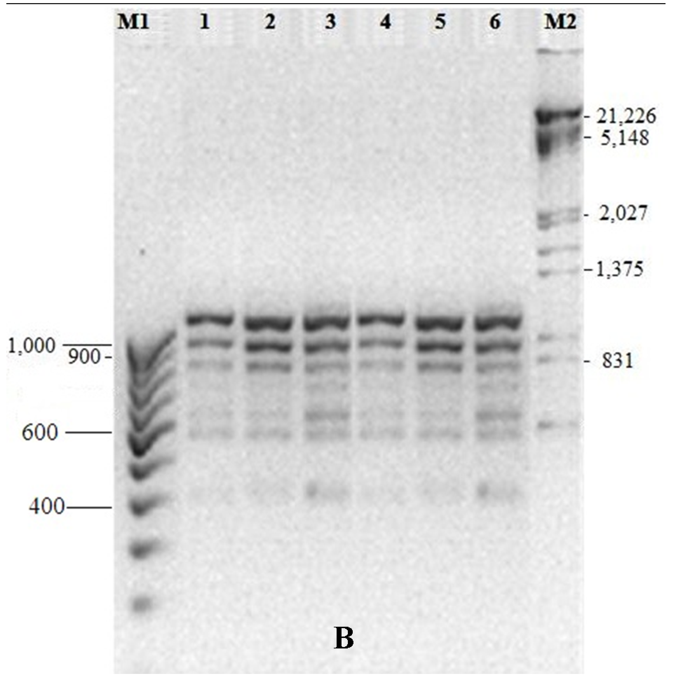
2.5. Genetic Fidelity Testing of Somaclones

The genomic DNA of in vitro regenerated leaf samples of C. indica was isolated prior to hardening. This was done by using Genelute Plant Genomic DNA kit (Sigma, St. Louis, MI, USA, Cat# G2N-70). Based on the spectrophotometric analysis, the DNA was diluted to a concentration of 25 ng∙µL−1.
A total of 20 RAPD and 10 ISSR primers were screened for PCR amplification (Table 1). Both RAPD and ISSR amplifications were performed using 25 µL of PCR mixture containing 12.5 µL PCR Master Mix 2×, 1.25 µL of primer (0.25 µM) and 2 µL of template DNA (25 ng∙µL−1). The PCR reactions were performed on a Perkin-Elmer Thermocycler 2400. The conditions of the thermal cycle for RAPD amplifications were 94 °C for 4 min, followed by 40 cycles of amplification with 1 min denaturation at 94 °C, 1 min annealing at 37 °C, 2 min primer extension at 72 °C, and a final extension at 72 °C for 10 min. The conditions of the thermal cycle for ISSR amplifications were 94 °C for 5 min, followed by 35 cycles of amplification with 45 s denaturation at 94 °C, 1 min annealing at 52 °C, 1 min primer extension at 72 °C, and a final extension at 72 °C for 7 min. The PCR products were separated on 1.5% (w/v) agarose gel with two DNA markers such as λ DNA/EcoRI/HindIII double digest and a 100 base pair (bp) DNA ladder (GeNeiTM), and were photographed with Gel Documentation system (UVi).

2.6. Statistical Analysis

All the experiments were repeated thrice, and the values were presented as mean ± standard error. Least significant difference (LSD) values of the treatments were determined between subcultures [13].

3. Results and Discussion

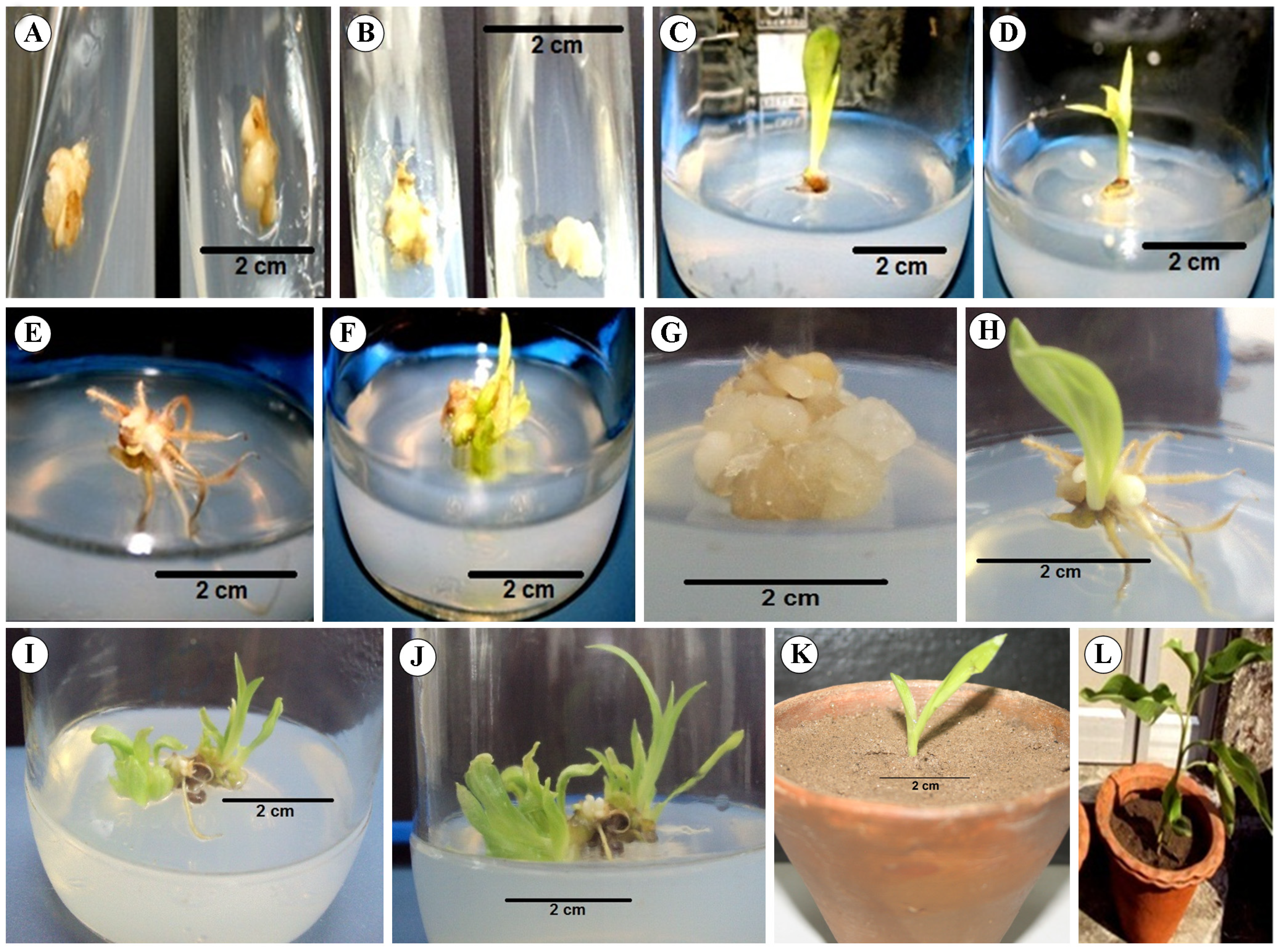
3.1. Establishment of Aseptic Culture

To manage the fungal and bacterial contamination problems a combination of surface sterilants like 70% ethanol for 45 s followed by 0.1% HgCl2 for 1.5 min showed the best result. There was no significant contamination problem during subculturing and maintaining of explants. Similar observations were noted by Bhattacharya and Sen in the micropropagation of ginger [14].

3.2. Callus Induction

Callus-like structures of the in vitro maintained leaves were observed after 15–20 days of culture (Figure 1). Out of various basal media, banana micropropagation media (BM) was found to be the most suitable. BM supplemented with 2 mg∙L−1 BAP and 0.5 mg∙L−1 NAA assisted profuse callusing. In contrast, 2,4-D at various concentrations i basal media without any hormone, yielded no result. Therefore, BM with BAP and NAA were the best for callus induction. Fully-developed callus was noticed after 10–15 days of callus induction (Figure 1).

3.3. Somatic Embryogenesis and Regeneration

3.3.1. Somatic Embryo Formation

Similar to the previous experiments, various hormone combinations for somatic embryogenesis were tried and 2 mg∙L−1 of BAP in BM was found most suitable. In fact, auxin (NAA) hindered somatic embryo formation. Somatic embryos were formed after 10–15 days following transfer of callus into BAP-assisted medium (Figure 1). An average 4–5 somatic embryos were produced per culture vessel. The number of embryos increased (9 ± 3) up to the 3rd subculture, while by the 4th subculture embryo number was reduced (6 ± 2).

3.3.2. Plantlet Regeneration

It was noted that root and shoot from the somatic embryos took place without changing the medium or hormones. In the BAP-assisted BM, rooting was initiated at 19 ± 3 days of somatic embryo formation while shoot development started a little later 28 ± 5 days. Early rooting had no affect on the formation of in vitro shoots [15]. However, root and shoot induction varied with different concentrations of BAP. Profuse rooting was observed in all cases. The earliest root and shoot formation (16 and 22 days, respectively) took place at 2 mg∙L−1 of BAP. Perhaps profuse rooting in Canna is a general phenomenon for the members of the order Zingiberales as Kambaska and Santilata [16] and Tyagi et al. [17] also reported a similar phenomenon in ginger and turmeric, respectively.

3.3.3. Subculturing and Hormone Effect on Shoot Growth

The number and length of shoots varied with the concentration of the phytohormone used (Table 2). The highest number of shoots was observed at 2 mg∙L−1 BAP. The number of shoots increased with BAP up to 2 mg∙L−1, and then gradually decreased thereafter. A higher concentration of BAP may have some inhibitory effect on the formation of shoots [18]. Shoot length also showed a similar pattern; the tallest shoot was recorded at 2 mg∙L−1 BAP. Subsequent subculturing of plantlets also had some effects on the growth rate of shoots (Table 2). The results indicated that in vitro growth of Canna gradually increased up to a certain limit in response to fresh medium. Decline in regeneration may be the effect of gradual aging and a decrease in totipotency of the explant [19].

3.3.4. Acclimatization of in vitro Regenerated Plants

The in vitro propagated plantlets with a well-developed shoot and root system were successfully transferred to clay pots containing an autoclaved mixture of soil and sand. They were maintained for about 2 months under plastic covers in order to avoid their desiccation (Figure 1). The survival rate of in vitro generated plantlets was as high as 80%.
The hardened plantlets were transferred to an outdoor garden. The survival rate of regenerated plantlets was as high as 72% in Canna. However, about 23% of the plantlets turned yellow. After about 2 months following transfer to the garden soil, an average of 5–8 leaves were evident and the height of the plants was around 30 cm.

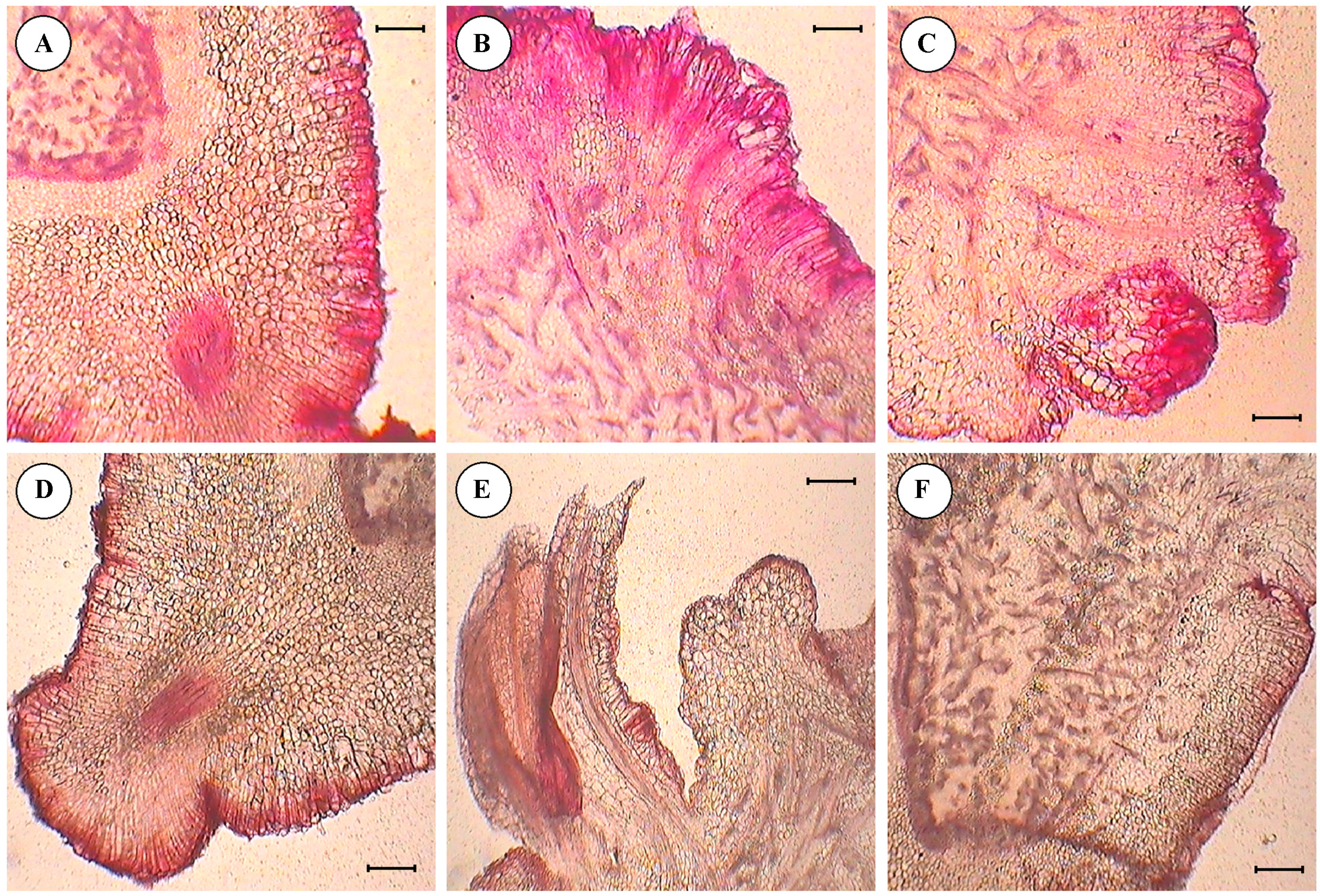
3.4. Histological Studies of Cultured Embryo

Transverse and longitudinal sections of somatic embryos generated from the leaf tissue revealed the followings under a light microscope in 10× magnification (Figure 2).
  • Anthocyanin pigmentation was visualized on the edges of the section in the form of red spots as stained by safranin.
  • Primordia of embryo development were observed from the surface of the cotyledon slices, in the form of compact masses of cells arranged sequentially on the edge of section.
  • Differentiation and separation of proembryonic globules which were had a well-differentiated epidermis.
  • Details of epidermal cell divisions, like periclinal and anticlinal walls and mitotically-active cells were observed.
  • Embryonic axes with shoot meristems, cortical tissues, central cylinders and procambial strands were clearly visible.

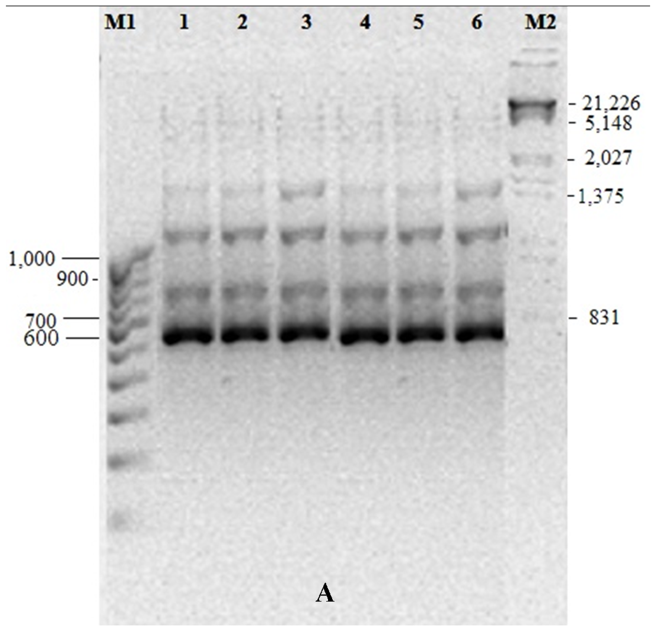
3.5. Genetic Fidelity among in vitro Raised Plantlets

Out of 20 RAPD and 10 ISSR primers, 10 and 7 primers produced amplification, respectively (Table 1). A total of 60 scorable bands with an average of 6 ± 2 bands per primer were obtained from the RAPD analysis, whereas a total 45 bands were produced from 6 primers of ISSR markers showing an average of 7 ± 2 bands per primer. Almost all of the bands generated through both RAPD and ISSR analysis were common in parental genotypes and the regenerated plants of different subcultures. Amplified products exhibited monomorphic patterns across all the subcultures for both markers. Representative RAPD and ISSR profiles are given in Figure 3A,B respectively. Though the plantlets were produced indirectly from the leaf tissue through the formation of callus, RAPD and ISSR analyses showed no detectable polymorphisms between parent plants and the regenerated plants. However, clonal fidelity was reported in other Zingiberales like banana and turmeric [17,20,21].

4. Conclusions

The present study is the first report of micropropagation of C. indica. It took about 12 weeks to obtain plantlets via somatic embryogenesis. The regenerated plantlets were healthy and RAPD and ISSR analysis showed that they were genetically stable and identical to their parental counterpart. The protocol developed for the rapid regeneration and multiplication of C. indica through in vitro callus and somatic embryogenesis may be used for the mass propagation of medicinally-important ornamental species.

Acknowledgments

We are thankful to the Department of Biotechnology, Government of India, for providing financial support to carry out our research. The authors are also grateful to Prof. Abhaya Prasad Das, University of North Bengal for authentication of plant material.

Author Contributions

Arnab Sen designed the experiment. Tanmayee Mishra and Arvind Kumar Goyal performed the experiment and wrote the manuscript. All the authors read and approved the final version of the manuscript.

Conflicts of Interest

The authors declare no conflict of interest.

References

  1. Nakornthap, A. Radiation-induced somatic mutations in the ornamental Canna. In Rome Meeting on the Uses of Induced Mutations in Plant; Breeding Pergamon Press: New York, NY, USA, 1965; pp. 707–721. [Google Scholar]
  2. Tanaka, N. The utilization of edible Canna plants in southeastern Asia and southern China. Econ. Bot. 2004, 58, 112–114. [Google Scholar] [CrossRef]
  3. Choudhury, M.D.; Bawari, M.; Singha, L.S. Some antipyretic ethno-medicinal plants of Manipuri Community of Barak Valley, Assam, India. Ethnobot. Leaflets 2010, 1, 21–28. [Google Scholar]
  4. Singh, P.K.; Kumar, V.; Tiwari, R.K.; Sharma, A.; Rao, C.V.; Singh, R.H. Medico-ethnobotany of Chatara’Block of District Sonebhadra, Uttar Pradesh, India. Advan. Biol. Res. 2010, 4, 65–80. [Google Scholar]
  5. Mishra, T.; Goyal, A.K.; Middha, S.K.; Sen, A. Antioxidative properties of Canna edulis Ker-Gawl. Ind. J. Nat. Prod. Res. 2011, 2, 315–321. [Google Scholar]
  6. Mishra, T.; Das, A.P.; Sen, A. Phytochemical screening and in vitro antioxidant profiling of solvent fractions of Canna edulis Ker Gawler. Free Radical. Antioxid. 2012, 2, 13–20. [Google Scholar] [CrossRef]
  7. Mishra, T.; Goyal, A.K.; Bhattacharya, M.; Kar, P.; Sen, A. Polyethylene glycol mediated protoplast fusion of medicinally important Canna. Res. Plant. Biol. 2015, 5, 20–24. [Google Scholar]
  8. Sakai, T.; Imai, K. The influences of growth regulators and culture medium composition on shoot-tip cultures of edible Canna. Environ. Control Biol. 2007, 45, 155–163. [Google Scholar] [CrossRef]
  9. Hosoki, T.; Sasaki, H. In vitro propagation of Canna edulis Ker by longitudinal shoot-split method. Plant Tissue Cult. Lett. 1991, 8, 175–178. [Google Scholar] [CrossRef]
  10. Murashige, T.; Skoog, F. A revised medium for rapid growth and bio assays with tobacco tissue cultures. Physiol. Plant. 1962, 15, 473–497. [Google Scholar] [CrossRef]
  11. Goyal, A.K.; Pradhan, S.; Basistha, B.C.; Sen, A. Micropropagation and assessment of genetic fidelity of Dendrocalamus strictus (Roxb.) nees using RAPD and ISSR markers. Biotech 2014. [Google Scholar] [CrossRef]
  12. Santra, S.C.; Chatterjee, T.P.; Das, A.P. College Botany Practical, 3rd ed.; New Central Book Agency Ltd.: Kolkata, India, 2006; pp. 321–322. [Google Scholar]
  13. Sit, A.K.; Bhattacharya, M.; Chenchaiah, K.C. Effect of benzyl amino purine on in vitro shoot multiplication of ginger (Zingiber officinale Rosc. cv. Garubathan). J. Plant. Crop. 2005, 33, 184–186. [Google Scholar]
  14. Bhattacharya, M.; Sen, A. Rapid in vitro multiplication of disease-free Zingiber officinale Rosc. Ind. J. Plant. Physiol. 2006, 11, 379–384. [Google Scholar]
  15. Mondal, M.; Gupta, S.; Mukherjee, B.B. In vitro propagation of shoot buds of Carica papaya L. (Caricaceae) var. Honey Dew. Plant Cell Rep. 1990, 8, 609–612. [Google Scholar] [CrossRef] [PubMed]
  16. Kambaska, K.B.; Santilata, S. Effect of plant growth regulator on micropropagtion of ginger (Zingiber officinale Rosc.) cv. Suprava and Suruchi. J. Agr. Tech. 2009, 5, 271–280. [Google Scholar]
  17. Tyagi, R.K.; Agrawal, A.; Mahalakshmi, C.; Hussain, Z.; Tyagi, H. Low-cost media for in vitro conservation of turmeric (Curcuma longa L.) and genetic stability assessment using RAPD markers. In Vitro Cell. Develop. Biol.-Plant 2007, 43, 51–58. [Google Scholar] [CrossRef]
  18. Hussey, G. In vitro propagation of Gladiolus by precocious axillary shoot formation. Sci. Hort. 1977, 6, 287–296. [Google Scholar] [CrossRef]
  19. Murashige, T.; Nakano, R. Morphogenetic behavior of tobacco tissue cultures and implication of plant senescence. Amer. J. Bot. 1965, 52, 819–827. [Google Scholar] [CrossRef]
  20. Rout, G.R.; Senapati, S.K.; Aparajita, S.; Palai, S.K. Studies on genetic identification and genetic fidelity of cultivated banana using ISSR markers. Plant Omics 2009, 2, 250–258. [Google Scholar]
  21. Lakshmanan, V.; Reddampalli Venkataramareddy, S.; Neelwarne, B. Molecular analysis of genetic stability in long-term micropropagated shoots of banana using RAPD and ISSR markers. Electron. J. Biotechnol. 2007, 10, 106–113. [Google Scholar] [CrossRef]
Figure 1. Stages of Callus induction and regeneration: (A) Callus induction; (B) Matured callus; (C) Formation of in vitro shoot; (D) Multiple shooting; (E) Profuse in vitro rooting; (F) Plantlet regeneration from callus. Stages of somatic embryogenesis and plantlet formation: (G) Somatic embryo formation; (H) In vitro shooting and rooting from embryo; (I) Fifteen days old plantlet; (J) Plantlet after 1 month; (K) Acclimatization of plant in clay pot containing mixture of soil and sand; (L) Transfer and hardening of in vitro grown plant.
Horticulturae 01 00003 g001
Figure 2. Histology of various stages of embryogenesis observed under light microscope (A) Embryonic axis; (B) & (C) Region of tissue differentiation; (D) Region of embryo induction; (E) Region of shoot primordia; (F) Part of embryonic axis. Scale = 20 μm.
Horticulturae 01 00003 g002
Figure 3. DNA fingerprinting pattern of in vitro callus regenerated plantlets of C. indica: (A) using RAPD primer OPB01; (B) using ISSR primer UBC815. Lane 2–6: micropropagated plantlets compared with mother plant (lane1); Lane M1: 100 bp molecular marker; Lane M2: λ DNA/EcoRI/HindIII double digest DNA ladder.
Horticulturae 01 00003 g003aHorticulturae 01 00003 g003b
Table 1. PCR amplification using RAPD (Sl. No. 1–10) and ISSR (Sl. No. 11–17) primers.
Total BandsSl. No.Primer IDPrimer TypeSequence (5’-3’)Band Size (bp)
71OPA01RAPD CAGGCCCTTC330–1120
620PA03RAPDAGTCAGCCAC400–1260
63OPA07RAPDGAAACGGGTG500–1550
840PA11RAPDCAATCGCCGT420–1380
75OPA17RAPDGACCGCTTGT760–1440
56OPB01RAPDGTTTCGCTCC410–1350
67OPF09RAPDCCAAGCTTCC610–1500
78OPG19RAPDGTCAGGGCAA500–1280
49OPH04RAPDGGAAGTCGCC300–1050
410OPN04RAPDGACCGACCCA450–1300
311UBC810ISSR(GA)8T550–800
612UBC815ISSR(CT)8G375–1100
813UBC822ISSR(TC)8A390–1280
614UBC824ISSR(TC)8G450–1110
815UBC825ISSR(AC)8T540–920
516UBC856ISSR(AC)8YA410–980
917UBC873ISSR(GACA)4390–1240
Table 2. Effect of subculture and concentration of phytohormones on shoot growth.
MediaBAP(mg∙L−1)Shoot Length (cm)
15 D #30 D45 D60 D75 D
BMControl0.20 ± 0.320.48 ± 0.211.23 ± 0.581.62 ± 0.821.81 ± 0.76
10.66 ± 0.38 ns1.80 ± 1.22 ns3.50 ± 1.61 **5.10 ± 0.91 **5.90 ± 1.02 **
21.04 ± 1.07 ns3.22 ± 1.81 **4.96 ± 0.92 **6.50 ± 1.12 **7.35 ± 0.84 **
30.51 ± 0.77 ns2.40 ± 1.54 **3.20 ± 1.43 **4.32 ± 0.98 **4.88 ± 0.67 **
40.42 ± 0.26 ns1.70 ± 0.93 *2.61 ± 0.78 *3.80 ± 1.31 **4.64 ± 1.95 **
Shoot No.
Control1.24 ± 0.882.31 ± 0.753.47 ± 0.924.84 ± 0.895.16 ± 0.80
13.29 ± 1.12 **8.96 ± 0.89 **13.44 ± 1.15 **18.88 ± 1.26 **19.56 ± 0.49 **
24.38 ± 1.79 **11.98 ± 1.93 **16.56 ± 0.95 **22.24 ± 1.84 **24.32 ± 1.77 **
33.43 ± 1.56 **9.01 ± 0.99 **12.88 ± 1.77 **17.02 ± 0.97 **19.42 ± 0.88 **
42.27 ± 0.44 ns7.99 ± 0.77 **10.42 ± 1.31 **13.62 ± 0.55 **15.31 ± 0.81 **
Standard deviation was calculated on 3 replications, each with sets of 10. # D= No. of days, ns = not significant, * = significant at 5% level of probability, ** = significant at 1% level of probability.

