Article

Functional Investigation of Mutant Vespa tropica Hyaluronidase Produced in Pichia pastoris: In Silico Studies and Potential Applications

by
Piyapon Janpan
1,2,
Bernhard Schmelzer
3,
Anuwatchakij Klamrak
1,2,
Jaran Nabnueangsap
4,
Yutthakan Saengkun
1,2,
Shaikh Shahinur Rahman
1,5,
Prapenpuksiri Rungsa
2,
Diethard Mattanovich
3 and
Sakda Daduang
1,2,*
1
Division of Pharmacognosy and Toxicology, Faculty of Pharmaceutical Sciences, Khon Kaen University, Khon Kaen 40002, Thailand
2
Protein and Proteomics Research Center for Commercial and Industrial Purposes (ProCCI), Khon Kaen University, Khon Kaen 40002, Thailand
3
Institute of Microbiology and Microbial Biotechnology, Department of Biotechnology and Food Science, BOKU University, 1190 Vienna, Austria
4
Salaya Central Instrument Facility RSPG, Research Management and Development Division, Office of the President, Mahidol University, Nakhon Pathom 73170, Thailand
5
Department of Applied Nutrition and Food Technology, Faculty of Biological Sciences, Islamic University, Kushtia 7000, Bangladesh
*
Author to whom correspondence should be addressed.
Fermentation 2025, 11(9), 497; https://doi.org/10.3390/fermentation11090497
Submission received: 20 July 2025 / Revised: 19 August 2025 / Accepted: 24 August 2025 / Published: 25 August 2025
(This article belongs to the Section Microbial Metabolism, Physiology & Genetics)

Abstract

The hyaluronidase enzyme derived from Vespa tropica (VesT2a) venom contains two putative catalytic residues. Herein, a double mutation was introduced into VesT2a at its catalytic sites by substituting Asp107 and Glu109 with Asn and Gln, respectively, to assess their essential roles in enzymatic function. We used Pichia pastoris to produce the mutated version of the VesT2a (mVesT2a) protein, and the process was more efficient when employing the methanol-inducible promoter (PAOX1) compared to the constitutive promoter (PGAP). In bioreactor scale-up, P. pastoris harboring the pAOX1-αMF-mVesT2a plasmid secreted 34.03 ± 2.31 mg/L of mVesT2a, with an apparent molecular mass of 46.6 kDa, retaining only 2.9% of hyaluronidase activity, thus indicating successful mutation. The newly developed indirect ELISA-based method using mVesT2a demonstrated its potential as an alternative approach for measuring hyaluronic acid (HA) at low concentrations and was also used to confirm HA-binding capacity. In silico docking and molecular dynamics simulations further supported the stable interaction of the mVesT2a–HA complex while suggested other surrounded acidic amino acid residues, which may play a minor role in HA degradation, supporting the remaining activity observed in the in vitro experiments.

1. Introduction

Hyaluronidases are classes of glycoside hydrolases that specifically breakdown hyaluronic acid (HA) to various hyaluronic acid oligomers [1]. Hyaluronidases are classified into three main groups based on biochemical analysis and generated end products, which consist of hyaluronate 4-glycanohydrolases (EC 3.2.1.35), hyaluronate 3-glycanohydrolases (EC 3.2.1.36), and microbial hyaluronidases (EC 4.2.2.1) [2]. For hyaluronate 4-glycanohydrolases, this group is endo-β-N-acetyl hexosaminidases, which randomly cleave the β-1–4 glycoside bond of HA to generated various chain lengths, especially tetra- and hexa-hyaluronic acid [3]. This glycosidase is commonly found in various living organisms, including humans, cattle, snakes, bees, and wasps, among others. In the catalytic mechanism, the hyaluronidase enzyme is generally understood as an acid–base catalytic process. The catalytic residues act as a proton donor to facilitate bond cleavage, then allowing a water molecule to perform a nucleophilic attack to complete hydrolysis [4,5,6]. Obviously, aspartate (Asp) and glutamate (Glu) residues, which are highly conserved in the substrate-binding region, have been reported as the primary catalytic residues in mammalian and venomous hyaluronidases, such as Asp129 and Glu131 in human hyaluronidase 1 (HYAL1); Glu148 in sperm plasma-membrane protein (PH-20); Asp107 and Glu109 in Vespa affinis, Vespula vulgaris, and Polybia paulista hyaluronidases; and Asp111 and Glu113 in bee venom hyaluronidase [4,6,7,8,9,10]. Thus, these two catalytic amino acids are key residues that influence hyaluronidase activity.
Vespa tropica is considered one of the most dangerous wasps and is commonly found in northeastern Thailand. Hyaluronidase (VesT2a) derived from V. tropica venom acts as a spreading factor, facilitating the dispersion of wasp venom toxins into prey for hunting, self-defense, and colony protection [9]. Compared to other venomous animals, the hyaluronidase in V. tropica venom exhibits higher activity than that of snakes (Naja siamensis), scorpions (Heterometrus laoticus), and other wasps (Vespa affinis), indicating its high enzymatic efficiency. Our previous studies have shown that VesT2a gene consisted of 1486 base pairs encoding 356 amino acids, showing the predicted molecular mass of mature VesT2a of 39,119.7 Da and an isoelectric point (pI) of 8.91. Two amino acids, Asp107 and Glu109, have been identified as catalytic residues, along with five putative glycosylation sites (Asn79, Asn99, Asn127, Asn187, and Asn325) and two disulfide bridges (Cys19–Cys308 and Cys185–Cys197) [11].
Although the putative residues governing its virulent activity have been identified, the crucial roles of its two catalytic residues in enzymatic function have not yet been investigated in this wasp species. Hyaluronidases from various other sources, however, have shown significant changes in activity following mutations at their catalytic residues. For example, mutations of D111N and E113Q in bee venom hyaluronidase (BVH) resulted in no detectable activity [10]. Similarly, in PH-20, the E113Q mutation caused complete loss of activity, while the D111N mutation retained only 3% of enzymatic activity [7]. In human HYAL1, mutation of the catalytic residue led to the retention of less than 2% of hyaluronidase activity [6]. To address this, the catalytic residues of V. tropica hyaluronidase were mutated (D107N and E109Q) to assess their impact on enzyme activity in the absence of these residues. Meanwhile, its binding capability was examined using a newly developed indirect ELISA-based method for detecting hyaluronic acid, due to the method’s sensitivity, selectivity, and specificity [12,13].
Herein, this study highlights the significant potential of using P. pastoris, a Generally Recognized as Safe (GRAS) expression host widely used for producing peptides or proteins [14,15], to produce a mutated hyaluronidase (mVesT2a) protein under the use of an appropriate promoter. The recombinant plasmid containing the mVesT2a gene was constructed using the GoldenPiCS system, specifically designed for expression in P. pastoris using the same strategies as recently established by our groups [16,17]. The secreted mVesT2a was characterized to foresee if it exerts strong hyaluronic binding activity with dramatically reduced HA degradation, which was clearly confirmed by the newly developed indirect ELISA technique. Furthermore, the interactions and stability of the mVesT2a–HA complex were explored through molecular docking and molecular dynamics simulation analyses, providing valuable insights into the binding mechanism and paving the way of the mutated protein for an HA detection assay, with promising utility in quality control for hyaluronic acid extraction and biomedical diagnostics.

2. Materials and Methods

2.1. Strains, Reagents, and Media

All microbial strains, including E. coli DH10B and P. pastoris CBS2612, as well as the GoldenPiCS system kit, were obtained as previously described by Prielhofer et al. [16]. The synthetic gene (Fs2_αMF_mVesT2a_GG-6xHis tag_Fs3), with codon optimization for expression in P. pastoris, was sourced from TWIST Bioscience (South San Francisco, CA, USA). Specific primers listed in Table 1 were obtained from Integrated DNA Technologies (IDT, Coralville, IA, USA).

2.2. Plasmid Construction via the Golden Gate-Derived P. pastoris Cloning System (GoldenPiCS)

The synthetic gene was amplified by Q5 High-Fidelity DNA Polymerase (NEB, Ipswich, MA, USA) with specific primers, F_mVesT2a and R_mVesT2a, following the thermocycling condition: initial denaturation at 98 °C for 30 s, 30 cycles of 98 °C for 10 s, 70 °C for 30 s, and 72 °C for 45 s, with final elongation at 72 °C for 2 min. The PCR product was investigated by the agarose gel electrophoresis technique and then purified through an innuPREP DOUBLEpure Kit (Analytik, Jena, Germany). The recombinant plasmids of the mVesT2a gene were constructed using the GoldenPiCS system, following [16]. The coding sequence (CDS), Fs2_αMF_mVesT2a_GG-6xHisTag_Fs3, was assembled into recipient BB1 through BsaI and T4 DNA ligase by incubating it with 30 cycles of 37 °C and 16 °C for 1 min and final incubation at 37 °C for 10 min. The BB1 plasmid was transformed into E. coli DH10B by using the heat shock method at 42 °C for 70 s. Transformants were selected on LB agar containing 25 µg/mL kanamycin. Subsequently, the BB1 plasmid was isolated and purified using the HiYield® Plasmid Mini Kit (Süd-Laborbedarf GmbH, Munich, Germany), following verification of the DNA sequence by Sanger cycle sequencing (Microsynth, Vienna, Austria). Next, the CDS in the BB1 plasmid was assembled with two different promoters, pGAP and pAOX1, and the ScCYC1tt terminator in the recipient direct BB3 plasmid via BpiI and T4 DNA ligase following the GGA-BB1 incubation condition. The BB3 plasmids were transformed into E. coli DH10B and selected on LB agar containing 50 µg/mL zeocin. Two recombinant BB3 plasmids, BB3aZ_pGAP_αMF_mVesT2a_GG-6xHisTag and BB3aZ_pAOX1_αMF_mVesT2a_GG-6xHisTag, were verified by Sanger cycle sequencing and showed the schematic maps in Figure S1 generated by using GenSmart Design (https://www.genscript.com/gensmart-design/# (accessed on 24 August 2024)). Afterward, each BB3 plasmid was linearized by AscI and integrated into the P. pastoris chromosome using electroporation. The transformants were selected on YPD agar containing 500 µg/mL zeocin.

2.3. Screening of P. pastoris Production and Bioreactor Cultivation

The screening of heterologous protein production by P. pastoris was performed in 96-deep-well plates (96-DWPs) using an enzymatic glucose release method according to the method described by Janpan et al. [17]. The Enpump200 kit (Enpresso, Berlin, Germany) was used to apply in the slowing glucose release condition.
The yeast transformants from each plasmid construct were inoculated in 300 µL YPD medium containing 500 µg/mL zeocin and incubated at 25 °C and 1200 rpm for 18 h. The cultures were centrifuged at 2000× g for 5 min, and the supernatants were removed. The pellets were resuspended in 150 µL of 2 × ASM.V6 minimal medium (6.3 g/L (NH4)2HPO4, 0.8 g/L (NH4)2SO4, 0.49 g/L MgSO4·7H2O, 2.64 g/L KCl, CaCl2·2H2O, 22 g/L citric acid monohydrate, 1.47 mL/L PTM1 trace metals, and 20 mL/L NH4OH (25%); pH was adjusted to 6.5 with KOH). Subsequently, 30 µL of suspended culture was transferred into a new 96-DWP containing 120 µL of 2 × ASM.V6 minimal medium. For PGAP screening, 150 µL of PSE solution (50 g/L polysaccharide, 0.7% amylase) was added to the main culture under glucose-limiting conditions and incubated at 25 °C and 1200 rpm for 48 h. For PAOX1 screening, 150 µL of PSE solution (25 g/L polysaccharide, 0.35% amylase) was added to the main culture, followed by methanol induction with 10, 20, 20, and 20 µL of absolute methanol after 3, 19, 27, and 43 h, respectively. After 48 h, recombinant protein production from different constructs was analyzed by gel capillary electrophoresis (LabChip® HT Protein Express, PerkinElmer, Waltham, MA, USA). The candidate clone, which has the best potential to produce mVesT2a protein, was selected for upscaled production via a bioreactor system.
Fed-batch fermentation with a sequential induction strategy was performed to produce the mVesT2a protein using the DASGIP® Parallel Bioreactor System (Eppendorf, Hamburg, Germany) [18]. The process began with a batch phase using 300 mL of modified BSM medium (glycerol as carbon source) and 10 mL of inoculum, maintained at 25 °C, pH 5.5, and 20% dissolved oxygen (DO) with 5% Glanapon to prevent foaming. After around 18 h, a glucose feeding phase started, supplying a medium with 50% glucose, 1% biotin, and 1% PTM0 trace salts solution at an exponential rate (y = 1.7313e0.0787x; µ = 0.079 h−1) for 18 h 30 min. Upon glucose depletion, methanol induction began with 0.5% (v/v) of methanol addition, followed by continuous feeding of 1% (v/v) methanol for 70 h at y = 3.0062e0.016x (µ = 0.016 h−1). Protein production during each phase was analyzed by SDS-PAGE using the NuPAGETM 4–12% Bis-Tris system (Invitrogen, Waltham, MA, USA).

2.4. Protein Purification

The supernatant was separated from the yeast culture by centrifugation at 8000× g and 4 °C for 30 min (Beckman Avanti J-20XP centrifuge, Beckman Coulter, Brea, CA, USA) and filtered via a 0.45 µm filter membrane (Merck, Darmstadt, Germany). The cell-free supernatant was then subjected to a 5 mL HisTrap HP column to purify the mVesT2a protein by using an ÄKTATM Protein Purification System (GE Healthcare, Chicago, IL, USA), following the manufacturer’s instruction. The purified protein sample was analyzed and characterized by SDS-PAGE, Western blot, and LC-MS/MS techniques.

2.5. SDS-PAGE and Western Blotting

The SDS-PAGE technique was performed to separate protein samples using a 13% polyacrylamide gel. Subsequently, the proteins were transferred onto a 0.45 μm nitrocellulose membrane (Mumbai, Maharashtra, India) using a TRANS-BLOT® SD semi-dry transfer system (Bio-Rad, Hercules, CA, USA). The membrane was then blocked with 5% skim milk in TBST buffer at 4 °C for 18 h to minimize non-specific binding, followed by incubation with anti-6xHis tag antibody conjugated to alkaline phosphatase (AP) (1:300 dilution in blocking solution) at room temperature for 2 h. The protein bands were visualized using an AP conjugate substrate kit (Bio-Rad, Hercules, CA, USA).

2.6. LC-MS/MS Analyses

The suspected protein band was investigated by LC-MS/MS using a Thermo Dionex Nano LC Ultimate 3000 system with an Acclaim PepMap RSLC C18 column (Thermo Scientific, Waltham, MA, USA) under a linear gradient of solvent A (0.1% formic acid in 2% acetonitrile) and solvent B (0.1% formic acid in 80% acetonitrile) at 0.3 µL/min. Mass spectrometry was then performed on a SCIEX Triple TOF 6600+ in positive ion mode, scanning MS at 350–1500 Da (AB SCIEX, Framingham, MA, USA). Candidate precursor ions exceeding 100 counts per second were selected for MS2 (100–1500 Da), and fragment spectra were matched to theoretical spectra in a protein database.

2.7. Hyaluronidase Activity Assay

The reaction mixture of the hyaluronidase activity assay was conducted in a 1.5 mL microcentrifuge tube, containing 2.0 µg of mVesT2a protein and 0.5 mg/mL HA in 0.2 M formate buffer pH 3.0 with 0.15 M NaCl. The reaction was incubated at 37 °C for 30 min, followed by stopping the reaction by adding two-fold volumes of CTAB reagent (2.5% cetyltrimethylammonium bromide in 2.0% NaOH) and incubating at 37 °C for 15 min. The absorbance was measured by a SPECTROstar Nano microplate reader at OD 405 nm (BMG LABTECH, Ortenberg, Germany). Based on international standard preparation, one turbidity-reducing unit is defined as the amount of hyaluronidase enzyme required to reduce the turbidity of 50 µg of hyaluronic acid by 50%. One unit of hyaluronidase activity is equivalent to one turbidity-reducing unit [8].

2.8. Development of mVesT2a Protein for Hyaluronic Acid Detection

To confirm the binding activity of the recombinant mVesT2a protein, an indirect ELISA-based hyaluronic acid (HA) detection method was employed. The HA detection method consist of four steps: plate coating, recombinant protein incubation, antibody incubation, and detection (Figure 1). Fifty microliters of HA standard solution or sample solution was added to a Nunclon Delta-treated 96-well plate (Thermo Fisher Scientific, Waltham, MA, USA) and incubated at 60 °C for 2 h and 30 min. This 96-well plate is modified to increase the hydrophilicity of the well surface, according to the product description. The HA sample was blocked with 200 µL of 1% BSA in phosphate-buffered saline (PBS) pH 7.4 at 25 °C for 1 h, followed by washing with 200 µL of PBST buffer (0.1% Tween 20 in PBS). Then, 100 µL of mVesT2a protein (1 µg/mL in PBST) was added to each well and incubated at 25 °C for 1 h. Subsequently, 100 µL of anti-6x-His tag Ab linked AP (1:1000 dilution) (Thermo Fisher Scientific, Waltham, MA, USA) was added and incubated at 25 °C for 1 h. Next, 100 µL of pNPP solution (1 mg/mL pNPP in 100 mM Tris-HCl, pH 9.5, containing 100 mM NaCl and 5 mM MgCl2) was added, and the mixture was incubated at 25 °C for 3 h. The concentration of HA was measured using a microplate reader at OD 405 nm.

2.9. In Silico Molecular Docking and Dynamics Simulations

AlphaFold2, a Google Colab notebook service (https://colab.research.google.com/github/sokrypton/ColabFold/blob/v1.2.0/AlphaFold2.ipynb (accessed on 2 January 2024)), was used to predict the 3D structure of the desired protein, which is the mVesT2a protein, based on multiple sequence alignments generated through MMseqs2. N-glycosylation sites and disulfide bond formation in the representative protein were predicted using NetNGlyc version 1.0 (https://services.healthtech.dtu.dk/services/NetNGlyc-1.0/ (accessed on 3 January 2024)) and Disulfide by Design version 2.0 (http://cptweb.cpt.wayne.edu/DbD2/index.php (accessed on 3 January 2024)), respectively. Exploring the HA binding site on mVesT2a, complexed with a hyaluronic acid hexamer (PDB ID: 4hya), was predicted using the CB-Dock2 server [19] (https://cadd.labshare.cn/cb-dock2/php/index.php (accessed on 3 January 2024)) based on AutoDock Vina. Self-docking the selected protein–ligand complex was conducted using GOLD Suite v.5.2.2 (Genetic Optimization of Ligand Docking) to optimize docking parameters and achieve an RMSD below 2 Å, prior to performing docking between mVesT2a protein and target ligands, as recommended in the GOLD user manual. The protein–ligand complex with the highest fitness score was chosen for dynamics simulation using the GROMACS simulation package, a versatile package to perform molecular dynamics, through the SiBioLead online molecular dynamics simulation web server (https://sibiolead.com/MDSIM (accessed on 1 February 2024)) to investigate the dynamic behavior, stability, and interactions of the complexes over the simulation time. Dynamics simulations were performed in four stages with appropriate modification [20]: (i) system setup using the optimized potentials for liquid simulations—all-atom (OPLS-AA) force field in a cubic box with an SPC water model and 0.15 M NaCl; (ii) energy minimization via the steepest descent algorithm for 5000 steps; (iii) equilibration under NVT and NPT ensembles at 1.0 bar and 300 K for 100 ps; and (iv) a 100 ns molecular dynamics simulation run using the leap-frog integrator, followed by trajectory analysis using standard GROMACS results analysis packages. The predicted interactions of protein–ligand complexes were identified and visualized using BIOVIA Discovery Studio 2021 Client and UCSF ChimeraX (version 1.5) software.

3. Results and Discussion

3.1. Recombinant Plasmids Construction and Small-Scale Production of mVesT2a Protein

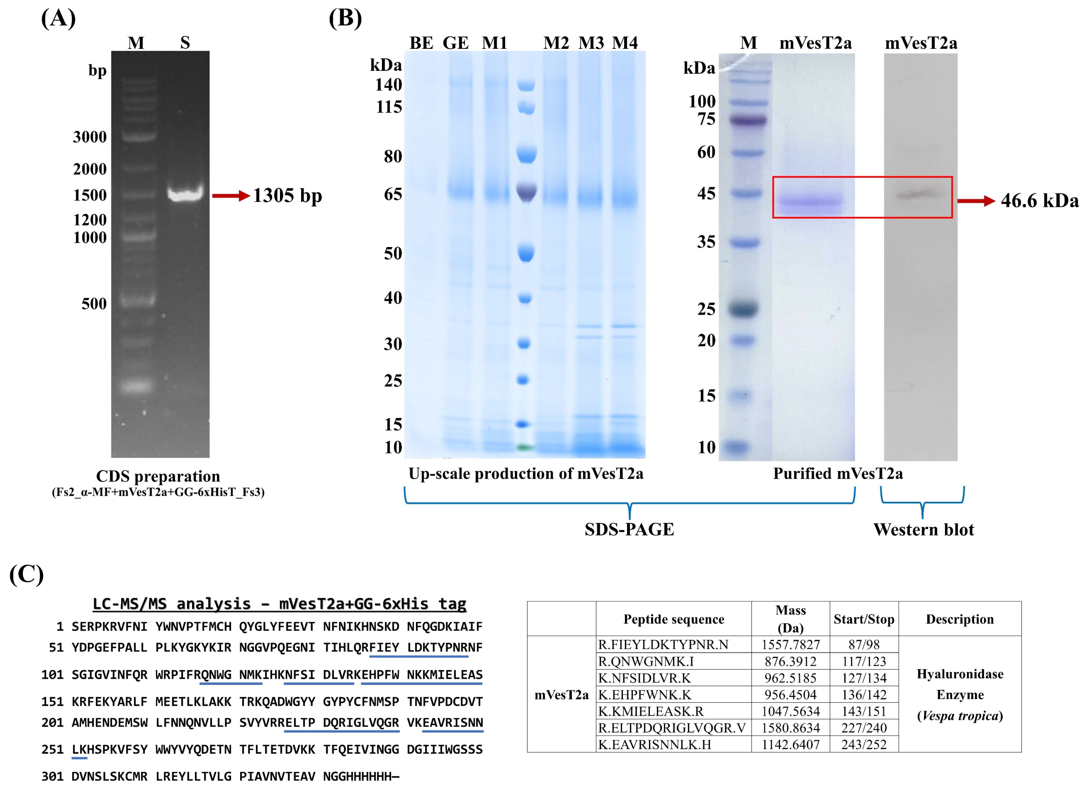
In this study, we have investigated the impact of catalytic amino acids on the hyaluronidase activity and the binding ability of the mVesT2a protein. We designed a mutated form of hyaluronidase derived from V. tropica venom by substituting two amino acids at the catalytic site—D107N and E109Q—to eliminate hyaluronidase activity. The mVesT2a gene was codon-optimized based on the codon usage frequency of P. pastoris for expressing the 318-amino-acid target protein, using the IDT codon optimization tool (https://www.idtdna.com/CodonOpt (accessed on 12 January 2022)), resulting in a codon adaptation index (CAI) of 0.75 (Figure S2). This optimization is expected to enhance expression by aligning codon usage with highly level expression and protein solubility in the yeast system [21,22,23]. The target gene fragment, mVesT2a (993 bp), which contained an alpha mating factor (αMF) at the 5′ end and a polyhistidine tag (6 × His) at the 3′ end to facilitate extracellular secretion and enable detection or purification, was synthesized and prepared before recombinant plasmid construction. This target gene, which is about 1305 bp, is shown in Figure 2A. Following gene preparation, recombinant plasmids of mVesT2a bearing different promoters were successfully constructed using the GoldenPiCS technique, an effective strategy for gene assembly in the P. pastoris expression system. The coding sequence was inserted into the yeast expression vector (direct BB3 plasmid) containing either the GAP or AOX1 promoter and the ScCYC1tt terminator via BpiI digestion, resulting in two distinct plasmids: pGAP_αMF_mVesT2a_GG-6xHis tag_ScCYC1tt and pAOX1_αMF_mVesT2a_GG-6xHis tag_ScCYC1tt (Figure S3). Sanger sequencing was utilized to confirm the mVesT2a sequence. Both recombinant plasmids were linearized using the AscI restriction enzyme prior to transformation into yeast cells via the electroporation technique. The candidate clones were selected on YPD agar containing 500 µg/mL zeocin. A high concentration of the resistance drug aids in selecting clones with a higher gene copy number and expression level compared to a low or normal concentration (approximately 50–100 µg/mL) of zeocin [24,25]. Subsequently, small-scale expression in a 96-deep-well plate using the enzymatic glucose release method showed that the selected P. pastoris clone containing the AOX1 promoter produced approximately 1.5 mg/L of secreted protein in the culture medium, which was about 3.57-fold higher than that obtained from the GAP promoter (Table 2), suggesting that the strong inducible AOX1 promoter is suitable for mVesT2a production in P. pastoris. As demonstrated in various reports, the methanol induction system, which relies on the tightly regulated AOX1 promoter, often yields significantly higher expression levels than the constitutive GAP promoter, such as the production of recombinant hyaluronidase (VesT2a) and β-fructofuranosidase proteins [17,26,27,28,29]. Additionally, expression of the target protein in methylotrophic yeast containing the AOX1 promoter allows for postgrowth activation, which begins after the yeast reaches high cell density during the growth phase through glucose or glycerol consumption [30,31]. The methanol induction system shifts resources from biomass production to protein synthesis, leading to a higher yield of the heterologous protein [32]. Therefore, P. pastoris containing the plasmid construct with the AOX1 promoter was chosen to produce the mVesT2a protein by methanol induction in upscaled production via a bioreactor system.

3.2. Upscaled Production, Detection, and Purification of mVestT2a Produced by P. pastoris

P. pastoris carrying the pAOX1_αMF + mVesT2a + GG-6xHis tag_ScCYC1tt construct was cultured with a fed-batch fermentation strategy in a 1 L bioreactor for upscaled production, which is an effective strategy for increasing recombinant protein yield. After 70 h of cultivation with 1% (v/v) methanol at 25 °C, pH 5.5, and 20% dissolved oxygen (DO), this engineered methylotrophic yeast secreted a heterologous protein with an expected molecular mass of approximately 44.7 kDa, yielding a total of 34 mg/L (Figure 2B, Table 1). The secreted protein sample was purified by using immobilized metal ion affinity chromatography (IMAC) and subsequently detected by Western blotting using a monoclonal antibody specific to the protein that fuses with a polyhistidine tag (6 × His tag) (Figure 2B). The identification of the purified protein sample was further confirmed by LC-MS/MS analysis, which matched the amino acid sequence of hyaluronidase (Figure 2C). The use of P. pastoris as an expression host, together with the plasmid construct containing an alpha mating factor (α-MF) secretion signal, is a key factor in enabling the secretion of heterologous proteins into the culture medium. P. pastoris possesses an efficient secretory pathway similar to higher eukaryotes, supporting proper protein folding and post-translational modifications such as glycosylation and disulfide bond formation [33,34,35]. These features contribute to the production of secreted proteins that more closely resemble the native structure. Moreover, the secretion signals, alpha mating factor, can serve as sorting signals to guide secretory proteins from the cytosol to the extracellular space [36,37], leading to the desired recombinant protein being conveniently harvested from the culture medium. This finding demonstrates the successful production of recombinant mVesT2a in a soluble form using the P. pastoris system. It also addresses the issue of inclusion body formation, which is commonly encountered when expressing recombinant hyaluronidase proteins in E. coli [7,38,39]. In the mVesT2a sequence, N-glycosylation site prediction revealed five putative glycosylation sites at Asn79, Asn99, Asn127, Asn187, and Asn325. The experimental molecular mass was estimated at 44.7 kDa by SDS-PAGE, which is approximately 4.65 kDa higher than the theoretical molecular mass of 40.05 kDa. The difference in molecular mass may be due to the presence of putative glycosylation, which is consistent with the production of rTsHya1 hyaluronidase in P. pastoris, resulting in a glycosylation-related increase in molecular mass of approximately 4.9 kDa [40].

3.3. Hyaluronidase Activity Assay and HA Detection Assay

The mutated hyaluronidase protein, which was mutated in two amino acids at the catalytic site (D107N, E109Q), was tested for hyaluronidase activity by using the CTAB turbidimetric method. The purified mVesT2a protein exhibited a hyaluronidase activity of approximately 119 U/mg at 37 °C (Table 3). Compared to our previous study, the double-point mutation reduced hyaluronidase activity about 97%. This result suggests that substituting two critically important residues in the active site affects the HA degradation ability, particularly by weakening the proton donation of the mutant residue to N-acetyl-D-glucosamine, which acts as a nucleophile [1]. This result has been supported by Zhang et al. (2009) [6], in whose study the mutated human hyaluronidase (Hyal1) retained approximately 0.25% and 1.40% of its activity compared to the wild type after point mutations at catalytic residues (D129N and E131Q), respectively. The single-point mutations of the catalytic residues (D107 and E109), however, will be investigated to clarify the specific role of each amino acid in governing hyaluronidase activity. According to our aspect, the binding capability of mVesT2a was evaluated via the developed method for HA detection. mVesT2a was applied to detect HA content based on indirect ELISA. The modified method demonstrated the binding activity of mVesT2a that can measure HA content in the range of 0.050–0.400 µg/mL (Y = 0.0606X + 0.0107, R2 = 0.9994), with an accuracy of 99% and precision (%RSD) of 16%. The LOD and LOQ were 0.095 µg/mL and 0.197 µg/mL, respectively (Table 4), while the conventional method (CTAB assay) for HA detection can detect HA in the range of 25–500 µg/mL with an LOD and LOQ of 1.52 and 4.60 µg/mL, respectively. Moreover, the newly developed method offers other advantages, including high sensitivity and specificity for detecting hyaluronic acid. Although this method can detect low signals of chondroitin sulfate, the levels are below the LOQ. This is likely due to structural differences between the substrates, as chondroitin sulfate contains sulfate groups on the N-acetylgalactosamine unit, exhibiting low binding affinity in the HA binding groove [41]. The high efficiency of this developed method may be caused by the utilization of the mVesT2a protein produced using P. pastoris due to appropriate protein folding. Compared to other HA detection methods, such as noncompetitive and competitive ELISA assays for HA detection, our developed method still has lower sensitivity, as both ELISA methods can measure hyaluronic acid in the range of 2–500 ng/mL [12,13]. However, our method reduces processing time by more than 50 h and has a low cost. This finding demonstrates the potential of mVesT2a for use in HA detection. Nevertheless, further improvements are needed, such as adapting other ELISA formats, like sandwich ELISA, to enhance detection sensitivity [42,43]. The use of fluorescence-based ELISA assays, such as those employing europium-labeled secondary antibodies, could provide higher sensitivity and a broader dynamic range compared to colorimetric detection.

3.4. Molecular Docking and Dynamics Simulation

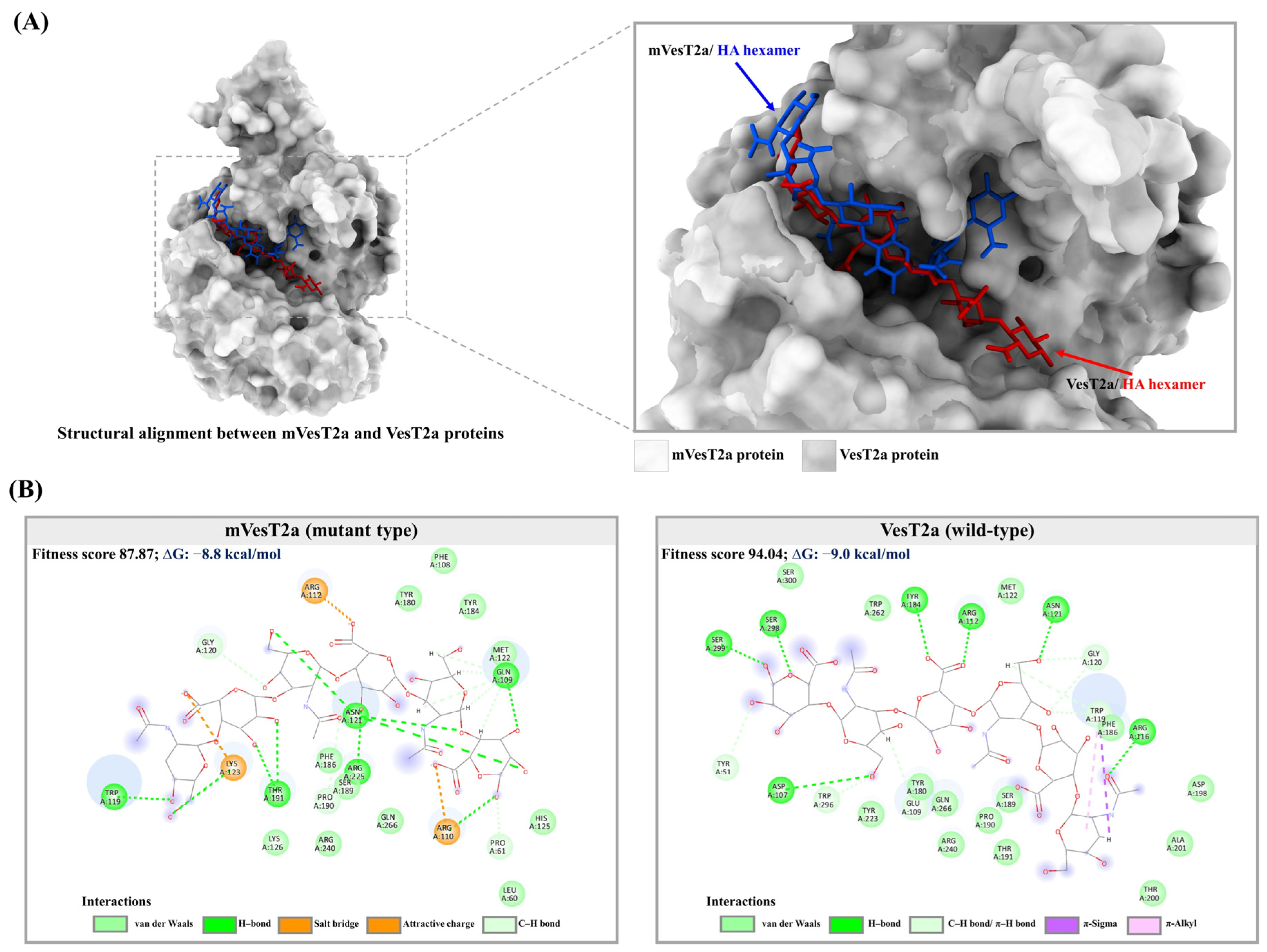
Molecular docking was performed to elucidate the differences in interactions between the VesT2a-HA hexamer and mVesT2a-HA hexamer (Figure 3). Alignment of the 3D structures of wild-type and mutant hyaluronidase proteins in complex with hyaluronic acid, as shown in Figure 3A, revealed a slight difference in the HA binding region. As depicted in Figure 3B, the HA hexamer interacts with the mVesT2a protein through hydrogen bonding, salt bridge, and electrostatic (attractive charge) interactions with key residues in the HA binding region, such as Arg112 (5.66 Å), Trp119 (2.63 Å), and residue 109 (2.35–2.81 Å), which corresponds to the E109Q mutation. Compared to the wild-type hyaluronidase, the mutant hyaluronidase still retains the ability to bind hyaluronic acid, with a docking score of 87.87, although this is lower than the wild-type hyaluronidase, which showed a docking score of 94.04. This finding aligns with our previous study, which reported stronger binding of wild-type hyaluronidase with the HA hexamer in the HA binding groove [17]. The docking result also suggests that the mutated VesT2a protein still maintains its binding ability to hyaluronic acid through key residues such as Gln109, Arg112, Trp119, Tyr180, Tyr184, and Gln266.
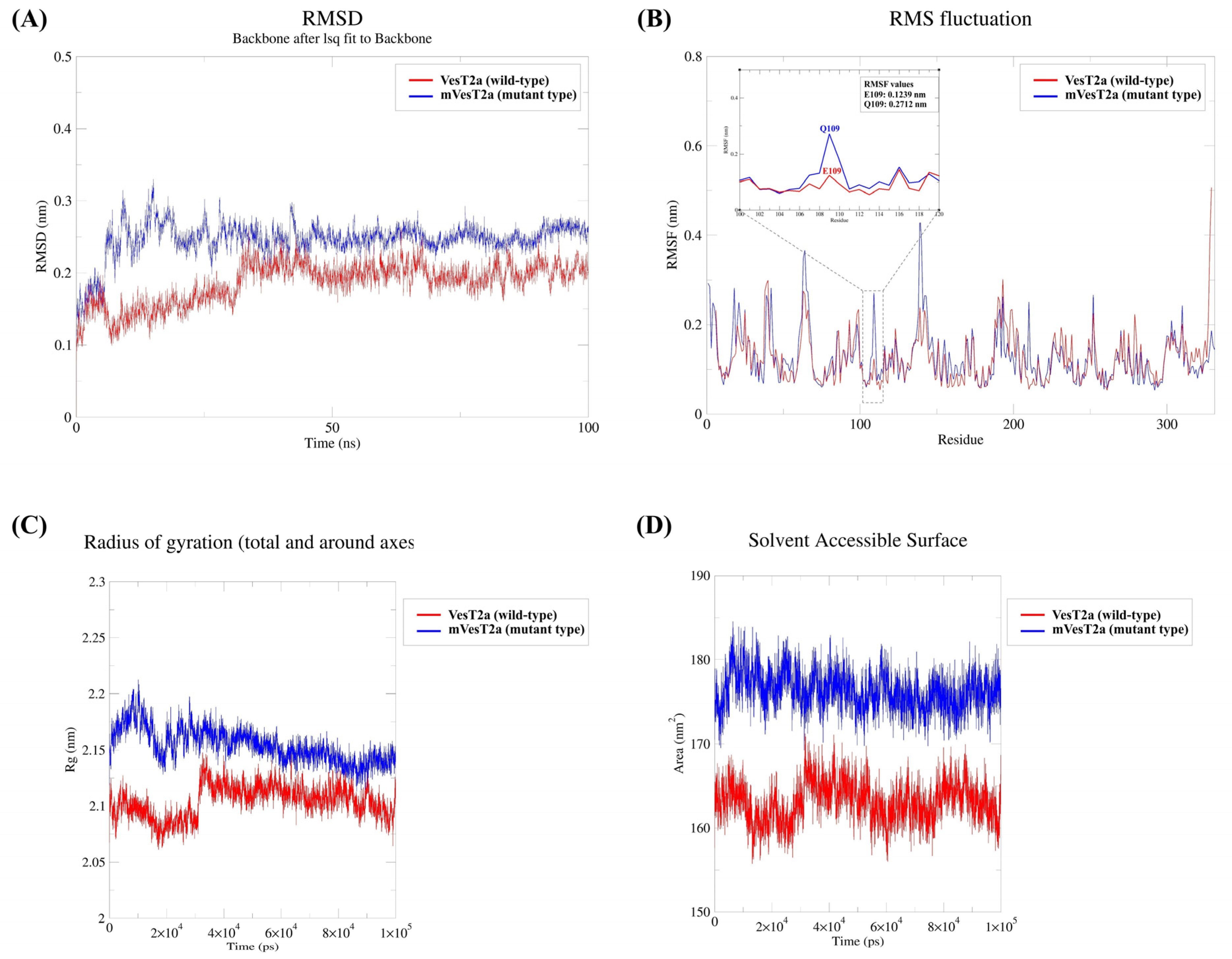
To validate the docking results, 100 ns of molecular dynamics simulation was carried out to evaluate the stability and binding interaction of two protein–ligand complexes, which are the VesT2a-HA hexamer (wild-type complex) and mVesT2a-HA hexamer (mutant complex) (Figure 4). Root mean squared deviation (RMSD) analysis indicated that the VesT2a-HA complex showed a slightly increased RMSD value and then stabilized after nearly 32 ns, with an average RMSD of 0.18 nm, while for the mVesT2a-HA complex, structural stability was observed after 42 ns of the simulation run with a higher average RMSD value of 0.25 nm. These results demonstrated the stability of each protein–ligand complex at a low RMSD under the given conditions, although the wild-type protein–ligand complex shows higher protein stability than the mutant protein–ligand complex (Figure 4A). Calculating the root mean squared fluctuation (RMSF) of two protein–ligand complexes, the RMSF analysis revealed comparable flexibility with minimal fluctuation between the wild-type protein–ligand complex and mutant protein–ligand complex. Notably, a catalytic residue, residue 109, was identified where the mutant protein (Q109: 0.27 nm) showed higher fluctuation than the wild-type protein (E109: 0.12 nm) (Figure 4B). This elevated fluctuation suggests that the E109Q mutation might affect chemical interaction between the mVesT2a protein and hyaluronic acid, potentially influencing the enzymatic function of hyaluronidase. Additionally, solvent-accessible surface area (SASA) and radius of gyration (Rg) are important parameters for evaluating the structural compactness and conformational stability of protein–ligand complexes during simulation. The average SASA value of the VesT2a-HA hexamer complex was approximately 176 nm2, whereas the mVesT2a-HA hexamer complex showed a lower average SASA value of 163 nm2 (Figure 4D). The wild-type protein complex displayed an average Rg value of 2.10 nm, which is close to the 2.15 nm of the mutant protein complex (Figure 4C). These findings indicate that the mutant protein complex shows a slightly more expanded conformation, reflecting the increasing Rg value, with a decrease in the area accessible to the surrounding solvent. This may indicate a minor effect on protein stability and ligand-binding interactions. Nevertheless, the minor differences in RMSD, RMSF, Rg, and SASA values indicate that both protein–ligand complexes maintained structural stability throughout the simulation, although the double-point mutations may have induced minor conformational changes.
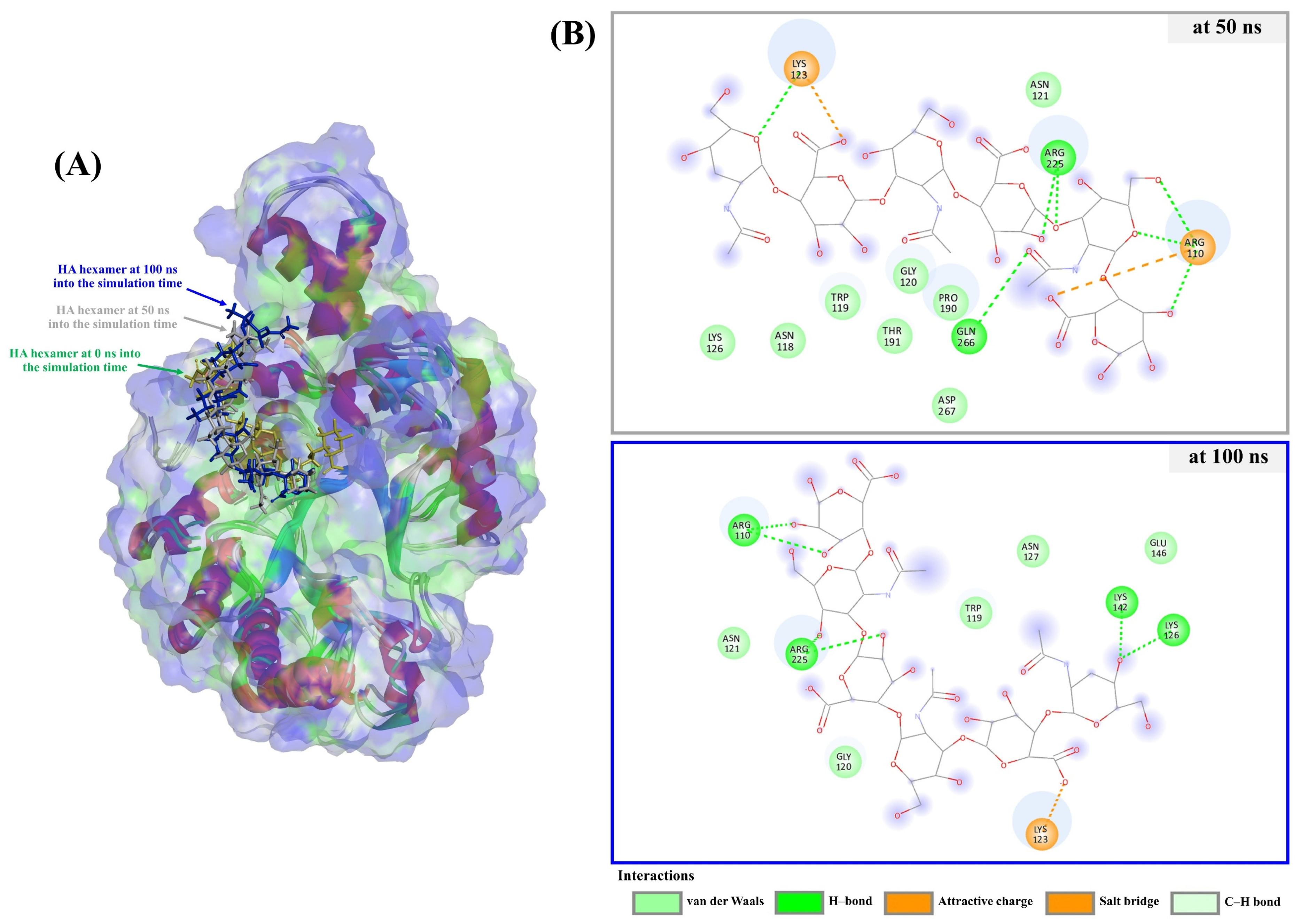
Despite Asp107 and Glu109 being designed to mutate to Asn (D107N) and Gln (E109Q), respectively, the results show a measurable 2.81% residual enzymatic activity, suggesting that other acidic amino acids presently surrounding the active pocket may play a minor role in the catalytic process, acting as those two residues—D107 and E109—that are well recognized in the usual catalytic mechanism. A similar pattern was also observed in glycoside hydrolase (GH) families, such as GH18, GH20, GH25, GH85, and GH56, which specifically cleave substrates containing an N-acetyl group linked to the C2 position of the pyranose ring moiety (C2-acetamido group) [44,45]. We then implemented MD simulation and found stable binding between mVesT2a–HA complexes at three different time points (0 ns [starting point], 50 ns, and 100 ns), (Figure 5). Despite slight reorientations of the HA hexamer observed at 50 ns and 100 ns, the binding was stable and sustained throughout the simulation. Asp267 (at 50 ns) and Glu146 (at 100 ns) appear to have the potential for alternative acid/base catalysis, with predicted pKa values of 2.50 and 3.14, respectively. Consequently, it is conceivable that one of the candidate residues, located within or near the active site, could presumably play a compensatory role under the altered electrostatic environment caused by the D107N and E109Q mutations, particularly if their pKa values are compatible with the acidic environment.

4. Conclusions

Here, we successfully produced the mutated hyaluronidase (mVesT2a) protein using a methanol-induction system in P. pastoris, thereby avoiding inclusion body formation typically observed in the E. coli expression system. mVesT2a exhibited greatly reduced hyaluronidase activity compared to the wild type, due to double-point mutations at the putatively conserved catalytic residues and with some intact activity remaining. MD simulation study also suggest that nearby acidic residues may partially compensate for the loss of catalytic function, despite the absence of the primary catalytic residues. These findings not only demonstrate the efficiency of mVesT2a expression and secretion in the yeast expression system but also highlight the critical role of catalytic residues in HA degradation. Additionally, the developed mutant hyaluronidase shows potential for various applications involving HA detection and quantification due to its high sensitivity and specificity. Future investigations will focus on kinetic analyses of wild-type and mutant V. tropica hyaluronidase under diverse conditions, providing key insights into their catalytic efficiency, and supporting the development of biotechnological applications.

Supplementary Materials

The following supporting information can be downloaded at: https://www.mdpi.com/article/10.3390/fermentation11090497/s1, Figure S1: The recombinant plasmids of mVesT2a gene. (A) BB3aZ_Fs-14_pAOX1_αMF_mVesT2a_GG-6xHis tag_ScCYC1tt; (B) BB3aZ_Fs-14_pGAP_αMF_mVesT2a_GG-6xHis tag_ScCYC1tt; Figure S2: Comparison of the DNA and amino acid sequences between the original V. tropica hyaluronidase (Ori.seq. (VesT2a)) and the codon-optimized mutant hyaluronidase (Opt.seq. (mVesT2a)) produced in P. pastoris, in which the catalytic residues Asp107 and Glu109 were substituted with Asn and Gln, respectively (red asterisks); Figure S3: The synthetic gene of the coding sequence (CDS) is composed of alpha-mating factor, mutated hyaluronidase (mVesT2a) and fusion tag (GG-6xHis tag). The mutations of catalytic residues, D107N and E109Q, were labeled with red asterisks (***).

Author Contributions

Conceptualization, P.J., B.S., A.K., D.M. and S.D.; methodology, P.J., B.S., A.K. and P.R.; software, P.J. and Y.S.; validation, P.J., B.S., A.K. and J.N.; formal analysis, P.J., B.S. and J.N.; investigation, P.J., B.S. and A.K.; resources, B.S., D.M. and S.D.; data curation, P.J., A.K., S.S.R. and P.R.; writing—original draft preparation, P.J., A.K. and S.S.R.; writing—review and editing, P.J., A.K. and S.S.R.; visualization, P.J. and Y.S.; supervision, D.M. and S.D.; project administration, S.D.; funding acquisition, S.D. All authors have read and agreed to the published version of the manuscript.

Funding

This research was funded by Research and Researcher for Industrial (RRi) Ph.D. Scholarship (Code: NRCT5-RRI63004-P03) awarded by the National Research Council of Thailand (NRCT). This research also received partial funding from the Fundamental Fund of Khon Kaen University (KKU), which in turn was financially supported by the National Science, Research, and Innovation Fund (NSRF), Thailand. The NSRF provided support for the research through the Basic Research Fund of Khon Kaen University.

Institutional Review Board Statement

Not applicable.

Informed Consent Statement

Not applicable.

Data Availability Statement

All data supporting the conclusions of this article are included in this article.

Acknowledgments

The authors are deeply grateful to the Faculty of Pharmaceutical Sciences, Khon Kaen University, Thailand, and the Institute of Microbiology and Microbial Biotechnology, Department of Biotechnology and Food Science, University of Natural Resources and Life Sciences, Austria. We also would like to acknowledge Kiattawee Choowongkomon from the Department of Biochemistry, Faculty of Sciences, Kasetsart University, Bangkok, Thailand, for providing the GOLD program used in molecular docking analyses. We also thank Patthana Tastub and Tewa Upathanpreecha from Betagro Science Center Co., Ltd.

Conflicts of Interest

The authors declare no conflicts of interest.

References

  1. El-safory, N.S.; Fazary, A.E.; Lee, C. Hyaluronidases, a Group of glycosidases: Current and future perspectives. Carbohydr. Polym. 2010, 81, 165–181. [Google Scholar] [CrossRef]
  2. Girish, K.S.; Kemparaju, K. The magic glue hyaluronan and its eraser hyaluronidase: A biological overview. Life Sci. 2007, 80, 1921–1943. [Google Scholar] [CrossRef]
  3. Šimek, M.; Lemr, K.; Hermannová, M.; Havlíček, V. Analysis of Hyaluronan and Its Derivatives Using Chromatographic and Mass Spectrometric Techniques. Carbohydr. Polym. 2020, 250, 117014. [Google Scholar] [CrossRef]
  4. Justo Jacomini, D.L.; Campos Pereira, F.D.; dos Santos Pinto, J.R.A.; dos Santos, L.D.; da Silva Neto, A.J.; Giratto, D.T.; Palma, M.S.; de Lima Zollner, R.; Brochetto Braga, M.R. Hyaluronidase from the venom of the social wasp Polybia paulista (Hymenoptera, Vespidae): Cloning, structural modeling, purification, and immunological analysis. Toxicon 2013, 64, 70–80. [Google Scholar] [CrossRef]
  5. Bordon, K.C.F.; Wiezel, G.A.; Amorim, F.G.; Arantes, E.C. Arthropod venom hyaluronidases: Biochemical properties and potential applications in medicine and biotechnology. J. Venom. Anim. Toxins Incl. Trop. Dis. 2015, 21, 4. [Google Scholar] [CrossRef]
  6. Zhang, L.; Bharadwaj, A.G.; Casper, A.; Barkley, J.; Barycki, J.J.; Simpson, M.A. Hyaluronidase activity of human Hyal1 requires active site acidic and tyrosine residues. J. Biol. Chem. 2009, 284, 9433–9442. [Google Scholar] [CrossRef]
  7. Arming, S.; Strobl, B.; Wechselberger, C.; Kreil, G. In vitro mutagenesis of PH-20 hyaluronidase from human sperm. Eur. J. Biochem. 1997, 247, 810–814. [Google Scholar] [CrossRef]
  8. Rungsa, P.; Janpan, P.; Saengkun, Y.; Jangpromma, N.; Klaynongsruang, S.; Patramanon, R.; Uawonggul, N.; Daduang, J.; Daduang, S. Heterologous expression and mutagenesis of recombinant Vespa affinis hyaluronidase protein (rVesA2a). J. Venom. Anim. Toxins Incl. Trop. Dis. 2019, 25, e20190030. [Google Scholar] [CrossRef] [PubMed]
  9. Rungsa, P.; Incamnoi, P.; Sukprasert, S.; Uawonggul, N.; Klaynongsruang, S.; Daduang, J.; Patramanon, R.; Roytrakul, S.; Daduang, S. Comparative proteomic analysis of two wasps venom, Vespa tropica and Vespa affinis. Toxicon 2016, 119, 159–167. [Google Scholar] [CrossRef] [PubMed]
  10. Marković-Housley, Z.; Miglierini, G.; Soldatova, L.; Rizkallah, P.J.; Müller, U.; Schirmer, T. Crystal structure of hyaluronidase, a major allergen of bee venom. Structure 2000, 8, 1025–1035. [Google Scholar] [CrossRef]
  11. Rungsa, P.; Incamnoi, P.; Sukprasert, S.; Uawonggul, N.; Klaynongsruang, S.; Daduang, J.; Patramanon, R.; Roytrakul, S.; Daduang, S. Cloning, structural modelling and characterization of VesT2s, a wasp venom hyaluronidase (HAase) from Vespa tropica. J. Venom. Anim. Toxins Incl. Trop. Dis. 2016, 22, 28. [Google Scholar] [CrossRef] [PubMed]
  12. Martins, J.R.M.; Passerotti, C.C.; Maciel, R.M.B.; Sampaio, L.O.; Dietrich, C.P.; Nader, H.B. Practical determination of hyaluronan by a new noncompetitive fluorescence-based assay on serum of normal and cirrhotic patients. Anal. Biochem. 2003, 319, 65–72. [Google Scholar] [CrossRef] [PubMed]
  13. Rössler, A. An Ultrasensitive, nonisotopic immunoassay for hyaluronan using the streptavidin-biotin system. Clin. Chim. Acta 1998, 270, 101–114. [Google Scholar] [CrossRef]
  14. Barone, G.D.; Emmerstorfer-Augustin, A.; Biundo, A.; Pisano, I.; Coccetti, P.; Mapelli, V.; Camattari, A. Industrial production of proteins with Pichia pastorisKomagataella phaffii. Biomolecules 2023, 13, 441. [Google Scholar] [CrossRef]
  15. Spohner, S.C.; Müller, H.; Quitmann, H.; Czermak, P. Expression of enzymes for the usage in food and feed industry with Pichia pastoris. J. Biotechnol. 2015, 202, 118–134. [Google Scholar] [CrossRef]
  16. Prielhofer, R.; Barrero, J.J.; Steuer, S.; Gassler, T.; Zahrl, R.; Baumann, K.; Sauer, M.; Mattanovich, D.; Gasser, B.; Marx, H. GoldenPiCS: A golden gate-derived modular cloning system for applied synthetic biology in the yeast Pichia pastoris. BMC Syst. Biol. 2017, 11, 123. [Google Scholar] [CrossRef]
  17. Janpan, P.; Schmelzer, B.; Klamrak, A.; Tastub, P.; Upathanpreecha, T.; Rahman, S.S.; Nabnueangsap, J.; Saengkun, Y.; Rungsa, P.; Mattanovich, D.; et al. Production of Vespa tropica hyaluronidase by Pichia pastoris. J. Fungi 2024, 10, 854. [Google Scholar] [CrossRef]
  18. Zavec, D.; Gasser, B.; Mattanovich, D. Characterization of methanol utilization negative Pichia pastoris for secreted protein production: New cultivation strategies for current and future applications. Biotechnol. Bioeng. 2020, 117, 1394–1405. [Google Scholar] [CrossRef]
  19. Liu, Y.; Yang, X.; Gan, J.; Chen, S.; Xiao, Z.X.; Cao, Y. CB-Dock2: Improved protein-ligand blind docking by integrating cavity detection, docking and homologous template fitting. Nucleic Acids Res. 2022, 50, 159–164. [Google Scholar] [CrossRef]
  20. Amin, M.; Barzegari, E.; Pourshohod, A.; Zeinali, M.; Jamalan, M. 3D structure prediction, dynamic investigation and rational construction of an epitope-masked thermostable bovine hyaluronidase. Int. J. Biol. Macromol. 2021, 187, 544–553. [Google Scholar] [CrossRef] [PubMed]
  21. Fu, H.; Liang, Y.; Zhong, X.; Pan, Z.L.; Huang, L.; Zhang, H.L.; Xu, Y.; Zhou, W.; Liu, Z. Codon optimization with deep learning to enhance protein expression. Sci. Rep. 2020, 10, 17617. [Google Scholar] [CrossRef]
  22. Xu, Y.; Liu, K.; Han, Y.; Xing, Y.; Zhang, Y.; Yang, Q.; Zhou, M. Codon usage bias regulates gene expression and protein conformation in yeast expression system P. pastoris. Microb. Cell Fact. 2021, 20, 91. [Google Scholar] [CrossRef]
  23. Yuankai, H.; Chen, Y.; Mo, D.; Cong, P.; He, Z. Attenuated secretion of the thermostable xylanase XynB from Pichia pastoris using synthesized sequences optimized from the preferred codon usage in yeast. J. Microbiol. Biotechnol. 2011, 22, 316–325. [Google Scholar] [CrossRef]
  24. Naseem, M.U.; Tajti, G.; Gaspar, A.; Szanto, T.G.; Borrego, J.; Panyi, G. Optimization of Pichia pastoris expression system for high-level production of margatoxin. Front. Pharmacol. 2021, 12, 733610. [Google Scholar] [CrossRef] [PubMed]
  25. Nordén, K.; Agemark, M.; Danielson, J.J.T.; Alexandersson, E.; Kjellbom, P.; Johanson, U. Increasing gene dosage greatly enhances recombinant expression of aquaporins in Pichia pastoris. BMC Biotechnol. 2011, 11, 47. [Google Scholar] [CrossRef] [PubMed]
  26. Potvin, G.; Ahmad, A.; Zhang, Z. Bioprocess engineering aspects of heterologous protein production in Pichia pastoris: A review. Biochem. Eng. J. 2012, 64, 91–105. [Google Scholar] [CrossRef]
  27. Waterham, H.R.; Digan, M.E.; Koutz, P.J.; Lair, S.V.; Cregg, J.M.; Bernardi, A. Isolation of the Pichia pastoris glyceraldehyde-3-phosphate dehydrogenase gene and regulation and use of its promoter. Gene 1997, 186, 37–44. [Google Scholar] [CrossRef]
  28. Anane, E.; van Rensburg, E.; Görgens, J.F. Comparison of constitutive and inducible β-fructofuranosidase production by recombinant Pichia pastoris in fed-batch culture using defined and semi-defined media. S. Afr. J. Chem. Eng. 2016, 22, 17–22. [Google Scholar] [CrossRef]
  29. Kim, S.; Warburton, S.; Boldogh, I.; Svensson, C.; Pon, L.; d’Anjou, M.; Stadheim, T.A.; Choi, B.K. Regulation of alcohol oxidase 1 (AOX1) promoter and peroxisome biogenesis in different fermentation processes in Pichia pastoris. J. Biotechnol. 2013, 166, 174–181. [Google Scholar] [CrossRef]
  30. Jia, L.; Tu, T.; Huai, Q.; Sun, J.; Chen, S.; Li, X.; Shi, Z.; Ding, J. Enhancing monellin production by Pichia pastoris at low cell induction concentration via effectively regulating methanol metabolism patterns and energy utilization efficiency. PLoS ONE 2017, 12, e0184602. [Google Scholar] [CrossRef]
  31. Hartner, F.S.; Glieder, A. Regulation of methanol utilisation pathway genes in yeasts. Microb. Cell Fact. 2006, 5, 39. [Google Scholar] [CrossRef] [PubMed]
  32. Gidijala, L.; Uthoff, S.; Kampen, S.J.; Steinbüchel, A.; Verhaert, R.M.D. Presence of protein production enhancers results in significantly higher methanol-induced protein production in Pichia pastoris. Microb. Cell Fact. 2018, 17, 112. [Google Scholar] [CrossRef] [PubMed]
  33. Karbalaei, M.; Rezaee, S.A.; Farsiani, H. Pichia pastoris: A highly successful expression system for optimal synthesis of heterologous proteins. J. Cell. Physiol. 2020, 235, 5867–5881. [Google Scholar] [CrossRef]
  34. Pan, Y.; Yang, J.; Wu, J.; Yang, L.; Fang, H. Current advances of Pichia pastoris as cell factories for production of recombinant proteins. Front. Microbiol. 2022, 13, 1059777. [Google Scholar] [CrossRef]
  35. Zha, J.; Liu, D.; Ren, J.; Liu, Z.; Wu, X. Advances in metabolic engineering of Pichia pastoris strains as powerful cell factories. J. Fungi 2023, 9, 1027. [Google Scholar] [CrossRef]
  36. Zou, C.; Lu, L.; Wang, S.; Zhang, C.; Chen, X.; Lin, Y.; Huang, Y. The α-mating factor secretion signals and endogenous signal peptides for recombinant protein secretion in Komagataella phaffii. Biotechnol. Biofuels Bioprod. 2022, 15, 140. [Google Scholar] [CrossRef]
  37. Ahmad, M.; Hirz, M.; Pichler, H.; Schwab, H. Protein expression in Pichia pastoris: Recent achievements and perspectives for heterologous protein production. Appl. Microbiol. Biotechnol. 2014, 98, 5301–5317. [Google Scholar] [CrossRef]
  38. Schwaighofer, A.; Ablasser, S.; Lux, L.; Kopp, J.; Herwig, C.; Spadiut, O.; Lendl, B.; Slouka, C. Production of active recombinant hyaluronidase inclusion bodies from Apis mellifera in E. coli BL21(DE3) and characterization by FT-IR spectroscopy. Int. J. Mol. Sci. 2020, 21, 3881. [Google Scholar] [CrossRef]
  39. Justo Jacomini, D.L.; Gomes Moreira, S.M.; Campos Pereira, F.D.; Zollner, R.D.L.; Brochetto Braga, M.R. Reactivity of IgE to the allergen hyaluronidase from Polybia paulista (Hymenoptera, Vespidae) venom. Toxicon 2014, 82, 104–111. [Google Scholar] [CrossRef]
  40. Amorim, F.G.; Boldrini-França, J.; de Castro Figueiredo Bordon, K.; Cardoso, I.A.; De Pauw, E.; Quinton, L.; Kashima, S.; Arantes, E.C. Heterologous expression of rTsHyal-1: The first recombinant hyaluronidase of scorpion venom produced in Pichia pastoris system. Appl. Microbiol. Biotechnol. 2018, 102, 3145–3158. [Google Scholar] [CrossRef]
  41. Iliás, A.; Liliom, K.; Greiderer-Kleinlercher, B.; Reitinger, S.; Lepperdinger, G. Unbinding of hyaluronan accelerates the enzymatic activity of bee hyaluronidase. J. Biol. Chem. 2011, 286, 35699–35707. [Google Scholar] [CrossRef]
  42. Kohl, T.O.; Ascoli, C.A. Immunometric double-antibody sandwich enzyme-linked immunosorbent assay. Cold Spring Harb. Protoc. 2017, 2017, 458–462. [Google Scholar] [CrossRef] [PubMed]
  43. Anagu, L.O.; Andoh, N.E. Vaccine development: From the laboratory to the field. In Vaccinology and Methods in Vaccine Research; Ashfield, R., Ed.; Elsevier: London, UK, 2022; pp. 95–131. [Google Scholar] [CrossRef]
  44. Vuong, T.V.; Wilson, D.B. Glycoside hydrolases: Catalytic base/nucleophile diversity. Biotechnol. Bioeng. 2010, 107, 195–205. [Google Scholar] [CrossRef] [PubMed]
  45. Jitonnom, J. Carbohydrate hydrolysis by glycosidase: Mechanistic aspects and applications. KKU Sci. J. 2013, 41, 582–594. [Google Scholar]
Figure 1. Schematic flowchart illustrating the development of the mVesT2a protein for hyaluronic acid detection using an indirect ELISA.
Fermentation 11 00497 g001
Figure 2. Protein production of recombinant mVesT2a protein using P. pastoris. LC-MS/MS analysis of mutated hyaluronidase (mVesT2a). (A) Coding sequence (CDS) gene preparation; M: 1kb DNA ladder marker; S: synthetic gene (Fs2_αMF_mVesT2a_GG-6xHis tag_Fs3). (B) mVesT2a protein characterizations using SDS-PAGE and Western blot techniques; M: protein molecular weight marker; BE: batch end phase; GE: glucose feeding end phase; M1–M4: samples collected during the methanol feeding phase; mVesT2a: mutated hyaluronidase protein. (C) Protein sequencing analysis via LC-MS/MS revealed the amino acid sequence of the target protein, highlighted with a blue underline.
Fermentation 11 00497 g002
Figure 3. Molecular docking analysis illustrates the structural orientation of mVesT2a and VesT2a proteins with a hyaluronic acid hexasaccharide. (A) Three-dimensional structure of the protein–ligand complex. (B) Two-dimensional representation of ligand interactions.
Fermentation 11 00497 g003
Figure 4. Molecular dynamics simulation analysis of mutant hyaluronidase (mVesT2a) and wild-type hyaluronidase (VesT2a) proteins with a hyaluronic acid hexasaccharide. (A) Root mean squared deviation (RMSD). (B) Root mean squared fluctuation (RMSF). (C) Radius of gyration (Rg). (D) Solvent-accessible surface area (SASA).
Fermentation 11 00497 g004
Figure 5. Illustration of mVesT2a protein–HA hexamer interaction at different stages of the simulation. (A) Comparison of the 3D structure of the protein–ligand complex at the starting point (0 ns) (green) and 50 ns (gray) and 100 ns (blue) into the simulation. (B) Two-dimensional representation of ligand interactions at 50 ns and 100 ns into the simulation.
Fermentation 11 00497 g005
Table 1. Specific primers for gene cloning of mutated hyaluronidase.
NameSequence (5′ to 3′)Purpose
F_mVesT2aGATCGGTCTCGTCAGAAAGGCCCAAAAGAGTGTTTAACATTTACTGmVesT2a gene
R_mVesT2aGATCGGTCTCCAAGCCTATTAGTGATGGTGGTGGTGATGTCCACmVesT2a gene
F_BB1CAGGAAACAGCTATGACSequencing
R_BB1GTAAAACGACGGCCAGTTSequencing
F_GAPACCAGAATCGAATATAAASequencing
F_AOX1CTTTCATAATTGCGACTGGTTCSequencing
R_BB3CGAGCGTCCCAAAACCSequencing
Table 2. Comparison of recombinant mVesT2a production using different promoter systems.
PromoterProtein conc. (mg/L)
Small-scale productionPGAP0.42
PAOX11.50
Upscaled production PAOX134.03 ± 2.31
Table 3. Comparison of hyaluronidase activity between mutant and wild-type hyaluronidase proteins produced by P. pastoris.
mVesT2a (Mutant Type)VesT2a (Wild Type)
Specific activity
(U/mg)
119.28 ± 10.75
(This study)
4238.37 ± 135.65
[17]
Table 4. Comparison of analytical performance between the developed indirect ELISA method and the conventional CTAB assay for hyaluronic acid detection.
ParametersDeveloped Method
(Indirect ELISA)
Conventional Method
(CTAB Assay)
Linearity and range0.050–0.400 µg/mL25–500 µg/mL
Linear equationY = 0.0606X + 0.0107Y = 0.0015X + 0.0524
R-squaredR2 = 0.9994R2 = 0.9911
Accuracy99.34%118.34%
Precision (%RSD)16.61%2.44%
Limit of Detection (LOD)0.095 µg/mL1.52 µg/mL
Limit of quantitation (LOQ)0.197 µg/mL4.60 µg/mL
SpecificityHAHA
Interferences (% recovery)
Glucose (0.400 µg/mL)N.D.N.D.
Chondroitin sulfate (0.400 µg/mL)Below LOQ98.97%
Chitosan (0.400 µg/mL)N.D.N.D.
Disclaimer/Publisher’s Note: The statements, opinions and data contained in all publications are solely those of the individual author(s) and contributor(s) and not of MDPI and/or the editor(s). MDPI and/or the editor(s) disclaim responsibility for any injury to people or property resulting from any ideas, methods, instructions or products referred to in the content.