Article

Metabolic Engineering of Escherichia coli Nissle 1917 for the Production of Heparosan Using Mixed Carbon Sources

State Key Laboratory of Green Biomanufacturing, College of Life Science and Technology, Beijing University of Chemical Technology, Beijing 100029, China
*
Author to whom correspondence should be addressed.
Fermentation 2025, 11(5), 289; https://doi.org/10.3390/fermentation11050289
Submission received: 18 April 2025 / Revised: 13 May 2025 / Accepted: 14 May 2025 / Published: 16 May 2025
(This article belongs to the Special Issue Microbial Cell Factories for the Production of Functional Compounds)

Abstract

:
Heparosan, a microbially synthesized capsular polysaccharide, possesses a polysaccharide backbone structurally analogous to heparin. Its biosynthesis holds significant importance for achieving the chemoenzymatic synthesis of heparin. Here, we developed a systematic metabolic engineering strategy in Escherichia coli Nissle 1917 to establish an efficient heparosan production platform. Through the systematic engineering of the glycolytic pathway involving the targeted knockout of zwf, pfkAB, pgi, and fruA (or alternatively fbaA) genes, we generated recombinant strains that lost the capacity to utilize glucose or fructose as sole carbon sources in a minimal medium. This metabolic reprogramming established glycerol as the exclusive carbon source for cell growth, thereby creating a tripartite carbon allocation system, including glycerol for biomass, glucose for UDP-glucuronic acid, and fructose for UDP-N-acetylglucosamine. Therefore, heparosan production was significantly improved from 137.68 mg/L in the wild type to 414.40 mg/L in the recombinant strain. Building upon this foundation, the overexpression of glmM, pgm, and galU genes in the biosynthetic pathway enabled a heparosan titer of 773.78 mg/L in shake-flask cultures. Temporal induction optimization further enhanced titers to 1049.96 mg/L, representing a 7.60-fold enhancement compared to the wild-type strain. This study establishes a triple-carbon-source co-utilization strategy, which holds promising implications for the biosynthesis of heparosan-like microbial polysaccharides.

1. Introduction

Heparin is a class of highly sulfated glycosaminoglycans whose polysaccharide backbone is composed of alternating residues of iduronic acid (IdoA)/glucuronic acid (GlcA) and N-acetylglucosamine (GlcNAc), connected by α-1,4/β-1,4 glycosidic bonds [1]. In the Golgi apparatus and endoplasmic reticulum of mammalian cells, the heparin polysaccharide backbone is synthesized and undergoes isomerization and multi-step sulfation modifications to form bioactive heparin molecules [2]. The introduction of 3-O-sulfated glucosamine residues is critical for imparting anticoagulant activity [3]. The anticoagulant function of heparin originates from its unique pentasaccharide sequence, which specifically binds to antithrombin III and induces a conformational change that significantly enhances the activities of factor IIa and Xa, thereby exerting anticoagulant effects [4]. Additionally, heparin exhibits various other biological functions, including anti-tumor and immunomodulatory activities [5,6]. Currently, the industrial production of heparin primarily relies on extraction from animal tissues, with raw materials mainly sourced from porcine intestinal mucosa and bovine lung tissues [1]. This method is characterized by low production costs and ease of operation. However, it is susceptible to fluctuations in raw material supply and carries the risk of viral contamination. Moreover, the production process generates large amounts of wastewater, and the variability between different batches may affect clinical efficacy [7].
The capsular polysaccharide of Escherichia coli O10:K5:H4 was isolated and characterized by a unique repeating structure of -4) GlcA-(beta 1-4)-GlcNAc (alpha 1- which was consistent with that of the heparin backbone [8]. This polysaccharide was regarded as a K5 polysaccharide or heparosan. Subsequently, E. coli Nissle 1917 (EcN) [9], and Pasteurella multocida [10] were also found to synthesize heparosan naturally. Heparosan can be converted into bioactive heparin through a chemoenzymatic modification process [11]. In detail, the N-acetylation of GlcNAc residues is removed through either NaOH treatment or enzymatic catalysis by N-deacetylase/N-sulfotransferase (NDST). The GlcA residues subsequently undergo stereochemical conversion via C5-epimerase to generate IdoA. The polysaccharide chain was further modified by specific sulfotransferases; 2-O-sulfotransferase (2-OST) primarily modifies the C2 position of IdoA residues, while 6-O-sulfotransferase (6-OST) and 3-O-sulfotransferase (3-OST) catalyze sulfation at the C6 and C3 positions of glucosamine (GlcN) residues, respectively. This multi-enzyme cascade precisely regulates the sulfation pattern essential for heparin’s bioactivity [12].
Heparosan synthesis has been achieved through metabolic engineering and synthetic biology approaches using microorganisms including E. coli BL21 [13], E. coli K12 [14], Bacillus megaterium [15], Bacillus subtilis [16], Corynebacterium glutamicum [17], and Pichia pastoris [7]. Among the natural and engineered producers, EcN exhibits significant antagonistic effects against various pathogenic enterobacteria and is a well-recognized probiotic [18]. EcN has been developed into microbial preparations used for the treatment of gastrointestinal disorders such as infectious diarrhea, chronic constipation, and inflammatory bowel disease [19]. While EcN demonstrates good biosafety, its heparosan production titer is relatively low due to the inefficient natural system for heparosan biosynthesis and secretion [9]. Heparosan is composed of repeating disaccharide units of GlcNAc and GlcA, whose biosynthesis originates from distinct metabolic precursors, i.e., glucose-6-phosphate (Glu-6-P) for UDP-GlcA and fructose-6-phosphate (Fru-6-P) for UDP-GlcNAc (Figure 1). Conventional fermentation using glucose may cause metabolic competition between these two biosynthetic pathways, leading to imbalanced UDP-sugar precursor pools that consequently constrain heparosan production [20]. In addition, a previous study has identified cyclic-AMP receptor protein recognition sequences near the heparosan biosynthesis operon in EcN, where transcriptional activity is inhibited under high-glucose conditions [21].
In this study, we developed an innovative carbon source allocation strategy that directs glycerol exclusively for biomass formation, glucose for UDP-GlcA production, and fructose for UDP-GlcNAc generation. This rationally designed triple-carbon-source co-utilization system ensures a balanced UDP-GlcA and UDP-GlcNAc precursor supply while eliminating carbon source competition. Furthermore, combined with the overexpression of key enzymes in the synthetic pathway, the heparosan titer in shake-flask cultures reached 1049.96 mg/L, thereby establishing an optimized platform for efficient heparosan production and the chemoenzymatic synthesis of heparin.

2. Materials and Methods

2.1. Bacterial Strains and Plasmids

Table 1 summarizes the bacterial strains and plasmids used in this study. E. coli DH5α was employed for plasmid construction and maintenance, while genetic modifications aimed at heparosan synthesis were introduced into the probiotic strain E. coli Nissle 1917 (EcN). The cultivation of E. coli strains was routinely carried out in Luria–Bertani (LB) medium, which contains 10 g of Bacto tryptone, 5 g of yeast extract, and 10 g of NaCl per liter. All cultures were incubated aerobically at 37 °C. To maintain plasmid stability, the media were supplemented with the appropriate antibiotics, ampicillin (100 μg/mL), kanamycin (50 μg/mL), or chloramphenicol (34 μg/mL), in accordance with the resistance markers present in each construct.

2.2. Deletion of E. coli Chromosomal Genes

Gene deletion in EcN strains was performed using the λ-Red recombinase-mediated homologous recombination technique, following established protocols [22]. For each target gene (pgi, fruA, fbaA, and fbaB), specific primer pairs (KOpgi_fw/KOpgi_rv, KOfruA_fw/KOfruA_rv, KOfbaA_fw/KOfbaA_rv, and KOfbaB_fw/KOfbaB_rv) were designed to amplify an antibiotic resistance cassette flanked by 60 bp-homologous sequences to the target loci from the template plasmid pKD13 (see Supplementary Table S1). These linear DNA fragments were then electroporated into the recipient strain carrying pKD46, which harbors the arabinose-induced λ-Red recombinase. All gene deletions were confirmed by colony PCR and subsequent DNA sequencing using the verification primers pgi_fw/pgi_rv, fruA_fw/fruA_rv, fbaA_fw/fbaA_rv, and fbaB_fw/fbaB_rv.

2.3. Plasmid Construction

Plasmid constructs were generated using a Gibson assembly to enable the seamless cloning of target genes. All gene fragments, including glmS, glmM, kfiD, kfiA, kfiC, ndk, and pgm from EcN, galU from B. subtilis 168, and nox from L. rhamnosus GG, were amplified by PCR with primers (see Supplementary Table S2 for details). For single-gene expression, fragments were cloned into the expression vector pEC-XK99E. Multigene co-expression constructs were created by simultaneously inserting multiple fragments into compatible pETDuet and pRSFDuet vectors. All constructs were verified by Sanger sequencing and analyzed for correct assembly using SnapGene software (Version 7.0).

2.4. Culture Conditions for Shake-Flask Experiments

Seed cultures were prepared using LB medium and incubated at 37 °C with shaking at 200 rpm for 14 h. For carbon source utilization experiments, a 2% (v/v) aliquot of the seed culture was transferred into 250 mL baffled flasks containing 20 mL of M9 minimal medium with 10 g/L glucose, fructose, glycerol, or carbon mixtures. For the production of heparosan, shake-flask cultivations were carried out using R medium, which is formulated with the following components (per liter): 4 g Bacto tryptone, 6 g yeast extract, 13.5 g KH2PO4, 4 g (NH4)2HPO4, 1.4 g MgSO4·7H2O, 1.7 g citrate, 0.1 g thiamine-HCl, and 10 mL of a trace element solution. The trace element solution was prepared by dissolving 2 g CaCl2, 2.2 g ZnSO4·7H2O, 0.5 g MnSO4·4H2O, 10 g FeSO4·7H2O, 0.02 g Na2B4O7·10H2O, 1 g CuSO4·5H2O, and 0.1 g (NH4)6Mo7O24·4H2O in 1 L of deionized water. A carbon mixture of glucose, fructose, and glycerol was employed in heparosan production studies.

2.5. Analytical Methods

During the shake-flask experiments, aliquots of the culture were periodically withdrawn to monitor cell growth and metabolite concentrations. Growth was tracked by measuring the optical density at 600 nm (OD600). After collection, the cultures were centrifuged at 8000× g for 10 min to pellet cells, and the supernatants were filtered through 0.22 μm syringe filters for the analysis of carbon utilization and acetate production using high-performance liquid chromatography (HPLC). The HPLC system (LC-20A, Shimadzu, Kyoto, Japan) was equipped with an Aminex HPX-87H ion-exchange column (Bio-Rad, Hercules, CA, USA) and a refractive index detector, with the column maintained at 55 °C and a mobile phase of 5 mM sulfuric acid at a flow rate of 0.6 mL/min.
Heparosan was extracted and quantified based on previously reported methods [23]. Briefly, 50 mL of the culture was centrifuged at 10,000× g for 10 min, and the supernatant was combined with four volumes of ice-cold ethanol and incubated at −20 °C overnight. The precipitate was recovered by centrifugation at 10,000× g for 10 min at 4 °C, dried, and then re-dissolved in deionized water. Insoluble residues were removed by an additional centrifugation step. The heparosan concentration was determined spectrophotometrically via the carbazole assay, using D-glucuronic acid as a standard [23]. Briefly, purified heparosan (1 mL) was mixed with 5 mL of sulfuric acid reagent and incubated in boiling water for 10 min. After cooling on ice, 250 μL of carbazole reagent (0.125% w/v) was added, followed by a second incubation in boiling water for 15 min. Absorbance at 530 nm was measured to calculate the heparosan concentration.

3. Results and Discussion

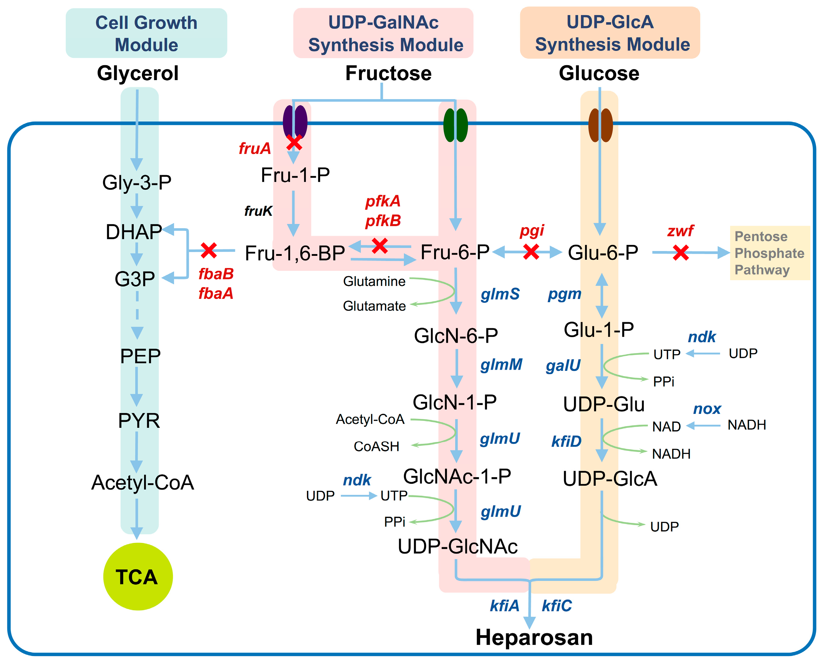
3.1. Design of Co-Utilization Pathways for Glucose, Fructose, and Glycerol

To improve heparosan biosynthesis in EcN, a rational redesign of the glycolytic pathway was carried out, so that glucose and fructose are separately channeled toward the synthesis of Glu-6-P and Fru-6-P, respectively. In detail, the disruption of pgi gene encoding phosphoglucose isomerase prevents the conversion between Glu-6-P and Fru-6-P, and the deletion of the zwf gene encoding Glu-6-P dehydrogenase blocks the entry of Glu-6-P into the pentose phosphate pathway, ensuring that the metabolic flux of Glu-6-P predominantly enters the UDP-GlcA synthesis pathway. For fructose utilization, two alternative approaches were implemented based on previous studies [24,25]. First, the fructose-specific phosphotransferase gene fruA was knocked out, while simultaneously deleting the phosphofructokinase genes pfkA and pfkB to block the entry of Fru-6-P into glycolysis, thereby directing its flux mainly towards the UDP-GlcNAc synthesis pathway. Second, on the basis of pfkAB deletion, knocking out the fbaA/fbaB encoding fructose-1,6-bisphosphate aldolase can also impede the catabolic utilization of fructose. Glycerol was supplied as an auxiliary carbon source to support cell growth, thereby achieving the synergistic utilization of glucose, fructose, and glycerol (Figure 1). The strategy demonstrates how rational pathway engineering can overcome inherent metabolic bottlenecks in heparosan biosynthesis.

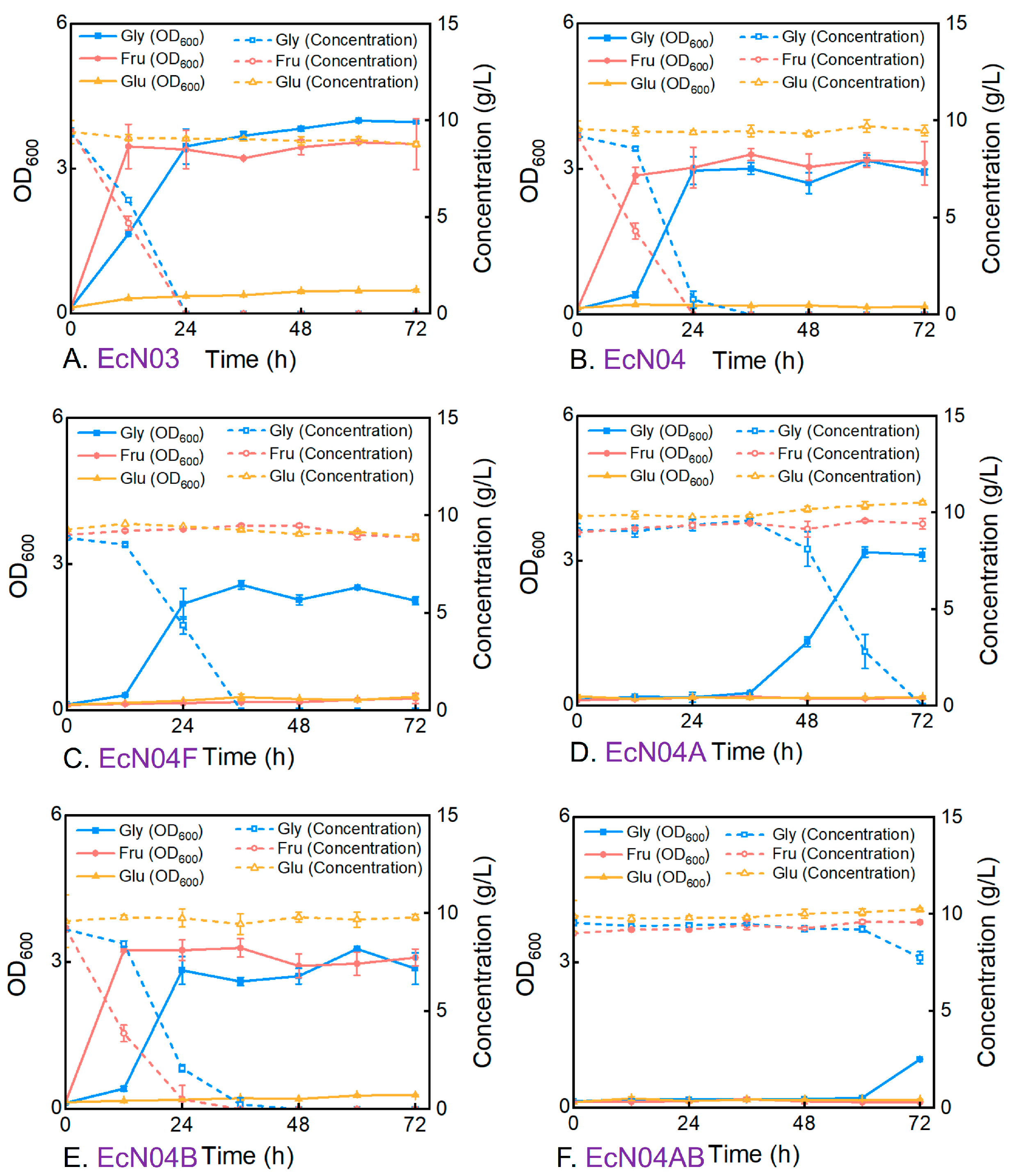
3.2. Metabolic Characteristics of Engineered Mutants During Cultivation on a Single Carbon Source

To verify the feasibility of the aforementioned strategy, the relevant genes were individually deleted to construct a series of E. coli mutants designated EcN03, EcN04, EcN04F, EcN04A, EcN04B, and EcN04AB (Table 1). The M9 minimal medium was supplemented with 10 g/L of either glucose, fructose, or glycerol as the sole carbon source. The cell growth and carbon source consumption profiles of the strains were investigated to evaluate their metabolic efficiencies for different carbon sources. Strain EcN03, with zwf and pfkAB deletions, showed the complete loss of the glucose utilization capacity with negligible biomass accumulation over 72 h, while maintaining normal cell growth and the consumption of both glycerol and fructose (Figure 2A). The subsequent introduction of the pgi knockout in EcN04 resulted in similar glucose and fructose utilization patterns as observed in EcN03. However, glycerol utilization showed a significant lag phase of 12 h (Figure 2B). This observation suggests that the pgi deletion impacts the gluconeogenic pathway, which is essential for generating six-carbon sugars during growth on glycerol [26].
Further genetic modifications revealed distinct functional roles of fructose metabolic enzymes. Strain EcN04F, harboring fruA deletion, showed similar metabolic behavior for glucose and glycerol to that of EcN04, yet its fructose utilization capacity was lost (Figure 2C). This result indicated that deleting pfkAB and fruA genes blocked the formation of fructose-1,6-bisphosphate, thus completely abolishing fructose consumption. In terms of the inactivation of fructose-1,6-bisphosphate aldolase, strain EcN04A, harboring fbaA deletion, showed no significant fructose consumption over 72 h, indicating that the deletion of fbaA, similarly to fruA deletion, effectively blocks fructose utilization (Figure 2D). It is worth noting that the glycerol utilization was further decreased, with glycerol consumption beginning only after 36 h. Since fructose-1,6-bisphosphate aldolase plays roles in both glycolysis and gluconeogenesis, its absence hinders the synthesis of six-carbon sugars when glycerol is employed as the sole carbon source, resulting in an even lower growth rate. In contrast, the deletion of the fbaB knockout showed minimal metabolic effects, and the glucose, fructose, and glycerol utilization profiles were similar to those of EcN04, suggesting that fbaB is not the predominant aldolase (Figure 2E). The fbaAB double knockout EcN04AB exhibited the most severe phenotype with glycerol utilization, with cell growth only after 60 h (Figure 2F). This indicates that the reaction catalyzed by fructose-1,6-bisphosphate aldolase plays a critical role in gluconeogenesis and that the deletion of both isoenzymes severely reduces the rate of glycerol utilization [27]. The above results indicate that in the EcN04F, EcN04A, and EcN04AB strains, the carbon metabolism characteristics adhere to the designed strategy for directional carbon allocation. Neither glucose nor fructose can serve as the primary carbon source for cell growth, while glycerol provides the energy and materials necessary for maintaining cell growth and metabolism.

3.3. Metabolic Characteristics of Engineered Mutants During Cultivation on Mixed Carbon Sources

To systematically evaluate the metabolic efficiency and carbon source allocation patterns of engineered strains under mixed carbon conditions, EcN04F, EcN04A, and EcN04AB were cultivated in M9 minimal medium supplemented with 10 g/L each of glucose, fructose, and glycerol. Growth curves and carbon source consumption profiles were measured to assess metabolic adaptations. In the mixed carbon source medium, EcN04F exhibited a 12 h lag phase followed by exponential growth, reaching a maximum OD600 of 3.6 at 36 h (Figure 3A). This represented a significant improvement over its performance in the glycerol-only medium, suggesting that the presence of glucose and fructose partially compensated for gluconeogenic deficiencies [28]. Notably, the strain preferentially utilized glycerol for growth, with a negligible consumption of glucose or fructose in the M9 medium.
Similar to EcN04F, the EcN04A strain also displayed a markedly shortened growth lag phase under mixed carbon source conditions, achieving a maximum OD600 of 3.42 at 36 h (Figure 3B). The availability of glucose and fructose alleviated gluconeogenic constraints, accelerating both growth initiation and glycerol utilization. In terms of fbaB deletion, EcN04B demonstrated similar cell growth and carbon utilization patterns to those of EcN04, which aligns with previous observations under single carbon source conditions, providing further evidence that fbaB does not play a major role in these metabolic processes (Figure 3C). EcN04AB also exhibited a shortened growth lag phase, with the onset of exponential growth advancing from 60 h to 36 h (Figure 3D). Notably, this lag period remained longer than that observed for EcN04A, demonstrating that while the presence of multiple carbon sources can partially compensate for metabolic deficiencies, the complete knockout of both fbaA and fbaB genes imposes more severe constraints on cellular metabolic adaptation than the single fbaA deletion [29]. This differential response further confirms the hierarchical importance of aldolase isozymes in supporting gluconeogenic flux, with fbaA playing the predominant role and fbaB providing an auxiliary function when fbaA is absent.

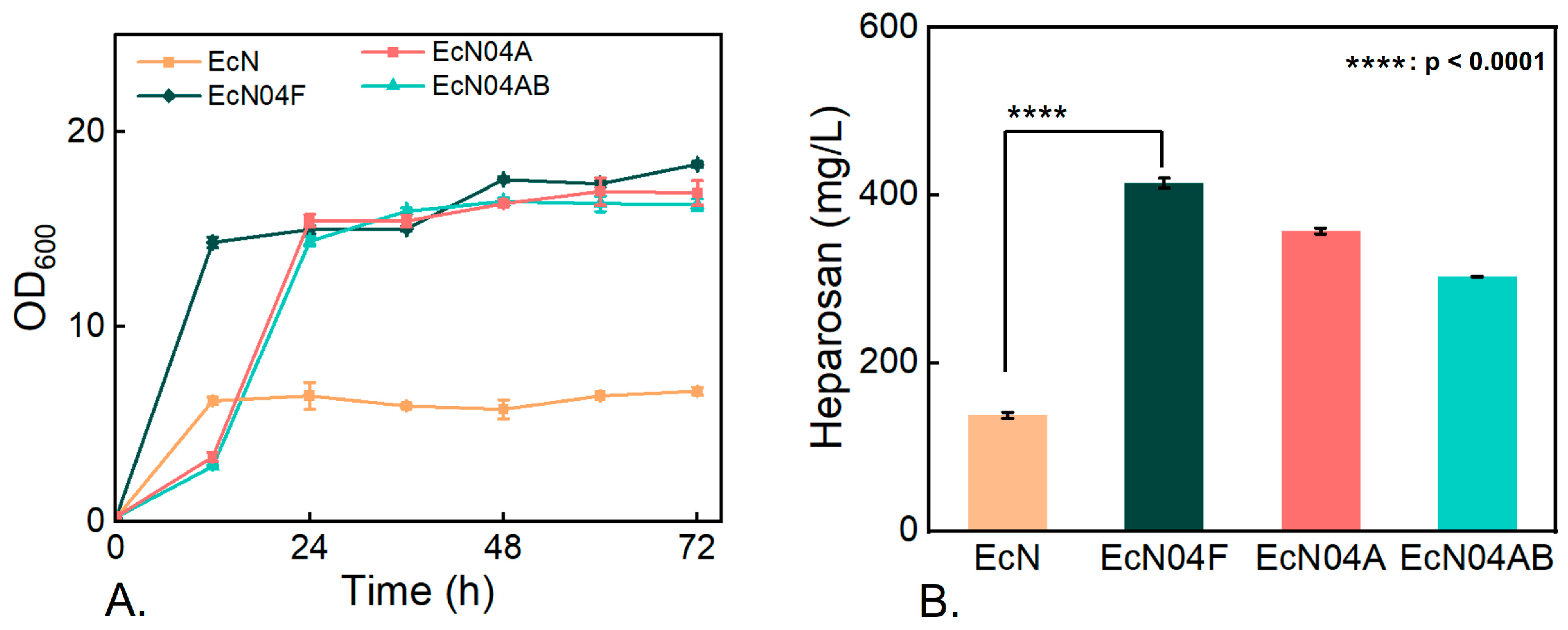
3.4. Effect of Carbon-Flux Allocation on Heparosan Production

Next, a comparative evaluation of heparosan production was conducted using wild-type EcN and the recombinant strains EcN04F, EcN04A, and EcN04AB in shake-flask cultures with the R medium. While EcN utilized 20 g/L glucose as the sole carbon source, each recombinant strain was grown with 20 g/L glycerol to support cell growth, supplemented with 2 g/L glucose and 2 g/L fructose as the designated carbon sources for heparosan synthesis. As shown in Figure 4, the three engineered strains achieved a significantly higher maximum biomass compared to wild-type EcN. Notably, EcN04F exhibited exponential growth during the first 0–12 h and rapidly reached its peak cell density. In comparison, EcN04A and EcN04AB displayed a delayed growth phase during the initial 0–12 h, entering a rapid proliferation stage only after 12 h and ultimately achieving the highest biomass by 24 h. These results demonstrate that the directed carbon-flux strategy markedly enhances maximum cell density, laying a strong foundation for subsequent high-density fermentations.
The metabolic engineering approach yielded substantial improvements in heparosan production, with all engineered strains outperforming the wild-type EcN of 137.68 mg/L. Specifically, EcN04F produced 414.40 mg/L, while EcN04A and EcN04AB achieved 357.21 mg/L and 302.82 mg/L, respectively (Figure 4). This pronounced increase in the product titer confirms that rerouting intracellular carbon flux effectively strengthens the biosynthetic pathway for heparosan. Due to the blocked glycolytic pathway, the conversion efficiency from sugars to heparosan was significantly improved, reaching a yield of 0.10 g/g in strain EcN04F. The superior performance of EcN04F provides important insights for future strain optimization, indicating that the targeted disruption of fructose phosphotransferase systems may be more physiologically favorable than the complete glycolysis blockade for maintaining metabolic balance during heparosan production.

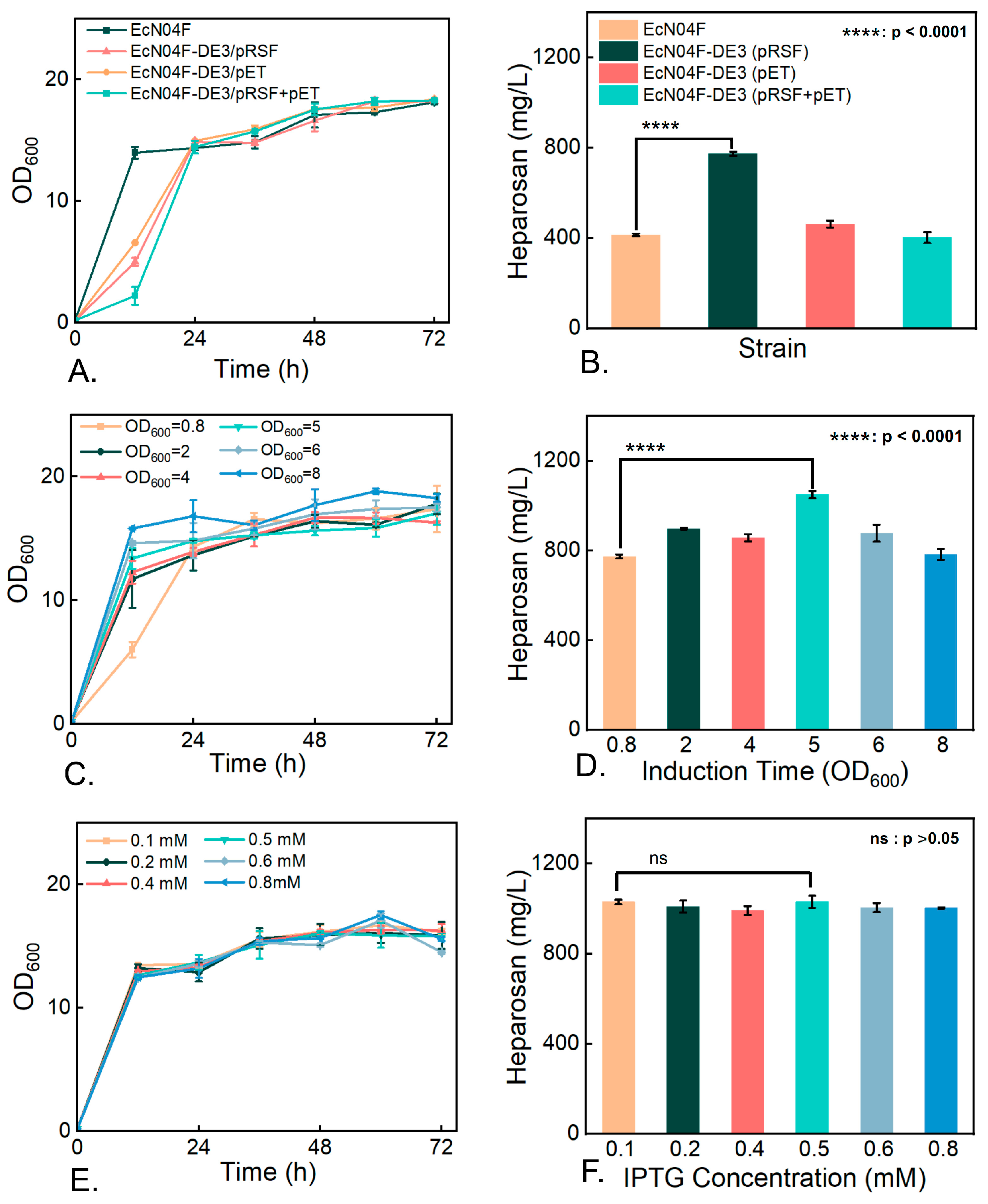
3.5. Impact of Key Enzyme Overexpression on Heparosan Biosynthesis

Finally, the effects of overexpressing key enzymes involved in the heparosan biosynthetic pathway were investigated in strain EcN04F. The selected genes included glmS (glutamine-fructose-6-phosphate aminotransferase), glmM (phosphoglucosamine mutase), kfiD (UDP-glucose dehydrogenase), pgm (phosphoglucomutase), galU (glucose-1-phosphate uridylyltransferase), kfiAC (glycosyltransferases A and C), ndk (nucleoside diphosphate kinase), and nox (NADH oxidase). The effect of individual gene overexpression was evaluated first (see Supplementary Figure S1). Among these, the overexpression of galU yielded the most significant improvement, increasing heparosan production by 31.8% to 578.69 mg/L. Moderate enhancements were observed with glmM (561.63 mg/L), pgm (526.59 mg/L), and kfiD (512.71 mg/L), whereas glmS overexpression showed no statistically significant effect (438.65 mg/L). Glycosyltransferase engineering revealed differential impacts; kfiA overexpression increased the titer by 15.3% to 477.94 mg/L, while kfiC showed negligible effects.
The UDP-sugar biosynthetic process requires cofactors such as UTP and NAD. Nucleoside diphosphate kinase catalyzes the transfer of phosphate groups between nucleoside diphosphates and triphosphates, maintaining the dynamic equilibrium of intracellular UTP pools [30]. Its overexpression may theoretically enhance UTP availability for UDP-sugar precursor synthesis. NADH oxidase directly oxidizes NADH to NAD and may facilitate the rapid regeneration of oxidized cofactors [31]. Unexpectedly, ndk overexpression failed to enhance production, and nox overexpression significantly reduced yields, suggesting that redox imbalance may disrupt precursor biosynthesis.
The poxB gene encodes pyruvate oxidase. Its knockout reduces acetate production with minimal effects on cell growth [32]. The pyruvate oxidase gene poxB in the genome of strain EcN04F was replaced with the DE3 cassette containing the T7 RNA polymerase gene [33], generating a T7-compatible host strain EcN04F-DE3. Using this strain, a dual-plasmid expression system driven by the T7 promoter was employed to co-express five genes that had previously shown positive effects when overexpressed individually. The highest yield was achieved by expressing pRSF-glmM-pgm-galU, resulting in a titer of 773.78 mg/L, which represents an 86.7% increase compared to the original strain EcN04F (Figure 5). The further optimization of IPTG induction conditions revealed that the optimal induction time was when OD600 reached 5, at which point the heparosan titer increased to 1049.96 mg/L, reaching a yield of 0.26 g/g. The IPTG concentration (0.1–0.8 mM), however, had no significant impact on production (Figure 5). These results suggest that delayed induction can effectively reduce the metabolic burden and enhance the biosynthetic efficiency of heparosan.
In previous studies, various metabolic engineering strategies have been employed to enhance heparosan biosynthesis in EcN. Through the overexpression optimization of key genes galU, kfiD, glmM, kfiA, and kfiC in the heparosan synthesis pathway, the titer reached 1.29 g/L in shake-flask cultures, with a conversion yield of 0.0255 g/g. The production titer was further improved to 11.50 g/L via fed-batch fermentation [23]. Chromosome evolution driven by antibiotics was employed to amplify the kps locus, which contains genes for the synthesis and transport of heparosan. The engineered strain with a 24-fold amplified kps copy number achieved a titer of 9.1 g/L in fed-batch fermentation [34]. By reconstructing the glycolytic pathway, we successfully separated the carbon fluxes for biomass formation and heparosan synthesis. The combination of this approach with existing engineering strategies holds potential for additional productivity gains. For instance, experimental evidence confirms that the coordinated overexpression of KfiA/KfiC synthases promotes heparosan chain elongation, positioning them as promising modification targets in future study [35].

4. Conclusions

This study established an optimized E. coli Nissle 1917 platform for heparosan biosynthesis through carbon flux redirection and enzyme engineering. The rational redesign of the glycolytic pathway effectively blocked the catabolic routes of glucose and fructose, channeling their metabolic flux exclusively into the two distinct monosaccharide biosynthesis branches, thereby achieving the balanced partitioning of these divergent metabolic pathways. The combinatorial reinforcement of key enzymes further substantially enhanced the heparosan biosynthetic capability, culminating in a shake-flask titer of 1049.96 mg/L. Through optimized fed-batch fermentation at the bioreactor scale with refined culture conditions and carbon feeding strategies, the heparosan production in EcN is expected to reach higher levels. These findings validate synthetic biology approaches for overcoming pathway competition in complex glycan biosynthesis, offering a sustainable alternative to animal-derived heparin production.

Supplementary Materials

The following supporting information can be downloaded at: https://www.mdpi.com/article/10.3390/fermentation11050289/s1, Table S1: Oligonucleotides used for gene deletion in this study; Table S2: Oligonucleotides used for plasmid construction in this study; Figure S1: Effect of single gene overexpression on heparosan production.

Author Contributions

Conceptualization, Z.-J.L.; methodology, F.S. and R.W.; formal analysis, F.S. and R.W.; investigation, F.S. and R.W.; writing—original draft preparation, Z.-J.L.; writing—review and editing, Z.-J.L.; supervision and project administration, Z.-J.L. All authors have read and agreed to the published version of the manuscript.

Funding

This research was funded by grants from the National Key R&D Program of China, grant number 2021YFC2103100.

Institutional Review Board Statement

Not applicable.

Informed Consent Statement

Not applicable.

Data Availability Statement

The original contributions presented in this study are included in the article/Supplementary Materials. Further inquiries can be directed to the corresponding author.

Conflicts of Interest

The authors declare no conflicts of interest.

References

  1. Sultana, R.; Kamihira, M. Bioengineered heparin: Advances in production technology. Biotechnol. Adv. 2024, 77, 108456. [Google Scholar] [CrossRef] [PubMed]
  2. Stevens, R.L.; Adachi, R. Protease–proteoglycan complexes of mouse and human mast cells and importance of their β-tryptase–heparin complexes in inflammation and innate immunity. Immunol. Rev. 2007, 217, 155–167. [Google Scholar] [CrossRef] [PubMed]
  3. Wander, R.; Kaminski, A.M.; Wang, Z.; Stancanelli, E.; Xu, Y.; Pagadala, V.; Li, J.; Krahn, J.M.; Pham, T.Q.; Liu, J.; et al. Structural and substrate specificity analysis of 3-O-sulfotransferase isoform 5 to synthesize heparan sulfate. ACS Catal. 2021, 11, 14956–14966. [Google Scholar] [CrossRef] [PubMed]
  4. Suzuki, T.; Ishii-Watabe, A.; Hashii, N.; Nakagawa, Y.; Takahashi, T.; Ebisawa, A.; Nishi, S.; Fujita, N.; Bando, A.; Sekimoto, Y.; et al. The establishment and validation of efficient assays for anti-IIa and anti-Xa activities of heparin sodium and heparin calcium. Biologicals 2013, 41, 415–423. [Google Scholar] [CrossRef]
  5. Hogwood, J.; Mulloy, B.; Lever, R.; Gray, E.; Page, C.P. Pharmacology of heparin and related drugs: An update. Pharmacol. Rev. 2023, 75, 328–379. [Google Scholar] [CrossRef]
  6. Wang, P.; Chi, L.; Zhang, Z.; Zhao, H.; Zhang, F.; Linhardt, R.J. Heparin: An old drug for new clinical applications. Carbohydr. Polym. 2022, 295, 119818. [Google Scholar] [CrossRef]
  7. Zhang, Y.; Wang, Y.; Zhou, Z.; Wang, P.; Xi, X.; Hu, S.; Xu, R.; Du, G.; Li, J.; Chen, J.; et al. Synthesis of bioengineered heparin by recombinant yeast Pichia pastoris. Green Chem. 2022, 24, 3180–3192. [Google Scholar] [CrossRef]
  8. Vann, W.F.; Schmidt, M.A.; Jann, B.; Jann, K. The structure of the capsular polysaccharide (K5 antigen) of urinary-tract-infective Escherichia coli 010:K5:H4. A polymer similar to desulfo-heparin. Eur. J. Biochem. 1981, 116, 359–364. [Google Scholar] [CrossRef]
  9. Datta, P.; Fu, L.; Brodfuerer, P.; Dordick, J.S.; Linhardt, R.J. High density fermentation of probiotic E. coli Nissle 1917 towards heparosan production, characterization, and modification. Appl. Microbiol. Biotechnol. 2021, 105, 1051–1062. [Google Scholar] [CrossRef]
  10. DeAngelis, P.L.; White, C.L. Identification and molecular cloning of a heparosan synthase from Pasteurella multocida type D. J. Biol. Chem. 2002, 277, 7209–7213. [Google Scholar] [CrossRef]
  11. Sheng, L.L.; Cai, Y.M.; Li, Y.; Huang, S.L.; Sheng, J.Z. Advancements in heparosan production through metabolic engineering and improved fermentation. Carbohydr. Polym. 2024, 331, 121881. [Google Scholar] [CrossRef] [PubMed]
  12. Deng, J.Q.; Li, Y.; Wang, Y.J.; Cao, Y.L.; Xin, S.Y.; Li, X.Y.; Xi, R.M.; Wang, F.S.; Sheng, J.Z. Biosynthetic production of anticoagulant heparin polysaccharides through metabolic and sulfotransferases engineering strategies. Nat. Commun. 2024, 15, 3755. [Google Scholar] [CrossRef] [PubMed]
  13. Zhang, C.; Liu, L.; Teng, L.; Chen, J.; Liu, J.; Li, J.; Du, G.; Chen, J. Metabolic engineering of Escherichia coli BL21 for biosynthesis of heparosan, a bioengineered heparin precursor. Metab. Eng. 2012, 14, 521–527. [Google Scholar] [CrossRef] [PubMed]
  14. Barreteau, H.; Richard, E.; Drouillard, S.; Samain, E.; Priem, B. Production of intracellular heparosan and derived oligosaccharides by lyase expression in metabolically engineered E. coli K-12. Carbohydr. Res. 2012, 360, 19–24. [Google Scholar] [CrossRef]
  15. Williams, A.; Gedeon, K.S.; Vaidyanathan, D.; Yu, Y.; Collins, C.H.; Dordick, J.S.; Linhardt, R.J.; Koffas, M.A.G. Metabolic engineering of Bacillus megaterium for heparosan biosynthesis using Pasteurella multocida heparosan synthase, PmHS2. Microb. Cell Fact. 2019, 18, 132. [Google Scholar] [CrossRef]
  16. Jin, P.; Zhang, L.; Yuan, P.; Kang, Z.; Du, G.; Chen, J. Efficient biosynthesis of polysaccharides chondroitin and heparosan by metabolically engineered Bacillus subtilis. Carbohydr. Polym. 2016, 140, 424–432. [Google Scholar] [CrossRef]
  17. Hu, L.; Wang, Y.; Hu, Y.; Yin, J.; Wang, L.; Du, G.; Chen, J.; Kang, Z. Biosynthesis of non-sulfated high-molecular-weight glycosaminoglycans and specific-sized oligosaccharides. Carbohydr. Polym. 2022, 295, 119829. [Google Scholar] [CrossRef]
  18. Grozdanov, L.; Raasch, C.; Schulze, J.; Sonnenborn, U.; Gottschalk, G.; Hacker, J.; Dobrindt, U. Analysis of the genome structure of the nonpathogenic probiotic Escherichia coli strain Nissle 1917. J. Bacteriol. 2004, 186, 5432–5441. [Google Scholar] [CrossRef]
  19. Sonnenborn, U.; Schulze, J. The non-pathogenic Escherichia coli strain Nissle 1917—features of a versatile probiotic. Microb. Ecol. Health Dis. 2009, 21, 122–158. [Google Scholar]
  20. Zhang, Q.; Yao, R.; Chen, X.; Liu, L.; Xu, S.; Chen, J.; Wu, J. Enhancing fructosylated chondroitin production in Escherichia coli K4 by balancing the UDP-precursors. Metab. Eng. 2018, 47, 314–322. [Google Scholar] [CrossRef]
  21. Yan, H.; Bao, F.; Zhao, L.; Yu, Y.; Tang, J.; Zhou, X. Cyclic AMP (cAMP) receptor protein-cAMP complex regulates heparosan production in Escherichia coli strain Nissle 1917. Appl. Environ. Microbiol. 2015, 81, 7687–7696. [Google Scholar] [CrossRef] [PubMed]
  22. Datsenko, K.A.; Wanner, B.L. One-step inactivation of chromosomal genes in Escherichia coli K-12 using PCR products. Proc. Natl. Acad. Sci. USA 2000, 97, 6640–6645. [Google Scholar] [CrossRef] [PubMed]
  23. Hu, S.; Zhao, L.; Hu, L.; Xi, X.; Zhang, Y.; Wang, Y.; Chen, J.; Chen, J.; Kang, Z. Engineering the probiotic bacterium Escherichia coli Nissle 1917 as an efficient cell factory for heparosan biosynthesis. Enzyme. Microb. Technol. 2022, 158, 110038. [Google Scholar] [CrossRef] [PubMed]
  24. Kornberg, H.L. Routes for fructose utilization by Escherichia coli. J. Mol. Microbiol. Biotechnol. 2001, 3, 355–359. [Google Scholar]
  25. Prior, T.I.; Kornberg, H.L. Nucleotide sequence of fruA, the gene specifying enzyme IIfru of the phosphoenolpyruvate-dependent sugar phosphotransferase system in Escherichia coli K12. J. Gen. Microbiol. 1988, 134, 2757–2768. [Google Scholar] [CrossRef]
  26. Shimizu, K.; Matsuoka, Y. Redox rebalance against genetic perturbations and modulation of central carbon metabolism by the oxidative stress regulation. Biotechnol. Adv. 2019, 37, 107441. [Google Scholar] [CrossRef]
  27. Abernathy, M.H.; Zhang, Y.; Hollinshead, W.D.; Wang, G.; Baidoo, E.E.K.; Liu, T.; Tang, Y.J. Comparative studies of glycolytic pathways and channeling under in vitro and in vivo modes. AIChE J. 2019, 65, 483–490. [Google Scholar] [CrossRef]
  28. Martínez-Gómez, K.; Flores, N.; Castañeda, H.M.; Martínez-Batallar, G.; Hernández-Chávez, G.; Ramírez, O.T.; Gosset, G.; Encarnación, S.; Bolivar, F. New insights into Escherichia coli metabolism: Carbon scavenging, acetate metabolism and carbon recycling responses during growth on glycerol. Microb. Cell Fact. 2012, 11, 46. [Google Scholar] [CrossRef]
  29. Macomber, L.; Elsey, S.P.; Hausinger, R.P. Fructose-1,6-bisphosphate aldolase (class II) is the primary site of nickel toxicity in Escherichia coli. Mol. Microbiol. 2011, 82, 1291–1300. [Google Scholar] [CrossRef]
  30. Yu, H.; Rao, X.; Zhang, K. Nucleoside diphosphate kinase (Ndk): A pleiotropic effector manipulating bacterial virulence and adaptive responses. Microbiol. Res. 2017, 205, 125–134. [Google Scholar] [CrossRef]
  31. Gao, H.; Li, J.; Sivakumar, D.; Kim, T.-S.; Patel, S.K.S.; Kalia, V.C.; Kim, I.-W.; Zhang, Y.-W.; Lee, J.-K. NADH oxidase from Lactobacillus reuteri: A versatile enzyme for oxidized cofactor regeneration. Int. J. Biol. Macromol. 2019, 123, 629–636. [Google Scholar] [CrossRef] [PubMed]
  32. Ye, T.; Ding, W.; An, Z.; Zhang, H.; Wei, X.; Xu, J.; Liu, H.; Fang, H. Increased distribution of carbon metabolic flux during de novo cytidine biosynthesis via attenuation of the acetic acid metabolism pathway in Escherichia coli. Microb. Cell Fact. 2025, 24, 36. [Google Scholar] [CrossRef] [PubMed]
  33. Du, F.; Liu, Y.Q.; Xu, Y.S.; Li, Z.J.; Wang, Y.Z.; Zhang, Z.X.; Sun, X.M. Regulating the T7 RNA polymerase expression in E. coli BL21 (DE3) to provide more host options for recombinant protein production. Microb. Cell Fact. 2021, 20, 189. [Google Scholar] [CrossRef] [PubMed]
  34. Yu, Y.; Gong, B.; Wang, H.; Yang, G.; Zhou, X. Chromosome evolution of Escherichia coli Nissle 1917 for high-level production of heparosan. Biotechnol. Bioeng. 2023, 120, 1081–1096. [Google Scholar] [CrossRef]
  35. Wang, Z.; Dordick, J.S.; Linhardt, R.J. Escherichia coli K5 heparosan fermentation and improvement by genetic engineering. Bioeng. Bugs 2011, 2, 63–67. [Google Scholar] [CrossRef]
Figure 1. Metabolic engineering strategy for heparosan biosynthesis via a tripartite carbon allocation strategy.
Fermentation 11 00289 g001
Figure 2. Cell growth and carbon utilization profiles of engineered E. coli strains cultivated with a single carbon source in M9 medium.
Fermentation 11 00289 g002
Figure 3. Cell growth and carbon utilization profiles of engineered E. coli strains cultivated with mixed carbon sources in M9 medium.
Fermentation 11 00289 g003
Figure 4. Improved heparosan production through carbon flux redirection. (A) Cell growth. (B) Heparosan production.
Fermentation 11 00289 g004
Figure 5. Effect of key enzyme overexpression on heparosan production. (A,B) Effects of multi-enzyme overexpression on cell growth and heparosan production. (C,D) Effects of induction time on cell growth and heparosan production. (E,F) Effects of IPTG concentration on cell growth and heparosan production.
Fermentation 11 00289 g005
Table 1. Strains and plasmids used in this study.
Strains/PlasmidsDescriptionReferences
Strains
E. coli DH5αWild-type strain for gene cloningTakara Bio
E. coli Nissle 1917Wild-type strain for heparosan productionDSMZ *
Bacillus subtilis 168Wild-type strain for cloning galU geneDSMZ
E. coli EcN03Nissle 1917 Δzwf ΔpfkB ΔpfkAThis study
E. coli EcN04Nissle 1917 Δzwf ΔpfkB ΔpfkA ΔpgiThis study
E. coli EcN04FNissle 1917 Δzwf ΔpfkB ΔpfkA Δpgi ΔfruAThis study
E. coli EcN04ANissle 1917 Δzwf ΔpfkB ΔpfkA Δpgi ΔfbaAThis study
E. coli EcN04BNissle 1917 Δzwf ΔpfkB ΔpfkA Δpgi ΔfbaBThis study
E. coli EcN04ABNissle 1917 Δzwf ΔpfkB ΔpfkA Δpgi ΔfbaA ΔfbaBThis study
E. coli EcN04F-DE3Nissle 1917 Δzwf ΔpfkB ΔpfkA Δpgi ΔfruA ΔpoxB::DE3This study
Plasmids
pKD13Plasmid harboring KanR and FLP recognition targetYale CGSC **
pKD46λ-Red recombinase expression helper plasmidYale CGSC
pCP20FLP recombinase helper plasmidYale CGSC
pEC-XK99EExpression vector, trc promoter, KanRSangon Biotech
pETDuetExpression vector, T7 promoter, AmpRSangon Biotech
pRSFDuetExpression vector, T7 promoter, KanRSangon Biotech
pEC-kfiDpEC-XK99E derived, harboring kfiD from EcNThis study
pEC-glmMpEC-XK99E derived, harboring glmM from EcNThis study
pEC-glmSpEC-XK99E derived, harboring glmS from EcNThis study
pEC-pgmpEC-XK99E derived, harboring pgm from EcNThis study
pEC-galUpEC-XK99E derived, harboring galU from B. subtilis 168This study
pEC-kfiApEC-XK99E derived, harboring kfiA from EcNThis study
pEC-kfiCpEC-XK99E derived, harboring kfiC from EcNThis study
pEC-ndkpEC-XK99E derived, harboring ndk from EcNThis study
pEC-nox1pEC-XK99E derived, harboring nox1 from L. rhamnosus GGThis study
pEC-nox2pEC-XK99E derived, harboring nox2 from L. rhamnosus GGThis study
pEC-nox3pEC-XK99E derived, harboring nox3 from L. rhamnosus GGThis study
pEC-nox4pEC-XK99E derived, harboring nox4 from L. rhamnosus GGThis study
pET-kfiD-kfiApETDuet derived, harboring kfiA and kfiCThis study
pRSF-glmM-pgm-galUpRSFDuet derived, harboring glmM, pgm, and galUThis study
* DSMZ, German Collection of Microorganisms and Cell Cultures. ** Yale CGSC, The E. coli Genetic Stock Center.
Disclaimer/Publisher’s Note: The statements, opinions and data contained in all publications are solely those of the individual author(s) and contributor(s) and not of MDPI and/or the editor(s). MDPI and/or the editor(s) disclaim responsibility for any injury to people or property resulting from any ideas, methods, instructions or products referred to in the content.

