Next Article in Journal
In-Depth Characterization of the Volatile Aroma Profile and Other Characteristics of White Wine Produced by Sequential Inoculation with a Lachancea thermotolerans Starter Yeast Strain
Next Article in Special Issue
Correction: Wang et al. Antibacterial Efficacy of Feline-Derived Lactic Acid Bacteria against Enteropathogenic Escherichia coli: A Comprehensive In Vitro Analysis. Fermentation 2024, 10, 514
Previous Article in Journal
Bioprospecting of Metschnikowia pulcherrima Strains, Isolated from a Vineyard Ecosystem, as Novel Starter Cultures for Craft Beer Production
 
 
Correction published on 6 December 2024, see Fermentation 2024, 10(12), 622.
Article

Antibacterial Efficacy of Feline-Derived Lactic Acid Bacteria against Enteropathogenic Escherichia coli: A Comprehensive In Vitro Analysis

by
Weiwei Wang
1,2,3,
Hao Dong
1,3,
Qianqian Chen
1,
Xiaohan Chang
1,
Longjiao Wang
2,4,
Chengyi Miao
1,
Shuxing Chen
1,
Lishui Chen
1,
Ran Wang
4,
Shaoyang Ge
4 and
Wei Xiong
1,2,*
1
Food Laboratory of Zhongyuan, Luohe 462300, China
2
Henan Zhiyuan Henuo Technology Co., Ltd., Luohe 462300, China
3
College of Food Science and Technology, Henan University of Technology, Zhengzhou 450001, China
4
Key Laboratory of Precision Nutrition and Food Quality, Department of Nutrition and Health, China Agricultural University, Beijing 100083, China
*
Author to whom correspondence should be addressed.
Fermentation 2024, 10(10), 514; https://doi.org/10.3390/fermentation10100514
Submission received: 11 September 2024 / Revised: 3 October 2024 / Accepted: 8 October 2024 / Published: 10 October 2024 / Corrected: 6 December 2024
(This article belongs to the Special Issue Antimicrobial Metabolites: Production, Analysis and Application)

Abstract

This study evaluated the antibacterial efficacy of 700 feline-derived lactic acid bacteria (LAB) strains against enteropathogenic Escherichia coli (EPEC), a common cause of diarrhea in cats. Following comprehensive screening, strains ZY25 and ZY35 were identified as the most effective, with inhibition zones of ≥22 mm. These strains demonstrated strong tolerance against stress conditions, such as low pH, bile salts, and gastrointestinal fluids, alongside high hydrophobicity and auto-aggregation abilities. Safety evaluations confirmed the absence of hemolytic activity, virulence factors, and antibiotic resistance genes. The antibacterial activity of these strains is attributed to the production of organic acids, particularly lactic acid and acetic acid. These findings suggest that strains ZY25 and ZY35 have potential as natural and effective probiotic treatments for managing EPEC-induced diarrhea in cats, thus offering an alternative to conventional antibiotics.

1. Introduction

Enteropathogenic Escherichia coli (EPEC) is a significant cause of diarrhea in cats, particularly affecting kittens. EPEC leads to severe intestinal damage, resulting in symptoms such as vomiting, lethargy, and dehydration, which can be fatal if untreated. Kittens with EPEC have significantly greater intestinal damage and higher quantities of the pathogen compared to those without diarrhea [1]. Additionally, EPEC is known to cause similar severe diarrheal disease and intestinal damage in other animals, indicating its broad pathogenic potential [2]. Infection with EPEC results in significant fluid and electrolyte losses, exacerbating the risk of dehydration and necessitating medical intervention [3]. This infection poses a substantial economic burden due to the costs of veterinary care, decreased productivity in breeding operations, and emotional distress experienced by pet owners [4,5]. The conventional treatment for EPEC-induced diarrhea includes antibiotics; however, the growing prevalence of antibiotic-resistant EPEC strains complicates these treatments. This resistance necessitates the search for alternative solutions, with probiotics emerging as a promising approach due to their natural and sustainable benefits [6,7].
Probiotics, which are live microorganisms that confer health benefits to the host, have demonstrated effectiveness in treating diarrhea caused by Escherichia coli (E. coli) in various animal models. These benefits are derived from mechanisms such as the competitive exclusion of pathogens, enhancement of the host immune response, and production of antimicrobial substances. Several studies have highlighted the efficacy of probiotics in addressing E. coli-induced diarrhea. For instance, research has shown that exopolysaccharides produced by Bifidobacterium animalis can mitigate E. coli-induced damage in intestinal epithelial cells by inhibiting apoptosis and restoring autophagy [8]. In another study, a screening of over 1100 Lactobacillus plantarum strains identified several with potent inhibitory effects against enterotoxigenic Escherichia coli (ETEC) K88 in weaned piglets, suggesting the potential of these strains to reduce E. coli infections [9]. Furthermore, strains of Lactobacillus isolated from various sources have demonstrated significant antimicrobial activity against uropathogenic Escherichia coli (UPEC), effectively reducing biofilm formation by up to 50% [10]. Comparative studies on different probiotics also revealed that a multi-strain synbiotic that contained various Lactobacillus and Bifidobacterium strains exhibited significantly stronger inhibition of E. coli and other pathogens compared with single-strain probiotics [11].
Given the unique gut microbiota of cats, it is crucial to develop probiotics from cat-derived strains. Host-specific probiotics are more likely to survive in, colonize, and exert beneficial effects on the feline gastrointestinal tract. Research has shown that these species-specific probiotics tend to be more effective than those derived from non-host sources. For example, studies have demonstrated that probiotics isolated from the same species provide better colonization and health benefits compared with commercial probiotics from different species [12]. Further supporting this concept, genetic variation and host-specific adaptation studies revealed that Lactobacillus johnsonii strains exhibit host-specific genetic variations when isolated from different animal hosts, suggesting a co-evolution with their hosts that enhances their effectiveness [13]. Additionally, Enterococcus hirae F2, a strain isolated from the gut of Catla catla fish, showed significant probiotic potential by surviving under highly acidic and bile salt conditions and exhibiting strong antimicrobial activity against pathogens [14]. These findings highlight the importance of using host-specific probiotics for achieving maximum efficacy in treating and maintaining the health of different species.
This study focused on isolating and screening lactic acid bacteria (LAB) strains from healthy cats’ feces to find probiotics that effectively combat EPEC. Initially, 700 LAB strains were isolated. From these, 200 randomly selected strains underwent 16S rRNA sequencing. These isolates were then tested for antibacterial activity against EPEC. The selected strains were further evaluated for their physiological and biochemical properties, such as their tolerance to different temperatures, salt concentrations, and pH levels, which ensured they can be easily propagated and maintained during production and storage. The safety assessments included testing for hemolytic activity, antibiotic susceptibility, and the absence of virulence and biogenic amine genes, which confirmed the strains’ safety. This research is significant for developing effective, host-specific probiotics for cats, thus offering a natural and sustainable solution to manage EPEC-induced diarrhea and reducing reliance on antibiotics.

2. Materials and Methods

2.1. Sample Collection and LAB Isolation

Three hundred fresh fecal samples from healthy cats were collected from pet stores and catteries in Luohe, China, between September and December 2023. Fecal samples were collected using sterile sampling spoons from the upper part of naturally expelled fecal pellets to avoid contamination. Each sample was placed in a sterile EP tube, labeled, and immediately transported to the laboratory for analysis in a cold chain box with sufficient dry ice. The time from sampling to analysis did not exceed 3 h, ensuring each sample’s integrity. All procedures involving the collection and handling of fecal samples were conducted in compliance with ethical guidelines approved by the Institutional Animal Care and Use Committee of China Agricultural University (AW20704202-5-4). In the laboratory, the fecal samples were serially diluted with sterile distilled water and plated on de Man, Rogosa, and Sharpe (MRS) agar (Merck, Darmstadt, Germany). The plates were incubated at 37 °C for 48 h under anaerobic conditions to culture single LAB colonies. Colonies that were round, raised or flat, creamy white or slightly yellow, moist, medium sized, and neatly edged were preliminarily identified as LAB. These isolates were stored at −80 °C for further testing and analysis.

2.2. Preliminary Identification of Strains

Among the approximately 700 preserved LAB strains, 200 strains were randomly selected for a diversity analysis.

2.2.1. Morphological Characteristics

The morphological characteristics of the LAB were identified using Gram staining. A drop of sterile water was placed on a slide, and a small amount of a single colony was smeared onto the slide and then air-dried over an alcohol lamp. The smear was stained with ammonium oxalate crystal violet for 1 min and rinsed with sterile water, followed by air-drying over an alcohol lamp. An iodine solution was then added to the slide for approximately 1 min for mordanting, rinsed with sterile water, and air dried. The slide was decolorized with 95% alcohol for 20 s, rinsed with sterile water, and air-dried. Subsequently, the smear was counterstained with safranin for 1 min, rinsed with sterile water, and air-dried. Finally, the slide was examined under a light microscope.

2.2.2. Physiological Experiments

The experiments used to identify the physiological characteristics of LAB mainly included the catalase and glucose gas production tests.
Catalase test: Place 100 μL of 3% H2O2 on a blank culture dish. Using an inoculating loop, pick a single colony and place it into the H2O2. Observe whether gas bubbles are produced. If bubbles are formed, the result is catalase-positive; if no bubbles are formed, the result is catalase-negative.
Glucose gas production test: Culture the lactic acid bacteria by inoculating a single colony into MRS broth and incubating at 30 °C for 24 h. Invert a Durham tube in a test tube containing the cultured lactic acid bacteria inoculated at 1% into MRS broth and incubate statically at 30 °C for 7 days. Observe the Durham tube for the presence of gas bubbles. The presence of gas bubbles indicates heterofermentative fermentation, while the absence of bubbles indicates homofermentative fermentation.

2.2.3. 16 S rRNA Gene Analysis

The selected 200 LAB strains were identified through genetic analysis using PCR and 16S rRNA gene sequencing. The universal primers 27 F (5′-AGAGTTTGATCCTGGCTC AG-3′) and 1492 R (5′-GGTTACCTTGTTACGACTT-3′) were utilized for the PCR amplification of the 16S rRNA gene. The amplified products were then analyzed by Sangon Biotech Co., Ltd., Shanghai, China. The sequence similarities of each contig were assessed by comparing their homologies in the GenBank database using BLAST (http://www.ncbi.nlm.nih.gov accessed on 12 January 2024). A phylogenetic tree of the selected high-performance strains was subsequently constructed using the neighbor-joining method with MEGA-X software (version 10.1.5).

2.3. Screening of LAB with Strong Antibacterial Activity against EPEC

The well diffusion technique was adapted from the method described by Sirichokchatchawan et al. [15], with modifications to suit this study. The target bacterium, EPEC, was cultured in a nutrient liquid medium and incubated at 37 °C with shaking at 180 rpm for 12 h until the concentration reached 1 × 10⁸ CFU/mL. Then, 200 µL of the bacterial suspension was spread on LB agar plates. Separately, 200 µL of cultures of different LAB strains, each at a concentration of 1 × 10⁸ CFU/mL, that had been incubated for 16 h was added to wells (10 mm in diameter) punched into the LB agar plates. Uninoculated MRS broth and penicillin served as the negative and positive controls, respectively. The EPEC strain was kindly provided by China Agricultural University. The LAB strains that produced inhibition zones greater than 18 mm in diameter were selected for further physiological and biochemical characterization.

2.4. Physiological and Biochemical Characteristics of Selected LAB Isolates

Following the methodology described by Zhang et al. [16], the physiological and biochemical characteristics of the selected LAB strains, including pH, salt, and temperature tolerance, were evaluated using MRS broth. Single LAB colonies were initially picked and inoculated into 20 mL of sterile MRS liquid medium. The cultures were incubated at 37 °C for 16 h, after which the OD was adjusted to 0.8 at 600 nm using sterile water. Then, 100 µL of each LAB suspension was mixed with 9.9 mL of MRS broth.
Acid and alkaline resistance: The LAB strains were tested for acid and alkaline resistance by culturing them in MRS broth adjusted to various pH levels (3.0, 3.5, 4.0, 4.5, 5.0, 6.0, 9.0, and 10.0). These cultures were incubated at 37 °C for 7 days.
Salt tolerance: The salt tolerance was assessed by culturing the LAB strains in MRS broth containing 3.0% and 6.5% NaCl, with the incubation at 37 °C for 48 h.
Temperature tolerance: The temperature tolerance of the LAB strains was determined by incubating them in MRS broth at different temperatures (5 °C, 10 °C, 45 °C, and 50 °C) for 7 days.
Post-incubation, the growth rates of the LAB strains were measured using the turbidimetry method by recording absorbance values using the OD at 600 nm. The visual turbidity was also noted. A sterile MRS medium without inoculation served as the control, with its OD600 value recorded as 0. The growth was categorized as follows based on the OD600 readings: 0 ≤ OD600 ≤ 0.2 indicated no growth (“−”); 0.2 < OD600 ≤ 0.6 indicated weak growth (“w”); and OD600 > 0.6 indicated growth (“+”).

2.5. Hydrophobicity and Auto-Aggregation Ability of Selected LAB Isolates

The adhesion ability of the selected LAB strains, as indicated by the cell surface hydrophobicity and auto-aggregation, was tested using the methods described by Wang et al. [9]. The strains that exhibited high cell surface hydrophobicity and auto-aggregation were further analyzed for gastrointestinal tolerance.

2.6. Survival of Representative Strains in GI Fluids

The simulated gastric fluid (SGF) and simulated intestinal fluid (SIF) tests were performed following the methods described by Zhang et al. [17]. The LAB strains were exposed to SGF for 3 h and SIF for 4 h. The strains that demonstrated strong tolerance to both SGF and SIF were selected for further safety analysis.

2.7. Safety Evaluation

2.7.1. Hemolytic Activity

Hemolytic activity was assessed using blood agar plates according to the manufacturer’s instructions. Fresh bacterial strains ZY25, ZY33, and ZY35 were streaked on Columbia blood agar plates. Staphylococcus aureus ATCC 29213T was used as the positive control. The plates were incubated, and the hemolytic activity was evaluated by observing the clear zones around the bacterial colonies, which indicated hemolysis.

2.7.2. Antibiotic Susceptibility

The antibiotic susceptibility of LAB isolates was determined using a modified disk diffusion method based on the protocol described by Niu et al. [18]. The antibiotics tested included gentamicin (GEN, 10 μg/disk), ciprofloxacin (CIP, 5 μg/disk), ceftriaxone (CTR, 30 μg/disk), erythromycin (E, 15 μg/disk), ampicillin (AMP, 10 μg/disk), tetracycline (TET, 30 μg/disk), compound sulfamethoxazole (SXT, 25 μg/disk), chloramphenicol (C, 30 μg/disk), lincomycin (MY, 2 μg/disk), and penicillin (PEN, 10 μg/disk). The results were expressed in millimeter diameters of the inhibition zones. The susceptibility of the isolates was classified as resistant, intermediate resistant, or susceptible according to the cutoff values proposed by de Souza et al. [19]. Each test was conducted in triplicate to ensure accuracy and reproducibility.

2.7.3. PCR Screening of Strains for Virulence Factors, Biogenic Amines, and Antibiotic Resistance Genes

The genetic traits related to virulence factors, biogenic amines, and antibiotic resistance in strains ZY25 and ZY35 were screened using PCR protocols following the method outlined by Wang et al. [9]. Enterococcus faecalis ATCC 29212T, which harbors the target virulence genes (ace, cylA, and gelE), was used as the positive control, while Milli-Q water was used as the negative control.

2.8. Broad-Spectrum Antibacterial Activity and Antibacterial Substances

2.8.1. Broad-Spectrum Antibacterial Activity

The antimicrobial activity of ZY25 and ZY35 against various pathogenic bacteria was evaluated using the agar well diffusion method, as referenced from Section 2.3 of the standard protocol. The broad-spectrum antimicrobial efficacy of ZY25 and ZY35 were tested against the following bacterial strains: Pseudomonas aeruginosa CICC 23694T, Staphylococcus aureus ATCC 29213T, Listeria monocytogenes CICC 23929T, Escherichia coli CICC 24189T, Bacillus subtilis CICC 10275T, and Shigella dysenteriae CICC 23829T.

2.8.2. Antimicrobial Substance Exploration

The antimicrobial substances produced by strains ZY25 and ZY35 were investigated using the method described by Ni et al. [20]. The experiments aimed to eliminate the effects of acid, hydrogen peroxide, and protease hydrolysis. First, the pH of the fermentation broths was adjusted within a range of 2.5 to 10.0 using 0.2 M hydrochloric acid and 0.2 M sodium hydroxide solutions. To remove the hydrogen peroxide, the broths were treated with a 0.5 mg/mL catalase solution and incubated at 37 °C for 2 h. For the protease treatment, the pH was adjusted to 6.0, and the broths were mixed with 1 mg/mL of proteinase K, trypsin, and pepsin, respectively, and then incubated at 37 °C for 2 h. Following these treatments, the broths were centrifuged at 8000 rpm for 10 min, and the supernatants were collected. The antimicrobial activity of the supernatants against EPEC was assessed using the agar well diffusion method, with untreated MRS broth supernatants as controls. Each experiment was performed in triplicate to ensure accuracy.

2.8.3. Growth Curve and Acid Production Curve

Each LAB colony was isolated and cultured in 20 mL of sterile MRS broth. The optical density at 600 nm (OD600) and colony forming units (CFU/mL) were measured at 2-h intervals up to 24 h post-inoculation at 37 °C. Additionally, the pH of each fermentation solution was recorded at 6-h intervals up to 48 h post-inoculation at 37 °C.

2.8.4. Determination of Organic Acid Content by High-Performance Liquid Chromatography (HPLC)

Chemicals and reagents (methanol, acetonitrile, formic acid, 1-(3-Dimethylaminopropyl)-3-ethylcarbodiimide, chloroform, and 3-nitrophenylhydrazine) were sourced from ANPEL (Shanghai, China), with all solvents being LC-MS grade. Ultra-pure water was prepared using a Milli-Q system (Merck Millipore, Burlington, MA, USA). The samples were extracted with 1 mL of a methanol–chloroform (7:3) solution on ice for 30 min; then, 600 μL of H2O was added, and the mixture was centrifuged at 12,000 rpm at 4 °C for 10 min. The supernatant was collected, and the process was repeated. For the derivatization, 10 μL of 0.1 M EDC and 10 μL of 0.1 M 3NPH were added, and the reaction proceeded for 30 min at 40 °C. The extracts were analyzed using a UPLC-Orbitrap-MS system with a Waters BEH C18 column (50 × 2.1 mm, 1.8 μm) at 40 °C, a flow rate of 0.35 mL/min, and an injection volume of 2 μL. The solvent system was water (0.1% formic acid)–acetonitrile (0.1% formic acid) with a gradient program from 90:10 to 10:90 and back to 90:10. HRMS data were recorded on a Q Exactive hybrid Q-Orbitrap mass spectrometer using Fullms-ms2 methods with the following parameters: −2.8 kV spray voltage, 40 arb sheath gas, 10 arb aux gas, 320 °C capillary temperature, and 350 °C aux gas heater temperature. The data were processed with Xcalibur 4.1 (Thermo Scientific, Waltham, MA, USA) and TraceFinder™ 4.1 Clinical (Thermo Scientific, Waltham, MA, USA), and the results were outputted in Excel format [21].

2.9. Statistical Analyses

Each test was performed in triplicate. The data analysis was conducted using one-way ANOVA or a paired t-test in SPSS 22.0 (IBM Corp., Armonk, NY, USA). The results are presented as the mean ± standard error of the mean (SEM), with p < 0.05 indicating statistical significance.

3. Results

3.1. Isolation and Screening of Lactic Acid Bacteria in Healthy Cat Feces

Approximately 700 LAB strains were isolated from the feces of 300 healthy cats. A random selection of 200 LAB strains underwent 16S rRNA sequencing. Statistical analysis was performed on the successfully sequenced strains, with the results shown in Figure 1. Among the 200 LAB strains, the top six strains identified were Ligilactobacillus animalis, Ligilactobacillus salivarius, Enterococcus hirae, Ligilactobacillus agilis, Enterococcus faecium, and Pediococcus acidilactici, with respective proportions of 34.5%, 19%, 13%, 7.5%, 6.5%, and 4%. The physiological characteristics of the strains are presented in Table 1. Among the LAB strains, 75% were rod-shaped, while 25% were cocci. Additionally, 96% of the strains were homofermentative, i.e., unable to produce gas from glucose, whereas 4% were heterofermentative, i.e., capable of producing gas from glucose. All the strains were Gram-positive and catalase-negative.

3.2. Inhibitory Activity against EPEC of LAB Strains Isolated

Table 2 presents the antibacterial activity of the LAB isolated from the feces of healthy cats against the pathogenic bacterium EPEC. Fifty strains (ZY1 to ZY50, which were sourced from the Food Laboratory of Zhongyuan and rebranded as the ZY series for better documentation and preservation) with inhibition zones of at least 18.00 mm (including the diameter of the hole puncher, which was 10.00 mm) were selected for further study. Among these strains, ZY1, ZY2, ZY3, ZY4, ZY7, ZY10, ZY12, ZY13, ZY14, ZY16, ZY17, ZY19, ZY21, ZY23, ZY24, ZY25, ZY29, ZY32, ZY33, ZY35, ZY37, ZY39, ZY42, ZY44, ZY46, ZY47, and ZY49 had inhibition zones greater than 22.00 mm. Strains with poor inhibition were excluded from the primary screening and are not listed in this manuscript.

3.3. Physiological and Biochemical Characteristics

The physiological and biochemical characteristics of the selected LAB isolates are shown in Table 3. All the strains were able to grow in 3.0 (w/v, %) NaCl; at 10 °C; and at pHs 4.5, 5.0, 5.5, 6.0, 9.0, and 10.0. Based on the comprehensive inhibitory activity, physiological, and biochemical results, ZY1, ZY3, ZY4, ZY7, ZY12, ZY16, ZY17, ZY21, ZY23, ZY24, ZY25, ZY29, ZY33, ZY35, ZY39, and ZY47 were selected for further research due to their stronger antibacterial activity and greater tolerance under different temperatures, salt concentrations, and pH conditions.

3.4. Cell Surface Hydrophobicity and Auto-Aggregation Ability of Selected LAB Isolates

The cell surface hydrophobicity and auto-aggregation measurements for the 16 screened lactic acid bacteria strains are presented in Figure 2. Notably, strains ZY21, ZY23, ZY24, ZY25, ZY33, ZY35, ZY39, and ZY47 exhibited cell surface hydrophobicity values that exceeded 30%, which were significantly higher compared with the other eight strains. Furthermore, the 8-h auto-aggregation rates for strains ZY21, ZY23, ZY25, ZY33, ZY35, and ZY39 were all above 40%, markedly surpassing the values observed in the other 10 strains. Considering these two parameters, strains ZY21, ZY23, ZY25, ZY33, ZY35, and ZY39 were selected for subsequent investigations.

3.5. 16S DNA Gene Sequence Analysis of Selected LAB Isolates

As shown in the phylogenetic tree (Figure 3), strains ZY21, ZY23, ZY25, ZY33, ZY35, and ZY39 were distinctly classified among various species of lactic acid bacteria. Strain ZY21 was placed within the Limosilactobacillus reuteri subgroup, closely related to Limosilactobacillus reuteri DSM 20016T, with a strong bootstrap support of 99%. Strain ZY39 fell within the Ligilactobacillus species, specifically clustered with Ligilactobacillus aviarius NRIC 101267T, supported by a bootstrap value of 100%. Similarly, strain ZY25 was categorized within the Ligilactobacillus agilis cluster, where it showed a close relationship with Ligilactobacillus agilis JCM 1187T, also with a strong bootstrap support of 100%. Strain ZY35 was grouped within the Ligilactobacillus salivarius cluster, closely associated with Ligilactobacillus salivarius JCM 1231T, with a bootstrap value of 99%. Strains ZY23 and ZY33 were placed in the Ligilactobacillus animalis cluster, closely related to Ligilactobacillus animalis KCTC 3501T.

3.6. Resistance to Simulated GIT Conditions of Selected LAB Isolates

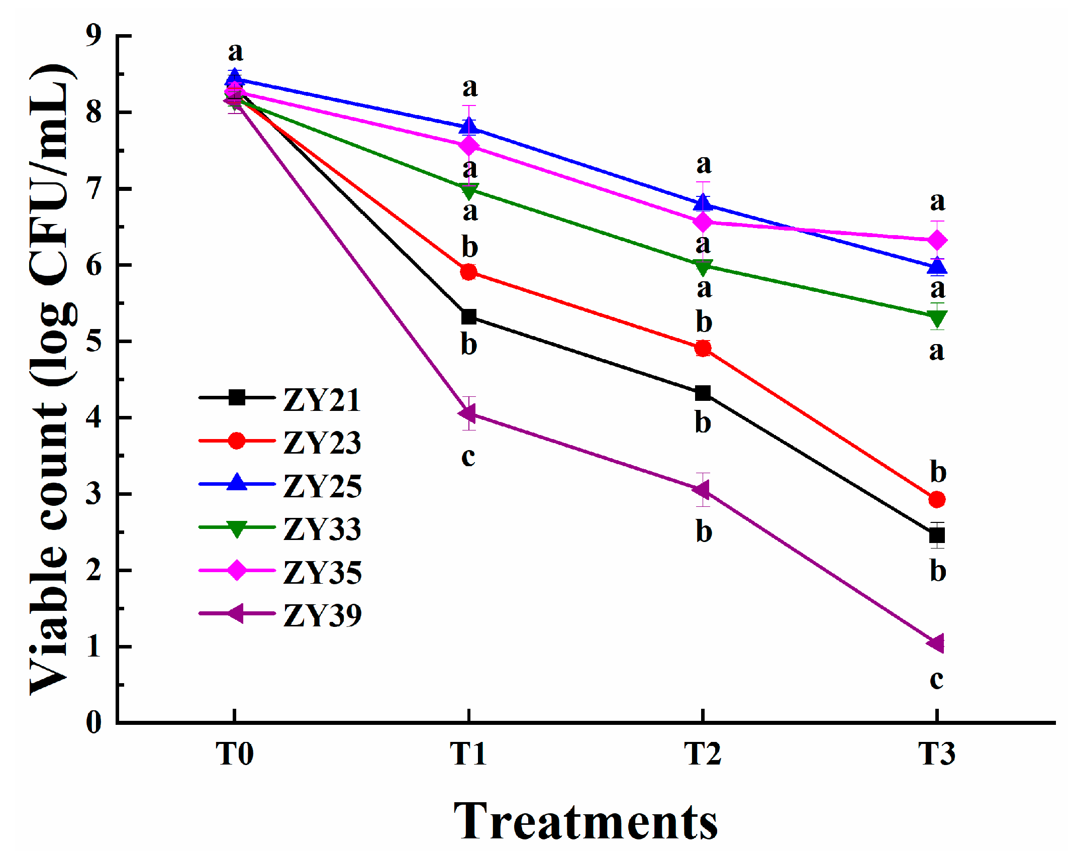
Figure 4 illustrates the viable count (log CFU/mL) of the LAB strains before and after the treatment with gastric and intestinal juices. The initial viable counts of strains ZY25, ZY33, and ZY35 before the gastric juice treatment were 8.44, 8.17, and 8.27 log CFU/mL, respectively. After 3 h of the gastric juice treatment, the viable counts were 7.80, 6.99, and 7.56 log CFU/mL, with survival rates of 92.4%, 85.6%, and 91.4%, respectively. The strains were then subjected to a 10-fold dilution and treated with intestinal juice. Before the intestinal juice treatment, the viable counts were 6.80, 5.99, and 6.56 log CFU/mL, respectively. After 4 h of the intestinal juice treatment, the viable counts were 5.97, 5.32, and 6.32 log CFU/mL, with survival rates of 87.8%, 88.8%, and 96.3%, respectively. However, the survival rates of strains ZY21, ZY23, and ZY39 after 3 h of the gastric juice treatment and 4 h of the intestinal juice treatment were significantly lower than those of strains ZY25, ZY33, and ZY35. Consequently, strains ZY25, ZY33, and ZY35 were selected for further analysis.

3.7. Safety Evaluation of Selected LAB Isolates

3.7.1. Hemolytic Activity of ZY25, ZY33, and ZY35

As shown in Figure 5, compared with the positive control, Staphylococcus aureus ATCC 29213T, which exhibited blood hemolysis activity in Figure 5A, strain ZY33 demonstrated similar hemolytic activity on the blood agar plates (Figure 5B). In contrast, strains ZY25 and ZY35 did not exhibit hemolytic activity (Figure 5C,D).

3.7.2. Profiles of Antibiotic Susceptibility of ZY25 and ZY35

The susceptibility pattern of the LAB isolates to different antibiotics is presented in Table 4. Strain ZY25 exhibited resistance against GEN, CIP, and SXT, and intermediate resistance against CTR, TET, and MY; it showed susceptibility to E, AMP, C, and PEN. Strain ZY35 displayed intermediate resistance against GEN, CIP, CTR, TET, and MY, while being resistant against CIP; it was susceptible to E, AMP, C, SXT, and PEN.

3.7.3. Virulence Factor, Biogenic Amine, and Antibiotic Resistance Genes of ZY25 and ZY35

The findings regarding the presence of virulence factor, biogenic amine, and antibiotic resistance genes in strains ZY25 and ZY35 are summarized in Table 5. According to the PCR analysis, neither ZY25 nor ZY35 carried virulence genes, including the collagen adhesion (ace), gelatinase (gelE), or cytolysin (cylA) genes. Additionally, both strains were devoid of biogenic amine genes, such as histidine decarboxylase (hdc), tyrosine decarboxylase (tdc), and ornithine decarboxylase (odc). Furthermore, no antibiotic resistance genes, including the vancomycin resistance (vanA) and tetracycline resistance (tetM) genes, were detected in either strain.

3.8. Broad-Spectrum Antibacterial Activity and Antibacterial Substances of ZY25 and ZY35

3.8.1. Antibacterial Spectra of ZY25 and ZY35

The results of the agar well diffusion assay, as shown in Table 6, revealed that ZY25 and ZY35 exhibited significant antibacterial activity against most of the tested microorganisms in this study. Strain ZY25 showed inhibition zones greater than 22.00 mm for Listeria monocytogenes CICC 23929T and Shigella dysenteriae CICC 23829T, and the inhibition zones ranged from 18.00 to 22.00 mm for Escherichia coli CICC 24189T, Bacillus subtilis CICC 10275T, and Staphylococcus aureus ATCC 29213T. However, ZY25 did not exhibit any inhibitory effect against Pseudomonas aeruginosa CICC 23694T. In contrast, strain ZY35 demonstrated inhibition zones between 18.00 and 22.00 mm for all the tested microorganisms, including Pseudomonas aeruginosa CICC 23694T.

3.8.2. The Influences of pH, Protease, and Catalase on Antimicrobial Activity

The antimicrobial activity of ZY25 and ZY35 against EPEC after various treatments is summarized in Table 7. Both strains exhibited strong antimicrobial activity in their fermentation liquids and supernatants, with inhibition zone diameters that consistently measured between 18.00 and 22.00 mm. The treatment with hydrogen peroxide did not diminish this activity, indicating that hydrogen peroxide was not a contributing factor. Furthermore, the neutralization of the samples resulted in a loss of antimicrobial activity, and the subsequent addition of proteinases to the neutralized samples failed to restore any antimicrobial effect. When the effects of pH were examined, the antimicrobial activity of both ZY25 and ZY35 remained robust at pH 2.5, with inhibition zones exceeding 22.00 mm. At pH 3.5, the activity was slightly reduced, with zones measuring between 18.00 and 22.00 mm. A further decline was noted at pH 4.5, where the inhibition zones measured between 14.00 and 18.00 mm. No antimicrobial activity was observed at pH values of 5.5 and higher, indicating that the antimicrobial substances produced by ZY25 and ZY35 were highly sensitive to alkaline conditions and remained effective primarily in acidic environments.
These results collectively demonstrate that the antimicrobial compounds of ZY25 and ZY35 are likely organic acids that exhibit optimal activity in acidic conditions and lose efficacy as the pH increases.

3.8.3. Growth Curves and Acid Production Capacity of ZY25 and ZY35

Figure 6 presents the 24-h growth curves and 48-h acid production curves of the strains. As illustrated in Figure 6A, strains ZY25 and ZY35 exhibited a lag phase from 0 to 4 h, followed by an exponential growth phase from 4 to 16 h. After 14 h, the growth rate of strain ZY25 slightly surpassed that of ZY35. The acid production curves shown in Figure 6B indicate no significant difference between the two lactic acid bacteria strains. At 12 h, the pH of the culture dropped below 4.5, and after 30 h, it fell below 3.85. Notably, the pH of strain ZY35 was slightly lower than that of ZY25.

3.8.4. Organic Acid Produced by Fermentation of ZY25 and ZY35

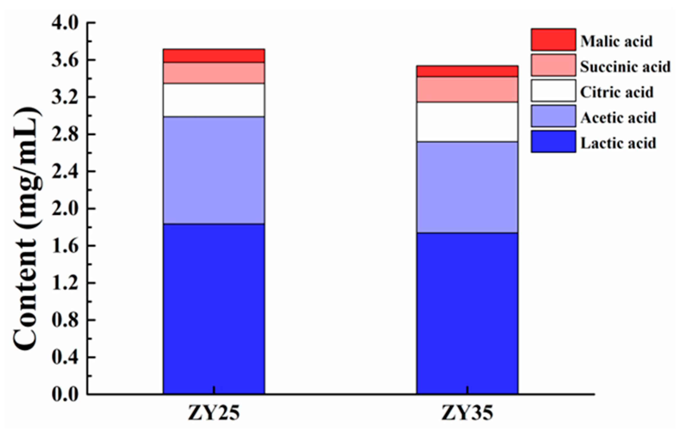
As illustrated in Figure 7, HPLC was employed to quantify the levels of 26 common organic acids in the fermentation broths of two bacterial strains: ZY25 and ZY35. The five most abundant organic acids identified were lactic, acetic, citric, succinic, and malic acids. The concentrations in the ZY25 fermentation broth were 1.834 mg/mL for lactic acid, 1.154 mg/mL for acetic acid, 0.359 mg/mL for citric acid, 0.226 mg/mL for succinic acid, and 0.142 mg/mL for malic acid. In the ZY35 broth, the levels were 1.737 mg/mL for lactic acid, 0.983 mg/mL for acetic acid, 0.426 mg/mL for citric acid, 0.273 mg/mL for succinic acid, and 0.116 mg/mL for malic acid.
Among the isolates tested, Ligilactobacillus agilis ZY25 and Ligilactobacillus salivarius ZY35 showed the highest antibacterial activity, with inhibition zones exceeding 22 mm. These strains demonstrated superior resilience in acidic conditions and tolerance to gastrointestinal fluids, making them promising candidates for probiotic applications.

4. Discussion

EPEC is a common pathogen responsible for causing diarrhea in cats, particularly in kittens. The management of such infections poses a significant challenge in feline health. Managing EPEC-induced diarrhea is challenging due to the pathogen’s ability to induce severe intestinal damage and its resistance to various antibiotics. Studies have shown that EPEC strains often display high levels of antimicrobial resistance, which complicates treatment strategies. These strains not only exhibit resistance to multiple antibiotics but also have the ability to form biofilms, which further complicates treatment and management efforts [22,23]. Probiotics have shown promising results in preventing and treating diarrhea across various animal species, including cats, dogs, and other livestock. However, many commercially available probiotics for pets are derived from non-host sources, and their efficacy remains uncertain. The efficacy of non-host-derived probiotics is often questioned due to the differences in the gut microbiota of different species [12]. Studies suggest that probiotics derived from the same host species are more likely to effectively colonize the gut and provide health benefits. This has led to a focus on developing probiotics specifically from the host species to improve their efficacy [8].
In this study, approximately 700 LAB strains were isolated from the feces of 300 healthy cats. The antibacterial activity of these isolates was tested against EPEC, and 50 strains with inhibition zones greater than 18 mm were selected for further study. Among these, several strains exhibited inhibition zones that exceeded 22 mm, indicating significant antibacterial activity against EPEC. Comparative studies have shown similar promising results with probiotics isolated from other sources. For example, Wang et al. evaluated over 1100 LAB strains and identified nine strains with inhibition zones of at least 22 mm against ETEC K88, highlighting the efficacy of specific Lactobacillus strains in pathogen inhibition [9]. Similarly, another study demonstrated that certain Lactobacillus strains isolated from kefir exhibited significant antimicrobial activity against UPEC, with inhibition zones that ranged from 62% to 75% in biofilm formation assays [24]. Additionally, Piątek et al. conducted head-to-head comparisons of different probiotics and found that a complex multi-strain synbiotic containing various Lactobacillus and Bifidobacterium strains exhibited significantly stronger inhibition of E. coli and other pathogens compared with single-strain probiotics [25]. These findings are consistent with our results, where selected LAB strains from healthy cats demonstrated strong inhibitory effects against EPEC. This underscores the potential of using host-specific probiotics to enhance antibacterial efficacy.
The physiological and biochemical characteristics of selected LAB isolates showed significant variability in their ability to grow under different stress conditions, including temperature, salt concentration, and pH. Sixteen strains (ZY1, ZY3, ZY4, ZY7, ZY12, ZY16, ZY17, ZY21, ZY23, ZY24, ZY25, ZY29, ZY33, ZY35, ZY39, and ZY47) demonstrated stronger antibacterial activity and greater tolerance, making them suitable candidates for further research. Studies have shown that the physiological and biochemical resilience of LAB is critical for their probiotic efficacy. For instance, Peng et al. emphasized the importance of whole-genome sequencing in understanding the genetic basis for probiotic properties, which includes resistance to various environmental stresses [26]. Similarly, the robust growth of LAB under different NaCl concentrations, temperatures, and pH levels was correlated with their ability to survive and function effectively in the gastrointestinal tracts of hosts [27].
A study by Huligere et al. also demonstrated that LAB strains with high tolerance to acid and bile salts showed significant probiotic potential, including strong antibacterial activity, high hydrophobicity, and auto-aggregation ability [28]. This is consistent with our findings, where strains such as ZY21, ZY23, ZY24, ZY25, ZY33, ZY35, ZY39, and ZY47 exhibited cell surface hydrophobicity values that exceeded 30% and auto-aggregation rates above 40%. Furthermore, research by Alameri et al. on LAB isolated from fresh vegetable products revealed that strains with high adhesion capability, acid and bile salt resistance, and antimicrobial activity demonstrated excellent probiotic characteristics [29]. This supports the selection criteria used in our study and highlights the importance of these physiological traits in determining the probiotic potential of LAB strains.
Accurate species identification is crucial for understanding bacterial habits, metabolism, and pathogenic patterns, which is essential for their effective applications in scientific research and probiotic development. This process ensures the selection of the most effective probiotic strains tailored to specific hosts or conditions, enhancing their efficacy and safety [30]. In this study, 16S rDNA sequences and the neighbor-joining method were used to construct phylogenetic trees for the selected LAB isolates. The analysis accurately classified strains such as ZY21, ZY39, ZY25, ZY35, ZY23, and ZY33 among various LAB species with strong bootstrap support. For instance, ZY21 was closely related to Limosilactobacillus reuteri DSM 20016T, and ZY39 was clustered with Ligilactobacillus aviarius NRIC 101267T. These robust classifications ensured reliable identification, facilitating their use in probiotic research. This approach aligns with other studies that emphasized the importance of precise species identification using 16S rDNA sequencing [31].
The safety evaluation of probiotic strains is crucial for their applications in food and health products. Hemolytic activity testing revealed that strain ZY33 exhibited hemolytic activity similar to the positive control Staphylococcus aureus ATCC 29213T, whereas strains ZY25 and ZY35 did not show hemolytic activity, indicating their safety in this regard [32]. Antibiotic susceptibility testing indicated that strain ZY25 exhibited resistance against gentamicin, ciprofloxacin, and sulfamethoxazole, but was susceptible to erythromycin, ampicillin, chloramphenicol, and penicillin. Strain ZY35 displayed intermediate resistance against several antibiotics but was susceptible to the most commonly used antibiotics, suggesting a lower risk of transferring antibiotic resistance genes [33]. PCR analysis confirmed that neither ZY25 nor ZY35 carried virulence genes (ace, gelE, cylA), biogenic amine production genes (hdc, tdc, odc), or antibiotic resistance genes (vanA, tetM), which further supports their safety for probiotic use [27,34]. The absence of biogenic amine genes is particularly important, as biogenic amines can cause adverse health effects, such as histamine poisoning [35]. These findings suggest that strains ZY25 and ZY35 are safe for use as probiotics, since they had no hemolytic activity, acceptable antibiotic susceptibility profiles, and an absence of virulence and biogenic amine genes. This comprehensive safety evaluation is consistent with other studies that emphasized the importance of thorough safety assessments for probiotic strains [36].
The antibacterial spectrum of LAB isolates ZY25 and ZY35 was tested using the agar well diffusion method. ZY25 showed significant inhibition zones against Listeria monocytogenes and Shigella dysenteriae (>22.00 mm) and moderate inhibitions against Escherichia coli, Bacillus subtilis, and Staphylococcus aureus (18.00–22.00 mm). ZY35 demonstrated broad-spectrum antibacterial activity against all tested pathogens, including Pseudomonas aeruginosa. These findings are consistent with the broad antimicrobial activity reported in other studies for LAB strains [37,38]. The antimicrobial activity of ZY25 and ZY35 against EPEC was examined under various conditions. Both strains maintained strong antimicrobial activity in their fermentation liquids and supernatants, which diminished upon neutralization, indicating pH dependence and the likely role of organic acids. The treatment with proteinases did not restore their activity, suggesting that bacteriocins were not the primary antimicrobial agents. These results align with findings from other studies that showed the significant role of organic acids produced by LAB in antimicrobial activity [39]. The growth curves and acid production profiles for ZY25 and ZY35 were evaluated over 48 h. Both strains exhibited a typical growth pattern with lag, exponential growth, and stationary phases. They also showed robust acid production, with pH values dropping significantly within the first 24 h. The ability of these strains to rapidly lower the pH is crucial for inhibiting pathogenic bacteria, as supported by studies on LAB’s role in inhibiting gut pathogens and promoting gut health through acidification [40].
HPLC was used to quantify the levels of organic acids produced by ZY25 and ZY35. The primary organic acids identified were lactic, acetic, citric, succinic, and malic acids. The high concentrations of lactic and acetic acids, which have strong antimicrobial properties, contributed significantly to the probiotic potential of these strains. This is in line with other research that demonstrated the role of organic acids in the antimicrobial activity of LAB [41,42]. These findings collectively demonstrate that ZY25 and ZY35 had broad-spectrum antibacterial activity primarily through organic acid production, robust growth, and acid production capabilities. Their effectiveness in acidic environments further supported their potential as effective probiotics. The results of this comprehensive analysis are consistent with those of other studies that highlighted the significant role of LAB in inhibiting pathogenic bacteria and promoting gut health [36,43].
Many studies focus on the use of probiotics like Lactobacillus spp., but our research emphasizes the efficacy of Ligilactobacillus agilis ZY25 and Ligilactobacillus salivarius ZY35. Ligilactobacillus salivarius has been shown to improve growth performance and antioxidative capacity in broiler chickens by modulating the microbiota–gut–brain axis, making it an effective probiotic for poultry. The strain also enhanced digestive enzyme secretion and positively impacted body weight and hormonal balance [44]. Similarly, Ligilactobacillus salivarius MP100, used in swine production, improved growth performance and fecal microbiota profiles without affecting meat quality, offering an alternative to metaphylactic antimicrobials and reducing antibiotic use [45]. Both strains exhibit substantial antibacterial activity against Escherichia coli, especially enteropathogenic strains, and their robust performance under simulated gastrointestinal conditions highlights their potential as host-specific probiotics, crucial for improving colonization and efficacy in feline gastrointestinal health.

5. Conclusions

This study successfully identified and characterized feline-derived LAB strains, specifically ZY25 and ZY35, which demonstrated significant antibacterial activity against EPEC. These strains exhibited strong tolerance to various stress conditions, including low pH, bile salts, and gastrointestinal fluids, alongside high hydrophobicity and auto-aggregation abilities, underscoring their potential as probiotics. Safety evaluations further confirmed the absence of hemolytic activity, virulence factors, and antibiotic resistance genes, reinforcing their suitability for safe probiotic use. The antimicrobial efficacy of ZY25 and ZY35 is primarily attributed to the production of organic acids, particularly lactic and acetic acids, which effectively inhibited the growth of EPEC and other pathogenic bacteria. Additionally, their robust growth and acid production capabilities under simulated gastrointestinal conditions suggest their potential in vivo efficacy. These findings highlight ZY25 and ZY35 as promising natural alternatives to conventional antibiotics for managing EPEC-induced diarrhea in cats, warranting further in vivo trials to validate their probiotic benefits and explore their commercial application in the veterinary field.

Author Contributions

Conceptualization, W.W.; methodology, H.D., X.C. and Q.C.; software, W.W., C.M. and R.W.; validation, S.C., L.C. and L.W.; formal analysis, W.W., H.D. and Q.C.; investigation, W.W.; resources, W.W., S.G. and W.X.; data curation, W.W.; writing—original draft preparation, W.W. and W.X.; writing—review and editing, W.X.; visualization, W.W.; supervision, W.W.; project administration, W.X.; funding acquisition, W.W. All authors have read and agreed to the published version of the manuscript.

Funding

This work was supported by the Foundation of Key Technology Research Project of Henan Province (Grant No. 242102110039).

Institutional Review Board Statement

All experimental procedures were authorized by the Animal Care and Use Committee prior to animal experimentation (AW20704202-5-4) and were performed following the guidelines of the Institute of Nutrition and Health, China Agricultural University.

Informed Consent Statement

Not applicable.

Data Availability Statement

The data generated from the study are clearly presented and discussed in the manuscript.

Conflicts of Interest

Weiwei Wang, Longjiao Wang, and Wei Xiong, co-authors of the article, are affiliated with Henan Zhiyuan Henuo Technology Co., Ltd. and are employed as technical consultants at the company. The authors declare no conflicts of interest.

Abbreviations

EPEC: enteropathogenic Escherichia coli, LAB: lactic acid bacteria, MRS: de Man, Rogosa, and Sharpe (medium), PCR: polymerase chain reaction, SGF: simulated gastric fluid, SIF: simulated intestinal fluid, CFU: colony forming unit, HPLC: high-performance liquid chromatography, OD: optical density, SEM: standard error of the mean.

References

  1. Watson, V.; Jacob, M.; Flowers, J.; Strong, S.; DebRoy, C.; Gookin, J. Association of atypical enteropathogenic Escherichia coli with diarrhea and related mortality in kittens. J. Clin. Microbiol. 2017, 55, 2719–2735. [Google Scholar] [CrossRef] [PubMed]
  2. Kjaergaard, A.; Carr, A.; Gaunt, M. Enteropathogenic Escherichia coli (EPEC) infection in association with acute gastroenteritis in 7 dogs from Saskatchewan. Can. Vet. J. 2016, 57, 964–968. [Google Scholar]
  3. Oliva, C.; Scaletsky, I.; Morais, M.; Neto, U. Severe acute diarrhea associated to classic enteropathogenic Escherichia coli (EPEC): Clinical features and fecal losses in hospitalized infants. Rev. Assoc. Med. Bras. 1997, 43, 283–289. [Google Scholar] [CrossRef] [PubMed][Green Version]
  4. Legge, S.; Taggart, P.; Dickman, C.; Read, J.; Woinarski, J. Cat-dependent diseases cost Australia AU$6 billion per year through impacts on human health and livestock production. Wildl. Res. 2020, 47, 731–746. [Google Scholar] [CrossRef]
  5. Spitznagel, M.; Cox, M.; Jacobson, D.; Albers, A.; Carlson, M. Assessment of caregiver burden and associations with psychosocial function, veterinary service use, and factors related to treatment plan adherence among owners of dogs and cats. J. Am. Vet. Med. Assoc. 2019, 254, 124–132. [Google Scholar] [CrossRef] [PubMed]
  6. Bhatt, S. Atypical enteropathogenic Escherichia coli: From kittens to humans and beyond! Infect. Immun. 2021, 89, e00752-20. [Google Scholar] [CrossRef]
  7. Jafari, E.; Mostaan, S.; Bouzari, S. Characterization of antimicrobial susceptibility, extended-spectrum β-lactamase genes and phylogenetic groups of enteropathogenic Escherichia coli isolated from patients with diarrhea. Osong Public Health Res. Perspect. 2020, 11, 327. [Google Scholar] [CrossRef]
  8. Yuan, L.; Chu, B.; Chen, S.; Li, Y.; Liu, N.; Zhu, Y.; Zhou, D. Exopolysaccharides from Bifidobacterium animalis Ameliorate Escherichia coli-Induced IPEC-J2 Cell Damage via Inhibiting Apoptosis and Restoring Autophagy. Microorganisms 2021, 9, 2363. [Google Scholar] [CrossRef]
  9. Wang, W.; Ma, H.; Yu, H.; Qin, G.; Tan, Z.; Wang, Y.; Pang, H. Screening of Lactobacillus plantarum subsp. plantarum with Potential Probiotic Activities for Inhibiting ETEC K88 in Weaned Piglets. Molecules 2020, 25, 4481. [Google Scholar] [CrossRef]
  10. Fernandes, M.; Rios, J.; Vasconcelos, B.; Lourenço, M.; Matos, M.; Cavalcante, R.; de Almeida, M.V.A.; Costa, R.A.; Carneiro, V. Effect of Lactobacillus spp. cell-free supernatant against planktonic growth and biofilm formation of foodborne Escherichia coli isolates. Lett. Appl. Microbiol. 2023, 76, 1. [Google Scholar] [CrossRef] [PubMed]
  11. Piatek, J.; Krauss, H.; Ciechelska-Rybarczyk, A.; Bernatek, M.; Wojtyla-Buciora, P.; Sommermeyer, H. In-Vitro Growth Inhibition of Bacterial Pathogens by Probiotics and a Synbiotic: Product Composition Matters. Int. J. Environ. Res. Public Health 2020, 17, 3332. [Google Scholar] [CrossRef] [PubMed]
  12. Tarkhani, R.; Imani, A.; Hoseinifar, S.H.; Ashayerizadeh, O.; Moghanlou, K.S.; Manaffar, R.; Van Doan, H.; Reverter, M. Comparative study of host-associated and commercial probiotic effects on serum and mucosal immune parameters, intestinal microbiota, digestive enzymes activity and growth performance of roach (Rutilus rutilus caspicus) fingerlings. Fish Shellfish Immunol. 2020, 98, 661–669. [Google Scholar] [CrossRef] [PubMed]
  13. Buhnik-Rosenblau, K.; Matsko-Efimov, V.; Jung, M.; Shin, H.; Danin-poleg, Y.; Kashi, Y. Indication for co-evolution of Lactobacillus johnsonii with its hosts. BMC Microbiol. 2012, 12, 149. [Google Scholar] [CrossRef] [PubMed]
  14. Adnan, M.; Patel, M.; Hadi, S. Functional and health promoting inherent attributes of Enterococcus hirae F2 as a novel probiotic isolated from the digestive tract of the freshwater fish Catla catla. PeerJ 2017, 5, e3085. [Google Scholar] [CrossRef] [PubMed]
  15. Sirichokchatchawan, W.; Pupa, P.; Praechansri, P.; Am-In, N.; Tanasupawat, S.; Sonthayanon, P.; Prapasarakul, N. Autochthonous lactic acid bacteria isolated from pig faeces in Thailand show probiotic properties and antibacterial activity against enteric pathogenic bacteria. Microb. Pathog. 2018, 119, 208–215. [Google Scholar] [CrossRef]
  16. Zhang, M.; Wang, X.; Cui, M.; Wang, Y.; Jiao, Z.; Tan, Z. Ensilage of oats and wheatgrass under natural alpine climatic conditions by indigenous lactic acid bacteria species isolated from high-cold areas. PLoS ONE 2018, 13, e0192368. [Google Scholar] [CrossRef]
  17. Zhang, B.; Wang, Y.; Tan, Z.; Li, Z.; Jiao, Z.; Huang, Q. Screening of probiotic activities of Lactobacilli strains isolated from traditional Tibetan Qula, a raw yak milk cheese. Anim. Biosci. 2016, 29, 1490–1499. [Google Scholar] [CrossRef]
  18. Niu, K.M.; Kothari, D.; Cho, S.B.; Han, S.G.; Song, I.G.; Kim, S.C.; Kim, S.K. Exploring the probiotic and compound feed fermentative applications of Lactobacillus plantarum SK1305 isolated from Korean green chili pickled pepper. Probiotics Antimicrob. Proteins. 2019, 11, 801–812. [Google Scholar] [CrossRef]
  19. De Souza, B.M.S.; Borgonovi, T.F.; Casarotti, S.N.; Todorov, S.D.; Penna, A.L.B. Lactobacillus casei and Lactobacillus fermentum strains isolated from mozzarella cheese: Probiotic potential, safety, acidifying kinetic parameters and viability under gastrointestinal tract conditions. Probiotics Antimicrob. Proteins 2019, 11, 382–396. [Google Scholar] [CrossRef]
  20. Ni, K.; Yang, H.; Hua, W.; Wang, Y.; Pang, H. Selection and characterisation of lactic acid bacteria isolated from different origins for ensiling Robinia pseudoacacia and Morus alba L. leaves. J. Integr. Agric. 2016, 15, 2353–2362. [Google Scholar] [CrossRef]
  21. Khan, N.; Ali, S.; Zandi, P.; Mehmood, A.; Babar, A. Role of sugars, amino acids and organic acids in improving plant abiotic stress tolerance. Pak. J. Bot. 2020, 52, 2. [Google Scholar] [CrossRef] [PubMed]
  22. Rodrigues, R.; Lima, N.C.S.; Taborda, R.L.M.; Esquerdo, R.P.; Gama, A.; Nogueira, P.; Orlandi, P.; Matos, N.B. Antibiotic resistance and biofilm formation in children with Enteropathogenic Escherichia coli (EPEC) in Brazilian Amazon. J. Infect. Dev. Ctries. 2019, 13, 698–705. [Google Scholar] [CrossRef]
  23. Shen, J.; Zhi, S.; Guo, D.; Jiang, Y.; Xu, X.; Zhao, L.; Lv, J. Prevalence, antimicrobial resistance, and whole genome sequencing analysis of Shiga toxin-producing Escherichia coli (STEC) and Enteropathogenic Escherichia coli (EPEC) from imported foods in China during 2015–2021. Toxins 2022, 14, 68. [Google Scholar] [CrossRef] [PubMed]
  24. Ghane, M.; Babaeekhou, L.; Ketabi, S.S. Antibiofilm Activity of Kefir Probiotic Lactobacilli against Uropathogenic Escherichia coli (UPEC). Avicenna J. Med. Biotechnol. 2020, 12, 221–229. [Google Scholar] [PubMed]
  25. Piatek, J.; Sommermeyer, H.; Ciechelska-Rybarczyk, A.; Bernatek, M. In-vitro pathogen inhibition: Comparing the inhibitory effects of a complex multistrain synbiotic with simple probiotics containing the yeast Saccharomyces boulardii or Lactobacillus rhamnosus bacteria. bioRxiv 2019. [Google Scholar] [CrossRef]
  26. Peng, X.; Ed-dra, A.; Yue, M. Whole genome sequencing for the risk assessment of probiotic lactic acid bacteria. Crit. Rev. Food Sci. Nutr. 2022, 63, 11244–11262. [Google Scholar] [CrossRef]
  27. Li, M.; Wang, Y.; Cui, H.; Li, Y.; Sun, Y.; Qiu, H. Characterization of lactic acid bacteria isolated from the gastrointestinal tract of a wild boar as potential probiotics. Front. Vet. Sci. 2020, 7, 49. [Google Scholar] [CrossRef]
  28. Huligere, S.; Kumari, V.; Alqadi, T.; Kumar, S.; Cull, C.; Amachawadi, R.; Ramu, R. Isolation and characterization of lactic acid bacteria with potential probiotic activity and further investigation of their activity by α-amylase and α-glucosidase inhibitions of fermented batters. Front. Microbiol. 2023, 13, 1042263. [Google Scholar] [CrossRef]
  29. Alameri, F.; Tarique, M.; Osaili, T.; Obaid, R.; Abdalla, A.; Masad, R.; Al-Sbiei, A.; Fernandez-Cabezudo, M.; Liu, S.; al-Ramadi, B.; et al. Lactic acid bacteria isolated from fresh vegetable products: Potential probiotic and postbiotic characteristics including immunomodulatory effects. Microorganisms 2022, 10, 389. [Google Scholar] [CrossRef]
  30. Celandroni, F.; Vecchione, A.; Cara, A.; Mazzantini, D.; Lupetti, A.; Ghelardi, E. Identification of Bacillus species: Implication on the quality of probiotic formulations. PLoS ONE. 2019, 14, e0217021. [Google Scholar] [CrossRef] [PubMed]
  31. Kim, E.; Yang, S.; Lim, B.; Park, S.; Rackerby, B.; Kim, H. Simultaneous detection of 37 Lactobacillus species using a real-time PCR assay based on whole-genome sequence analysis. Preprints 2020. [Google Scholar] [CrossRef]
  32. Kim, T.; Mondal, S.; Jeong, C.; Kim, S.; Ban, O.; Jung, Y.; Yang, J.; Kim, S. Safety evaluation of Lactococcus lactis IDCC 2301 isolated from homemade cheese. Food Sci. Nutr. 2021, 10, 67–74. [Google Scholar] [CrossRef] [PubMed]
  33. Hussain, N.; Tariq, M.; Saris, P.; Zaidi, A. Evaluation of the probiotic and postbiotic potential of lactic acid bacteria from artisanal dairy products against pathogens. J. Infect. Dev. Ctries. 2021, 15, 102–112. [Google Scholar] [CrossRef]
  34. Chen, W.; Yu, L.; Shi, Y. Safety evaluation of lactic acid bacteria. In Lactic Acid Bacteria; Springer Nature Singapore Pte Ltd. and Science Press: Singapore, 2019; pp. 371–409. [Google Scholar] [CrossRef]
  35. Elzeini, H.; Ali, A.; Nasr, N.; Hassan, M.; Hassan, A.; Elenany, Y. Probiotic capability of novel lactic acid bacteria isolated from worker honey bees gut microbiota. FEMS Microbiol. Lett. 2021. [Google Scholar] [CrossRef] [PubMed]
  36. Pradhan, D.; Mallappa, R.; Grover, S. Comprehensive approaches for assessing the safety of probiotic bacteria. Food Control 2020, 108, 106872. [Google Scholar] [CrossRef]
  37. Wang, X.; Wang, W.; Lv, H.; Zhang, H.; Liu, Y.; Zhang, M.; Wang, Y.; Tan, Z. Probiotic potential and wide-spectrum antimicrobial activity of lactic acid bacteria isolated from infant feces. Probiotics Antimicrob. Proteins 2020, 13, 90–101. [Google Scholar] [CrossRef]
  38. Tkhruni, F.; Israyelyan, A. Comparative antimicrobial activity of some metabiotics synthesized by lactic acid bacteria. New Arm. Med. J. 2023, 17. [Google Scholar] [CrossRef]
  39. Valdez-Salas, B.; Beltrán-Partida, E.; Zlatev, R.; Stoytcheva, M.; González-Mendoza, D.; Salvador-Carlos, J.; Moreno-Ulloa, A.; Cheng, N. Structure-activity relationship of diameter controlled Ag@Cu nanoparticles in broad-spectrum antibacterial mechanism. Mater. Sci. Eng. C Mater. Biol. Appl. 2021, 119, 111501. [Google Scholar] [CrossRef]
  40. Jung, J.; Han, S.-S.; Kim, Z.; Kim, M.H.; Kang, H.; Jin, H.; Lee, M.H. In-vitro characterization of growth inhibition against the gut pathogen of potentially probiotic lactic acid bacteria strains isolated from fermented products. Microorganisms 2021, 9, 2141. [Google Scholar] [CrossRef]
  41. Liu, Z.; Xu, C.; Tian, R.; Wang, W.; Ma, J.; Gu, L.; Liu, F.; Jiang, Z.; Hou, J. Screening beneficial bacteriostatic lactic acid bacteria in the intestine and studies of bacteriostatic substances. J. Zhejiang Univ. Sci. B 2021, 22, 533–547. [Google Scholar] [CrossRef]
  42. Faraki, A.; Rahmani, F. The antioxidant activity of lactic acid bacteria and probiotics: A review. J. Food Saf. Hyg. 2021, 6, 168–182. [Google Scholar] [CrossRef]
  43. Gizachew, S.; Beeck, W.; Spacova, I.; Dekeukeleire, M.; Alemu, A.; Woldemedhin, W.; Mariam, S.; Lebeer, S.; Engidawork, E. Antibacterial and immunostimulatory activity of potential probiotic lactic acid bacteria isolated from Ethiopian fermented dairy products. Fermentation 2023, 9, 258. [Google Scholar] [CrossRef]
  44. Yang, J.; Wang, J.; Liu, Z.; Chen, J.; Jiang, J.; Zhao, M.; Gong, D. Ligilactobacillus salivarius improve body growth and anti-oxidation capacity of broiler chickens via regulation of the microbiota-gut-brain axis. BMC Microbiol. 2023, 23, 395. [Google Scholar] [CrossRef]
  45. Alba, C.; Castejón, D.; Remiro, V.; Rodríguez, J.M.; Sobrino, O.J.; de María, J.; Fumanal, P.; Fumanal, A.; Cambero, M.I. Ligilactobacillus salivarius MP100 as an alternative to metaphylactic antimicrobials in swine: The impact on production parameters and meat composition. Animals 2023, 13, 1653. [Google Scholar] [CrossRef]
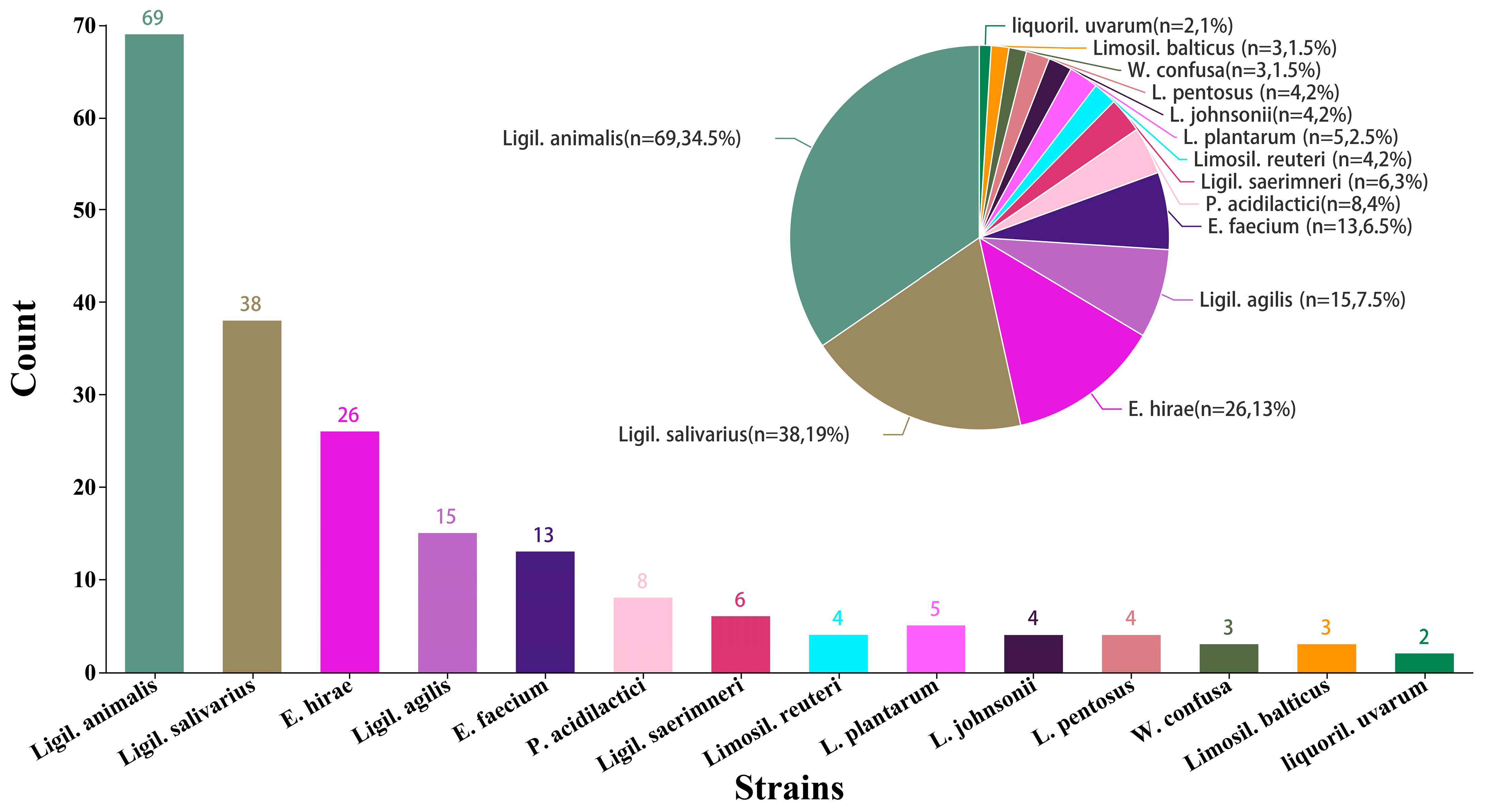
Figure 1. Distribution of the main LAB strains in the healthy cat feces. Ligilactobacillus animalis (Ligil. animalis), Ligilactobacillus salivarius (Ligil. salivarius), Enterococcus hirae (E. hirae), Ligilactobacillus agilis (Ligil. agilis), Enterococcus faecium (E. faecium), Pediococcus acidilactici (P. acidilactici), Ligilactobacillus saerimneri (Ligil. saerimneri), Limosilactobacillus reuteri (Limosil. reuteri), lactobacillus plantarum (L. plantarum), lactobacillus johnsonii (L. johnsonii), lactobacillus pentosus (L. pentosus), Weissella confuse (W. confusa), Limosilactobacillus balticus (Limosil. balticus), and liquorilactobacillus uvarum (liquoril. uvarum).
Fermentation 10 00514 g001
Figure 2. Cell surface hydrophobicity and auto-aggregation ability of LAB isolates: (A) hydrophobicity of selected LAB isolates; (B) auto-aggregation ability of selected LAB isolates. Different lowercase letters denote significant difference (p < 0.05).
Fermentation 10 00514 g002
Figure 3. Phylogenetic tree showing the relative positions of selected isolates ZY21, ZY23, ZY25, ZY33, ZY35, and ZY39 by the neighbor-joining method. Bacillus subtilis was used as the outgroup. Bootstrap values shown at the nodes of the tree are from 1000 replicates, and the bar indicates 1% sequence divergence.
Fermentation 10 00514 g003
Figure 4. Survival of selected LAB strains in the simulated gastrointestinal fluids. Viable count (log CFU/mL) of the selected LAB strains after simulated gastrointestinal tract (GIT) conditions. Gastric juice T0: viability at the beginning of the gastric juice treatment; gastric juice T1: viability after the simulation of gastric conditions; intestinal juice T2: viability at the beginning of the gastric juice treatment; intestinal juice T3: viability after the simulation of enteric conditions. Different lowercase letters on the same row denote significant differences (p < 0.05) during the assay.
Fermentation 10 00514 g004
Figure 5. Hemolytic activity of selected LAB isolates: (A) positive control—Staphylococcus aureus ATCC 29213T, (B) ZY33, (C) ZY25, and (D) ZY35.
Fermentation 10 00514 g005
Figure 6. Growth curves and acid production capacity of isolates ZY25 and ZY35: (A) growth curves of two LAB isolates and (B) acid production capacity.
Fermentation 10 00514 g006
Figure 7. The organic acids produced by fermentation of ZY25 and ZY35.
Fermentation 10 00514 g007
Table 1. Physiological properties of the LAB isolates.
StrainsShapeGluFermentationGramCAT
RodCocci-+HomoHetero
75%25%96%4%96%4%+-
Table 2. Antimicrobial activity against EPEC of representative LAB isolates.
IsolatesAntimicrobial ActivityIsolatesAntimicrobial Activity
ZY1++++ZY26+++
ZY2++++ZY27+++
ZY3++++ZY28+++
ZY4++++ZY29++++
ZY5+++ZY30+++
ZY6+++ZY31+++
ZY7++++ZY32++++
ZY8+++ZY33++++
ZY9+++ZY34+++
ZY10++++ZY35++++
ZY11+++ZY36+++
ZY12++++ZY37++++
ZY13++++ZY38+++
ZY14++++ZY39++++
ZY15+++ZY40+++
ZY16++++ZY41+++
ZY17++++ZY42++++
ZY18+++ZY43+++
ZY19++++ZY44++++
ZY20+++ZY45+++
ZY21++++ZY46++++
ZY22+++ZY47++++
ZY23++++ZY48+++
ZY24++++ZY49++++
ZY25++++ZY50+++
Notes: +++, diameter of the inhibition zone: 18.00–22.00 mm; ++++, more than 22.00 mm; the diameter of the inhibition zone included that of the hole puncher (10.00 mm).
Table 3. Growth of Selected LAB Isolates Under Different Temperatures, Salt, and pH Conditions.
IsolatesTemperature (°C)NaCl (w/v, %)pH
51045503.06.53.03.54.04.55.05.56.09.010.0
ZY1+++++w+++++++
ZY2++++w++++++
ZY3+++++w++++++++
ZY4+++++w+++++++
ZY5w+w++ww++++++
ZY6+++.+ww++++++
ZY7+++++w+++++++
ZY8++w+w++++++
ZY9++w++++++++
ZY10w+w+ww+++++++
ZY11w+++w+++++++
ZY12+++++w++++++++
ZY13w++++++++++
ZY14w++++w++++++
ZY15ww+++++++++
ZY16w+++++w++++++++
ZY17+w+++w++++++++
ZY18w++++w++++++
ZY19w+w+ww+++++++
ZY20w+++++++++++
ZY21w+++++w+++++++
ZY22++++ww++++++
ZY23+++++w++++++++
ZY24+++++w+++++++
ZY25w+++++w++++++++
ZY26+ww+++++++++
ZY27w++++ww++++++
ZY28++++++++++
ZY29w+++++ww+++++++
ZY30++w++w++++++
ZY31++++++++++
ZY32+++w+++++++
ZY33++++++++++++++
ZY34w+++ww+++++
ZY35w++++w+++++++++
ZY36++w++w++++++
ZY37w+++++++++++
ZY38+++++++++++
ZY39w+++++ww+++++++
ZY40w+++www++++++
ZY41+++www++++++
ZY42ww+w++w++++++
ZY43+++++w++++++
ZY44w++++ww++++++
ZY45++++www++++++
ZY46++w++++++++
ZY47w+++++++++++++
ZY48++++++++++
ZY49++w+ww++++++
ZY50++++w++++++
Notes: +, positive; −, negative; w, weakly positive.
Table 4. Antibiotic susceptibility of isolates ZY25 and ZY35.
IsolateGENCIPCTREAMPTETSXTCMYPEN
ZY25RRISSIRSIS
ZY35IRISSISSRS
Notes: S—susceptible; I—intermediate resistant; R—resistant. The concentrations of antibiotics are expressed in micrograms per disc (μg/disc): genmalicin (GEN, 10), ciprofloxacin (CIP, 5), ceftriaxone (CTR, 30), erythromycin (E, 15), ampicillin (AMP, 10), telracyclin (TET, 30), compound sulfamethoxa (SXT, 25), chloramphenicol (C, 30), lincomycin (MY, 2), and penicillin (PEN, 10).
Table 5. Detection of virulence factor, biogenic amine production, and antibiotic resistance genes in ZY25 and ZY35.
IsolatesVirulence Factor GenesBiogenic Amine GenesAntibiotic Resistance Genes
acegelEcylAHdcTdcOdcvanAtetM
ZY25--------
ZY35--------
Notes: -, the genes were not detected in the strains.
Table 6. Antibacterial spectra of ZY25 and ZY35.
IsolatesIndicator Bacteria
P. aeruginosaS. aureusL. monocytogenesE. coliB. subtilisS. dysenteriae
ZY25+++++++++++++++++
ZY35++++++++++++++++++
Notes: 1. −, no inhibition; +++, diameter of the inhibition zone: 18.00–22.00 mm; ++++, more than 22.00 mm; the diameter of the inhibition zone included that of the hole puncher (10.00 mm). 2. P. aeruginosa: Pseudomonas aeruginosa CICC 23694T, S. aureus: Staphylococcus aureus ATCC 29213T, L. monocytogenes: Listeria monocytogenes CICC 23929T, E. coli: Escherichia coli CICC 24189T, B. subtilis: Bacillus subtilis CICC 10275T, S. dysenteriae: Shigella dysenteriae CICC 23829T.
Table 7. Antimicrobial activity of ZY25 and ZY35 to EPEC after different treatments.
TreatmentZY25 Antimicrobial
Activity
ZY35 Antimicrobial
Activity
Fermentation liquid++++++
Supernatant++++++
Hydrogen peroxide++++++
Proteinase K
Pepsinum
Trypsase
2.5++++++++
3.5++++++
4.5++++
5.5
6.5
7
10
Note: ++, diameter of the inhibition zone: 14.00–18.00 mm; +++, 18.00–22.00 mm; ++++, more than 22.00 mm, −, no inhibition zone was detected; the diameter of the inhibition zone included that of the hole puncher (10.00 mm).
Disclaimer/Publisher’s Note: The statements, opinions and data contained in all publications are solely those of the individual author(s) and contributor(s) and not of MDPI and/or the editor(s). MDPI and/or the editor(s) disclaim responsibility for any injury to people or property resulting from any ideas, methods, instructions or products referred to in the content.