Article

Selective Growth of and Electricity Production by Marine Exoelectrogenic Bacteria in Self-Aggregated Hydrogel of Microbially Reduced Graphene Oxide

1
Center for Fostering Young and Innovative Researchers, Nagoya Institute of Technology, Nagoya 466-8555, Japan
2
Electronics-Inspired Interdisciplinary Research Institute (EIIRIS), Toyohashi University of Technology, Toyohashi, Aichi 441-8580, Japan
3
Department of Biomedical Science, College of Life and Health Science, Chubu University, Kasugai, Aichi 487-8501, Japan
4
Nagoya Municipal Industrial Research Institute, Nagoya, Aichi 456-0058, Japan
*
Author to whom correspondence should be addressed.
Submission received: 9 February 2016 / Revised: 9 May 2016 / Accepted: 17 May 2016 / Published: 20 May 2016
(This article belongs to the Special Issue Graphene Nanocomposite for Advanced Applications)

Abstract

:
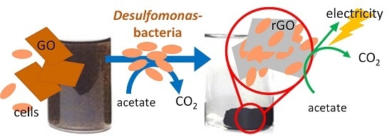
Graphene oxide (GO) has been shown to be reduced by several microorganisms. Recent studies of the growth of Geobacter species in the presence of GO and electricity production by recovery of electrons on the reduced form of GO (rGO) have indicated substantial benefits of GO and GO-respiring bacteria (GORB) in microbial electrochemical systems. In this study, we enriched GORB from a coastal sample to investigate the distribution and phylogenetic variety of GORB in seawater environments. X-ray photoelectron spectroscopy (XPS) and four-terminal probing revealed that the enriched microbial community (designated as CS culture) reduced GO and self-aggregated into a conductive hydrogel complex with rGO (the CS-rGO complex). In the process of GO reduction, certain bacterial populations grew in a manner that was dependent on GO respiration coupled with acetate oxidization. High-throughput sequencing of 16S rRNA as a biomarker revealed the predominance of Desulfomonas species at 92% of the total bacterial population in the CS culture. The CS-rGO complex produced electricity with acetate oxidization, exhibiting less than 1 Ω/cm3 of charge transfer resistance. Thus, these results suggested that Desulfomonas species could grow on rGO and produce electricity via the reduced form of GO.

Graphical Abstract

1. Introduction

Graphene oxide (GO) has been shown to be reduced by several microorganisms, such as Shewanella species [1,2], Escherichia coli [3], and baker’s yeast [4]. In addition, we recently showed that the growth of Geobacter species was dependent on GO reduction [5], i.e., respiration coupled with acetate oxidization and GO reduction. Bacteria within the genera Geobacter and Shewanella are capable of respiration with the utilization of extracellular electron acceptors, such as iron oxide. Shewanella reduces GO through the outer membrane cytochrome, a key protein involved in respiration through extracellular electron acceptors [6]. The mechanisms of GO reduction in microorganisms unable to undergo this type of respiration, e.g., Escherichia coli [3] and baker’s yeast [4], are unknown but may involve electron transfer from electron carriers, such as vitamin C [7], produced by these microorganisms. In such cases, GO may be used as an electron sink and may not permit cell growth.
Two-dimensional materials have attracted increasing interest in biosystems research [8,9,10,11]. Among them, microbial GO reduction has received much attention in bioelectrochemical engineering applications, such as the enhancement of electricity recovery in bioelectrochemical systems (BESs) using microorganisms as biocatalysts. In BESs, GO and the microbially reduced form of GO (rGO) have great advantages as anode materials in the recovery of electrons from microbial cells; these advantages include the large surface area of GO, the hydrophilicity of the materials to allow better access to microbial cells, and functions promoting bacterial growth and respiration through extracellular electrodes and GO. In our recent demonstration of GO reduction by Geobacter species, GO was shown to be self-aggregated into a conductive hydrogel complex with cells of Geobacter species in the reduction process [5]. The hydrogel complex of the microbially reduced GO and bacterial cells showed stable electricity with better growth of biofilm, less charge transfer resistance, and larger capacitance compared with that associated with graphite felt, a representative anode for BES. However, most bacteria known to reduce GO, except Shewanella species, have been obtained from freshwater environments. In addition, Shewanella species do not form hydrogel solids but instead exhibit a floccular form.
In this study, we aimed to enrich marine bacteria that could reduce GO and form a conductive hydrogel with the reduced GO for future applications in the production of electricity from waste-biomass in seawater. Our results are also expected to provide insights into the diversity of GO-respiring bacteria (GORB) in the natural environment.

2. Results

2.1. GO Reduction by the CS Culture, An Enrichment Culture of Marine GORB

We obtained an enrichment culture of marine GORB designated as the CS culture by incubation of the mixture of seawater and coastal sand with GO and acetate and serial transfer more than 20 times. Figure 1A shows the apparent changes in the CS culture supplemented with GO. At day 0, the GO in the CS culture was initially well dispersed across the entire culture, which was brown in color, and then changed into a black hydrogel complex over the course of 10 days. From the X-ray photoelectron spectroscopy (XPS) analysis data shown in Figure 1B, the GO in the CS culture showed C1s spectra with two major peaks at approximately 287 eV for C=O and 285 eV for C–C/C–H bonds at day 0. The C=O peak was initially dominant and then gradually decreased in the CS cultures. Moreover, the C–C/C–H peak became dominant, and the C=O peak almost disappeared. These results indicated that microorganisms in the CS culture reduced GO to rGO, which appeared as a black hydrogel. Electrical conductivity analysis of GO and the black hydrogel complex showed significant increases in electrical conductivity following incubation (Figure 1C). The black hydrogel complex exhibited an electrical conductivity of 25 mS/cm, although the GO showed no significant conductivity in the analysis. Collectively, these data indicated that the marine GORB in the CS culture transformed water-dispersed GO to a conductive hydrogel aggregate of rGO.

2.2. Microbial Growth Dependent on GO

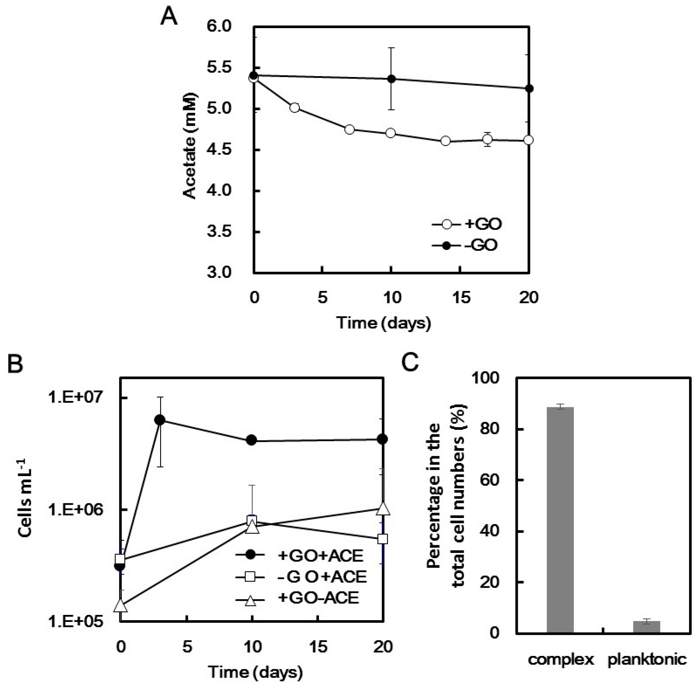
Changes in acetate concentrations in the CS culture were monitored as shown in Figure 2A. During the reduction of GO, acetate in the culture was significantly consumed from 5.4 ± 0.45 to 4.2 ± 0.58 mM but was not changed without GO. The correspondence of GO reduction and acetate consumption indicated the redox coupling of acetate oxidization and GO reduction. Direct counting of cells in the whole culture after homogenization (to break the hydrogel) showed that cells in the culture supplemented with both acetate and GO exhibited a 20-fold increase in numbers from 3.1 ± 2.2 × 10⁵ to 4.3 ± 2.2 × 10⁶ cells/mL in the cultures (Figure 2B), although acetate and GO did not support growth individually.
Because cell growth requires both GO and acetate, we could conclude that the growth of the cells was indeed attributable to GO respiration coupled with acetate oxidization. Among the grown cells in the CS culture, 90% ± 2.5% were present in the developed hydrogel complex (Figure 2C) rather than in planktonic liquid culture. The results clearly indicated that microbial cells in the CS culture grew well with GO respiration and that the grown cells were present in the hydrogel formed by rGO. Therefore, the hydrogel complex is referred to as the rGO-CS complex hereafter.

2.3. Microbial Composition in the CS Culture

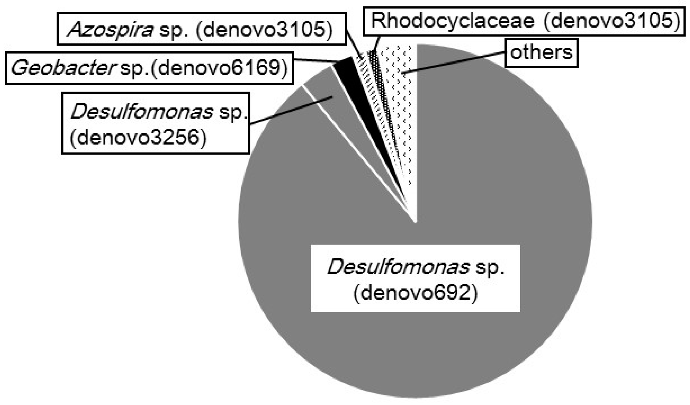
To identify the microorganisms grown in the rGO-CS complex at the genus level, we analyzed 16S rRNA genes by high-throughput sequencing. In the analysis using universal primer sets specific for prokaryotes, only bacteria were detected with a predominance of Desulfomonas species as major members having a population of more than 92% (Figure 3). Specifically, denovo692 and denovo3256 of operational taxonomic units (OTUs) having less than 97% sequence similarity were assigned to genus Desulfomonas and accounted for 89% and 3.2% of the total population, respectively. Desulfomonas species are known to reduce solid iron oxide [12,13] and electrodes and are frequently detected in marine microbial fuel cells [14,15]. Therefore, bacteria of the genus Desulfomonas, particularly those corresponding to denovo692, were the primary microorganisms reducing GO in the CS culture. In addition to Desulfomonas species, Geobacter (well-known exoelectrogenic bacteria) and Azospira species were detected, although these bacteria have been observed abundantly in GO-reducing enrichment cultures inoculated with freshwater samples [5].

2.4. Cyclic Voltanmetry (CV) and Electrochemical Impedance Spectroscopy (EIS) of the rGO-CS Complex

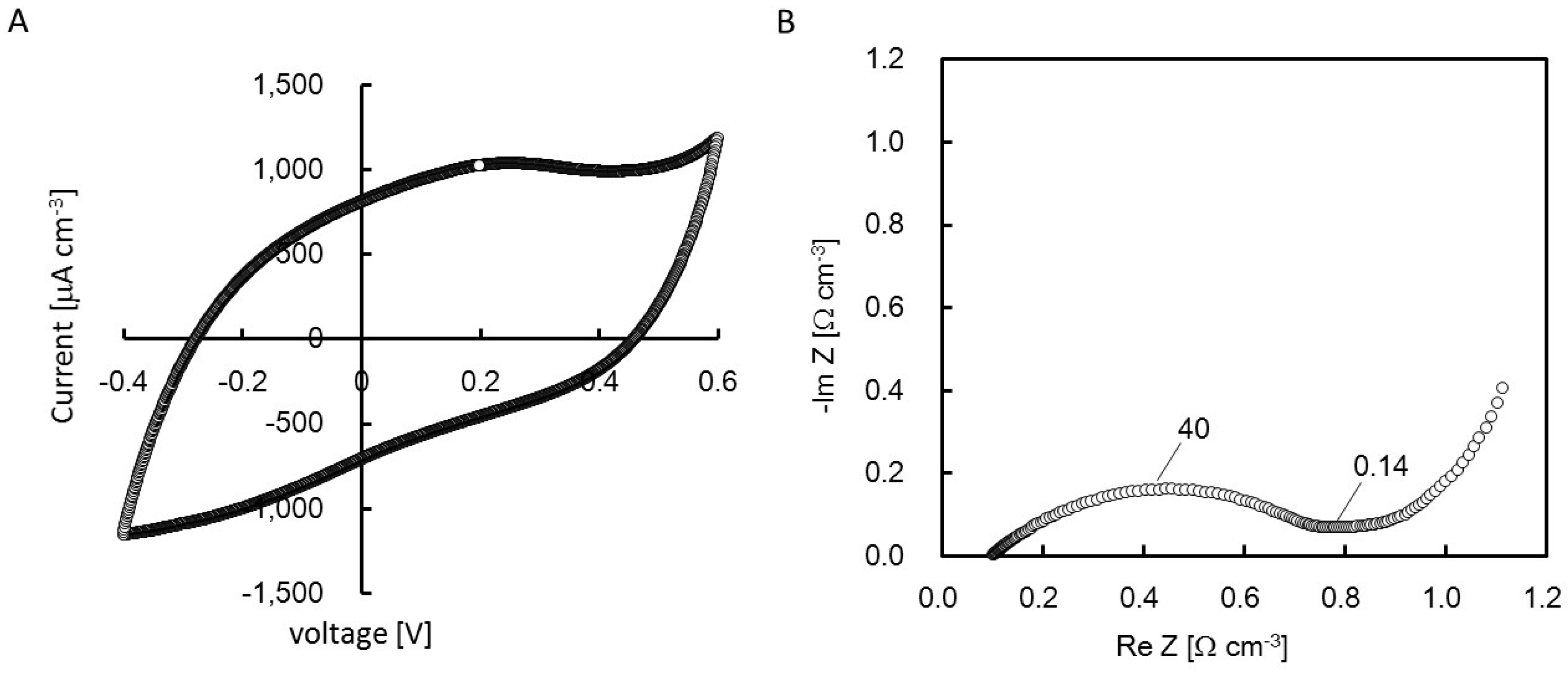
Figure 4A shows the CV for the CS-rGO complex. The CV showed the catalytic current of acetate oxidization, e.g., 1200–1700 μA/cm3 at 500 mV vs. Ag/AgCl with a 0.2 mV/s scan rate. No obvious redox peak was observed. The voltammogram showed a symmetric discharge of current, indicating electric double-layer capacitance due to the large surface area of the CS-rGO complex. The Nyquist plot yielded from EIS of the CS-rGO complex showed that the charge transfer resistance (Rct), indicated as a diameter of semicircles, was estimated to be less than 1.0 Ω/cm (Figure 4B). The catalytic current and charge transfer resistance were similar, as oberved in the complexes with pure culture of Geobacter sp. R4 and Geobacter-dominated cultures enriched from freshwater environments [5].

2.5. Electrochemical Cultivation

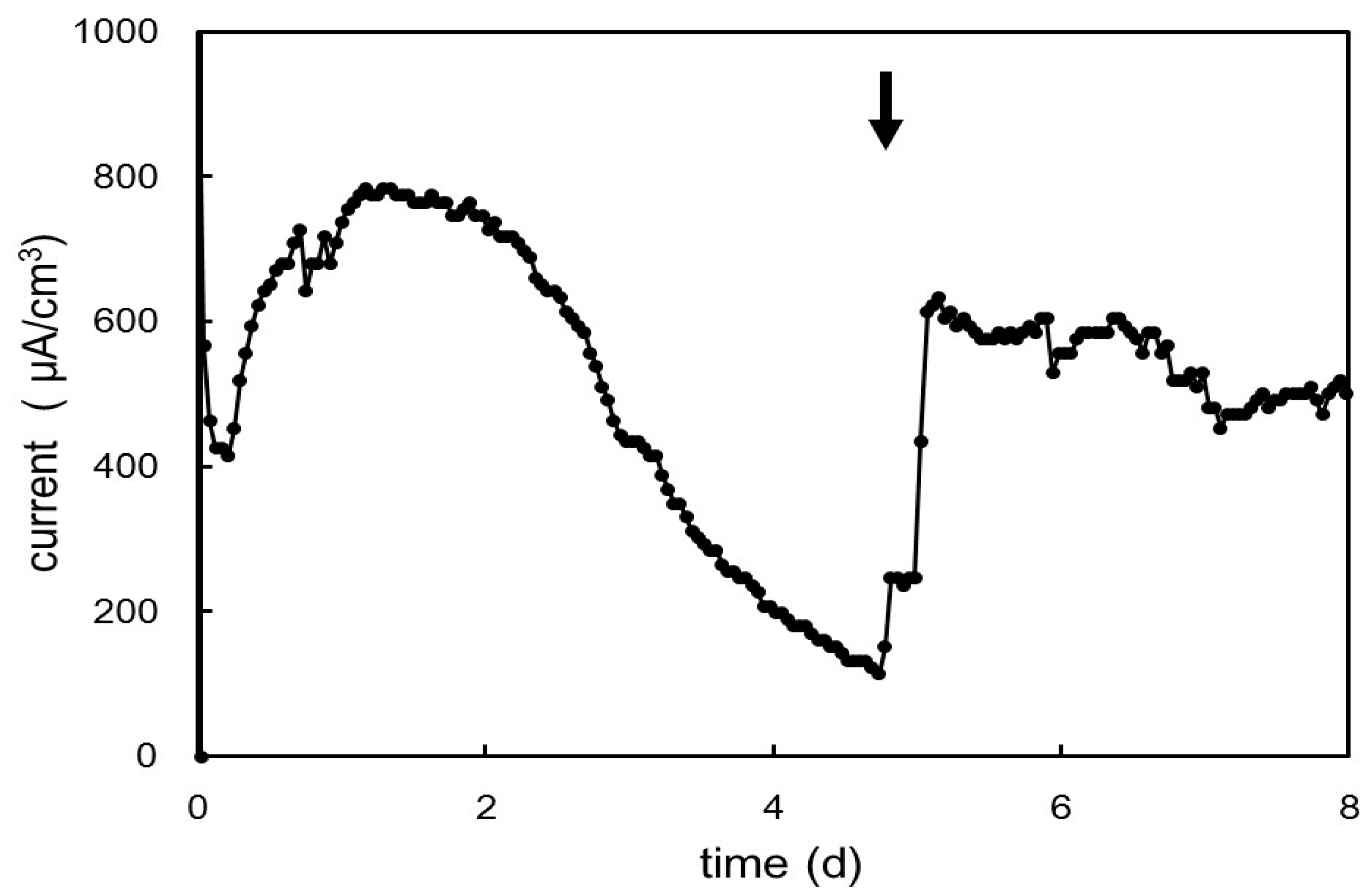
To analyze whether the rGO-CS complex with Desulfomonas predominace converted acetate to electricity, the rGO-CS complex was electrochemically cultivated at +200 mV (vs. Ag/AgCl). As shown in Figure 5, we observed immediate recovery of electricity in the CS culture and maximum peaks in the range of 800 μA/cm at one to two days. The electricity production gradually decreased and recovered after the addition of acetate on day 5. The immediate, stable production of electricity indicated the pre-growth of exoelectrogens during GO reduction and the stability in complexes after polarization, as observed in the rGO-complex with Geobacter dominance [5].

3. Discussion

GO is a nanomaterial that has been reported to have antibacterial or bactericidal effects toward a variety of bacteria [16,17,18,19,20] and to cause cytotoxicity in manmalian cells [21]. However, GO can also support bacterial growth, particularly for bacteria capable of exoelectron transfer. In this study, we examined the enrichment of GORB from a coastal sample to investigate the distribution and phylogenetic variety of GORB in seawater environments. Our results indicated that Desulfomonas and Geobacter species could grow on rGO and produce electricity via the reduced form of GO and provided important insights into the diversity of GORB in the natural environment.
The differences between the effects of GO reported in previous studies and this study may be associated with differences in bacterial species and physicochemical states of GO, such as sizes, dispersion in water, and coating on a dried surface [20]. Previously, GO or chemically reduced GO dispersed in aquous solutions has been reported to inactivate the cells due to oxidative stress [16,17], local heating by near-infrared irradiation [22], and membrane perturbation [19]. Potentially, some cells in rGO hydrogel were also inactivated via those mechanisms, while the total biomass in the culture was increased due to a balance of inactivated cells and the growth of surviving cells by GO respiration, which is a unique metabolism of exoelectrogens. For exoelectrogens, GO can function as a screening factor allowing the enrichment of exoelectrogens from natural environments. This study succesfully demonstrated the GO-dependent selectability of Desulfomonas species from coastal samples; this genus is a well-known representative marine exoelectrogen. The CS culture was obtained from the mixture of coastal sand and seawater using a procedure similar to that used for the enrichment of Geobacter sp. from freshwater environments, except for the changes in mineral compositions in the medium suitable for marine microorganisms. Therefore, the observed differences in the phylogenies of dominant bacteria may have been related to preferences regarding the sality of the environment.
The minor population of Shewanella spp. in the CS culture was unexpected because the species are known to produce electricity and reduce GO. Among more than 60 species of the genus Shewanella, countable species, e.g., S. oneidensis [23], S. putrefaciens [24], S. loihica [25], and S. electrodiphila [26], have been shown to generate electricity. Among these species, S. putrefaciens [24] and S. electrodiphila [26] can grow on acetate, while S. oneidensis [22] and S. loihica [27] cannot grow on acetate. Therefore, dominance of Desulfomonas species in the CS culture may be caused by the preference of the bacteria to redox coupling of acetate oxidization and GO reduction rather than Shewanella with limited capacity to reduce carbon electrodes by acetate oxidization.
The mechanisms through which Desulfomonas reduce GO are unknown. The electron transferring pathway from bacterial cells to GO has been studied using a range of mutants of S. oneidensis MR-1 lacking proteins of c-type cytochromes [1,6]. In the current model, electrons from acetate oxidation in the cytoplasm are passed via the menaquinone pool to outer membrane–anchored multiheme c-type cytochromes, MtrC and OmcA, together with an integral outer membrane scaffolding protein, MtrB. Forty-seven multiheme cytochromes are also encoded in the genome of a strain of Desulfomonas and are candidate proteins potentially involved in GO reduction [28].
The screening technique for GORB from natural environments used in this study, i.e., serial transferring enrichment using minimum medium supplemented with a simple electron donor and GO as the sole electron acceptor, is conventional but effective for enriching GORB with a preference for specific conditions. The agreement between the abilities to reduce GO and electricity production in both freshwater and marine environments indicated the wide applicability of GO for the enrichment of exoelectrogens and formation of a conductive hydrogel for different microbial inocula.

4. Materials and Methods

4.1. GO

GO was purchased as dried powder from Royal Elite New Energy Science & Technology Co., Ltd. (Shanghai, China) and used to enrich GORB. The GO was added to autoclaved MilliQ water and dispersed via ultrasonication for several hours. Most of the GO flakes were arranged in a single layer and had variable sizes (average: 0.26 ± 0.46 μm2), as analyzed by XPS and AFM imaging [5].

4.2. Enrichment of GORB

To enrich marine GORB, subsurface coastal sand and seawater were collected as an inoculum source from Ikobe coast in Toyohashi, Japan (34°39′27′′N, 137°23′31′′E). One gram wet weight of the coastal sand was mixed with 1 mL seawater, and the mixture was used as the inoculum solution. One milliliter of the inoculum solution was then introduced to serum bottles with a 60 mL capacity, including 20 mL DS-AGOFS medium containing following minerals in a basal medium (per liter): 20 g/L NaCl, 0.3 g/L KCl, 0.5 g/L NH4Cl, 0.1 g/L CaCl2·2H2O, 4 g/L MgCl2·6H2O, 0.6 g/L KH2PO4, 2.5 g/L NaHCO3, 1 mL/L SL-10, 1 mL/L Se/W solution, and 0.2 mg/L resazurin. This basal medium was prepared anaerobically under flashing N2:CO2 (80:20, v/v) gas [29] and then supplemented with 2 mM FeS solution, 10 mM acetate, and 0.67 g/L GO.
The microcosm of the mixture of environmental sample and the medium was incubated at 28 °C for seven days to allow microbial transformation of GO, which originally exhibited a brown color with dispersion in the liquid phase and then changed to black-colored aggregates of the rGO, as reported previously [30]. Then, the microcosm was transferred to fresh medium at 5% of the transfer rate and again incubated for seven days. The soil-free culture obtained by repeating the transfer more than 20 times was designated as the CS culture.

4.3. XPS and Electric Conductive Analysis

To determine chemical states of GO and rGO, samples deposited on glass slides were analyzed using XPS, as previously reported [1]. XPS spectra were acquired on an XPS instrument (Versa Probe PHI-5000; ULVAC-PHI Inc., Osaka, Japan) equipped with a monochromatic Al Ka X-ray source and operated at a bare pressure lower than 10−6 Pa. The electric conductivities of GO and rGO were determined by four-terminating sensing, as described previously [31].

4.4. Phylogenetic Identification of Bacteria in the CS Culture

For the identification of bacteria presented in the CS culture, high-throughput sequencing was performed for the 16S rRNA gene, a biomarker representing microbial phylogenetic affiliation. The partial hypervariable region of the 16S rRNA gene was amplified by polymerase chain reaction (PCR) [32] using the bacterial and archaeal consensus primers 515F and 806R. The amplicons were sequenced on an Illumina MiSeq platform at FASMAC Co., Ltd. (Atugi, Japan). A compiled set of the sequences was deposited in Short Read Archive database under accession number DRA004469.

4.5. Electrochemical Cultivation

For electrochemical cultivation, a hydrogel complex of rGO and microorganisms in the CS culture was prepared as follows. The CS culture was cultivated in a 0.93 L glass bottle (90 mm in diameter and 175 mm in height) with the following minor modifications. First, 1 L of DS-AGOFS medium was prepared as described previously. After autoclaving, the medium was mixed with 0.67 g/L GO and 15 mL of the CS culture, filled to the top of a glass bottle with removal of the headspace. The glass bottle was then incubated at 28 °C for one month. After incubation, the rGO-CS complex (approximately 30 mm in diameter and 10–20 mm in height) was obtained.
Electrochemical cultivation was conducted using a 0.93 L sterilized glass bottle (90 mm in diameter and 175 mm in height) as a cultivation cell. The bottle was filled with DS-AGOS medium, which had almost the same composition as the DS-AGOFS medium except for replacement of 0.075 mM FeS with 1 mM Na2S. Then, the rGO-CS complex was placed in a platinum cage in the bottle and connected with a platinum wire as the working electrode. An Ag/AgCl (KCl salt) electrode and another platinum wire were used as the reference and counter electrodes, respectively. Polarization was conducted by setting the working electrode potential at +200 mV vs. Ag/AgCl using a potentiostat (HA-1510; Hokuto Denko, Tokyo, Japan). During polarization, the current was recorded using a data logger (T&D Corporation, Nagano, Japan) every 60 min.

4.6. Electrochemical Analysis

Cyclic voltammetry (CV) analysis and electrochemical impedance spectroscopy (EIS) of the rGO-CS complex was conducted using an electrochemical measurement system (HZ-7000; Hokuto Denko). CV and EIS analyses were performed using the same bottle as that used for electrochemical cultivation described above after 2 h for stabilization following addition of 5 mM acetate. CV was conducted at a scan rate of 0.2 mV/s in the potential range from −400 to 600 mV (vs. Ag/AgCl). EIS was examined in a frequency range of 100 kHz to 0.5 mHz at 200 mV with 20 mV amplitude for the applied AC signal. Nyquist plots were obtained using ZSimpWin (Princeton Applied Research, Oak Ridge, TN, USA).

5. Conclusions

In this study, we successfully enriched a representative marine exoelectrogen, the Desulfomonas species, using GO. The Desulfomonas-dominated culture formed a hydrogel complex with rGO and generated electricity coupled with acetate oxidization by recovering electrons on rGO, as demonstrated using a representative freshwater exoelectrogen, Geobacter species. Our findings indicated the wide applicability of GO to enrich exoelectrogens in the hydrogel complex of rGO.

Acknowledgments

We thank Yuji Nagao for support with the experiments and Akira Hiraishi for discussion. This study was funded in part by the JSPS KAKENHI Grant-in-Aid for Young Scientists (A) (grant number: 26701010); the Program to Disseminate the Tenure Tracking System, MEXT, Japan; and the JST Accelerating Utilization of University IP Program.

Author Contributions

Naoko Yoshida designed and conducted experiments and wrote the paper; Yuko Goto performed enrichment experiments; Yasushi Miyata performed electrochemical analysis.

Conflicts of Interest

The authors report a potential competing financial interest in that they are currently applying for patents on the methods reported in the paper.

References

  1. Salas, E.C.; Sun, Z.; Luttge, A.; Tour, J.M. Reduction of graphen oxide via bacterial respiration. ACS Nano 2012, 4, 4852–4856. [Google Scholar] [CrossRef] [PubMed]
  2. Wang, G.; Qian, F.; Saltikov, C.W.; Jiao, Y.; Li, Y. Microbial reduction of graphene oxide by Shewanella. Nano Res. 2011, 4, 563–570. [Google Scholar] [CrossRef]
  3. Akhavan, O.; Ghaderi, E. Escherichia coli bacteria reduce graphene oxide to bactericidal graphene in a self-limiting manner. Carbon 2012, 50, 1853–1860. [Google Scholar] [CrossRef]
  4. Khanra, P.; Kuila, T.; Kim, N.H.; Bae, S.H.; Yu, D.-S.; Lee, J.H. Simultaneous bio-functionalization and reduction of graphene oxide by baker’s yeast. Chem. Eng. J. 2012, 183, 526–533. [Google Scholar] [CrossRef]
  5. Yoshida, N.; Miyata, Y.; Doi, K.; Goto, Y.; Nagao, Y.; Tero, R.; Hiraishi, A. Graphene oxide-dependent growth and self-aggregation into a hydrogel complex of exoelectrogenic bacteria. Sci. Rep. 2016, 21867. [Google Scholar] [CrossRef] [PubMed]
  6. Jiao, Y.; Qian, F.; Li, Y.; Wang, G.; Saltikov, C.W.; Gralnick, J.A. Deciphering the electron transport pathway for graphene oxide reduction by Shewanella oneidensis MR-1. J. Bacteriol. 2011, 193, 3662–3665. [Google Scholar] [CrossRef] [PubMed]
  7. Goto, Y.; Yoshida, N. Preliminary evaluation of a microbial fuelcell treating artificial dialysis wastewater using graphene oxide. AIP Conf. Proc. 2016, 1709, 020007. [Google Scholar]
  8. Wang, Y.; Li, Z.; Wang, J.; Li, J.; Lin, Y. Graphene and graphene oxide: biofunctionalization and applications in biotechnology. Trends Biotechnol. 2011, 19, 205–212. [Google Scholar] [CrossRef] [PubMed]
  9. Kalantar-zadeh, K.; Ou, J.Z.; Daeneke, T.; Strano, M.S.; Pumera, M.; Gras, S.L. Two-Dimensional Transition Metal Dichalcogenides in Biosystems. Adv. Funct. Mater. 2015, 25, 5086–5099. [Google Scholar] [CrossRef]
  10. Kuila, T.; Bose, S.; Khanra, P.; Mishra, A.K.; Kim, N.H.; Lee, J.H. Recent advances in graphene-based biosensors. Biosens. Bioelectron. 2011, 26, 4637–4648. [Google Scholar] [CrossRef] [PubMed]
  11. Sun, X.; Liu, Z.; Welsher, K.; Robinson, J.T.; Goodwin, A.; Zaric, S.; Dai, H. Nano-graphene oxide for cellular imaging and drug delivery. Nano Res. 2008, 1, 203–212. [Google Scholar] [CrossRef] [PubMed]
  12. Roden, E.E.; Lovley, D.R. Dissimilatory Fe (III) reduction by the marine microorganism Desulfomonas acetoxidans. Appl. Environ. Microbiol. 1993, 59, 734–742. [Google Scholar] [PubMed]
  13. Butler, J.E.; Young, N.D.; Lovley, D.R. Evolution from a respiratory ancestor to fill syntrophic and fermentative niches: Comparative genomics of six Geobacteraceae species. BMC Genom. 2009, 10, 103. [Google Scholar] [CrossRef] [PubMed]
  14. Bond, D.R.; Holmes, D.E.; Tender, L.M.; Lovley, D.R. Electrode-reducing microorganisms that harvest energy from marine sediments. Science 2002, 295, 483–485. [Google Scholar] [CrossRef] [PubMed]
  15. Holmes, D.; Bond, D.; O’neil, R.; Reimers, C.; Tender, L.; Lovley, D. Microbial communities associated with electrodes harvesting electricity from a variety of aquatic sediments. Microb. Ecol. 2004, 48, 178–190. [Google Scholar] [CrossRef] [PubMed]
  16. Liu, S.; Zeng, T.H.; Hofmann, M.; Burcombe, E.; Wei, J.; Jiang, R.; Kong, J.; Chen, Y. Antibacterial activity of graphite, graphite oxide, graphene oxide, and reduced graphene oxide: Membrane and oxidative stress. ACS Nano 2011, 5, 6971–6980. [Google Scholar] [CrossRef] [PubMed]
  17. Gurunathan, S.; Han, J.W.; Dayem, A.A.; Eppakayala, V.; Kim, J.-H. Oxidative stress-mediated antibacterial activity of graphene oxide and reduced graphene oxide in Pseudomonas aeruginosa. Int. J. Nanomed. 2012, 7, 5901–5914. [Google Scholar] [CrossRef] [PubMed]
  18. Akhavan, O.; Ghaderi, E. Toxicity of graphene and graphene oxide nanowalls against bacteria. ACS Nano 2010, 4, 5731–5736. [Google Scholar] [CrossRef] [PubMed]
  19. Chen, J.; Peng, H.; Wang, X.; Shao, F.; Yuan, Z.; Han, H. Graphene oxide exhibits broad-spectrum antimicrobial activity against bacterial phytopathogens and fungal conidia by intertwining and membrane perturbation. Nanoscale 2014, 6, 1879–1889. [Google Scholar] [CrossRef] [PubMed]
  20. Perreault, F.; de Faria, A.F.; Nejati, S.; Elimelech, M. Antimicrobial properties of graphene oxide nanosheets: why size matters. ACS Nano 2015, 9, 7226–7236. [Google Scholar] [CrossRef] [PubMed]
  21. Carpio, I.E.M.; Santos, C.M.; Wei, X.; Rodrigues, D.F. Toxicity of a polymer–graphene oxide composite against bacterial planktonic cells, biofilms, and mammalian cells. Nanoscale 2012, 4, 4746–4756. [Google Scholar] [CrossRef] [PubMed]
  22. Akhavan, O.; Ghaderi, E.; Esfandiar, A. Wrapping bacteria by graphene nanosheets for isolation from environment, reactivation by sonication, and inactivation by near-infrared irradiation. J. Phys. Chem. B 2011, 115, 6279–6288. [Google Scholar] [CrossRef] [PubMed]
  23. Ringeisen, B.R.; Henderson, E.; Wu, P.K.; Pietron, J.; Ray, R.; Little, B.; Biffinger, J.C.; Jones-Meehan, J.M. High power density from a miniature microbial fuel cell using Shewanella oneidensis DSP10. Environ. Sci. Technol. 2006, 40, 2629–2634. [Google Scholar] [CrossRef] [PubMed]
  24. Kim, H.J.; Park, H.S.; Hyun, M.S.; Chang, I.S.; Kim, M.; Kim, B.H. A mediator-less microbial fuel cell using a metal reducing bacterium, Shewanella putrefaciens. Enzyme Microb. Technol. 2002, 30, 145–152. [Google Scholar] [CrossRef]
  25. Newton, G.J.; Mori, S.; Nakamura, R.; Hashimoto, K.; Watanabe, K. Analyses of current-generating mechanisms of Shewanella loihica PV-4 and Shewanella oneidensis MR-1 in microbial fuel cells. Appl. Environ. Microbiol. 2009, 75, 7674–7681. [Google Scholar] [CrossRef] [PubMed]
  26. Zhang, J.; Burgess, J.G. Shewanella electrodiphila sp. nov., a psychrotolerant bacterium isolated from Mid-Atlantic Ridge deep-sea sediments. Int. J. Syst. Evol. Microbiol. 2015, 65, 2882–2889. [Google Scholar] [CrossRef] [PubMed]
  27. Gao, H.; Obraztova, A.; Stewart, N.; Popa, R.; Fredrickson, J.K.; Tiedje, J.M.; Nealson, K.H.; Zhou, J. Shewanella loihica sp. nov., isolated from iron-rich microbial mats in the Pacific Ocean. Int. J. Syst. Evol. Microbiol. 2006, 56, 1911–1916. [Google Scholar]
  28. Alves, A.S.; Paquete, C.M.; Fonseca, B.M.; Louro, R.O. Exploration of the ‘cytochromome’ of Desulfuromonas acetoxidans, a marine bacterium capable of powering microbial fuel cells. Metallomics 2011, 3, 349–353. [Google Scholar] [CrossRef] [PubMed]
  29. Yoshida, N.; Ye, L.; Liu, F.; Li, Z.; Katayama, A. Evaluation of biodegradable plastics as solid hydrogen donors for the reductive dechlorination of fthalide by Dehalobacter species. Biores. Technol. 2013, 130, 478–485. [Google Scholar] [CrossRef] [PubMed]
  30. Tanizawa, Y.; Okamoto, Y.; Tsuzuki, K.; Nagao, Y.; Yoshida, N.; Tero, R.; Iwasa, S.; Hiraishi, A.; Suda, Y.; Takikawa, H.; et al. Microorganism mediated synthesis of reduced graphene oxide films. J. Phy. Conf. Ser. 2012, 352, 012011. [Google Scholar] [CrossRef]
  31. Goto, Y.; Yoshida, N.; Umeyama, Y.; Yamada, T.; Tero, R.; Hiraishi, A. Enhancement of electricity production by graphene oxide in soil microbial fuel cells and plant microbial fuel cells. Front. Bioeng. Biotech. 2015, 3, 42. [Google Scholar] [CrossRef] [PubMed]
  32. Yoshida, N.; Iguchi, H.; Yurimoto, H.; Murakami, A.; Sakai, Y. Aquatic plant surface as a niche for methanotrophs. Front. Microbiol. 2014, 5, 30. [Google Scholar] [CrossRef] [PubMed]
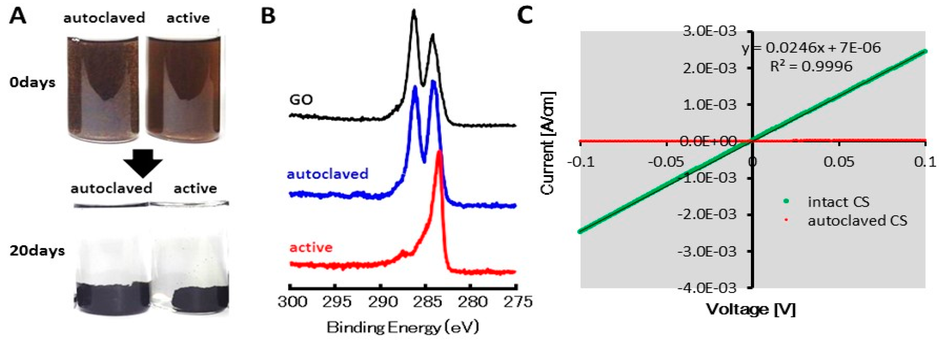
Figure 1. Reduction of GO in the CS culture. (A) Appearance of GO reduction in the CS culture; (B) XPS spectra of GO and GO in the cultures inoculated with autoclaved and intact cells; (C) Electrical conductivity of GO/rGO in the cultures inoculated with autoclaved and intact cells.
Carbon 02 00015 g001
Figure 2. Growth of microbial cells and acetate consumption depending on GO. (A) Acetate concentration in the CS cultures with and without GO; (B) Microbial cell counts in the cultures with and without GO and acetate; (C) Percentages of cells attached in complex and in planktonic liquid culture.
Carbon 02 00015 g002
Figure 3. Microbial composition of the rGO-CS complex determined by high-throughput sequencing of the 16S rRNA gene.
Carbon 02 00015 g003
Figure 4. CV (A) and EIS (B) data of the rGO-CS complex. The numbers shown in the graph (B) are frequencies: f (Hz) = ω/2π.
Carbon 02 00015 g004
Figure 5. Electricity production by the rGO-CS complex. The arrow indicates the time at which 5 mM acetate was added.
Carbon 02 00015 g005