Communication

DEAD-Box Helicase 3 Modulates the Non-Coding RNA Pool in Ribonucleoprotein Condensates During Stress Granule Formation

Department of Drug Discovery & Biomedical Sciences, College of Pharmacy, University of South Carolina Columbia, 715 Sumter St., Columbia, SC 29208, USA
*
Authors to whom correspondence should be addressed.
These authors contributed equally to this work.
Non-Coding RNA 2025, 11(4), 59; https://doi.org/10.3390/ncrna11040059
Submission received: 7 May 2025 / Revised: 11 July 2025 / Accepted: 17 July 2025 / Published: 1 August 2025

Abstract

Stress granule formation is a type of liquid–liquid phase separation in the cytoplasm, leading to RNA–protein condensates that are associated with various cellular stress responses and implicated in numerous pathologies, including cancer, neurodegeneration, inflammation, and cellular senescence. One of the key components of mammalian stress granules is the DEAD-box RNA helicase DDX3, which unwinds RNA in an ATP-dependent manner. DDX3 is involved in multiple steps of RNA metabolism, facilitating gene transcription, splicing, and nuclear export and regulating cytoplasmic translation. In this study, we investigate the role of the RNA helicase DDX3’s enzymatic activity in shaping the RNA content of ribonucleoprotein (RNP) condensates formed during arsenite-induced stress by inhibiting DDX3 activity with RK-33, a small molecule previously shown to be effective in cancer clinical studies. Using the human osteosarcoma U2OS cell line, we purified the RNP granule fraction and performed RNA sequencing to assess changes in the RNA pool. Our results reveal that RK-33 treatment alters the composition of non-coding RNAs within the RNP granule fraction. We observed a DDX3-dependent increase in circular RNA (circRNA) content and alterations in the granule-associated intronic RNAs, suggesting a novel role for DDX3 in regulating the cytoplasmic redistribution of non-coding RNAs.

1. Introduction

Stress granules (SGs) are biomolecular condensates formed from mRNA and RNA-binding proteins (RBPs) in the cytoplasm through liquid–liquid phase separation—a reversible process that leads to the formation of liquid-like droplets in cytoplasm—in response to various cellular stresses, including UV irradiation, heat, osmotic stress, endoplasmic reticulum stress, oxidative stress, and viral infections [1]. While SGs have traditionally been associated with translational shut-off and mRNA storage during stress, emerging research has revealed a more complex functional landscape. Recent findings highlight several key aspects of SG activity: (a) a portion of mRNAs recruited to SGs displays translational activity, and translational repression has been shown as a process co-occurring with SG formation [2]; (b) SGs maintain cellular homeostasis by preventing cell death pathways [3,4,5] and delaying premature senescence [6]; and (c) SGs regulate cellular metabolism, including inhibiting fatty acid oxidation during starvation through direct interactions with mitochondria and lipid droplets [7]. SG dysregulation is implicated in various pathologies [8], including cancer, where increased SG formation and persistence contribute to resistance to cell death, and senescence, where defects in SG dynamics are observed [9]. In neurodegeneration, SGs may seed pathological protein aggregation, while impaired SG transport along axons disrupts mRNA trafficking to synapses [10]. SGs are also exploited by viruses, which manipulate SG-associated RBPs to enhance replication and alter stress responses [11].
The diverse functions and associated pathologies of SGs arise from their complex and heterogeneous organization. According to the conventional model, mature SGs consist of dense cores surrounded by less dense shells [12], both composed of mRNA and proteins, with dynamic exchange occurring between the core, shell, and surrounding cytoplasm [13]. Comparative studies reveal a conserved SG protein pool, including RBPs, prion-like proteins, translation factors, and ~50% uncharacterized proteins [12] probably diffused in SGs during their cytosol journey [14]. SG-associated mRNAs encode proteins for ATP binding, transcription, cell cycle regulation, and splicing [15]. Despite extensive data collection, significant gaps remain in understanding the biophysical mechanisms of SG assembly, organization, and dynamics [12,13]; transcriptome [15] and protein composition [12,14]; and interactions with other biomolecular condensates (e.g., P-bodies [14], Q-bodies [16]) and organelles [7], particularly across different cell types and stress conditions [17,18].
Using inhibitors that block glycolysis and oxidative phosphorylation in U2OS cells, it was shown that ATP is required for SG assembly, SG fusion, and the dynamic exchange of G3BP—a marker and one of the key drivers of SG formation—between SGs and the cytoplasm [12]. Later, it was reported that proteins from the RNA-dependent DEAD-box ATPase (DDX) family—which interact with RNA and contain low-complexity domains (LCDs, loosely structured protein regions that are essential for oligomerization and enable the formation of protein networks)—can modulate the assembly and disassembly of biomolecular condensates such as stress SGs and P-bodies [19,20]. The proposed mechanism suggests that the formation of complexes between DDX family proteins (such as yeast Dhh1 and Ded1), ATP, and RNA drives liquid–liquid phase separation, resulting in the formation of DDX–RNA condensates, as shown in vitro [20]. Moreover, the reverse process, driven by the activation of Ded1 ATPase activity through the C-terminal region of eIF4G, was shown to lead to the disassembly of these complexes.
Our previous work indicates that the inhibition of ATPase activity in human Dead-Box Helicase 3 (DDX3), the orthologue of yeast Dhh1 and Ded1 and a component specific to mammalian SGs, using RK-33 or the knockdown of DDX3 in U2OS cells, leads to a decrease in the rate of SG assembly but does not affect disassembly [21]. DDX3, an ATP-dependent helicase, localizes to both the nucleus and cytoplasm, with a higher nuclear concentration in healthy cells [22]. DDX3 has been shown to hydrolyze ATP to unwind RNA-RNA and DNA-RNA duplexes and to recruit RNA into SGs in mammalian cells through the formation of DDX3–RNA complexes and protein oligomerization [20,23]. However, DDX3 activity extends to nearly all steps of RNA metabolism, including nuclear mRNA transport, ribosomal and nuclear small RNA export, 5′ UTR translation activation, and RNA splicing, although its precise roles remain unclear [22]. Therefore, we hypothesize that alterations in DDX3 activity can influence not only the SG dynamics but also lead to changes in the RNA pool, both in RNP granules, including SG and associated condensates such as P-bodies, and the cytoplasm. Here, we report that the analysis of RNP granule content purified from U2OS cells treated with sodium arsenite—a known inducer of both SG and P-bodies [24]—and RK-33, a small molecule inhibitor of DDX3 ATPases activity shown to be effective in breast cancer bone metastasis patients [25], reveals alterations in the non-coding RNA pools of both RNP granules and the cytoplasm. Specifically, we observe an increase in intron RNA and circular RNA (circRNA) within RNP granules.

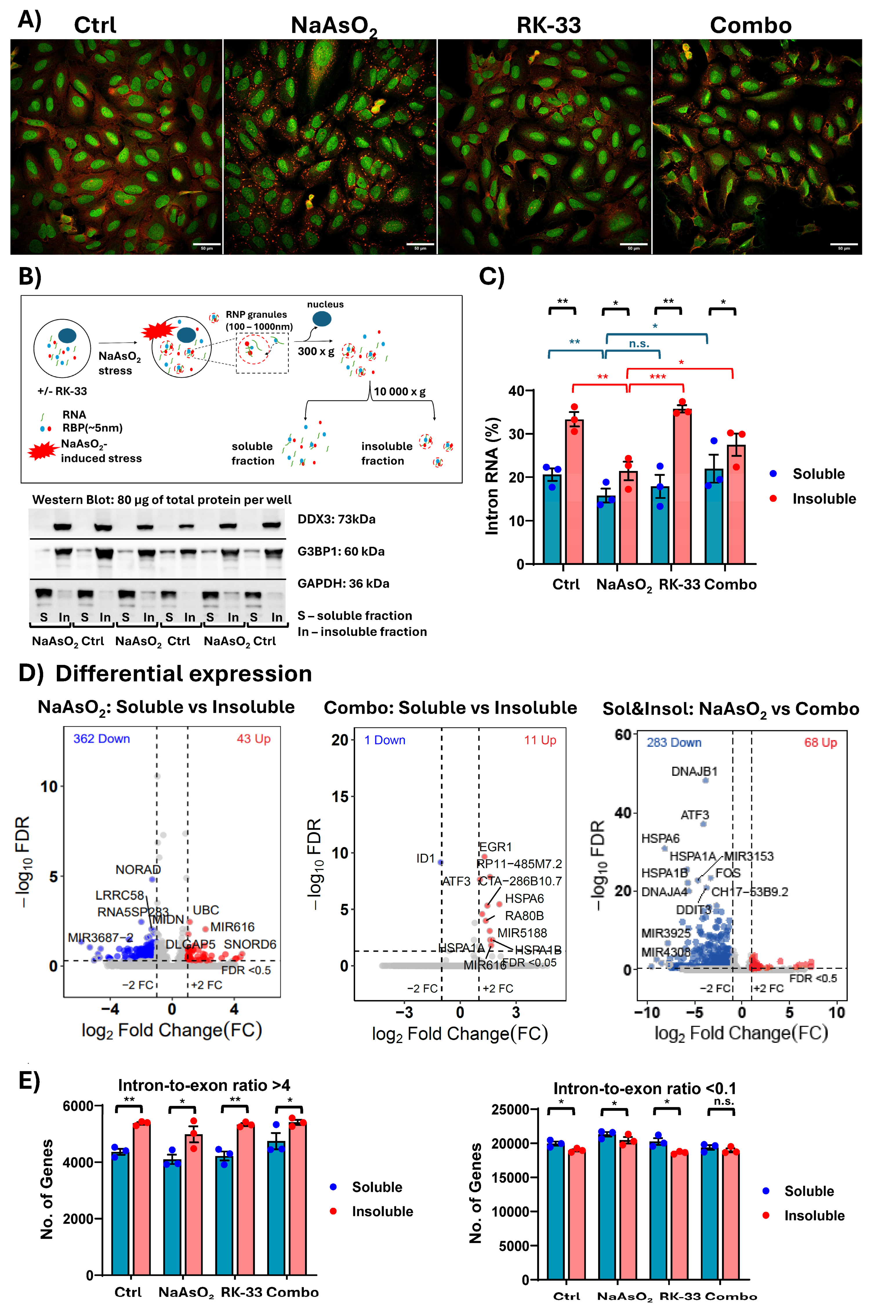
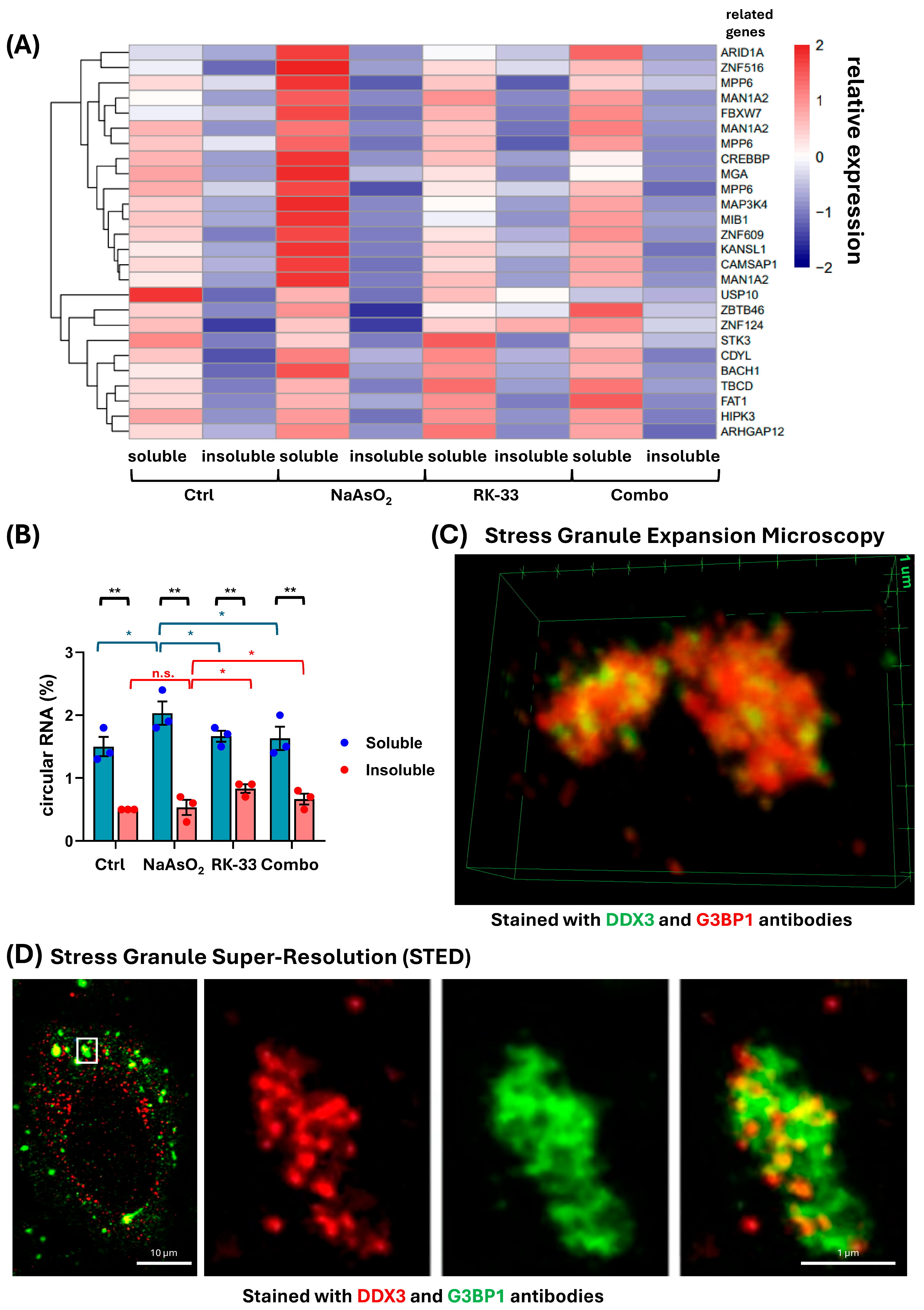
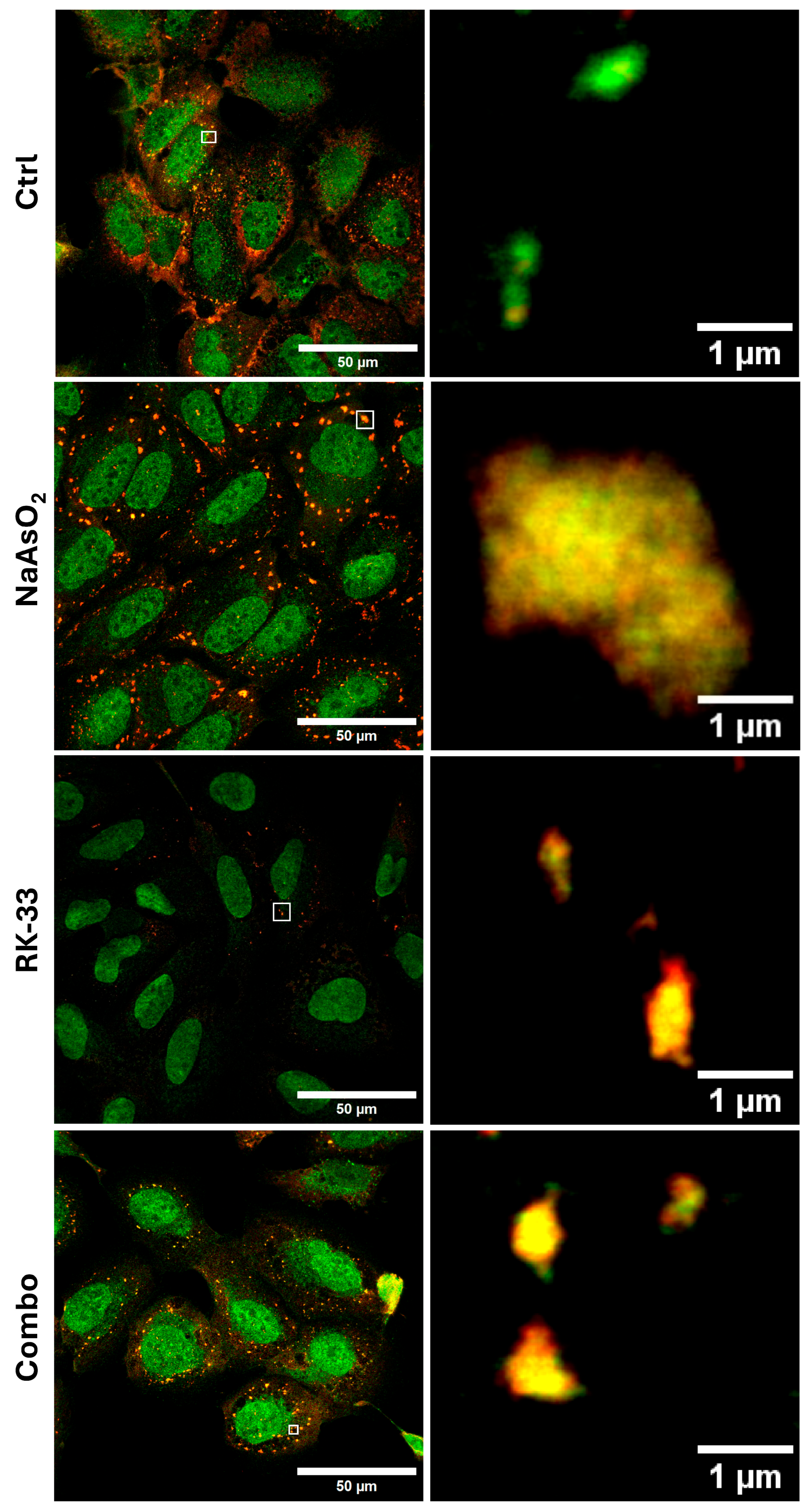
2. Results and Discussion

To investigate the impact of DDX3 activity on the RNA composition of RNP granules, we induced the formation of SGs and P-bodies in U2OS cells using sodium arsenite (NaAsO2), an inducer of oxidative stress [24]. The inhibition of DDX3 activity was achieved using the small molecule RK-33 before NaAsO2 treatment. RK-33 has been shown to specifically inhibit the ATPase and ATP-dependent RNA helicase activities of DDX3 by binding to its ATP-binding site [26,27], leading to decreased SG assembly as described in our previous work [21] (Figure 1A, Figure S1). Notably, a small subset of cells in the population exhibited stress granules even in the absence of induced stress. Following cell lysis, the RNP granule-enriched fraction was obtained through a series of centrifugation steps [28] (Figure 1B) from cells that were either untreated, treated with NaAsO2 or RK-33 alone, or co-treated with both compounds, as described in the Section 3. Western blot was used to validate the efficiency of the assay in isolating RNP-enriched stress granules, both under basal conditions and following NaAsO2 treatment. RNA was then purified from this fraction. The separation of G3BP1 between insoluble and soluble fractions corresponds to the behavior of G3BP1 as a canonical G3BP1 marker and repeats the DDX3 pattern in Western blot. Interestingly, it has been shown that G3BP1, while serving as a marker for stress granules, exhibits different behavior in its response to stress [29]. Recent publications have provided evidence that SGs are formed from pre-existing nanoscale seeds, which are present before the application of stress. The accumulation of both G3BP1 and DDX3 in SG-enriched fractions in both NaAsO2 naïve conditions pointed to the enrichment of cores [30,31,32]. The oxidative stress did not cause any further enrichment of G3BP1 and DDX3 in the SG-enriched fraction. This may suggest that most of these proteins are localized in nano-seeds, and SGs form by the redistribution of nano-seed proteins rather than the recruitment of soluble G3BP1 and DDX3. Additional extensive experiments are needed to confirm this. The RNA composition of the soluble and insoluble (SG- and P-body-enriched) fractions was determined after rRNA depletion using Illumina sequencing. The obtained reads were aligned to the genome, and the reads aligned to exons and introns were counted separately to determine processed mRNAs (or exonic) and intronic transcripts, which may represent intron-derived transcripts, lariat RNAs, or non-spliced mRNAs exported to the cytoplasm [33,34,35] (see the Section 3 for details). We observed an increase in intronic RNA levels between the soluble (~20%) and insoluble (~30%) cytoplasmic fractions across different conditions (Figure 1C). Moreover, we observed an increase in intronic RNA levels within the insoluble (RNP granule) fraction isolated from cells co-treated with RK-33 and NaAsO2, compared to the insoluble fraction from cells treated with NaAsO2 alone (Figure 1C, Supplementary Tables S1–S3). The differential expression analysis revealed a difference in RNA content between the soluble and insoluble fractions in NaAsO2-treated cells, as well as a significantly smaller difference between these fractions in cells co-treated with NaAsO2 and RK-33 (Figure 1D). The differential analysis of combined data from both the soluble and insoluble fractions revealed a decrease in cytoplasmic RNA content following co-treatment with NaAsO2 and RK-33. This observation is consistent with previous reports showing changes in the transcriptome landscape following RK-33 treatment [36,37] and, notably, shows further alterations between the RNP-enriched insoluble fraction and the soluble cytoplasmic fraction after RK-33 treatment. Further, co-treatment with RK-33 also resulted in the loss of the RNP-enriched fraction containing long non-coding RNAs (lncRNAs) such as NORAD—an lncRNA that regulates PUM protein activity and contributes to the formation of RNP condensates like stress granules [38]. Additional analysis revealed a higher intron-to-exon ratio in the insoluble RNP-enriched fraction compared to the soluble cytoplasmic fraction (Figure 1E, Supplementary Table S4).
Given the enrichment of non-coding intronic sequences, we analyzed the distribution of another class of ncRNAs, circRNAs. To determine circRNAs, we identified back-splice junctions in RNA sequencing data—a method that detects upstream exons appearing downstream, a characteristic feature of circRNAs (see the Section 3 for details). circRNA content was higher in the soluble fraction than in the insoluble fraction, associated with RNP granules, under all conditions (Figure 2A,B, Supplementary Tables S5–S7). As expected and consistent with previous reports, the overall expression of circRNAs remained relatively low [39]. Notably, NaAsO2 treatment led to an increase in circRNA content in the soluble fraction (Figure 2B), in line with the slow and inefficient biogenesis of circRNAs previously described [39]. Specifically, we observed an increase in circRNAs reported to be associated with proliferation or apoptosis signaling, linked to genes such as ARID1A [40,41], MPP6 [42], MAN1A2 [43,44], CREBBP [45,46], MGA [47], MAP3K4 [48], ZNF609 [49], and KANSL1 [50] (Figure 2A). Interestingly, the recently reported circMPP6 was found to interact with the RBP MEX3A [51], facilitating its association with P-body proteins, while ARID1A has been reported to interact with KHSRP [52,53], which can also be recruited into P-bodies. Both circMPP6 and circARID1A showed increased levels in the soluble fraction, accompanied by a decrease in the RNP granule fraction during NaAsO2 treatment. Subsequent treatment with RK-33 decreased circRNA content in the soluble fraction, and co-treatment with NaAsO2 and RK-33 prevented the stress-induced increase in circRNA content in the soluble fraction. Notably, in both cases, an increase in circRNA levels in the insoluble fraction was observed.
The molecular mechanism of DDX-dependent RNA redistribution is yet to be identified. A plausible explanation for the observed effects of RK-33, a DDX3 ATPase activity inhibitor, on the non-coding RNA pool is altered substrate preference and the RNA-binding efficiency of DDX3. To explore further, we analyze the distribution of DDX3 and the commonly used marker of SGs, G3BP1, in the NaAsO2-induced stress granules with expansion and super-resolution microscopy (Figure 2C,D and Figure 3). Strikingly, DDX3 forms distinct cores within SGs, which may or may not overlap with G3BP1 (Figure 2C,D). Moreover, STED super-resolution imaging suggested a reduction in stress granule size following co-treatment with RK-33 and NaAsO2, compared to NaAsO2 treatment alone (Figure 3). However, further quantitative analysis is required to confirm and quantify this observation. The results pointed out the possibility that DDX3 molecules form distinct domains in SGs, selectively enriched with specific RNAs. Recent reports have shown that DDX3 ATPase activity is required to enhance the dynamics of G3BP1-driven RNP granules, such as SGs, as well as to support ongoing translation in granules [54].
However, the activity of DDX3 is not restricted to RNP granules. The changes in intron and circRNA levels observed in our results (Figure 1 and Figure 2) may be linked to an alteration in DDX3’s role in splicing—a function that remains poorly characterized in the literature. Notably, the C-terminus of DDX3 contains a domain similar to those found in splicing factors [55], and DDX3 has been shown to interact with spliceosome and ribonucleoprotein complexes post-splicing [22]. Another potential explanation is that RK-33 alters DDX3’s role in nuclear export. DDX3 has been shown to interact with the TAP protein, which mediates the nuclear export of mRNA and may itself be transported via the TAP-dependent pathway [56]. Additionally, DDX3 has been shown to facilitate the export of intron-containing HIV-1 RNA through the CMR1-mediated nuclear export pathway [57]. Finally, DDX3 has also been reported to enhance transcription by interacting with transcription factors [58]. For example, circRNA Circ-CTNNB1 binds to DDX3, facilitating its interaction with the transcription factor Yin Yang 1, which leads to β-catenin activation [59]. Given the numerous activities of DDX3 in RNA processing and localization, further analysis is needed to dissect the effects.
Overall, our results are the first to determine the function of DDX3 activity in regulating the subcellular distribution of circRNAs and intron-associated RNAs in SG- and P-body-enriched fractions. Importantly, the SGs’ core structure persists in both naïve and stressed conditions; therefore, the results demonstrate the DDX3 dependence of the RNA compositions of seed nanocores and stress-induced stress granules. Interestingly, co-treatment with RK-33 and NaAsO2 led to the depletion of non-coding RNAs—such as lncRNA NORAD [38], circMPP6 [42], and ARID1A [40]—that promote RNP granule formation or interact with associated RBPs (Figure 1D and Figure 2A). This highlights DDX3 activity as a potential target for investigating the role of non-coding RNAs—especially circRNAs, whose involvement in regulating RNP granule formation was recently suggested [60]. Notably, RK-33 co-treatment with NaAsO2 decreased stress response RNAs—such as ATF3 [61], DNAJB1 [62], HSPA6 [63], and HSPA1B [64]—compared to arsenite alone, with reductions in ATF3, DNAJB1, and certain circRNAs associated with cancer corresponding to RK-33’s established anti-tumor effects [25]. However, our preliminary results do not provide a definitive answer regarding the specific mechanism by which DDX3 activity influences non-coding RNA content in RNP granules and their formation. This opens several avenues for future investigation, including the identification of RNAs that directly interact with DDX3—potentially using RNA editing enzyme fusions—as well as the application of FISH techniques to validate the localization [15,65] depletion of lncRNAs and circRNAs within RNP granules. In addition, testing different RK-33 pre-treatment time points prior to stress induction may help elucidate the temporal dynamics of its effects on the non-coding RNA pool, particularly cytoplasmic circRNAs, whose biogenesis has been reported to be inefficient [39].

3. Methods

3.1. Cell Culture and Stress Granule Induction

U2OS cells were maintained in high-glucose Dulbecco’s Modified Eagle Medium (ATCC, Manassas, VA, USA) supplemented with 10% fetal bovine serum (Mediatech Inc., Manassas, VA, USA) and 1% penicillin/streptomycin (HyClone, GE Healthcare Life Sciences, Pittsburgh, PA, USA) at 37 °C in a humidified atmosphere with 5% CO2, as previously described [21]. Cells were pre-treated with 6 μM RK-33 (MedChemExpress, Monmouth Junction, NJ, USA) (, stock solution 5mM DMSO (MilliporeSigma Burlington, MA, USA D8418) for 1 h. Sodium arsenite stress was induced by treating cells with sodium arsenite, 500 µM NaAsO2 (MilliporeSigma, Burlington, MA, USA S7400), for 60 min. Basal conditions were defined as treatment with 0.12% DMSO for 2 h, corresponding to the duration and timing of RK-33 addition in treated samples.

3.2. Expansion Microscopy

The expansion microscopy procedure was performed based on previously published work by Boyden’s laboratory, with some modifications [66]. Briefly, U2OS cells treated with NaAsO2 were fixed for 5 min at room temperature using a fixative solution containing PFA and Triton X-100, followed by an additional permeabilization step with 0.5% Triton X-100. Coverslips with the cells were carefully washed for 10 min in a sodium borohydride solution to reduce autofluorescence and potentially restore antigenicity after fixation. Cells were then blocked with 5% BSA at room temperature for one hour.
The primary antibody mixture—mouse anti-DDX3 (sc-365768, Lot# D0819, 200 µg/mL, Santa Cruz Biothechnology, Dallas, TX, USA) and rabbit anti-G3BP1 (A302-033A-T, Lot# 2, 200 µg/mL, Fortis Life Sciences, Boston, MA, USA)—was applied overnight at 4 °C. Secondary antibodies used for visualization were Abberior StarRed 639–conjugated anti-mouse for DDX3 and donkey anti-rabbit Alexa Fluor Plus 555 for G3BP1.
For anchoring, coverslips were pre-incubated with 100 mM sodium bicarbonate twice for 15 min each, followed by incubation in 0.1% (w/v) GMA in 100 mM sodium bicarbonate for 3 h at room temperature, and then transferred for overnight incubation at 37 °C.
The gelation procedure was performed in a cold room (4 °C) to prevent premature gelation. The gelling chamber was assembled according to the referenced protocol [66]. Samples were carefully submerged in a mixture composed of StockX (8.6% (w/v) sodium acrylate (SA), 2.5% (w/v) acrylamide (AA), 0.15% (w/v) N,N′-methylenebisacrylamide (Bis), 2 M sodium chloride (NaCl), and 1× PBS, with 10% (w/v) N,N,N′,N′-Tetramethylethylenediamine (TEMED) and 10% (w/v)) ammonium persulfate (APS) in a 47:1:1:1 ratio. The samples were left at 4 °C for 30 min.
Gelled samples were transferred into a humidified tip box and incubated at 37 °C for 2 h. After gelation, gels were trimmed and incubated overnight in digestion buffer consisting of 8 U/mL Proteinase K (ThermoFisher Scientific, Waltham, MA, USA), Catalog No. BP1700-100), 0.5% (w/v) Triton X-100, 1 mM EDTA, 2 M NaCl, and ddH2O.
The following day, coverslips were placed in 10 mL of ddH2O in Petri dishes and shaken at 60 rpm on an orbital shaker for 10 min at room temperature, in the dark (protected with aluminum foil). This washing step was repeated twice. Finally, the samples were trimmed to fit into glass-bottom 35 mm Petri dishes for image acquisition.
Microscopic data were acquired using a Carl Zeiss LSM 700 laser scanning confocal microscope (Carl Zeiss Meditec, Dublin, CA, USA) equipped with a 20× Plan APO 0.8 air objective. Imaging was performed in Z-stack mode with a total of 17 optical sections. Images were captured at 1× scanning zoom with a resolution of 20.15 pixels per micron in 16-bit depth. Excitation was performed at 555 nm and 639 nm wavelengths. Subsequent image processing and analysis were conducted using ImageJ 1.54p software (National Institutes of Health, USA).

3.3. STED and Confocal Microscopy

Stimulated Emission Depletion (STED) microscopy was performed using a Stedycon STED microscope on fixed U2OS cells treated with 500 μM NaAsO2 for 60 min and labeled for stress granule markers. G3BP1 was detected with Abberior STAR RED and DDX3 with Abberior STAR ORANGE secondary antibodies. Images were acquired at a pixel size of 30 nm, with line accumulations of 10 for STED and 5 for confocal channels. Pixel dwell times were 10 µs (STED) and 5 µs (confocal). Excitation lasers were 640 nm (STAR RED) and 561 nm (STAR ORANGE), with STED at 775 nm (28.1% for STAR RED, 50% for STAR ORANGE). Gated detection was used for STED (1–7 ns), while confocal channels had no gating. Images were acquired with a 100× objective, using a 32 µm pinhole, over a field of view of 34 × 63 µm (1123 × 2087 pixels).
Confocal microscopy (40×) and STED super-resolution microscopy (100×) were also performed using the STELLARIS 8 STED system on samples treated with NaAsO2, RK-33, or a combination of both, with concentrations as described above and 1% DMSO as the basal condition. G3BP1 was detected with Abberior STAR RED, DDX3—with Aberrior STAR ORANGE. Excitation lasers were 642 nm (STAR RED) and 589 nm (STAR ORANGE), with STED at 775 nm (80% for STAR RED, 90% for STAR ORANGE). Gated detection was used for STED STAR RED (1–7 ns), STAR ORANGE in Intensity mode, while confocal channels had no gating. Confocal microscopy (20x) of these samples was performed on a Zeiss LSM 700 microscope. Images were acquired at 2048 × 2048 resolution with a pixel size of 0.3126 μm. Detection was performed with PMT detectors, using 555 nm and 639 nm excitation lasers.

3.4. Stress Granule (SG) Isolation

Stress granule isolation was adapted from previous protocols [28,67]. Briefly, cells were grown in 20 mL culture dishes, washed once with ice-cold culture medium and once with ice-cold PBS, then scraped and collected by centrifugation at 200× g for 5 min at 4 °C. Pellets were washed with 500 µL ice-cold PBS, centrifuged at 300× g, and lysed in buffer [50 mM Tris (pH 7.6), 50 mM NaCl, 5 mM MgCl2, 0.5% NP-40 (MilliporeSigma Burlington, MA, USA 492016, Lot B21198), 1 mM β-mercaptoethanol (Bio-Rad, Hercules, CA, USA #1610710), 1× EDTA-free protease inhibitor cocktail (Roche, Indianapolis, IN, USA, 05 892 791 001), and 0.4 U/µL RNase inhibitor (New England Biolabs, Ipswich, MA, USA M0314L)]. Lysates were homogenized with 30 strokes using a Dounce homogenizer and centrifuged at 300× g for 3 min at 4 °C to remove nuclei. Then the supernatant was centrifugated at 10,000× g for 10 min at 4 °C. The soluble and insoluble (RNP granule) cytoplasmic fractions were separated, and protein and RNA concentrations were measured using a NanoDrop spectrophotometer.

3.5. Western Blot

After protein concentrations were measured using a NanoDrop spectrophotometer, as described in the Section 3.4, equal amounts of protein (approximately 80 μg from both soluble and insoluble fractions) were loaded onto SurePAGE 4–12% Bis-Tris gradient gels. Following electrophoretic separation, proteins were transferred onto PVDF membranes (Cat# 88518, Thermo Fisher Scientific, Waltham, MA, USA). The membranes were blocked in 1× TBS-T containing 5% non-fat dry milk and incubated overnight at 4 °C with primary antibodies. The primary antibodies applied included anti-G3BP1 (1:1000, A302-033A, Thermo Fisher Scientific), anti-DDX3 (1:3000, sc-365768, Santa Cruz Biotechnology, Dallas, TX, USA), and anti-GAPDH (1:5000, #2118, Cell Signaling Technology). HRP-conjugated secondary antibodies, anti-mouse (1:8000, #7076S, Cell Signaling Technology, Danvers, MA, USA), and anti-rabbit (1:8000, #7074S, Cell Signaling Technology, Danvers, MA) were used for detection. Protein bands were visualized using the SuperSignal West Femto Maximum Sensitivity Substrate (ThermoFisher Scientific).

3.6. Intron and Circular RNA Analysis

RNA and library preparation, the post-processing of the raw data, and data analysis were performed by the USC CTT COBRE Functional Genomics Core. RNAs were extracted with Zymo Direct-zol RNA Microprep Kits as per manufacturer recommendations (Zymo, Tustin, CA, USA), and RNA quality was evaluated on RNA-1000 chip using Bioanalyzer (Agilent, Santa Clara, CA, USA). RNA libraries were prepared using an established protocol with NEBNext Ultra II Directional Library Prep Kit (NEB, Lynn, MA, USA). Each library was made with one of the TruSeq barcode index sequences and had a 1:200 dilution of Spike-in SIRV_Set3 (Lexogen, Greenland, NH, USA) added. Illumina sequencing was performed by Medgenome (Foster City, CA, USA) with Illumina HiSeqX (150 bp, pair-ended).
Human genome GRCh38.p13 (GRCh38_r98.all, GENCODE release 40) and comprehensive gene annotation GTF (Regions CHR, GENCODE release 40) were downloaded from GENCODE.
Sequences were aligned to the human genome GRCh38.p13 (GRCh38_r98.all, GENCODE release 40) using STAR 2.7.2b [68]. SAMtools (v1.2) was used to convert aligned SAM files to BAM files, and reads were counted using the feature-Counts function of the Subreads package [69] using a homemade gencode_v40annotation_intron.gtf annotation file. CircRNA was detected from chimeric reads by DCC 0.5.0 [70], the tool identifying circRNA via back-splice junctions.
A comprehensive gene annotation GTF was downloaded (Regions CHR, GENCODE release 40) from GENCODE. Using bedtools 2.27.1 for a homemade annotation file, first use mergeBed to merge all exons. Then, subtractBed was used to subtract the exonic region from the genic region to define intronic regions.
Only reads that were mapped uniquely to the genome were used for gene expression analysis. Differential expression analysis was performed in R using the edgeR package [71]. The average read depth for the samples was 18 million reads, and only genes with at least one count per million average depth were considered for differential expression analysis. Raw counts were normalized using the Trimmed Mean of M-values (TMM) method, and the normalized read counts were then fitted to a generalized linear model using the function glmFit [72]. Genewise tests for significant differential expression were performed using the function glmLRT. The p-value was then corrected for testing using Benjamini–Hochberg’s FDR [73].
To compare expression levels between soluble and insoluble fractions within the same treatment condition, a paired t-test was used [74]. For comparisons between treatment conditions, an unpaired t-test was applied. The experiment with RNA sequencing of soluble and insoluble fractions was repeated three times for all experiment conditions.

4. Conclusions

Here, we have demonstrated that the treatment of U2OS cells with RK-33, an inhibitor of DDX3 ATPase activity, leads to a redistribution of non-coding RNAs within RNP granules and the cytoplasm. Specifically, we observed a depletion of RNA species in SGs, including the lncRNA NORAD, which is known to support RNP condensate formation. We also showed that RK-33 treatment prior to SG induction with NaAsO2 prevented the stress-induced accumulation of circRNAs associated with proliferation and apoptosis signaling in the soluble fraction. Notably, RK-33 treatment prior to SG induction led to the depletion of circRNAs such as circMPP6 and ARID1A in the soluble cytoplasmic fraction, both of which have been reported to interact with RBPs associated with P-bodies and SGs. These findings are consistent with the reduced SG assembly we previously reported and are further supported by representative confocal images showing reduced SG formation.
These results highlight the potential role of DDX3 in modulating the non-coding RNA landscape, particularly regulatory RNAs involved in RNP granule formation and signaling-related circRNAs. Interestingly, we also noted a tendency toward increased levels of circRNA and intronic RNA within RNP granules post-treatment. While preliminary, these trends warrant further investigation. Future studies could explore longer RK-33 incubation times to assess RNA dynamics more fully, compare responses in non-cancerous cell lines to distinguish cancer-specific effects, and incorporate FISH-based validation of affected RNA species in granules.

Supplementary Materials

The following supporting information can be downloaded at: https://www.mdpi.com/article/10.3390/ncrna11040059/s1, Figure S1. Effect of RK33 treatment on SGs formation, Table S1. Raw exon counts data of RNA-Seq samples; Table S2. Raw intron counts data of RNA-Seq samples; Table S3. Total counts numbers of exon and intron reads of RNA-Seq samples; Table S4. Number of genes meeting different intron-to-exon ratio selection criteria; Table S5. Raw circular RNA counts data of the RNASeq samples; Table S6. Raw linear RNA counts data of the RNASeq samples; Table S7. Percentage of circular RNA counts of the RNASeq samples.

Author Contributions

B.C.C., H.J., E.K. and M.S. initiated the study; B.C.C., H.J., E.K., S.S. and M.S. designed and performed the experiments; E.K., H.J., A.S., V.S., M.C., B.C.C. and M.S. analyzed the data; E.K., A.S. and V.S. performed image analysis; and E.K., H.J., A.S. and M.S. wrote the manuscript. All authors have read and agreed to the published version of the manuscript.

Funding

We thank COBRE Center for Targeted Therapeutics, Microscopy and Flow Cytometry Core for image analysis and Functional Genomics Core for transcriptomics analysis. The COBRE CTT cores are supported by NIH NIGMS P20GM109091, P30GM154632. The work was supported by awards from NIH NIDA R21DA058586, R01DA054992, NIH NIGMS P20AGM109091-10S3 (to MS).

Data Availability Statement

RNA sequencing data were deposited in the NCBI. GEO Accession number is GSE303136.

Conflicts of Interest

The authors declare no conflicts of interest.

References

  1. Kedersha, N.; Anderson, P. Regulation of Translation by Stress Granules and Processing Bodies. Prog. Mol. Biol. Transl. Sci. 2009, 90, 155–185. [Google Scholar] [CrossRef] [PubMed]
  2. Daniel, M.; Bastian, E.; Franka, V.; Jan, E.; Gregory, R.; Jeffrey, A.C. Single-Molecule Imaging Reveals Translation of Mrnas Localized to Stress Granules. Cell 2020, 183, 1801–1812. [Google Scholar] [CrossRef]
  3. Place, D.E.; Samir, P.; Malireddi, R.S.; Kanneganti, T.D. Integrated Stress Response Restricts Macrophage Necroptosis. Life Sci. Alliance 2022, 5, e202101260. [Google Scholar] [CrossRef] [PubMed]
  4. Samir, P.; Kesavardhana, S.; Patmore, D.M.; Gingras, S.; Malireddi, R.K.S.; Karki, R.; Guy, C.S.; Briard, B.; Place, D.E.; Bhattacharya, A.; et al. Ddx3x acts as a Live-or-Die Checkpoint in Stressed Cells by Regulating Nlrp3 Inflammasome. Nature 2019, 573, 590–594. [Google Scholar] [CrossRef] [PubMed]
  5. Arimoto, K.; Fukuda, H.; Imajoh-Ohmi, S.; Saito, H.; Takekawa, M. Formation of Stress Granules Inhibits Apoptosis by Suppressing Stress-Responsive Mapk Pathways. Nat. Cell Biol. 2008, 10, 1324–1332. [Google Scholar] [CrossRef] [PubMed]
  6. Omer, A.; Patel, D.; Lian, X.J.; Sadek, J.; Di Marco, S.; Pause, A.; Gorospe, M.; Gallouzi, I.E. Stress Granules Counteract Senescence by Sequestration of Pai-1. EMBO Rep. 2018, 19, e44722. [Google Scholar] [CrossRef] [PubMed]
  7. Amen, T.; Kaganovich, D. Stress Granules Inhibit Fatty Acid Oxidation by Modulating Mitochondrial Permeability. Cell Rep. 2021, 35, 109237. [Google Scholar] [CrossRef] [PubMed]
  8. Marcelo, A.; Koppenol, R.; de Almeida, L.P.; Matos, C.A.; Nóbrega, C. Stress Granules, Rna-Binding Proteins and Polyglutamine Diseases: Too Much Aggregation? Cell Death Dis. 2021, 12, 592. [Google Scholar] [CrossRef] [PubMed]
  9. Lee, J.I.; Namkoong, S. Stress Granules Dynamics: Benefits in Cancer. BMB Rep. 2022, 55, 577–586. [Google Scholar] [CrossRef] [PubMed]
  10. Ma, Y.; Farny, N.G. Connecting the Dots: Neuronal Senescence, Stress Granules, and Neurodegeneration. Gene 2023, 871, 147437. [Google Scholar] [CrossRef] [PubMed]
  11. White, J.P.; Lloyd, R.E. Regulation of Stress Granules in Virus Systems. Trends Microbiol. 2012, 20, 175–183. [Google Scholar] [CrossRef] [PubMed]
  12. Jain, S.; Wheeler, J.R.; Walters, R.W.; Agrawal, A.; Barsic, A.; Parker, R. ATPase-Modulated Stress Granules Contain a Diverse Proteome and Substructure. Cell 2016, 164, 487–498. [Google Scholar] [CrossRef] [PubMed]
  13. Streit, L.; Kuhn, T.; Vomhof, T.; Bopp, V.; Ludolph, A.C.; Weishaupt, J.H.; Gebhardt, J.C.M.; Michaelis, J.; Danzer, K.M. Stress Induced Tdp-43 Mobility Loss Independent of Stress Granules. Nat. Commun. 2022, 13, 5480. [Google Scholar] [CrossRef] [PubMed]
  14. Nicolas, C.; Olivia, L.; Juan-Pablo, C.; Fréderic, P.; Marie, T.; Emeline, S.; Carine, V.; Nathalie, S.-L.; Emeline, L.; Camille, L.; et al. The Proteome and Transcriptome of Stress Granules and P Bodies during Human T Lymphocyte Activation. Cell Rep. 2023, 42, 112211. [Google Scholar] [CrossRef] [PubMed]
  15. van Leeuwen, W.; VanInsberghe, M.; Battich, N.; Salmen, F.; van Oudenaarden, A.; Rabouille, C. Identification of the Stress Granule Transcriptome Via Rna-Editing in Single Cells and in Vivo. Cell Rep. Methods 2022, 2, 100235. [Google Scholar] [CrossRef] [PubMed]
  16. Xie, Y.; Liu, T.; Gresham, D.; Holt, L.J. Mrna Condensation Fluidizes the Cytoplasm. bioRxiv. 2023, 123, 219a–220a. [Google Scholar] [CrossRef]
  17. Law, J.O.; Jones, C.M.; Stevenson, T.; Williamson, T.A.; Turner, M.S.; Kusumaatmaja, H.; Grellscheid, S.N. A Bending Rigidity Parameter for Stress Granule Condensates. Sci. Adv. 2023, 9, eadg0432. [Google Scholar] [CrossRef] [PubMed]
  18. Aulas, A.; Fay, M.M.; Lyons, S.M.; Achorn, C.A.; Kedersha, N.; Anderson, P.; Ivanov, P. Stress-Specific Differences in Assembly and Composition of Stress Granules and Related Foci. J. Cell Sci. 2017, 130, 927–937. [Google Scholar] [CrossRef] [PubMed]
  19. Linsenmeier, M.; Hondele, M.; Grigolato, F.; Secchi, E.; Weis, K.; Arosio, P. Dynamic Arrest and Aging of Biomolecular Condensates Are Modulated by Low-Complexity Domains, Rna and Biochemical Activity. Nat. Commun. 2022, 13, 3030. [Google Scholar] [CrossRef] [PubMed]
  20. Hondele, M.; Sachdev, R.; Heinrich, S.; Wang, J.; Vallotton, P.; Fontoura, B.M.A.; Weis, K. Dead-Box Atpases Are Global Regulators of Phase-Separated Organelles. Nature 2019, 573, 144–148. [Google Scholar] [CrossRef] [PubMed]
  21. Cui, B.C.; Sikirzhytski, V.; Aksenova, M.; Lucius, M.D.; Levon, G.H.; Mack, Z.T.; Pollack, C.; Odhiambo, D.; Broude, E.; Lizarraga, S.B.; et al. Pharmacological Inhibition of Dead-Box Rna Helicase 3 Attenuates Stress Granule Assembly. Biochem. Pharmacol. 2020, 182, 114280. [Google Scholar] [CrossRef] [PubMed]
  22. Martina, S. Human Dead-Box Protein 3 Has Multiple Functions in Gene Regulation and Cell Cycle Control and Is a Prime Target for Viral Manipulation. Biochem. Pharmacol. 2010, 79, 297–306. [Google Scholar] [CrossRef]
  23. Ivan, I.R.E.S.; Juliana Helena Costa, S.; Juliana, F. A Comprehensive Review on Ddx3x Liquid Phase Condensation in Health and Neurodevelopmental Disorders. Int. J. Biol. Macromol. 2024, 259, 129330. [Google Scholar] [CrossRef]
  24. Kedersha, N.; Stoecklin, G.; Ayodele, M.; Yacono, P.; Lykke-Andersen, J.; Fritzler, M.J.; Scheuner, D.; Kaufman, R.J.; Golan, D.E.; Anderson, P. Stress Granules and Processing Bodies Are Dynamically Linked Sites of Mrnp Remodeling. J. Cell Biol. 2005, 169, 871–884. [Google Scholar] [CrossRef] [PubMed]
  25. Paul, T.W.; Farhad, V.; Guus, M.B.; Kathleen, L.G.; Georgia, C.-T.; Natalie, D.; Paul, J.; Venu, R. Targeting Rna Helicase Ddx3x with a Small Molecule Inhibitor for Breast Cancer Bone Metastasis Treatment. Cancer Lett. 2024, 604, 217260. [Google Scholar] [CrossRef] [PubMed]
  26. Bol, G.M.; Vesuna, F.; Xie, M.; Zeng, J.; Aziz, K.; Gandhi, N.; Levine, A.; Irving, A.; Korz, D.; Tantravedi, S.; et al. Targeting Ddx3 with a Small Molecule Inhibitor for Lung Cancer Therapy. EMBO Mol. Med. 2015, 7, 648–669. [Google Scholar] [CrossRef] [PubMed]
  27. Yang, S.N.Y.; Atkinson, S.C.; Audsley, M.D.; Heaton, S.M.; Jans, D.A.; Borg, N.A. Rk-33 Is a Broad-Spectrum Antiviral Agent That Targets Dead-Box Rna Helicase Ddx3x. Cells 2020, 9, 170. [Google Scholar] [CrossRef] [PubMed]
  28. Namkoong, S.; Ho, A.; Woo, Y.M.; Kwak, H.; Lee, J.H. Systematic Characterization of Stress-Induced Rna Granulation. Mol. Cell 2018, 70, 175–187.e8. [Google Scholar] [CrossRef] [PubMed]
  29. Gwon, Y.; Maxwell, B.A.; Kolaitis, R.-M.; Zhang, P.; Kim, H.J.; Taylor, J.P. Ubiquitination of G3bp1 Mediates Stress Granule Disassembly in a Context-Specific Manner. Science 2021, 372, eabf6548. [Google Scholar] [CrossRef] [PubMed]
  30. Glauninger, H.; Hickernell, C.J.W.; Bard, J.A.; Drummond, D.A. Stressful Steps: Progress and Challenges in Understanding Stress-Induced Mrna Condensation and Accumulation in Stress Granules. Mol. Cell 2022, 82, 2544–2556. [Google Scholar] [CrossRef] [PubMed]
  31. Glauninger, H.; Bard, J.A.M.; Wong Hickernell, C.J.; Airoldi, E.M.; Li, W.; Singer, R.H.; Paul, S.; Fei, J.; Sosnick, T.R.; Wallace, E.W.J.; et al. Transcriptome-Wide Mrna Condensation Precedes Stress Granule Formation and Excludes Stress-Induced Transcripts. bioRxiv 2024. [Google Scholar] [CrossRef]
  32. Demeshkina, N.A.; Ferré-D’Amaré, A.R. Large-Scale Purifications Reveal Yeast and Human Stress Granule Cores Are Heterogeneous Particles With Complex Transcriptomes and Proteomes. Cell Rep. 2025, 44, 115738. [Google Scholar] [CrossRef] [PubMed]
  33. Talhouarne, G.J.S.; Gall, J.G. Lariat Intronic Rnas in the Cytoplasm of Vertebrate Cells. Proc. Natl. Acad. Sci. USA 2018, 115, E7970–E7977. [Google Scholar] [CrossRef] [PubMed]
  34. St Laurent, G.; Shtokalo, D.; Tackett, M.R.; Yang, Z.; Eremina, T.; Wahlestedt, C.; Urcuqui-Inchima, S.; Seilheimer, B.; McCaffrey, T.A.; Kapranov, P. Intronic Rnas Constitute the Major Fraction of the Non-Coding Rna in Mammalian Cells. BMC Genom. 2012, 13, 504. [Google Scholar] [CrossRef] [PubMed]
  35. Armakola, M.; Higgins, M.J.; Figley, M.D.; Barmada, S.J.; Scarborough, E.A.; Diaz, Z.; Fang, X.; Shorter, J.; Krogan, N.J.; Finkbeiner, S.; et al. Inhibition of Rna Lariat Debranching Enzyme Suppresses Tdp-43 Toxicity in Als Disease Models. Nat. Genet. 2012, 44, 1302–1309. [Google Scholar] [CrossRef] [PubMed]
  36. Rao, S.; Lungu, C.; Crespo, R.; Steijaert, T.H.; Gorska, A.; Palstra, R.-J.; Prins, H.A.B.; van Ijcken, W.; Mueller, Y.M.; van Kampen, J.J.A.; et al. Selective Cell Death in Hiv-1-Infected Cells by Ddx3 Inhibitors Leads to Depletion of the Inducible Reservoir. Nat. Commun. 2021, 12, 2475. [Google Scholar] [CrossRef] [PubMed]
  37. Vesuna, F.; Akhrymuk, I.; Smith, A.; Winnard, P.T.; Lin, S.-C.; Panny, L.; Scharpf, R.; Kehn-Hall, K.; Raman, V. RK-33, a Small Molecule Inhibitor of Host Rna Helicase Ddx3, Suppresses Multiple Variants of Sars-Cov-2. Front. Microbiol. 2022, 13, 959577. [Google Scholar] [CrossRef] [PubMed]
  38. Elguindy, M.M.; Mendell, J.T. Norad-Induced Pumilio Phase Separation is Required for Genome Stability. Nature 2021, 595, 303–308. [Google Scholar] [CrossRef] [PubMed]
  39. Liang, D.; Tatomer, D.C.; Luo, Z.; Wu, H.; Yang, L.; Chen, L.L.; Cherry, S.; Wilusz, J.E. The Output of Protein-Coding Genes Shifts to Circular Rnas When the Pre-Mrna Processing Machinery Is Limiting. Mol. Cell 2017, 68, 940–954.e3. [Google Scholar] [CrossRef] [PubMed]
  40. Ma, Q.; Yang, F.; Huang, B.; Pan, X.; Li, W.; Yu, T.; Wang, X.; Ran, L.; Qian, K.; Li, H.; et al. Circarid1a Binds to Igf2bp3 in Gastric Cancer and Promotes Cancer Proliferation by Forming a Circarid1a-Igf2bp3-Slc7a5 Rna-Protein Ternary Complex. J. Exp. Clin. Cancer Res. 2022, 41, 251. [Google Scholar] [CrossRef] [PubMed]
  41. Li, B.; Chen, J.; Wu, Y.; Luo, H.; Ke, Y. Decrease of Circarid1a Retards Glioblastoma Invasion by Modulating Mir-370-3p/Tgfbr2 Pathway. Int. J. Biol. Sci. 2022, 18, 5123–5135. [Google Scholar] [CrossRef] [PubMed]
  42. Her, P.H.; Li, T.; Huang, Z.; Xu, X.; Yang, W.; Chen, M.; Teng, M.; Chen, S.; Zeng, Y.; Liu, S.; et al. Functional Landscape of Circular Rnas in Human Cancer Cells. bioRxiv 2025. [Google Scholar] [CrossRef]
  43. Guo, R.; Cui, X.; Li, X.; Zang, W.; Chang, M.; Sun, Z.; Liu, Z.; Sun, Y.; Jia, J.; Li, W. Circman1a2 is Upregulated by Helicobacter Pylori and Promotes Development of Gastric Cancer. Cell Death Dis. 2022, 13, 409. [Google Scholar] [CrossRef] [PubMed]
  44. Fan, C.M.; Wang, J.P.; Tang, Y.Y.; Zhao, J.; He, S.Y.; Xiong, F.; Guo, C.; Xiang, B.; Zhou, M.; Li, X.L.; et al. Circman1a2 Could Serve as a Novel Serum Biomarker for Malignant Tumors. Cancer Sci. 2019, 110, 2180–2188. [Google Scholar] [CrossRef] [PubMed]
  45. Ding, N.; Zhang, Y.; Huang, M.; Liu, J.; Wang, C.; Zhang, C.; Cao, J.; Zhang, Q.; Jiang, L. Circ-Crebbp Inhibits Sperm Apoptosis Via the Pi3k-Akt Signaling Pathway by Sponging Mir-10384 and Mir-143-3p. Commun. Biol. 2022, 5, 1339. [Google Scholar] [CrossRef] [PubMed]
  46. Sun, H.; Liu, F.; Song, X.; Sun, R.; Zhang, M.; Huang, J.; Gu, W.; Shao, Y. M6a-Modified Circcrebbp Enhances Radiosensitivity of Esophageal Squamous cell Carcinoma by Reducing the Stability of Myc through Interaction with Igf2bp3. Int. J. Biol. Macromol. 2025, 286, 138534. [Google Scholar] [CrossRef] [PubMed]
  47. Sun, J.; Zhang, H.; Wei, W.; Xiao, X.; Huang, C.; Wang, L.; Zhong, H.; Jiang, Y.; Zheng, F.; Yang, H.; et al. Regulation of Cd8+ T Cells Infiltration and Immunotherapy by Circmga/Hnrnpl Complex in Bladder Cancer. Oncogene 2023, 42, 1247–1262. [Google Scholar] [CrossRef] [PubMed]
  48. Duan, J.-L.; Chen, W.; Xie, J.-J.; Zhang, M.-L.; Nie, R.-C.; Liang, H.; Mei, J.; Han, K.; Xiang, Z.-C.; Wang, F.-W.; et al. A Novel Peptide Encoded by N6-Methyladenosine Modified Circmap3k4 Prevents Apoptosis in Hepatocellular Carcinoma. Mol. Cancer 2022, 21, 93. [Google Scholar] [CrossRef] [PubMed]
  49. Legnini, I.; Di Timoteo, G.; Rossi, F.; Morlando, M.; Briganti, F.; Sthandier, O.; Fatica, A.; Santini, T.; Andronache, A.; Wade, M.; et al. Circ-Znf609 Is a Circular Rna That Can Be Translated and Functions in Myogenesis. Mol. Cell 2017, 66, 22–37.e9. [Google Scholar] [CrossRef] [PubMed]
  50. Ayyildiz, D.; Bergonzoni, G.; Monziani, A.; Tripathi, T.; Döring, J.; Kerschbamer, E.; Di Leva, F.; Pennati, E.; Donini, L.; Kovalenko, M.; et al. Cag Repeat Expansion in the Huntington’s Disease Gene Shapes Linear and Circular Rnas Biogenesis. PLoS Genet. 2023, 19, e1010988. [Google Scholar] [CrossRef] [PubMed]
  51. Chen, R.-X.; Xu, S.-D.; Deng, M.-H.; Hao, S.-H.; Chen, J.-W.; Ma, X.-D.; Zhuang, W.-T.; Cao, J.-H.; Lv, Y.-R.; Lin, J.-L.; et al. Mex-3 Rna Binding Family Member A (Mex3a)/Circmpp6 Complex Promotes Colorectal Cancer Progression by Inhibiting Autophagy. Signal Transduct. Target. Ther. 2024, 9, 80. [Google Scholar] [CrossRef] [PubMed]
  52. Fuchs, S.; Danßmann, C.; Klironomos, F.; Winkler, A.; Fallmann, J.; Kruetzfeldt, L.-M.; Szymansky, A.; Naderi, J.; Bernhart, S.H.; Grunewald, L.; et al. Defining the Landscape of Circular Rnas in Neuroblastoma Unveils a Global Suppressive Function of Mycn. Nat. Commun. 2023, 14, 3936. [Google Scholar] [CrossRef] [PubMed]
  53. Taniuchi, K.; Ogasawara, M. Khsrp-Bound Small Nucleolar Rnas Associate with Promotion of Cell Invasiveness and Metastasis of Pancreatic Cancer. Oncotarget 2020, 11, 131–147. [Google Scholar] [CrossRef] [PubMed]
  54. Irmela, R.E.A.T.; Andreas, H.; Christine, D.A.; Janani, N.; Charlotte, M.F.; Marta, A.; Hannes, A.; Tuomas, P.J.K.; Michael, S.; Titus, M.F.; et al. G3bp-Driven Rnp Granules Promote Inhibitory Rna-Rna Interactions Resolved by Ddx3x to Regulate Mrna Translatability. Mol. Cell 2025, 85, 585–601.e511. [Google Scholar] [CrossRef]
  55. Ania, M.O.; Arvind, H.P. Hepatitis C Virus Core Protein Interacts with a Human Dead Box Protein Ddx3. Virology 1999, 257, 330–340. [Google Scholar] [CrossRef] [PubMed]
  56. Lai, M.C.; Lee, Y.H.; Tarn, W.Y. The Dead-Box Rna Helicase Ddx3 Associates with Export Messenger Ribonucleoproteins as Well as Tip-Associated Protein and Participates in Translational Control. Mol. Biol. Cell 2008, 19, 3847–3858. [Google Scholar] [CrossRef] [PubMed]
  57. Venkat, S.R.K.Y.; Christine, N.; Ya-hui, C.; Lawrence, K.; Kuan-Teh, J. Requirement of Ddx3 Dead Box Rna Helicase for Hiv-1 Rev-Rre Export Function. Cell 2004, 119, 381–392. [Google Scholar] [CrossRef] [PubMed]
  58. Mo, J.; Liang, H.; Su, C.; Li, P.; Chen, J.; Zhang, B. Ddx3x: Structure, Physiologic Functions and Cancer. Mol. Cancer 2021, 20, 38. [Google Scholar] [CrossRef] [PubMed]
  59. Yang, F.; Fang, E.; Mei, H.; Chen, Y.; Li, H.; Li, D.; Song, H.; Wang, J.; Hong, M.; Xiao, W.; et al. Cis-Acting Circ-Ctnnb1 Promotes β-Catenin Signaling and Cancer Progression Via Ddx3-Mediated Transactivation of Yy1. Cancer Res. 2019, 79, 557–571. [Google Scholar] [CrossRef] [PubMed]
  60. Chen, S.; Zhang, J.; Zhao, F. Screening Linear and Circular Rna Transcripts from Stress Granules. Genom. Proteom. Bioinform. 2023, 21, 886–893. [Google Scholar] [CrossRef] [PubMed]
  61. Zhao, J.; Li, X.; Guo, M.; Yu, J.; Yan, C. The Common Stress Responsive Transcription Factor Atf3 Binds Genomic Sites Enriched with P300 and H3k27ac for Transcriptional Regulation. BMC Genom. 2016, 17, 335. [Google Scholar] [CrossRef] [PubMed]
  62. Batra, J.; Tripathi, S.; Kumar, A.; Katz, J.M.; Cox, N.J.; Lal, R.B.; Sambhara, S.; Lal, S.K. Human Heat Shock Protein 40 (Hsp40/Dnajb1) Promotes Influenza a Virus Replication by Assisting Nuclear Import of Viral Ribonucleoproteins. Sci. Rep. 2016, 6, 19063. [Google Scholar] [CrossRef] [PubMed]
  63. Ramirez, V.P.; Stamatis, M.; Shmukler, A.; Aneskievich, B.J. Basal and Stress-Inducible Expression of Hspa6 in Human Keratinocytes is Regulated by Negative and Positive Promoter Regions. Cell Stress. Chaperones 2015, 20, 95–107. [Google Scholar] [CrossRef] [PubMed]
  64. Solarz, A.; Majcher-Maślanka, I.; Kryst, J.; Chocyk, A. A Search for Biomarkers of Early-Life Stress-Related Psychopathology: Focus on 70-kDa Heat Shock Proteins. Neuroscience 2021, 463, 238–253. [Google Scholar] [CrossRef] [PubMed]
  65. McMahon, A.C.; Rahman, R.; Jin, H.; Shen, J.L.; Fieldsend, A.; Luo, W.; Rosbash, M. Tribe: Hijacking an Rna-Editing Enzyme to Identify Cell-Specific Targets of Rna-Binding Proteins. Cell 2016, 165, 742–753. [Google Scholar] [CrossRef] [PubMed]
  66. Cui, Y.; Yang, G.; Goodwin, D.R.; O’Flanagan, C.H.; Sinha, A.; Zhang, C.; Kitko, K.E.; Park, D.; Aparicio, S.; Consortium, I.; et al. A Multifunctional Anchor for Multimodal Expansion Microscopy. bioRxiv 2022. [Google Scholar] [CrossRef]
  67. Anthony, K.; Saumya, J.; Tyler, M.; Joshua, R.W.; Roy, P. Isolation of Mammalian Stress Granule Cores for Rna-Seq Analysis. Methods 2018, 137, 49–54. [Google Scholar] [CrossRef] [PubMed]
  68. Dobin, A.; Davis, C.A.; Schlesinger, F.; Drenkow, J.; Zaleski, C.; Jha, S.; Batut, P.; Chaisson, M.; Gingeras, T.R. Star: Ultrafast Universal Rna-Seq Aligner. Bioinformatics 2013, 29, 15–21. [Google Scholar] [CrossRef] [PubMed]
  69. Liao, Y.; Smyth, G.K.; Shi, W. The Subread Aligner: Fast, Accurate and Scalable Read Mapping by Seed-and-Vote. Nucleic Acids Res. 2013, 41, e108. [Google Scholar] [CrossRef] [PubMed]
  70. Cheng, J.; Metge, F.; Dieterich, C. Specific Identification and Quantification of Circular Rnas from Sequencing Data. Bioinformatics 2016, 32, 1094–1096. [Google Scholar] [CrossRef] [PubMed]
  71. Robinson, M.D.; McCarthy, D.J.; Smyth, G.K. edgeR: A Bioconductor Package for Differential Expression Analysis of Digital Gene Expression Data. Bioinformatics 2009, 26, 139–140. [Google Scholar] [CrossRef] [PubMed]
  72. McCarthy, D.J.; Chen, Y.; Smyth, G.K. Differential Expression Analysis of Multifactor Rna-Seq Experiments with Respect to Biological Variation. Nucleic Acids Res. 2012, 40, 4288–4297. [Google Scholar] [CrossRef] [PubMed]
  73. Benjamini, Y.; Hochberg, Y. Controlling the False Discovery Rate: A Practical and Powerful Approach to Multiple Testing. J. R. Stat. Society. Ser. B Methodol. 1995, 57, 289–300. [Google Scholar] [CrossRef]
  74. Stevens, J.R.; Herrick, J.S.; Wolff, R.K.; Slattery, M.L. Power in Pairs: Assessing the Statistical Value of Paired Samples in Tests for Differential Expression. BMC Genom. 2018, 19, 953. [Google Scholar] [CrossRef] [PubMed]
Figure 1. (A) Confocal images (40×) of U2OS cells immunostained for G3BP1 (STAR RED, red) and DDX3 (STAR ORANGE, green). Yellow regions indicate areas of colocalization between the two proteins. The picture demonstrates the formation of SG under four different conditions. (B) Schematic of RNA granule isolation: Cells were lysed, and the nuclear fraction was removed by centrifugation at 300× g. The cytoplasmic fraction was then separated into soluble and insoluble fractions by centrifugation at 10,000× g. Insoluble fractions were enriched for RNP granules such as SGs and P-bodies, based on the previous literature. Western blotting was performed in three replicas to verify the enrichment of SG markers in the corresponding fractions prior to sequencing analyses. (C) Level of intron RNA in soluble and insoluble fractions was estimated as the percentage of intron reads relative to total reads. (D) Differential analysis of the whole RNA content normalized to spike-in control between soluble and insoluble fractions: (left) cells treated with NaAsO2; (middle) cells co-treated with RK-33 and NaAsO2; (right) differential analysis of the whole RNA normalized to spike-in control dataset combined from soluble and insoluble fractions between cells treated with NaAsO2 and co-treated with RK-33 and NaAsO2. Reads-log10(FDR) < 0.5 and log2 (FC) > 2 were estimated as significant. FDR—false discovery rate, FC—fold change. (E) For each gene, the intron-to-exon ratio was estimated in both soluble and insoluble fractions, focusing on genes with ratios greater than 4 (left) and less than 0.1 (right). (C,E) Statistical comparisons between soluble and insoluble fractions within the same condition were performed using a paired t-test. Unpaired t-tests were used to compare soluble (blue) or insoluble (red) fractions between the NaAsO2 condition and other conditions across different experiments. p-value < 0.005 (***), p-value < 0.05 (**), p-value < 0.5 (*), p-value > 0.5 (n.s.) (A,C,E) Conditions: Ctrl–U2OS cell line without treatment; NaAsO2–U2OS cell line treated with sodium arsenite to cause stress granule formation; RK-33–U2OS cell line treated with RK-33, inhibitor of DDX3; Combo–U2OS cell line treated with RK-33 and sodium arsenite.
Ncrna 11 00059 g001
Figure 2. (A) Heatmap of circRNA levels in soluble and insoluble fractions under different treatment conditions; (B) level of circRNA in soluble and insoluble fractions was estimated as the percentage of circRNA reads relative to total reads. Statistical comparisons between soluble and insoluble fractions within the same condition were performed using a paired t-test. Unpaired t-tests were used to compare soluble (blue) or insoluble (red) fractions between the NaAsO2 condition and other conditions across different experiments. p-value < 0.05 (**), p-value < 0.5 (*), p-value > 0.5 (n.s.) Conditions: Ctrl–U2OS cell line without treatment; NaAsO2–U2OS cell line treated with sodium arsenite to cause stress granule formation; RK-33–U2OS cell line treated with RK-33, inhibitor of DDX3; Combo–U2OS cell line treated with RK-33 and sodium arsenite. (C) Three-dimensional image from expansion microscopy showing SGs in U2OS stained with DDX3 (STAR REd, green) and G3BP1 (Alexa Fluor Plus 555, red) antibodies. (D) STED super-resolution images of a U2OS cell stained with antibodies against DDX3 (STAR RED, red) and G3BP1 (STAR ORANGE, green). Insets show magnified views of stress granules (SGs) with individual channels for DDX3 and G3BP1, as well as the merged image. (C,D) SG formation was caused by treatment with NaAsO2. Yellow corresponds to the colocalization of DDX3 and G3BP1.
Ncrna 11 00059 g002
Figure 3. STED images (100×) of U2OS cells exhibiting SG formation, immunostained for G3BP1 (STAR RED, red channel) and DDX3 (STAR ORANGE, green channel). Yellow regions indicate areas of colocalization between the two proteins. White square is associated with the corresponding SG shown on the right. Conditions: Ctrl–U2OS cell line without treatment; NaAsO2–U2OS cell line treated with sodium arsenite to cause stress granule formation; RK-33–U2OS cell line treated with RK-33, inhibitor of DDX3; Combo–U2OS cell line treated with RK-33 and sodium arsenite.
Ncrna 11 00059 g003
Disclaimer/Publisher’s Note: The statements, opinions and data contained in all publications are solely those of the individual author(s) and contributor(s) and not of MDPI and/or the editor(s). MDPI and/or the editor(s) disclaim responsibility for any injury to people or property resulting from any ideas, methods, instructions or products referred to in the content.

