1. Introduction
Materials in the system SiO
2-ZnO have been attracting considerable attention due to their optical properties such as green-yellow band emissions and photoluminescence [
1,
2,
3,
4], as well as the applicability in the sensor field [
5,
6], their antibacterial activity [
7,
8] and anticorrosive resistance [
9]. At ambient temperature, pure ZnO is a semiconductor with a large direct band gap of roughly 3.3 eV. ZnO films are transparent across the visible spectrum [
10].
There are several studies on SiO
2-ZnO materials reported in the literature, some of which are presented below. Our target was to obtain sol–gel SiO
2-ZnO powders and to investigate the effect of gel synthesis and heat treatment on their structural and optical properties. Sol–gel was used to create ZnO nanocrystallites that were contained in mesoporous silica with a ZnO:SiO
2 ratio of 20:80 [
4]. It was found that the SiO
2 matrix was a very successful coating medium that only produced an amount of ZnO nanoparticles with quite small particle sizes (1.42–1.47 nm). Due to the efficient integration of the nanoparticles in the SiO
2 matrix, the growth in granule size in the ZnO-SiO
2 composite films stopped at 773 K. The optical properties of nanoparticles are extremely limited indicating high stability after annealing treatments at 773 K post-deposition. XRD indicated the wurtzite structure on predominant axe (002). A variation in the range 4.23–4.29 eV occurred with the variation in the particle 1.42–1.47 nm. When one of the films was annealed at 1073 K for 7 h, the prohibited band abruptly decreased from 4.3 eV to 3.33 eV (bulk value) as a result of the quick grain development. This resulted in high UV luminescence for the ZnO nanoparticles in the silica matrix after annealing at 773 K for 30 min in an oxygen-rich environment [
4].
Based on a ZnO-SiO
2 composite film obtained using the sol–gel method, Wang [
5] created an NH
3 gas sensor with a surface acoustic wave. The sensor with the highest detection capabilities has a maximum frequency shift of 1.132 kHz below 10 ppm ammonium gas and a ratio of ZnO:SiO
2 = 1:2. By altering the molar ratio of ZnO and SiO
2, the sensor’s sensitivity can be raised. A significant response can also be influenced by the presence of water on the surface and the creation of Si-O-H bonds since water can absorb more ammonia molecules. The less crystallized film was the best.
Ali [
6] looked at how heat treatment affected the structural and optical characteristics of ZnO:SiO
2 nanocomposite films. The sol–gel technique was used to create thin films on Si substrates. Tetraethoxysilane, water, C
2H
5OH, and HCl had an equimolar ratio of 1:4:5:0.25. Zinc acetate Zn(CH
3COO)
2 was added to silica sol while being intensively stirred for three hours in order to produce composites of 5 wt percent ZnO:SiO
2. Treatment at high temperatures results in an increase in the c-axis orientation of ZnO. There were two optical band gaps found, at about 3.0 eV and 4.2 eV. The first peak’s origin may be associated with ZnO, whereas the second peak’s origin may be associated with SiO
2. ZnO:SiO
2 nanoparticles grow in size as the temperature rises. A lower detection limit and good sensitivity were displayed by the ZnO:SiO
2 film. The cyclic test demonstrated that the sensor for liquid–solid chemical detection was stable throughout time [
6].
Antireflective coatings often consist of single- or multi-layer structures. High reflection causes light loss, which lowers the efficiency of optoelectronics including photodetectors, solar cells, and light-emitting devices [
7,
8,
9,
10,
11]. To reduce reflection and enhance performance in optical and optoelectronic applications, antireflection films are necessary. A number of methods, including photolithography [
8], imprinting [
7], etc., can be used to create thin films. These technologies make use of previously created high-purity oxides. Excellent production rates, low energy costs, and high material purity are all benefits of the sol–gel process.
For the deposition of meso- and microporous silica-based films, the sol–gel technique provides a quick and inexpensive procedure [
12]. ZnO nanoparticles having a hexagonal shape and a desired orientation (100) are very effectively incorporated into the silica matrix in ZnO:SiO
2 films with crack-free mesoporous morphology. SiO
2:ZnO had an equivalent molar ratio of 76:24, and TEOS, C
2H
5OH, H
2O, HCl, and CTAB had molar ratios of 1:25:7:0.14:0.05. Using the dip-coating process, the films were applied to a Corning 1737 glass substrate at withdrawal speeds of 10, 20, and 30 cm/min. With optical transmission ranging from 75% to 95% in the visible and infrared, the films are extremely transparent. The computed Eg values are much higher than those of the typical ZnO bulk material (3.3 eV) and the band gap of ZnO:SiO
2 composite decreases with increase in thickness and deposition speed (from 5.26 eV to 4.18 eV) [
12]. The potential for porous ZnO:SiO
2 films as catalysts and gas-sensing materials is tremendous [
12].
He et al. looked into the photoluminescence behavior of ZnO particles embedded in sol–gel silica at ambient temperature. Approximately, 3.25 eV (ultraviolet (UV)), 2.8 eV (blue), and 2.45 eV (green) ZnO emissions were found. Solutions of ethanol, zinc acetate (ZA), HNO
3, and tetraethoxysilane (TEOS) were utilized as precursors. According to the XRD results, crystalline ZnO was produced at temperatures above 800 °C, and several peaks associated with Zn
2SiO
4 were seen [
13].
Mohamed et al. prepared ZnO-SiO
2 mixed oxides via a sol–gel approach using TEOS and Zn (NO
3)
2·6H
2O. They exhibit several crystalline peaks mainly composed of the ZnO crystallites’ phase. The obtained xerogel material was used for photocatalytic degradation of methylene blue dye. The optimal condition was to maintain a 30:70 molar ratio for the ZnO:SiO
2 at room temperature for 60 min [
14].
By developing a gel-drying technique and simultaneously controlling ZnO grain formation by optimizing the molar ratio of ethylene glycol (EG)/nitrates in its precursors, Chao Yang presented a simple sol–gel strategy for the manufacture of ZnO/SiO
2 adsorbents for effective H
2S removal. The created adsorbent has a high H
2S removal capacity of 108.9 mg S/g sorbent due to the well-dispersed ZnO nanograins (57 wt percent ZnO loading) that were incorporated into a SiO
2 matrix [
15].
Due to the lack of information on the amorphous SiO2-ZnO powders produced by the sol–gel method and the fact that a material’s properties are strongly influenced by its microstructure, it is necessary to investigate how the thermal treatment and the order in which the precursors are added during the synthesis influences the structure and morphology of the final product.
In this study, we looked at how heat treatment and gel preparation affected the structural and optical characteristics of sol–gel-prepared SiO2-ZnO materials. The produced powders can be utilized in additive manufacturing to create dense or porous films for use in optoelectronics and catalysis. Based on the literature presented above, the novelty of the paper consists of obtaining different optical properties for non-crystalline solids as 90%SiO2-10%ZnO by modifying the synthesis steps.
2. Materials and Methods
2.1. Synthesis
In this article, we looked at how the different reaction steps affected the characteristics of the gels that were produced. With TEOS (tetraethyl ortosilicate) [Si(OC2H5)4], zinc acetate dehydrate, ethanol, distillated water, and HCl as a starting point, the solutions are made using the sol–gel method. All solutions had the following molar ratio: C2H5OH:TEOS:H2O:HCl = 10:1:3:0.03. To obtain non-crystalline (vitreous) materials, high dilution was employed.
Synthesis 1: Hydrochloric-acid-acidified water was added after mixing TEOS and ethanol for 30 min. The zinc acetate dehydrate powder was dissolved in the solution and agitated for an additional 30 min.
Synthesis 2: Hydrochloric-acid-acidified water was added after mixing TEOS and ethanol for 30 min. After that, it was filled with a water-dissolved zinc acetate dehydrate solution, which was agitated for 30 min.
Synthesis 3: Involved mixing TEOS and ethanol for 30 min before adding and stirring hydrochloric-acid-acidified water. After 30 min of stirring, a solution of ethanol and zinc acetate dehydrate was poured on top.
Synthesis 4: TEOS was added to a solution of zinc acetate dehydrate, water, HCl, and ethanol after it had been agitated for 30 min.
The ageing time of the solutions was 18 h and they were dried at 200 °C in order to obtain the gels noted G1, G2, G3 and G4. A part of each solution was kept at room temperature to determine the gelling time. Solution 1 gelled in two weeks and became solid after one month. Solution 2 and 3 remained liquid and transparent after three months and became solids in one year. Solution 4 became a transparent gel after 1 month and solidified after three months. The final concentration for all syntheses is 90 mol% SiO2 and 10 mol% ZnO. The bulk gels were dried in an oven, grounded and heat-treated at 700 °C and 900 °C for 2 h.
In
Figure 1, the synthesis schemes of G1–G4 powders are presented.
2.2. Characterization
Gels and treated powders were characterized by Fourier-transform infrared spectroscopy (FTIR); X-ray diffraction (XRD); scanning electron microscopy (SEM) and diffuse reflectance UV-VIS.
Using a Rigaku Ultima IV diffractometer (Rigaku Corporation, Tokyo, Japan) in parallel beam geometry with CuK radiation (wavelength 1.5406 Å) in the 2θ range between 10 and 70 with a speed of 2°/min and a step size of 0.02°, the gels and treated powders were subjected to X-ray diffraction (XRD). Thermo Fisher Scientific Inc., Waltham, MA, USA, Nicolet 6700 instrument was used to record the Fourier-transform infrared (FTIR) spectra of the gels and treated powders in the 400–4000 cm−1 domain with a sensibility of 4 cm−1. One milligram of the finely powdered sample was combined with 200 milligrams of KBr and pressed into a clear pellet. The morphology of the gels was studied using scanning electron microscopy (SEM) using a Quanta 250 (fitted with EDAX detector, FEI Company, Eindhoven, The Netherlands) model of microscope. In order to prepare, the sample was immobilized using double-sided carbon tape with no coating. In order to study optical properties, UV-VIS spectroscopy was used. With the aid of an integrating sphere-equipped spectrophotometer, a Perkin Elmer Lambda 35, diffuse reflectance UV-VIS spectra were acquired. With spectralon as a reference, measurements were made on powders in the wavelength range of 1100–200 nm. The Kubelka–Munk function, F(R), was used to translate the reflectance values into absorption spectra.
3. Results and Discussion
In
Figure 2a–c, the X-ray diffraction patterns for powders thermally treated at 200 °C, 700 °C and 900 °C, are presented. All powders have non-crystalline structures as can be seen in the figures. Even after heat treatment at 900 °C, no crystallization of any compound is observed.
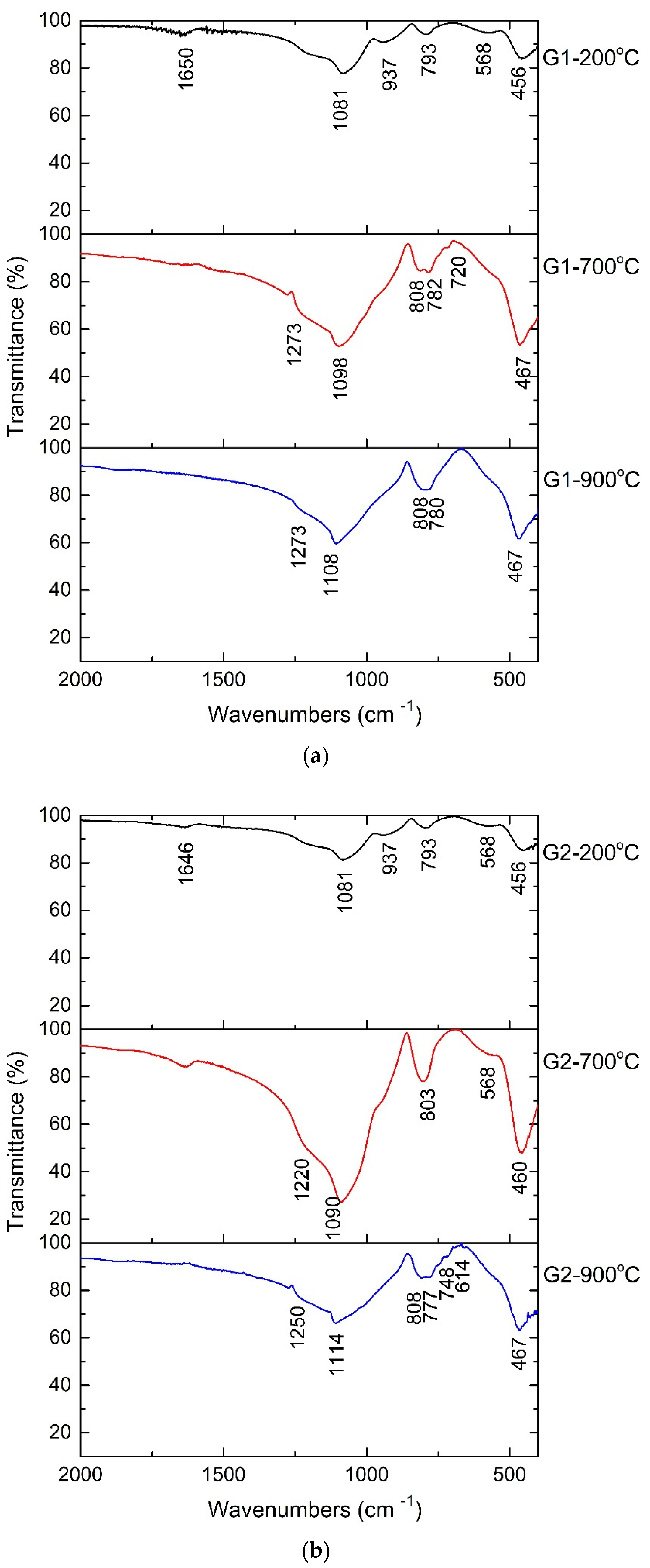
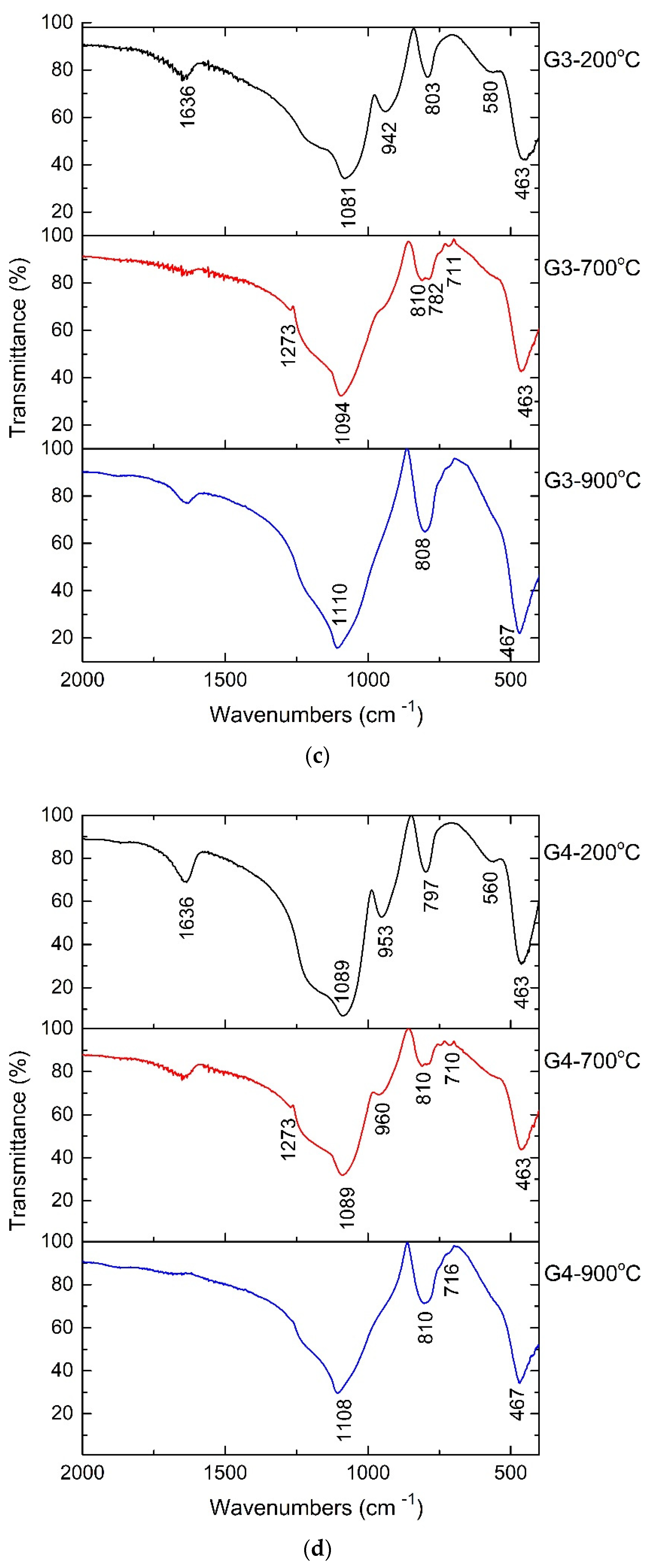
The FTIR spectra for treated powders are shown in
Figure 3. The bands that are typical of Si-O vibrations are visible in the recorded spectra and are identical to those that have been described in the literature [
16,
17,
18,
19,
20,
21]. Small bands at 560 cm
−1 or 568 cm
−1 may be attributed to the vibration of the Si-O-Zn bonds, whereas the main bands at 1273 cm
−1, 1080 cm
−1, 1114 cm
−1, 793 cm
−1, and 467 cm
−1 are assigned to Si-O-Si bonds in networks and chains. Band at 937 cm
−1 is characteristic of isolated [SiO
4] tetrahedra. Due to its wide bands, the FTIR spectrum reveals the formation of a vitreous material. In the non-crystalline solids, the structural units are ordered at small distances such as isolated SiO
44− tetrahedra (structural unit); pairs of tetrahedra, chains of tetrahedra, double chains (rings) of tetrahedra and network (3D). The bands between 800 and 900 cm
−1 are characteristic to stretching motions of Si-O in isolated tetrahedra with 4 non-bridging oxygen/tetrahedra. Bands at 943 and 1094 cm
−1 are characteristic of Si-O-Si vibrations in chains and sheet (ring) of tetrahedra with 1 or 2 non-bridging oxygen. The framework vibrations were observed in 1108–1273 cm
−1 domain. Si-O and Zn-O bond’s band position of vibration can be seen in the spectrum, but they cannot be attributed to crystalline forms. As a result, the powders also have a vitreous structure. The O-H vibration of the surface-absorbent water is attributed to the bands at 1650 cm
−1 and 1636 cm
−1. In
Table 1 are presented all the bands for initial and treated powders and their assignations.
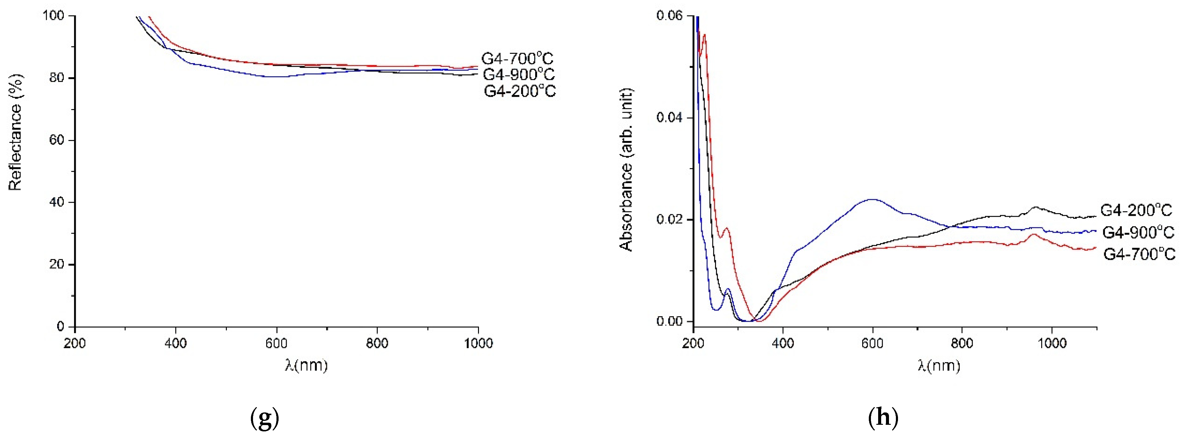
The amorphous powders show good transmittance. G1 powder shows the best transmittance value, while the structure of G4 powder is the most ordered of all the studied samples.
Figure 4 shows the reflectance spectra of powders produced in different synthesis modes (1–4). For G1–G4 at 200 °C all reflectance values were above 6o% from 200 to 1100 nm wavelength (λ).
Figure 4 a,c show the decrease in reflectance below 30% for the G1 and G2 heat-treated powders at 700 °C and 900 °C. The reflectance below 390 nm wavelength was decreased due to the intrinsic absorption of ZnO as Chang showed in reference [
7]. So, the synthesis for G1 and G2 powders is suitable for materials used for printing antireflective coatings. The reflectance was above 60% for heat-treated G3 and G4 powders. They are highly reflective materials and can be ideal for UV and lighting systems used in water disinfection. On the other part, the absorbance spectra of powders present one absorption band in visible domain: at 450 nm (violet region) for G1, G2 and G3 treated at 700 °C and 900 °C; and at 600 nm (orange region) for G4-900 °C. In the case of powders obtained by synthesis 1, synthesis 2 and synthesis 3 absorption increases after heat treatment at 700 °C and 900 °C, which means that the transmittance decreases. For non-crystalline materials, the high transmittance is desired. In conclusion, powders such as G1-900 °C, G2-700 °C, G3-900 °C and G4-700 °C with lower absorbance and higher transmittance are desirable for solar cell coatings.
Table 2 gives the band gap values estimated from Tauc plot and the Klubelka–Munk relation. The values vary between 1.2 eV for G1 heat-treated at 900 °C and 5.9 eV for G4 heat-treated at 900 °C.
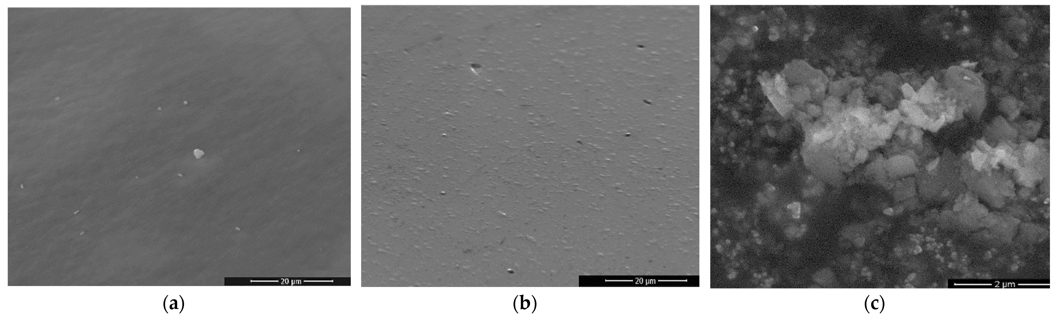
Figure 5 shows the morphology of gels G1, G2 and G4 after heat treatment at 900 °C. Powders G1 and G2 have a matrix morphology specific to vitreous materials. In
Figure 5c, an agglomeration on nanostructures can be seen indicating the beginning of nucleation of G4-900 °C powder. The observation on the morphology of the powders is in agreement with their structure determined by XRD and FTIR spectroscopy.


 ,
, 


