

An Evaluation of Cellulose Hydrogels Derived from tequilana Weber Bagasse for the Regeneration of Gingival Connective Tissue in Lagomorphs

 , , , and
Abstract

1. Introduction
2. Results and Discussion
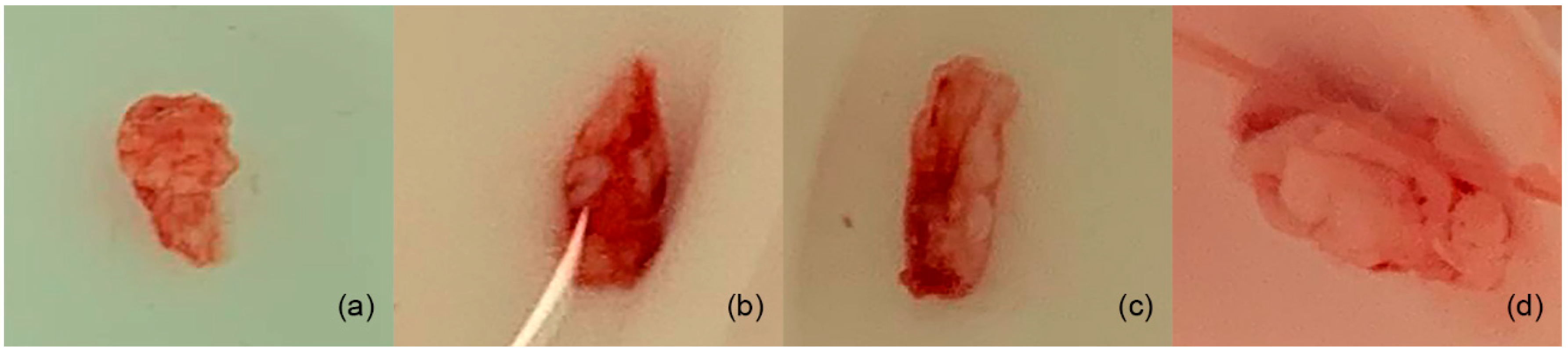
2.1. Hydrogel
2.2. General Clinical Characteristics of the Study Groups
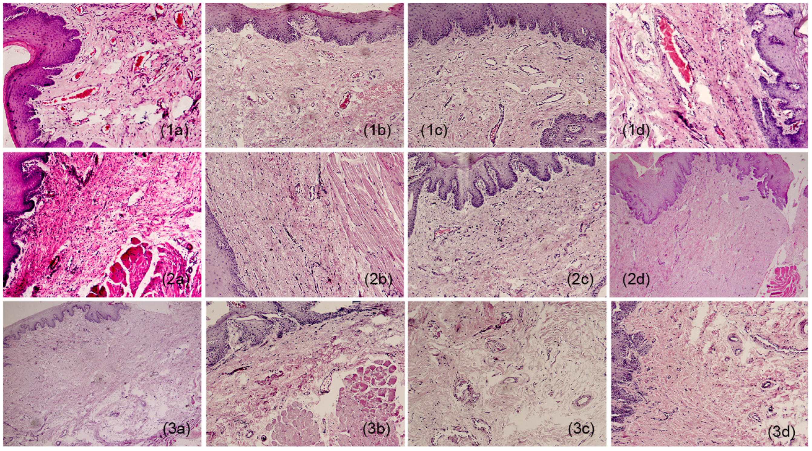
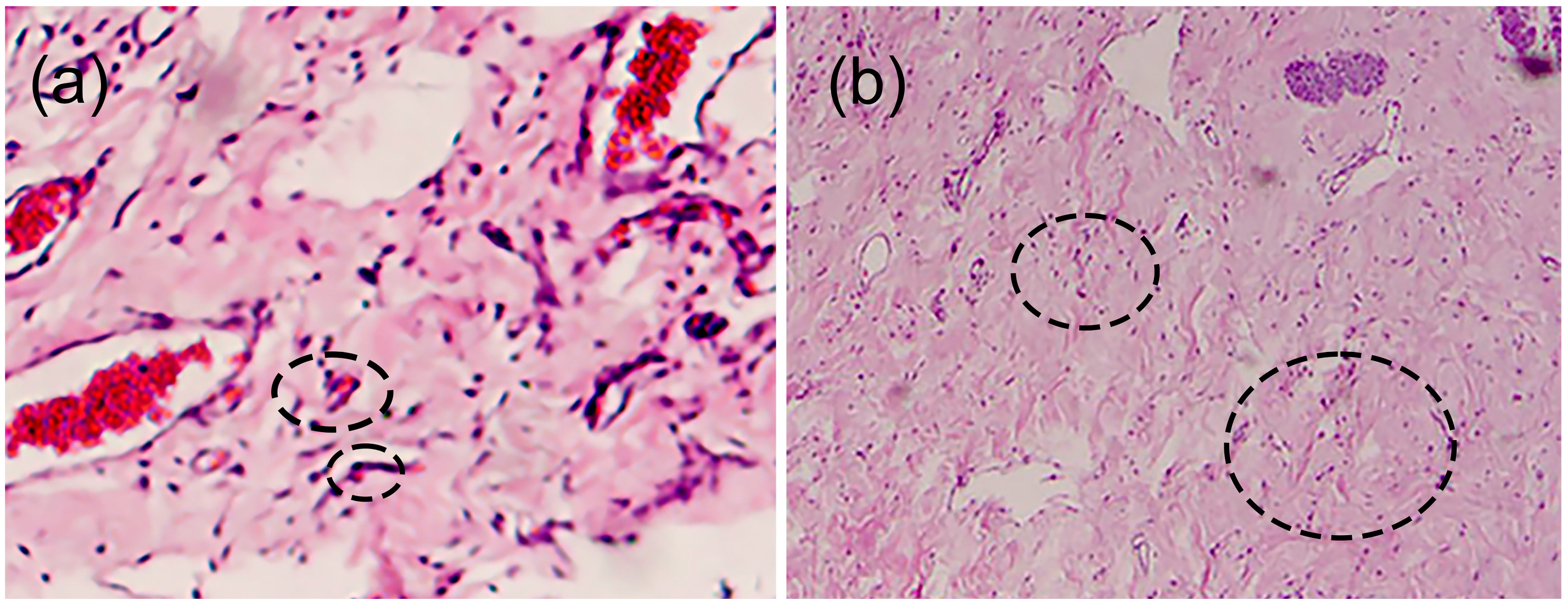
2.3. Histopathology
3. Conclusions
4. Materials and Methods
4.1. Materials
4.2. Hydrogel Elaboration
4.2.1. Agave Bagasse Fiber Treatment
4.2.2. Elaboration of Hydrogel Films
4.3. Experimental Rabbit Model
4.3.1. In Vivo Assay
4.3.2. Anesthesia Protocol (Performed by a Veterinarian)
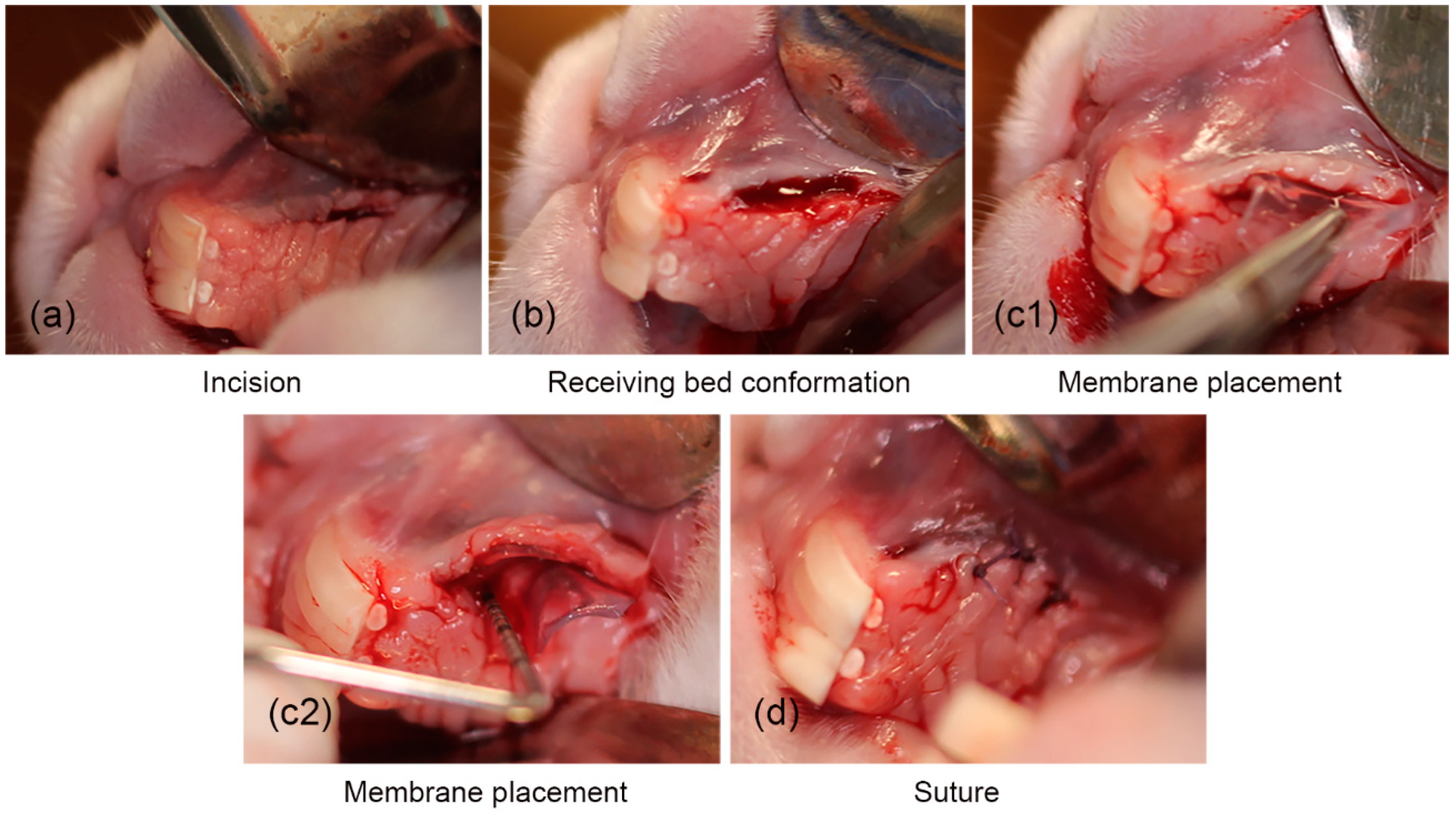
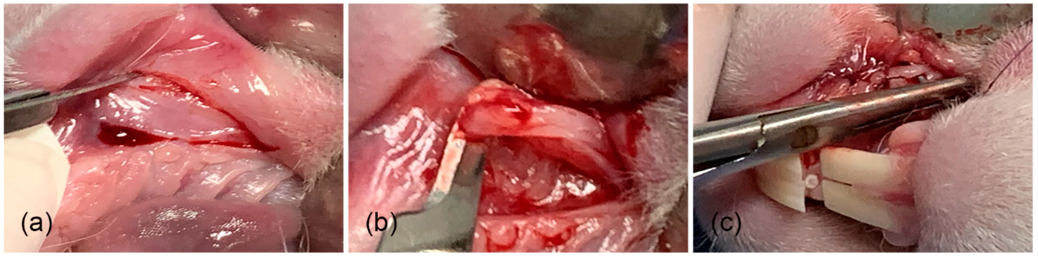
4.3.3. Surgical Procedure and Biomaterial Implantation (Day 0)
4.3.4. Analgesia and Antibiotic Therapy Protocol
4.4. Histopathology
Histopathology Analysis
4.5. Statistical Analysis
Author Contributions
Funding
Institutional Review Board Statement
Informed Consent Statement
Data Availability Statement
Acknowledgments
Conflicts of Interest
References
- Mehta, P.; Sharma, M.; Devi, M. Hydrogels: An overview of its classication, properties, and applications. J. Mech. Behav. Biomed. Mater. 2023, 147, 106145–106155. [Google Scholar] [CrossRef] [PubMed]
- Kapusta, O.; Jarosz, A.; Stadnik, K.; Ginnakoudakis, D.A.; Barczynski, B.; Barczak, M. Antibacterial natural hydrogels in biomedicine: Properties, appliactions, and challenges—A concise Review. Int. J. Mol. Sci. 2023, 24, 2191. [Google Scholar] [CrossRef] [PubMed]
- Carton, F.; Rizzi, M.; Canciani, E.; Sieve, G.; Di Francesco, D.; Casarella, S.; Di Nunno, L.; Boccafoschi, F. Use of hydrogels in regenerative medicine: Focus on mechanical properties. Int. J. Mol. Sci. 2024, 25, 11426. [Google Scholar] [CrossRef]
- Jagur-Grodzinski, J. Polymeric gels and hydrogels for biomedical and pharmaceutical appliations. Polym. Adv. Technol. 2010, 21, 27–47. [Google Scholar] [CrossRef]
- Wichterle, O.; Lim, D. hydrophilic gels for biological use. Nature 1960, 185, 117–118. [Google Scholar] [CrossRef]
- Hoang, T.T.; Lee, Y.; Ryu, S.B.; Sung, H.J.; Park, K.D. Oxidezed cyclodextrin-functionalized injectable gelatin hydrogels as a new platform for tissue-adhesive hydrophobic drug delivery. RSC Adv. 2017, 7, 34053–34062. [Google Scholar] [CrossRef]
- Das, D.; Pal, S. Modified biopolymer-dextrin based crosslinked hydrogels: Application in controlled drug delivery. RSC Adv. 2015, 5, 25014–25050. [Google Scholar] [CrossRef]
- Peppas, N.A.; Hilt, J.Z.; Khademhosseini, A.; Langer, R. Hydrogels in biology and medicine: From molecular principles to bionanotechnology. Adv. Mater. 2006, 18, 1345–1360. [Google Scholar] [CrossRef]
- Silva, L.P. Current trends and challangers in biofabrication using biomaterials and nanomaterials: Future perspectives for 3D/4D bioprinting. 3D 4D Print Biomed. Appl. 2018, 15, 373–421. [Google Scholar]
- Mancha-Sanchez, E.; Gomez-Blanco, J.C.; Lopez-Nieto, E.; Casado, J.; Macias-Garcia, A.; Diaz-Diez, M.A.; Carrasco-Amador, J.P.; Torrejon-Martin, D. Hydrogels for bioprinting: A systematic review of hydrogels syntehsis, bioprinting parameters, and bioprinted structures behavior. Front. Bioeng. Biotechnol. 2020, 8, 776. [Google Scholar] [CrossRef]
- Tovar-Carrillo, K.L.; Sueyoshi, S.S.; Tagaya, M.; Kobayashi, T. Fibroblast compatibility on scaffold hydrogels prepared from agave tequilana weber bagasse for tissue regeneration. Ind. Eng. Chem. Res. 2013, 33, 11607–11613. [Google Scholar] [CrossRef]
- Tovar-Carrillo, K.L.; Nakasone, K.; Tagaya, M.; Kobayashi, T. Effects of sodium hypochlorite on agave teuilana weber bagasse fibers used to elaborate cyto and biocompatible hydrogel films. Mater. Sci. Eng. C 2014, 42, 808–815. [Google Scholar] [CrossRef] [PubMed]
- Khan, F.; Tare, R.S.; Oreffo, R.O.C.; Bradley, M. Versatile biocompatible polymer hydrogels: Scaffolds for cell growth. Angew. Chem. Int. Ed. 2009, 48, 978–982. [Google Scholar] [CrossRef] [PubMed]
- Varaprasad, K.; Raghavebdra, G.M.; Jayaramudu, T.; Yallapu, M.M.; Sadiku, R.A. Mini review on hydrogels classification and recent developments in miscellaneous applications. Mater. Sci. Eng. C 2017, 79, 958–971. [Google Scholar] [CrossRef] [PubMed]
- Chen, N.; Wang, H.; Ling, C.; Vermerris, W.; Wang, B.; Tong, Z. Cellulose-based injectable hydrogel composite for pH-responsive and controllable drug delivery. Carbohydr. Polym. 2019, 225, 115207–115220. [Google Scholar] [CrossRef]
- Jia, Y.; Wang, X.; Huo, M.; Zhai, X.; Li, F.; Zhong, C. Preparation and characterization of novel bacterial cellulose/chitosan bio-hydrogel. Nanomater. Nanotech. 2017, 7, 1847980417707172. [Google Scholar] [CrossRef]
- Peppas, N.A.; Khare, A.R. Preparation, structure and diffusional behavior of hydrogels in controlled release. Adv. Drug Deliv. Rev. 1993, 11, 1–35. [Google Scholar] [CrossRef]
- Buwalda, S.J.; Boere, K.W.M.; Dijkstra, P.J.; Feijen, J.; Vermonden, T.; Hennink, W.E. Hydrogels in a historical perspective: From simple networks to smart materials. J. Control Release 2014, 190, 254–273. [Google Scholar] [CrossRef] [PubMed]
- Astrini, N.; Anah, L.; Haryono, A. Crosllinking parameter on the preparation of cellulose based hydrogel with divinylsulfone. Procedia Chem. 2012, 4, 275–281. [Google Scholar] [CrossRef]
- Boyer, C.; Rethore, G.; Weiss, P.; d’Arros, C. A self-setting hydrogel of sylated chitosan and cellulose for repair of osteochondral defects: From in vitro characterization to preclinical evaluation in dogs. Front. Bioeng. Biotecnol. 2020, 8, 23. [Google Scholar]
- Rodrigues da Silva, I.G.; dos Santos Pantoja, B.T.; Rodrigues Almeida, G.H.; Oliveira Carreira, A.C.; Miglino, M.A. Bacterial cellulose and ECM hydrogels: An innovative approach for cardiovascular regenerative medicine. Int. J. Mol. Sci. 2022, 23, 3955. [Google Scholar] [CrossRef] [PubMed]
- Nakasone, K.; Ikematsu, S.; Kobayashi, T. Biocompatibility evaluation of cellulose hydrogel film regenerated from sugar cane bagasse waste and its in vivo behavior in mice. Ind. Eng. Chem. Res. 2015, 55, 30–37. [Google Scholar] [CrossRef]
- Salem, A.K.; Stevents, R.; Pearson, R.G. Interactions of 3T3 fibroblast and endothelial cells with defined pore features. J. Biomed. Mater. Res. 2002, 61, 212–217. [Google Scholar] [CrossRef] [PubMed]
- Lee, K.Y.; Mooney, D.J. hydrogels for tissue engineering. Chem. Rev. 2001, 101, 1869–1879. [Google Scholar] [CrossRef] [PubMed]
- Cicero, L.; Fazzotta, S.; Palumbo, V.D.; Cassata, G.; Lo Monte, A.I. Anesthesia protocols in laboratory animals used for scientific purposes. Acta Biomed. 2018, 89, 337–342. [Google Scholar] [PubMed]
- Travelli, L.; McGuire, M.K.; Zucchelli, G.; Rasperini, G.; Feinberg, S.E.; Wang, H.L. Extracellular matrix-based scaffolding technologies for periodontal and peri-implant soft tissue regeneration. J. Periodontol. 2020, 91, 17–25. [Google Scholar] [CrossRef] [PubMed]
- Gerber, D.A.; Rosenberg, E.S. The endentulous ridge in fixed prosthodontics. Compend. Cont. Educ. Dent. 1981, 2, 212–233. [Google Scholar]
- Zuhr, O.; Baumer, D.; Hurzeler, M. The addition of soft tissue replacement grafts in plastic periodontal and implant surgery: Critical elements in design and execution. J. Clin. Periodontol. 2014, 41, 123–142. [Google Scholar] [CrossRef]
- Balamurugan, R.; Mohamed, M.; Pandey, V.; Katikaneni, H.K.R.; Kumar, K.A. Clinical and histological comparison of polyglycolic acid suture with black silk suture after minor oral surgical procedure. J. Contemp. Dent. Pract. 2012, 13, 521–527. [Google Scholar] [CrossRef]
- McCarthy, W.H. A new synthetic absorbable suture material: A clinical trial of polyglycol acid suture in general surgery. Aust. N. Z. J. Surg. 1970, 39, 422–424. [Google Scholar] [CrossRef]
- Sortino, F.; Lambardo, C.; Sciacca, A. Silk and polyglycolic acid in oral surgery: A comparable study. Oral Surgery. Oral Med. Oral Pathol. Oral Radiol. Endodontology 2008, 105, 12–22. [Google Scholar]
- Sheikh, Z.; Brooks, P.J.; Barzilay, O.; Fine, N.; Glogauer, M. macrophages, forein bady giant cells and their response to implantable biomaterials. Materials 2015, 8, 5671–5701. [Google Scholar] [CrossRef] [PubMed]
- Benato, L.; Rooney, N.J.; Murrel, J.C. Pain and analgesia in pet rabbits within the veterinary environment: A review. Vet. Anaesth. Analg. 2019, 46, 151–162. [Google Scholar] [CrossRef] [PubMed]
- Romano, M.; Ruggiero, A.; Squeglia, F.; Maga, G.; Berisio, R. A structural view of SARS-CoV-2 RNA replication machinery: RNA synthesis, proofreading and final capping. Cells 2020, 9, 2110–2120. [Google Scholar] [CrossRef] [PubMed]
- Cheng, Z.; Nolan, A.; McKellar, Q.A. Anti’inflmmatory effects of caprofen, caprofen enantiomers, and NG-nitro-L-arginine methyl ester in sheep. Am. J. Veter. 2002, 63, 782–788. [Google Scholar] [CrossRef] [PubMed]
- Helmus, M.N. Biomaterials in Design and Reliability of Medical Devices; Springer: Berlin/Heidelberg, Germany, 2000; Chapter 1; p. 23. [Google Scholar]

| Variables | Membrane | Surgery | Control | p-Value |
|---|---|---|---|---|
| χ ± DS | χ ± SD | χ ± SD | ||
| n = 8 | n = 4 | n = 4 | ||
| Weight (kg) | ||||
| Initial | 2.412 ± 0.269 | 2.090 ± 0.462 | 2.335 ± 0.286 | 0.304 |
| Final | 2.971 ± 0.156 | 2.815± 0.199 | 2.850 ± 0.252 | 0.371 |
| Water intake (mL) | ||||
| Initial | 1458 ± 96 | 1428 ± 267 | 1488 ± 224 | 0.898 |
| Final | 2693 ± 403 | 2670 ± 823 | 2778 ± 900 | 0.969 |
| Food intake (g) | ||||
| Initial | 380 ± 26 | 324.0 ± 86 | 389.25 ± 30 | 0.137 |
| Final | 806 ± 53 | 800.0 ± 53 | 819.2 ± 62 | 0.880 |
| Variables | Membrane | Surgery | Control | p-Value |
|---|---|---|---|---|
| n = 8 Rabbits | n = 4 Rabbits | n = 4 Rabbits | ||
| (%) | (%) | (%) | ||
| Inflammatory infiltrates | ||||
| No presence (0%) | 5 (62.5) | 4 (100) | 4 (100) | |
| Slight (<25%) | 3 (37.5) | 0 (0) | 0 (0) | 0.092 |
| Mild (26–50%) | 0 (0) | 0 (0) | 0 (0) | |
| Generalized (<50%) | 0 (0) | 0 (0) | 0 (0) | |
| Angiogenesis | ||||
| No presence (0%) | 1 (12.5) | 3 (75) | 4 (100) | |
| Focused (<30%) | 4 (50) | 1 (25) | 0 (0) | 0.006 * |
| Diffuse (30–60%) | 3 (37.5) | 0 (0) | 0 (0) | |
| Generalized (>60%) | 0 (0) | 0 (0) | 0 (0) | |
| Fibrogenesis | ||||
| No presence (0%) | 1 (12.5) | 3 (75) | 4 (100) | |
| Focused (<30%) | 0 (0) | 0 (0) | 0 (0) | 0.004 * |
| Diffuse (30–60%) | 5 (62.5) | 1 (25) | 0 (0) | |
| Generalized (>60%) | 2 (25) | 0 (0) | 0 (0) |
| Variables | Four Weeks | Eight Weeks | Twelve Weeks | Sixteen Weeks | p-Value |
|---|---|---|---|---|---|
| n = 4 Rabbits | n = 4 Rabbits | n = 4 Rabbits | n = 4 Rabbits | ||
| (%) | (%) | (%) | (%) | ||
| Inflammatory infiltrates | |||||
| No presence (0%) | 3 (75) | 3 (75) | 3 (75) | 4 (100) | |
| Slight (<25%) | 1 (25) | 1 (25) | 1 (25) | 0 (0) | 0.405 |
| Mild (26–50%) | 0 (0) | 0 (0) | 0 (0) | 0 (0) | |
| Generalized (<50%) | 0 (0) | 0 (0) | 0 (0) | 0 (0) | |
| Angiogenesis | |||||
| No presence (0%) | 3 (75) | 2 (50) | 2 (50) | 1 (25) | |
| Focused (<30%) | 0 (0) | 1 (25) | 2 (50) | 2 (50) | 0.481 |
| Diffuse (30–60%) | 1 (25) | 1 (25) | 0 (0) | 1 (25) | |
| Generalized (>60%) | 0 (0) | 0 (0) | 0 (0) | 0 (0) | |
| Fibrogenesis | |||||
| No presence (0%) | 3 (75) | 2 (50) | 2 (50) | 1 (25) | |
| Focused (<30%) | 0 (0) | 0 (0) | 0 (0) | 0 (0) | 0.194 |
| Diffuse (30–60%) | 1 (25) | 1 (25) | 2 (50) | 2 (50) | |
| Generalized (>60%) | 0 (0) | 1 (25) | 0 (0) | 1 (25) |
| Variables | Four Weeks | Eight Weeks | Twelve Weeks | Sixteen Weeks | p-Value |
|---|---|---|---|---|---|
| n = 4 Rabbits | n = 4 Rabbits | n = 4 Rabbits | n = 4 Rabbits | ||
| (%) | (%) | (%) | (%) | ||
| Inflammatory infiltrates | |||||
| No presence (0%) | 2 (50) | 2 (50) | 2 (50) | 4 (100) | |
| Slight (<25%) | 2 (50) | 2 (50) | 2 (50) | 0 (0) | 0.359 |
| Mild (26–50%) | 0 (0) | 0 (0) | 0 (0) | 0 (0) | |
| Generalized (<50%) | 0 (0) | 0 (0) | 0 (0) | 0 (0) | |
| Angiogenesis | |||||
| No presence (0%) | 2 (50) | 0 (0) | 0 (0) | 0 (0) | |
| Focused (<30%) | 0 (0) | 2 (50) | 4 (100) | 2(50) | 0.655 |
| Diffuse (30–60%) | 2 (50) | 2 (50) | 0 (0) | 2 (50) | |
| Generalized (>60%) | 0 (0) | 0 (0) | 0 (0) | 0 (0) | |
| Fibrogenesis | |||||
| No presence (0%) | 2 (50) | 0 (0) | 0 (0) | 0 (0) | |
| Focused (<30%) | 0 (0) | 0 (0) | 0 (0) | 0 (0) | 0.172 |
| Diffuse (30–60%) | 2 (50) | 2 (50) | 0 (0) | 2 (50) | |
| Generalized (>60%) | 0 (0) | 2 (50) | 0 (0) | 2 (50) |
| Variables | Four Weeks | Eight Weeks | Twelve Weeks | Sixteen Weeks | p-Value |
|---|---|---|---|---|---|
| n = 4 Rabbits | n = 4 Rabbits | n = 4 Rabbits | n = 4 Rabbits | ||
| (%) | (%) | (%) | (%) | ||
| Inflammatory infiltrates | |||||
| No presence (0%) | 1 (100) | 1 (100) | 1 (100) | 1 (100) | |
| Slight (<25%) | 0 (0) | 0 (0) | 0 (0) | 0 (0) | --- |
| Mild (26–50%) | 0 (0) | 0 (0) | 0 (0) | 0 (0) | |
| Generalized (<50%) | 0 (0) | 0 (0) | 0 (0) | 0 (0) | |
| Angiogenesis | |||||
| No presence (0%) | 1 (100) | 1 (100) | 1 (100) | 0 (0) | |
| Focused (<30%) | 0 (0) | 0 (0) | 0 (0) | 1 (100) | 0.180 |
| Diffuse (30–60%) | 0 (0) | 0 (0) | 0 (0) | 0 (0) | |
| Generalized (>60%) | 0 (0) | 0 (0) | 0 (0) | 0 (0) | |
| Fibrogenesis | |||||
| No presence (0%) | 1 (100) | 1 (100) | 1 (100) | 0 (0) | |
| Focused (<30%) | 0 (0) | 0 (0) | 0 (0) | 0 (0) | 0.180 |
| Diffuse (30–60%) | 0 (0) | 0 (0) | 0 (0) | 1 (100) | |
| Generalized (>60%) | 0 (0) | 0 (0) | 0 (0) | 0 (0) |
| Variables | Four Weeks | Eight Weeks | Twelve Weeks | Sixteen Weeks | p-Value |
|---|---|---|---|---|---|
| n = 4 Rabbits | n = 4 Rabbits | n = 4 Rabbits | n = 4 Rabbits | ||
| (%) | (%) | (%) | (%) | ||
| Inflammatory infiltrates | |||||
| No presence (0%) | 1 (100) | 1 (100) | 1 (100) | 1 (100) | |
| Slight (<25%) | 0 (0) | 0 (0) | 0 (0) | 0 (0) | --- |
| Mild (26–50%) | 0 (0) | 0 (0) | 0 (0) | 0 (0) | |
| Generalized (<50%) | 0 (0) | 0 (0) | 0 (0) | 0 (0) | |
| Angiogenesis | |||||
| No presence (0%) | 1 (100) | 1 (100) | 1 (100) | 1 (100) | |
| Focused (<30%) | 0 (0) | 0 (0) | 0 (0) | 0 (0) | --- |
| Diffuse (30–60%) | 0 (0) | 0 (0) | 0 (0) | 0 (0) | |
| Generalized (>60%) | 0 (0) | 0 (0) | 0 (0) | 0 (0) | |
| Fibrogenesis | |||||
| No presence (0%) | 1 (100) | 1 (100) | 1 (100) | 1 (100) | |
| Focused (<30%) | 0 (0) | 0 (0) | 0 (0) | 0 (0) | --- |
| Diffuse (30–60%) | 0 (0) | 0 (0) | 0 (0) | 0 (0) | |
| Generalized (>60%) | 0 (0) | 0 (0) | 0 (0) | 0 (0) |
| Drug | Acepromazine Maleate | Atropine Sulfate | Ketamine Hydrochloride | Xylazine Hydrochloride |
|---|---|---|---|---|
| Dose and route of administration | 0.75 mg/kg | 0.044 mg/kg | 50 mg/kg | 10 mg/kg |
| IM | IM | IM | IM |
| Drug | Carprofen | Sulfamethoxazole with Trimethoprim |
|---|---|---|
| Dose | 0.75 mg/kg/24 h | 0.044 mg/kg/12 h |
| by | IM | IM |
Disclaimer/Publisher’s Note: The statements, opinions and data contained in all publications are solely those of the individual author(s) and contributor(s) and not of MDPI and/or the editor(s). MDPI and/or the editor(s) disclaim responsibility for any injury to people or property resulting from any ideas, methods, instructions or products referred to in the content. |
© 2025 by the authors. Licensee MDPI, Basel, Switzerland. This article is an open access article distributed under the terms and conditions of the Creative Commons Attribution (CC BY) license (https://creativecommons.org/licenses/by/4.0/).
Share and Cite
López-Domínguez, S.; Cuevas-González, J.C.; Espinosa-Cristóbal, L.F.; Ríos-Arana, J.V.; Saucedo Acuña, R.A.; Cuevas-González, M.V.; Zaragoza-Contreras, E.A.; Tovar Carrillo, K.L. An Evaluation of Cellulose Hydrogels Derived from tequilana Weber Bagasse for the Regeneration of Gingival Connective Tissue in Lagomorphs. Gels 2025, 11, 75. https://doi.org/10.3390/gels11010075
López-Domínguez S, Cuevas-González JC, Espinosa-Cristóbal LF, Ríos-Arana JV, Saucedo Acuña RA, Cuevas-González MV, Zaragoza-Contreras EA, Tovar Carrillo KL. An Evaluation of Cellulose Hydrogels Derived from tequilana Weber Bagasse for the Regeneration of Gingival Connective Tissue in Lagomorphs. Gels. 2025; 11(1):75. https://doi.org/10.3390/gels11010075
Chicago/Turabian StyleLópez-Domínguez, Silvia, Juan Carlos Cuevas-González, León Francisco Espinosa-Cristóbal, Judith Virginia Ríos-Arana, Rosa Alicia Saucedo Acuña, María Verónica Cuevas-González, Erasto Armando Zaragoza-Contreras, and Karla Lizette Tovar Carrillo. 2025. "An Evaluation of Cellulose Hydrogels Derived from tequilana Weber Bagasse for the Regeneration of Gingival Connective Tissue in Lagomorphs" Gels 11, no. 1: 75. https://doi.org/10.3390/gels11010075
APA StyleLópez-Domínguez, S., Cuevas-González, J. C., Espinosa-Cristóbal, L. F., Ríos-Arana, J. V., Saucedo Acuña, R. A., Cuevas-González, M. V., Zaragoza-Contreras, E. A., & Tovar Carrillo, K. L. (2025). An Evaluation of Cellulose Hydrogels Derived from tequilana Weber Bagasse for the Regeneration of Gingival Connective Tissue in Lagomorphs. Gels, 11(1), 75. https://doi.org/10.3390/gels11010075