Article

Identification and Functional Analysis of CAP Genes from the Wheat Stripe Rust Fungus Puccinia striiformis f. sp. tritici

1
State Key Laboratory of Crop Stress Biology for Arid Areas, College of Plant Protection, Northwest A&F University, Xianyang 712100, China
2
College of Life Sciences, Northwest A&F University, Xianyang 712100, China
3
Key Laboratory of Plant Hormones and Development Regulation of Chongqing, School of Life Sciences, Chongqing University, Chongqing 401331, China
*
Authors to whom correspondence should be addressed.
J. Fungi 2023, 9(7), 734; https://doi.org/10.3390/jof9070734
Submission received: 4 May 2023 / Revised: 28 June 2023 / Accepted: 6 July 2023 / Published: 7 July 2023
(This article belongs to the Section Fungal Pathogenesis and Disease Control)

Abstract

:
Cysteine-rich secretory proteins (C), antigen 5 (A), and pathogenesis-related 1 proteins (P) comprise widespread CAP superfamily proteins, which have been proven to be novel virulence factors of mammalian pathogenic fungi and some plant pathogens. Despite this, the identification and function of CAP proteins in more species of plant pathogens still need to be studied. This work presents the identification and functional analysis of CAP superfamily proteins from Puccinia striiformis f. sp. tritici (Pst), an important fungal pathogen that causes wheat stripe rust on wheat worldwide. A total of six CAP genes were identified in the Pst genome, designated as PsCAP1PsCAP6. Five PsCAP proteins, including PsCAP1, PsCAP2, PsCAP3, PsCAP4, and PsCAP5, have N-terminal signal peptides secreted with the yeast signal sequence trap assay. Single-nucleotide polymorphism (SNP) analysis indicated that they showed a low level of intraspecies polymorphism. The expression abundance of PsCAP genes at different Pst infection stages was detected by RT-qPCR, and most of them were highly expressed during Pst infection on wheat and also Pst sexual reproduction on barberry (Berberis shensiana). Noticeably, the silencing of these six PsCAP genes by BSMV-mediated HIGS indicated that PsCAP1, PsCAP4, and PsCAP5 contribute significantly to Pst infection in wheat. These results indicate that PsCAP proteins may act as virulence factors during Pst infection, which also provides insights into Pst pathogenicity.

1. Introduction

The CAP protein superfamily is derived from proteins with great sequence similarity, including cysteine-rich secretory protein (CRISP) in vertebrates, antigen 5 (Ag5) in insects, and pathogenicity-related protein 1 (PR-1) in plants [1,2]. All members of the CAP protein superfamily contain a highly conserved CAP domain that exhibits a unique alpha–beta–alpha sandwich fold [2]. The biochemical activity and action mechanism of the CAP domain remained a mystery until the finding in the budding yeast Saccharomyces cerevisiae [3]. S. cerevisiae has three CAP proteins designated as PRY1–3, and biochemical analysis showed that they can bind sterols and assist in protein export [3]. In addition, expression of the mammalian CRISP2 protein can restore the defects of PRY mutants, indicating that the conserved CAP domain is necessary and sufficient for sterol binding and lipid export [3]. Moreover, many CAP proteins have been proven to have N-terminal signaling peptides and can be secreted outside of the cell [1], while some of them are deficient in secretion function [1], which suggests that CAP proteins may have functional diversity due to different localizations. These structural components of CAP proteins form the basic ingredients for the biological functions of CAP proteins. Incorporating the signature sequences and motifs, more CAP proteins in different species can be identified using Pfam to advance the study of the identification of CAP proteins in different species.
CAP proteins are found in the majority of living organisms and are involved in multiple cellular processes, including reproduction, prostate and brain cancer in mammals, and immune defense in plants [1]. In addition, CAP proteins have also been characterized as novel virulence factors in mammalian pathogenic fungi [2]. In the ascomycete Fusarium oxysporum, the mutant-lacking Fpr1 that encodes a secreted CAP protein reduced virulence in mammalian hosts [4]. The PR-1-like proteins of Rbe1 and Rbt4 from the mammalian pathogen Candida albicans were reported to interact with the host neutrophils contributing to pathogenesis and impeding mammalian cell clearance of the pathogen [5]. Many plant fungal pathogens display expansion of CAP proteins [2], and some CAP genes from Moniliophthora perniciosa were proven to be highly and specifically expressed during the interaction with the host cacao [6], which indicates that CAP proteins may also play important roles in the virulence of the plant fungal pathogen. The PR-1-like protein FgPR-1L-4 from wheat pathogen Fusarium graminearum is involved in pathogen–host interaction and affects fungal virulence in the host, which is the first example that proves non-plant PR-1 protein’s role as a pathogenicity factor to a host [7]. New research reported that CcCAP1 from Cytospora chrysosperma causes canker disease in poplar, mainly localized to the plant nucleus to suppress plant immunity, and the conserved CAP domain was sufficient for its function [8]. These results indicate that CAP proteins play pathogenic roles in plant-pathogenic fungi. Despite this, the identification and function of CAP proteins in more plant pathogenic fungi still need to be studied.
Rust fungi (Uredinales or Pucciniales) comprise the largest group of plant pathogenic fungi and mainly infect wheat, oat, barley, and other cereal crops and weeds [9]. Wheat stripe rust is caused by Puccinia striiformis f. sp. tritici (Pst), which is one of the main yield-limiting factors affecting wheat production globally [9,10]. The wheat stripe rust can cause 10–70% of wheat yield loss, which mainly depends on epidemiology, environmental climate, wheat cultivars, and race of pathogens [9,11,12]. The evolution and migration of newly generated Pst virulent races are occurring at an increasing rate. These leads to the loss of disease resistance in multiple wheat varieties. Thus, a better understanding of the molecular mechanism used by Pst pathogenicity is important for the development of more effective and durable control of wheat stripe rust. Pst is an obligate biotrophic parasite that is entirely dependent on the host plant to complete its growth and reproduction. The main stage of the life cycle of Pst is the asexual reproduction stage, that is, from the inoculation of urediniospores in wheat plants to the production of new urediniospores [12,13]. This asexual stage is usually divided into three stages: penetration stage, biotrophic/parasitic stage, and sporulation stage [14]. In the parasitic stage, it is very important to establish the elaborate relationships between Pst and wheat, and some of the up-regulated genes have been proven to be virulence factors for Pst infection at this stage [15,16,17].
Given the important role of CAP proteins in the virulence of plant fungal pathogens, this study identified six genes encoding putative CAP protein in the Pst genome, designated as PsCAP1PsCAP6, which have the conserved CAP domains and belong to the PR-1-like subfamily in plants. Sequences and structural features of these genes and their expression profile throughout Pst development were analyzed. The functions of PsCAP genes were investigated by gene silencing using BSMV-mediated HIGS. We revealed that CAP proteins are novel virulence factors during the Pst infection of wheat plants.

2. Materials and Methods

2.1. Bioinformatics Analysis of Pst CAP Genes

The sequences of Pst CAP genes were obtained by comparing the amino acid sequences of three CAP proteins (PRY1–3) from the model fungus S. cerevisiae with the NCBI database. CAP proteins in different species were searched in the Broad Institute Web Server and NCBI website by Blastp searches. The signal peptides of CAP proteins were predicted using SignalP 5.0, an online signal peptide prediction website. CLUSTALW was used to conduct multiple sequence alignment among Pst CAP proteins and CAP proteins in other species, and the results were viewed using JALVIEW 2.8 [18]. MEGA5 was used for creating phylogenetic trees [19], and the sequences displayed in the phylogenetic tree were downloaded from the NCBI database.

2.2. Sequence Polymorphism Analysis of Pst CAP Proteins

The local blast was used to identify sequences of the coding regions of each PsCAP gene among seven Pst isolates, including CYR32, PST-21, PST-43, PST-78, PST-130, PST-08/21, and PST-87/7. The genomes of the seven isolates were all downloaded from the NCBI database. DNAMAN6.0 was used to conduct multiple sequence alignments, which were then manually adjusted to minimize the number of implied mutations. MEGA5 was used to calculate the nonsynonymous substitutions (dN) and synonymous substitutions (dS), respectively, and then the ratio of dN/dS was obtained manually.

2.3. Strains and Plants, Gene Cloning, and Plasmid Construction

The stripe rust isolate used in this study was CYR32, and the wheat cultivar used was Suwon11, which is highly susceptible to CYR32 (CYR32 and Suwon11 form a compatible interaction). Wheat was cultured at 16 °C with a standard light–dark cycle of 16 h of light (60 μmol m−2·s−1). Yeast isolate YTK12 used for secretion validation was cultured in the YPDA liquid medium at 30 °C. BSMV isolate ND-18 was used for gene silencing [20]. The pSUC2T7M13ORI (hereinafter referred to as pSUC2) vector was used to verify the yeast secretion system [21]. Specific segments of PsCAP genes were inserted into the γ vector for the silencing system, respectively. The CYR32-infected Suwon11 cDNA samples were used as templates to amplify PsCAP genes by PCR. Primer 5.0 and online NCBI Primer-BLAST were used to design specific primers listed in Supplementary Materials Table S2.

2.4. Yeast Signal Peptide Secretion Validation

To identify the secretion feature of Pst CAP proteins, a Yeast Signal Peptide Screen Trap (YSST) assay was carried out as previously described [21,22]. Each of the N-terminal signal peptide sequences of four PsCAP proteins (PsCAP1, PsCAP2, PsCAP4, and PsCAP5) and the first 30 amino acids of the other two PsCAP proteins (PsCAP3 and PsCAP6) were fused to the vector pSUC2T7M13ORI (hereinafter referred to as pSUC2). The pSUC2-derived plasmids were transformed into yeast strain YTK12, which is defective for tryptophan biosynthesis using the lithium acetate method [23]. The pSUC2 vector, which carries the sucrose invertase gene, lacks the signal peptide, and the sucrose invertase gene can only be switched on to convert exogenous sucrose into glucose when the signal peptide is inserted. A CMD-W medium with sucrose in place of glucose was used for all transformants. To assay the secretion of invertase, positive colonies were cultured on the YPRAA medium with raffinose as the carbohydrate source. The pSUC2-Avr1b (a secreted signal peptide of Avr1b from Phytophthora sojae) and pSUC2-Mg87 (25bp in the N-terminal of Magnaporthe oryzae protein Mg87) were used as positive and negative controls, separately [24].

2.5. RNA Isolation and RT-qPCR Analysis

Wheat cultivar Suwon11, which grew to the two-leaf stage, was used to inoculate CYR32 as previously described [25]. Samples were collected at 12, 24, 48, 72, 120, 168, and 216 hpi (hours post inoculation), respectively. Urediniospores were incubated for 10 h in sterile distilled water at 9 °C for harvesting germinated urediniospores [26]. Infected barberry (Berberis shensiana) leaves were sampled at 11 dpi [27]. Total cellular RNA was extracted using the Quick RNA Isolation Kit (Huayueyang Biotech Co., Ltd., Beijing, China), and the operation method was referred to the product manual protocol. Then the first-strand cDNA was synthesized using an RT-PCR system (Promega, Madison, WI, USA). RT-qPCR was performed to measure the gene transcription level. The housekeeping gene of PsEF1 (Elongation factor 1) was used as an endogenous control. Reactions were performed on a Bio-Rad CFX Manager (version 3.1). The comparative 2−ΔΔCT method was used to quantify relative gene expression [28].

2.6. BSMV-Mediated Gene Silencing of PsCAP Genes

Barley stripe mosaic virus (BSMV)-mediated gene silencing was conducted to identify the virulence of PsCAP genes [20]. Capped in vitro transcripts were prepared with linearized plasmids containing the three-part genome of BSMV (α, β, γ, or recombinant γ gene) using the RiboMAX™ Large Scale RNA Production Systems-T7 (Promega, Madison, WI, USA). BSMV:α, BSMV:β, and BSMV:γ or recombinant γ vectors were mixed with 1× Fes buffer at the ratio of 1:1:1 to inoculate the two-leaf wheat seedlings, which then were cultured at 25 °C until the leaves showed the typical chlorotic mosaic symptom [20]. The phenotype was observed and photographed. BSMV:TaPDS (TaPDS, the wheat phytoene desaturase) was used as an index for BSMV infection, and the wheat seedlings inoculated with a 1× Fes buffer were used as a negative control (MOCK). The fourth leaves were further inoculated with the Pst isolate of CYR32 and then maintained at 16 °C. The Pst-infected leaves should be sampled at 24 and 120 hpi for RNA isolation, and RT-qPCR was performed to evaluate the silencing efficiency; this method was described previously. Pst infection phenotypes (urediospore sporulation) were recorded and photographed at 14 dpi.

3. Results

3.1. Pst Contains Six CAP Genes That Are Specifically Expanded in Rust Fungi

From a BLAST search using the three CAP proteins (PRY1-3) from the model fungus S. cerevisiae, the queries revealed six genes encoding putative CAP proteins (designated PsCAP1PsCAP6) in the Pst genome (Supplemental Table S1). The six genes were heterogeneous in size and gene structure (Supplemental Table S1), but all predicted proteins contained a C-terminal CAP domain of about 120 amino acids in size (Figure 1A and Supplemental Table S1) and shared significant sequence similarity over the CAP domain (Figure 1B). In addition to the CAP domain, four Pst CAP proteins, including PsCAP1, PsCAP2, PsCAP4, and PsCAP5, also contained a putative N-terminal signal peptide (Figure 1A and Supplemental Table S1).
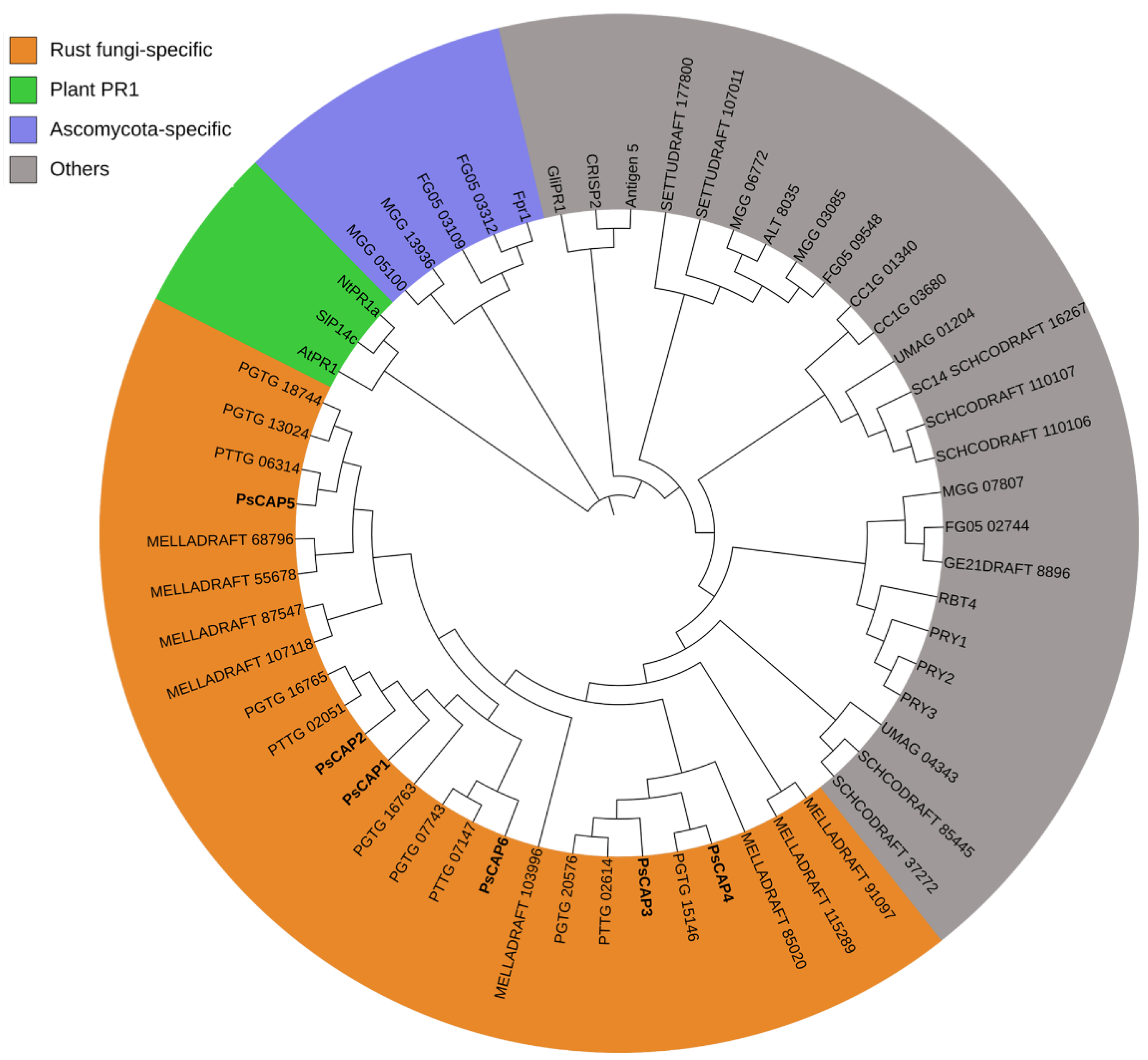
To check the phylogenetic relationship among the six CAP proteins from Pst and CAP proteins from other organisms, we performed a phylogenetic analysis according to sequence alignment. We included the plant PR-1 proteins P14c from tomato [30], PR1a from tobacco [31], and AtPR-1 from Arabidopsis [32] in the analysis. Phylogenetic analysis indicated that all PsCAP proteins belong to a clade of rust fungi-specific CAP proteins (Figure 2). In addition, we found a clade of ascomycota-specific CAP proteins (Figure 2).

3.2. Intraspecific Variation of PsCAP Genes

To identify the intra-species polymorphism of the six PsCAP genes, we compared their coding regions in seven different Pst isolates, and the results are shown in Table 1. A total of 3–12 nucleotide substitutions but only 1–3 nonsynonymous nucleotide substitutions were observed in the six CAP genes (Table 1). Additionally, PsCAP4 contains 15 nucleotide insertions/deletions among different Pst isolates (Supplemental Figure S1). We also calculated the ratio of nonsynonymous (dN) to synonymous (dS) substitution [33] of the six CAP genes. None of the six PsCAP genes had a ratio of dN/dS significantly greater than 1.0 (Table 1), suggesting that all these six genes are under negative selection. These results indicated that each CAP family member shows a low level of polymorphism among different Pst isolates.

3.3. Secretion Validation of Predicted Signal Peptides of CAP Proteins

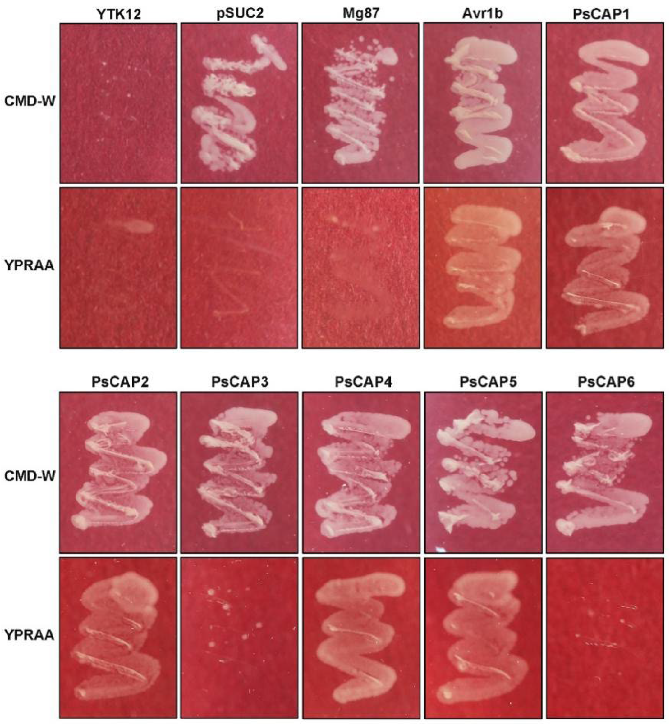
Many CAP proteins from other organisms have been clarified to have secreted signaling peptides [2]. We performed a yeast signal peptide screening trap [21,22] to validate the putative secretion function of the six PsCAP proteins. As shown in Figure 3, all YTK12 strains carrying pSUC2 or pSUC2-recombinant plastids can grow on a sucrose-contained CMD-W medium, indicating that the vectors of the yeast strain were successfully transformed. YTK12 strains carrying PsCAP1, PsCAP2, PsCAP4, and PsCAP5 could grow on the YPRAA medium (with raffinose as the only carbohydrate source, which requires secreted sucrose invertase for yeast growth). By contrast, PsCAP3 and PsCAP6, which have no N-terminal signal peptides, could not enable YTK12 to grow on the YPRAA medium (Figure 3). The pSUC2-Avr1b construct (Avr1b, a secreted protein from Phytophthora sojae) and pSUC2-Mg87 (25bp in the N-terminal of Magnaporthe oryzae protein Mg87) [24] were used as positive and negative controls separately (Figure 3). The results indicated that the signal peptides of PsCAP1, PsCAP2, PsCAP4, and PsCAP5 are active in the yeast-secretion system.

3.4. PsCAP Genes Are Highly Expressed in Pst Parasitic Stages

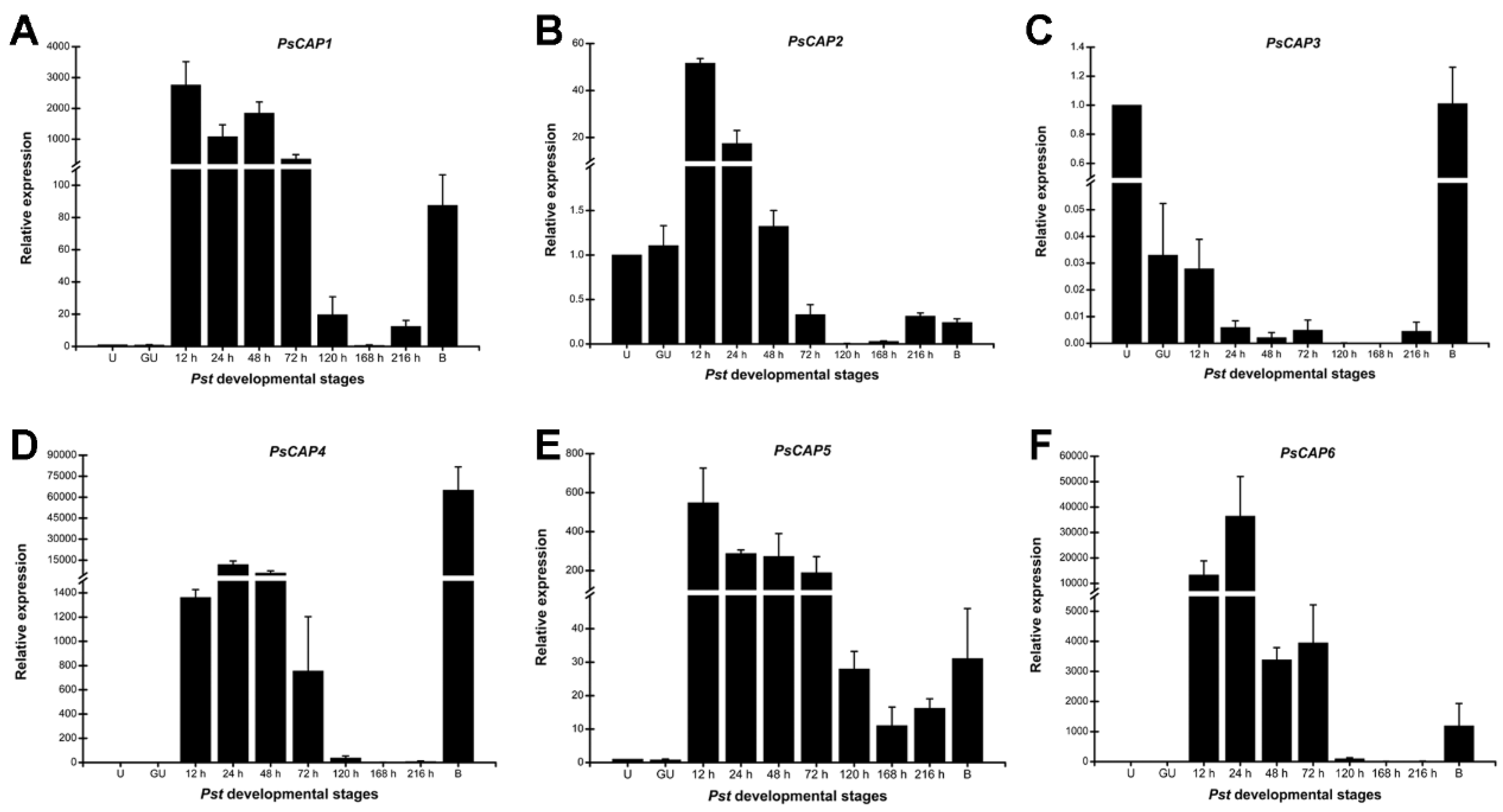
Several CAP proteins from mammals exhibit significant expression abundance in immune-related cells and tissues [34]. In plants, PR-1 family proteins are strikingly up-regulated during pathogens infection [35]. RT-qPCR was conducted to verify the expression profile of PsCAP genes in different infection stages during the Pst life cycle. A specific expression pattern was shown by each PsCAP gene during the interaction of Pst CYR32 with wheat cultivar Suwon11. The transcript levels of four PsCAP genes (PsCAP1, PsCAP4, PsCAP5, and PsCAP6) proliferated during the period of 12–72 hpi, the key biotrophic stage for Pst–wheat interaction (Figure 4A,D–F). PsCAP2 was up-regulated in 12 and 24 hpi (Figure 4B). In addition, we detected the expression levels of PsCAP genes in Pst-infected barberry (Berberis shensiana), which was the alternate host of Pst. Noticeably, PsCAP1, PsCAP4, PsCAP5, and PsCAP6 were also highly up-regulated in Pst-infected barberry (Figure 4). Thus, the results indicated that CAP genes seem to be preferentially expressed during the infection of host plants, hinting at their possible role in Pst virulence.

3.5. Three PsCAP Genes Are Required for Pst Infection on Wheat

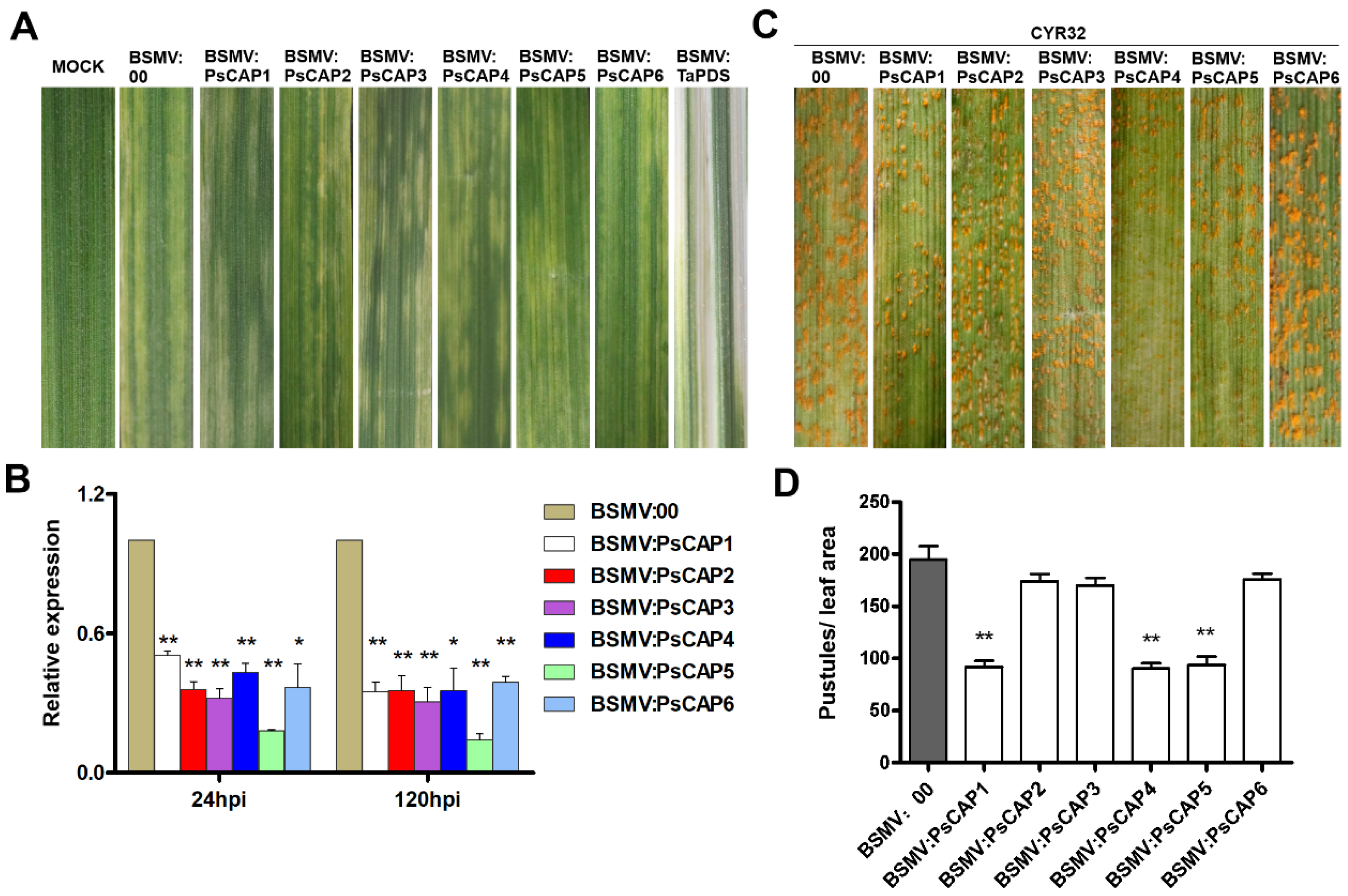
In order to analyze whether CAP genes can affect the infection of Pst on wheat, transient silencing of these PsCAP genes in wheat was performed by the BSMV-induced gene-silencing technique [20,36]. The phenotypes after inoculation of recombinant BSMV on wheat seedlings are shown in Figure 5A, showing chlorotic mosaic symptoms but without significant leaf damage. Meanwhile, wheat seedlings inoculated with BSMV:TaPDS showed a severe chlorophyll bleaching phenotype, which suggested that the wheat phytoene desaturase gene TaPDS was successfully silenced (Figure 5A). Then, the Pst isolate CYR32 was inoculated on wheat seedlings pre-infected with BSMV. As shown in Figure 5C, urediospores of different degrees were generated on the leaves of the target gene-silenced wheat seedlings. The statistical results showed that BSMV:PsCAP1, BSMV:PsCAP4, and BSMV:PsCAP5 had significantly reduced pustules per unit area compared to BSMV:00 (the negative control) leaves (Figure 5D). RT-qPCR was conducted to determine the gene silencing efficiency of the six CAP genes, and all of them were effectively silenced (Figure 5B). These results suggested that PsCAP1, PsCAP4, and PsCAP5 are required for Pst virulence by contributing to the Pst infection of wheat plants.

4. Discussion

The CAP protein superfamily is reported to be found in more than 2500 species, including most prokaryotes and eukaryotes such as bacteria, fungi, plants, and animals [2]. CAP proteins have long been reported to be involved in multiple cellular processes, including reproduction, prostate and brain cancer in mammals, and immune defense in plants [1]. In addition, there is increasing evidence that CAP proteins play an important role in the pathogenicity of both mammalian pathogenic fungi and some plant pathogens. Despite this, the identification and function of CAP proteins in more plant pathogens still need to be studied. One such plant pathogen is Pst, a fungus that severely threatens wheat yield worldwide. This study identified six Pst proteins with conserved CAP domains and homologous to plant PR-1 proteins. The characterized functions revealed that PsCAP proteins are novel virulence factors during the Pst infection of wheat plants.
The CAP motif is characteristic and definitive of the entire CAP superfamily and has been proven necessary and sufficient for the CAP protein’s biochemical activity and mode of action [1,3]. As shown in our results, all six PsCAP proteins contain the CAP motif with a conserved secondary structure of the alpha–beta–alpha sandwich fold [37,38]. Phylogenetic analysis in our study indicated that all PsCAP proteins belong to a clade of rust fungi-specific CAP proteins. In addition, we found there is also a clade of ascomycota-specific CAP proteins. These results indicate the evolutionary diversity of CAP superfamilies across species, which is consistent with the results described previously [39]. We also predicted the signal peptides among the six genes and, as shown in the results, PsCAP1, PsCAP2, PsCAP4, and PsCAP5 had predicted signal peptides. The secretion function of the four predicted signal peptides was confirmed using a yeast-secretion trap assay. This study suggests that these PsCAP proteins with signal peptides are likely to be secreted by Pst and then may enter host plants upon Pst infection. Compared with CAP proteins in other species, all CRISP proteins contain a predicted signal peptide consistent with their extracellular function [1]. However, mammalian GLIPR2 proteins do not contain a predicted signal sequence, and further investigation has proven their functional sites in the intracellular Golgi membrane [40]. Considering the difference among PsCAP proteins in sequence and structure, we presumed that PsCAP proteins may have functional diversity.
In this study, RT-qPCR revealed that five PsCAP genes (PsCAP1, PsCAP2, PsCAP4, PsCAP5, and PsCAP6) were strikingly up-regulated in the early stage of Pst infection in wheat. The early stage of Pst infection is the key period for haustoria formation. During this period, Pst could secrete a variety of pathogenicity factors, including effectors through haustoria [17,41,42], thus affecting plant immunity and establishing a parasitic relationship between Pst and wheat. This strongly hints at the potential role of PsCAP proteins in the biotrophic stage of Pst. In addition, previous studies have reported that the majority of CAP proteins in other species showed a notable expression bias in immunity-related activities [1]. The largest number of CAP proteins (11 in total) were identified from M. perniciosa, a pathogen causing witches’ broom disease in cacao plants, and most of them are highly and specifically induced during infection [6]. This is consistent with the results of our study. In addition, PsCAP1, PsCAP4, PsCAP5, and PsCAP6 were also highly up-regulated in Pst sexual reproduction in barberry, indicating that some of the PsCAP proteins might play a role in Pst infection of alternate host barberry.
To investigate the role of CAP proteins in Pst pathogenicity, we silenced six PsCAP genes with the BSMV-mediated virus-silencing system separately. The results indicated that the sporulation of PsCAP1, PsCAP4, and PsCAP5 was significantly reduced, indicating their virulence roles in Pst infection on wheat. Similar results were reported in CAP superfamily proteins from other species. CAP proteins have been reported as novel virulence factors in pathogenic fungi during infection of the mammalian hosts, such as RBT4 in C. albicans and Fpr1 in F. oxysporum [4,43]. Afterward, similar functions were reported from the CAP proteins of some plant pathogenic fungi. The CAP protein FgPR1L-4 from F. graminearum, a pathogenic fungus that causes wheat head blight, can promote fungal virulence during wheat infection [7]. Two of the three CAP proteins (VmPR1a and VmPR1c) identified in Valsa mali are virulence factors in the pathogenic infection course of host apples [44]. Deleting the CAP protein CcCAP1 from C. chrysosperma, the agent that causes poplar canker disease, decreases pathogen virulence during the infection process [8]. Importantly, the authors found that the conserved CAP domain is essential for CcCAP1′s virulence activity [8]. However, in our results, there was no significant sporulation reduction in PsCAP2-, PsCAP3-, and PsCAP6-silenced wheat seedlings, implying that they do not affect Pst pathogenicity. Presumably, this is due to functional redundancy as described in C. albicans and C. chrysosperma [5,8]. According to the results presented above, it seems that the secreted PsCAP proteins act as effectors to attack the host’s immune system, thereby causing susceptibility. In plant species, PR-1 proteins have been characterized as markers of induced defense against pathogens. Gamir and colleagues provided genetic and biochemical evidence for the capacity of PR-1 proteins to bind sterols and demonstrated that the inhibitory effect of plant PR-1 on pathogen growth is caused by sterol sequestration from pathogens [45]. Therefore, it is speculated that the pathogenic mechanism of secreted PsCAP proteins may be that PsCAP proteins act as fungal effectors by sequestering host plant sterols. The precise regulatory mechanism of each PsCAP family member needs further investigation.
In conclusion, this study identified six genes (PsCAP1PsCAP6) encoding CAP subfamily proteins and homologous to plant PR-1 proteins in Pst. We clarified their virulence roles in the Pst–wheat interaction. Five of the six PsCAP proteins have N-terminal signaling peptides that may be secreted by Pst during infection. The expression profile indicated that most of the PsCAP genes are preferentially up-regulated during the infection of host plants. BSMV-mediated HIGS revealed that the sporulation of PsCAP1, PsCAP4, and PsCAP5 was significantly reduced in the gene-silenced plants, indicating their roles in Pst infection. Our results suggest that CAP proteins may act as virulence factors during Pst infection, which also provides significant insights into Pst pathogenicity.

Supplementary Materials

The following supporting information can be downloaded at: https://www.mdpi.com/article/10.3390/jof9070734/s1, Figure S1: Sequence alignment of PsCAP4 among 7 Pst isolates; Table S1: Characteristics of six PsCAP genes identified in the Pst genome; Table S2: List of specific primers designed in this study.

Author Contributions

Y.C., Z.K. and M.Z. conceived and designed the experiments; M.Z., Y.Z., H.G. and M.C. performed the experiments; M.Z., M.C. and P.G. wrote the paper; Y.C. and Z.K. revised the manuscript. All authors have read and agreed to the published version of the manuscript.

Funding

This work was funded by the National Natural Science Foundation of China (grant number 32161143023), the Shaanxi Innovation Team Project (grant number 2018TD-004), and the 111 Project from the Ministry of Education of China (grant number BP0719026).

Institutional Review Board Statement

Not applicable.

Informed Consent Statement

Not applicable.

Data Availability Statement

All data generated or analyzed in this research process are included in this published article and its Supplementary Materials.

Conflicts of Interest

The authors declare no conflict of interest.

References

  1. Gibbs, G.; Roelants, K.; O’Bryan, M. The CAP superfamily: Cysteine-rich secretory proteins, antigen 5, and pathogenesis-related 1 proteins-roles in reproduction, cancer, and immune defense. Endocr. Rev. 2008, 29, 865–897. [Google Scholar] [CrossRef] [PubMed]
  2. Schneiter, R.; Di Pietro, A. The CAP protein superfamily: Function in sterol export and fungal virulence. Biomol. Concepts 2013, 4, 519–525. [Google Scholar] [CrossRef]
  3. Choudhary, V.; Schneiter, R. Pathogen-Related Yeast (PRY) proteins and members of the CAP superfamily are secreted sterol-binding proteins. Proc. Natl. Acad. Sci. USA. 2012, 109, 16882–16887. [Google Scholar] [CrossRef] [PubMed]
  4. Prados-Rosales, R.; Roldán-Rodríguez, R.; Serena, C.; López-Berges, M.; Guarro, J.; Martínez-del-Pozo, A.; Di Pietro, A. A PR-1-like Protein of Fusarium oxysporum functions in virulence on mammalian hosts. J. Biol. Chem. 2012, 287, 21970–21979. [Google Scholar] [CrossRef] [PubMed] [Green Version]
  5. Rohm, M.; Lindemann, E.; Hiller, E.; Ermert, D.; Lemuth, K.; Trkulja, D.; Sogukpinar, O.; Brunner, H.; Rupp, S.; Urban, C.F.; et al. A family of secreted pathogenesis-related proteins in Candida albicans. Mol. Microbiol. 2013, 87, 132–151. [Google Scholar] [CrossRef]
  6. Teixeira, P.J.; Thomazella, D.P.; Vidal, R.O.; Prado, P.F.; Reis, O.; Baroni, R.M.; Franco, S.F.; Mieczkowski, P.; Pereira, G.A.; Mondego, J.M. The fungal pathogen Moniliophthora perniciosa has genes similar to plant PR-1 that are highly expressed during its interaction with cacao. PLoS ONE 2012, 7, e45929. [Google Scholar] [CrossRef]
  7. Lu, S.; Edwards, M.C. Molecular characterization and functional analysis of PR-1-like proteins identified from the wheat head blight fungus Fusarium graminearum. Phytopathology 2018, 108, 510–520. [Google Scholar] [CrossRef] [Green Version]
  8. Han, Z.; Xiong, D.; Xu, Z.; Liu, T.; Tian, C. The Cytospora chrysosperma virulence effector CcCAP1 mainly localizes to the plant nucleus to suppress plant immune responses. mSphere 2021, 6, e00883-e00820. [Google Scholar] [CrossRef]
  9. Begum, S.; Iqbal, M.; Ahmed, I.; Fayyaz, M.; Shahzad, A.; Ali, G.M. Allelic variation at loci controlling stripe rust resistance in spring wheat. J. Genet. 2014, 93, 579–586. [Google Scholar] [CrossRef]
  10. Chen, W.; Wellings, C.; Chen, X.; Kang, Z.; Liu, T. Wheat stripe (yellow) rust caused by Puccinia striiformis sp. tritici. Mol. Plant Pathol. 2014, 15, 433–446. [Google Scholar] [CrossRef]
  11. Wellings, C.R. Global status of stripe rust: A review of historical and current threats. Euphytica 2011, 179, 129–141. [Google Scholar] [CrossRef]
  12. Chen, X. Pathogens which threaten food security: Puccinia striiformis, the wheat stripe rust pathogen. Food Secur. 2020, 12, 239–251. [Google Scholar] [CrossRef]
  13. Petersen, R.H. The rust fungus life cycle. Bot. Rev. 1974, 40, 453–513. [Google Scholar] [CrossRef]
  14. Gan, P.H.; Dodds, P.N.; Hardham, A.R. Plant Infection by biotrophic fungal and oomycete pathogens. In Signaling and Communication in Plant Symbiosis; Perotto, S., Baluška, F., Eds.; Springer: Berlin/Heidelberg, Germany, 2011; pp. 183–212. [Google Scholar]
  15. Cheng, Y.; Wang, X.; Yao, J.; Voegele, R.T.; Zhang, Y.; Wang, W.; Huang, L.; Kang, Z. Characterization of protein kinase PsSRPKL, a novel pathogenicity factor in the wheat stripe rust fungus. Environ. Microbiol. 2015, 17, 2601–2617. [Google Scholar] [CrossRef]
  16. Wang, B.; Sun, Y.; Song, N.; Zhao, M.; Liu, R.; Feng, H.; Wang, X.; Kang, Z. Pst-milR1, an important pathogenicity factor of Puccinia striiformis (Pst), impairs wheat resistance to Pst by suppressing wheat pathogenesis related gene 2. N. Phytol. 2017, 215, 338–350. [Google Scholar] [CrossRef] [PubMed] [Green Version]
  17. Tang, C.; Xu, Q.; Zhao, J.; Yue, M.; Wang, J.; Wang, X.; Kang, Z.; Wang, X. A rust fungus effector directly binds plant pre-mRNA splice site to reprogram alternative splicing and suppress host immunity. Plant Biotechnol. J. 2022, 20, 1167–1181. [Google Scholar] [CrossRef]
  18. Waterhouse, A.M.; Procter, J.B.; Martin, D.M.; Clamp, M.; Barton, G.J. Jalview Version 2-multiple sequence alignment editor and analysis workbench. Bioinformatics 2009, 25, 1189–1191. [Google Scholar] [CrossRef] [Green Version]
  19. Tamura, K.; Peterson, D.; Peterson, N.; Stecher, G.; Nei, M.; Kumar, S. MEGA5: Molecular evolutionary genetics analysis using maximum likelihood, evolutionary distance, and maximum parsimony methods. Mol. Biol. Evol. 2011, 28, 2731–2739. [Google Scholar] [CrossRef] [Green Version]
  20. Holzberg, S.; Brosio, P.; Gross, C.; Pogue, G.P. Barley stripe mosaic virus-induced gene silencing in a monocot plant. Plant J. 2002, 30, 315–327. [Google Scholar] [CrossRef]
  21. Jacobs, K.A.; Collins-Racie, L.A.; Colbert, M.; Duckett, M.K.; Golden-Fleet, M.; Kelleher, K.; Kriz, R.; LaVallie, E.; Merberg, D.; Spaulding, V.; et al. A genetic selection for isolating cDNAs encoding secreted proteins. Gene 1997, 198, 289–296. [Google Scholar] [CrossRef]
  22. Oh, S.-K.; Young, C.; Lee, M.; Oliva, R.; Bozkurt, T.; Cano, L.; Win, J.; Bos, J.; Liu, H.; Damme, M.; et al. In planta expression screens of Phytophthora infestans RXLR effectors reveal diverse phenotypes, including activation of the Solanum bulbocastanum disease resistance protein Rpi-blb2. Plant Cell Online 2009, 21, 2928–2947. [Google Scholar] [CrossRef] [PubMed] [Green Version]
  23. Gietz, R.D.; Schiestl, R.H.; Willems, A.R.; Woods, R.A. Studies on the transformation of intact yeast cells by the liac/ss-dna/peg procedure. Yeast 1995, 11, 355–360. [Google Scholar] [CrossRef] [PubMed]
  24. Gu, B.; Kale, S.D.; Wang, Q.; Wang, D.; Pan, Q.; Cao, H.; Meng, Y.; Kang, Z.; Tyler, B.; Shan, W. Rust secreted protein Ps87 is conserved in diverse fungal pathogens and contains a RXLR-like motif sufficient for translocation into plant cells. PLoS ONE 2011, 6, e27217. [Google Scholar] [CrossRef] [PubMed]
  25. Kang, Z.; Huang, L.; Buchenauer, H. Ultrastructural changes and localization of lignin and callose in compa and tible and incompatible interactions between wheat and Puccinia striiformis. J. Plant Dis. Prot. 2002, 109, 25–37. [Google Scholar]
  26. Zhang, Y.; Qu, Z.; Zheng, W.; Liu, B.; Wang, X.; Xue, X.; Xu, L.; Huang, L.; Han, Q.; Zhao, J.; et al. Stage-specific gene expression during urediniospore germination in Puccinia striiformis f. sp. tritici. BMC Genom. 2008, 9, 203. [Google Scholar] [CrossRef] [Green Version]
  27. Zhao, J.; Wang, L.; Wang, Z.; Chen, X.; Zhang, H.; Yao, J.; Zhan, G.; Chen, W.; Huang, L.; Kang, Z. Identification of eighteen berberis species as alternate hosts of Puccinia striiformis f. sp tritici and virulence variation in the pathogen isolates from natural infection of barberry plants in China. Phytopathology 2013, 103, 927–934. [Google Scholar] [CrossRef] [Green Version]
  28. Livak, K.J.; Schmittgen, T.D. Analysis of relative gene expression data using real-time quantitative PCR and the 2(-Delta Delta C(T)) Method. Methods 2001, 25, 402–408. [Google Scholar] [CrossRef]
  29. Breen, S.; Williams, S.J.; Outram, M.; Kobe, B.; Solomon, P.S. Emerging insights into the functions of pathogenesis-related protein 1. Trends Plant Sci. 2017, 22, 871–879. [Google Scholar] [CrossRef]
  30. Niderman, T.; Genetet, I.; Bruyere, T.; Gees, R.; Stintzi, A.; Legrand, M.; Mosinger, E. Pathogenesis-related PR-1 proteins are antifungal. Isolation and characterization of three 14-kilodalton proteins of tomato and of a basic PR-1 of tobacco with inhibitory activity against Phytophthora infestans. Plant Physiol. 1995, 108, 17–27. [Google Scholar] [CrossRef] [Green Version]
  31. Van Loon, L.C.; Van Kammen, A. Polyacrylamide disc electrophoresis of the soluble leaf proteins from Nicotiana tabacum var. ‘Samsun’ and ‘Samsun NN’: II. Changes in protein constitution after infection with tobacco mosaic virus. Virology 1970, 40, 199–211. [Google Scholar] [CrossRef]
  32. Uknes, S.; Mauch-Mani, B.; Moyer, M.; Potter, S.; Williams, S.; Dincher, S.; Chandler, D.; Slusarenko, A.; Ward, E.; Ryals, J. Acquired resistance in Arabidopsis. Plant Cell 1992, 4, 645–656. [Google Scholar] [CrossRef] [PubMed] [Green Version]
  33. Nei, M.; Gojobori, T. Simple methods for estimating the numbers of synonymous and nonsynonymous nucleotide substitutions. Mol. Biol. Evol. 1986, 3, 418–426. [Google Scholar] [CrossRef] [PubMed]
  34. Reddy, T.; Gibbs, G.M.; Merriner, D.J.; Kerr, J.B.; O’Bryan, M.K. Cysteine-rich secretory proteins are not exclusively expressed in the male reproductive tract. Dev. Dyn. 2008, 237, 3313–3323. [Google Scholar] [CrossRef] [PubMed]
  35. Loon, L.C.V.; Rep, M.; Pieterse, C.M.J. Significance of inducible defense-related proteins in infected plants. Annu. Rev. Phytopathol. 2006, 44, 135–162. [Google Scholar] [CrossRef] [PubMed] [Green Version]
  36. Nowara, D.; Gay, A.; Lacomme, C.; Shaw, J.; Ridout, C.; Douchkov, D.; Hensel, G.; Kumlehn, J.; Schweizer, P. HIGS: Host-induced gene silencing in the obligate biotrophic fungal pathogen Blumeria graminis. Plant Cell 2010, 22, 3130. [Google Scholar] [CrossRef] [PubMed] [Green Version]
  37. Asojo, O.A.; Goud, G.; Dhar, K.; Loukas, A.; Zhan, B.; Deumic, V.; Liu, S.; Borgstahl, G.; Hotez, P. X-ray structure of Na-ASP-2, a pathogenesis-related-1 protein from the nematode parasite, Necator americanus, and a vaccine antigen for human hookworm infection. J. Mol. Biol. 2005, 346, 801–814. [Google Scholar] [CrossRef]
  38. Serrano, R.L.; Kuhn, A.; Hendricks, A.; Helms, J.B.; Sinning, I.; Groves, M.R. Structural analysis of the human Golgi-associated plant pathogenesis related protein GAPR-1 implicates dimerization as a regulatory mechanism. J. Mol. Biol. 2004, 339, 173–183. [Google Scholar] [CrossRef]
  39. Szyperski, T.; Fernandez, C.; Mumenthaler, C.; Wüthrich, S.K. Structure comparison of human glioma pathogenesis-related protein GliPR and the plant pathogenesis-related protein P14a indicates a functional link between the human immune system and a plant defense system. Proc. Natl. Acad. Sci. USA 1998, 95, 2262–2266. [Google Scholar] [CrossRef]
  40. Eberle, H.B.; Serrano, R.L.; Füllekrug, J.; Schlosser, A.; Lehmann, W.D.; Lottspeich, F.; Kaloyanova, D.; Wieland, F.T.; Helms, J.B. Identification and characterization of a novel human plant pathogenesis-related protein that localizes to lipid-enriched microdomains in the Golgi complex. J. Cell Sci. 2002, 115, 827–838. [Google Scholar] [CrossRef]
  41. Cheng, Y.; Wu, K.; Yao, J.; Li, S.; Wang, X.; Huang, L.; Kang, Z. PSTha5a23, a candidate effector from the obligate biotrophic pathogen Puccinia striiformis f. sp. tritici, is involved in plant defense suppression and rust pathogenicity. Environ. Microbiol. 2017, 19, 1717–1729. [Google Scholar] [CrossRef]
  42. Wei, J.; Wang, X.; Hu, Z.; Wang, X.; Wang, J.; Wang, J.; Huang, X.; Kang, Z.; Tang, C. Puccinia striiformis effector Hasp98 facilitates pathogenicity by blocking the kinase activity of wheat TaMAPK4. J. Integr. Plant Biol. 2023, 65, 249–264. [Google Scholar] [CrossRef]
  43. Braun, B.R.; Head, W.S.; Wang, M.X.; Johnson, A.D. Identification and characterization of TUP1-regulated genes in Candida albicans. Genetics 2000, 156, 31–44. [Google Scholar] [CrossRef] [PubMed]
  44. Wang, C.; Yin, Z.; Nie, J.; Lin, Y.; Huang, L. Identification and virulence analysis of CAP superfamily genes in Valsa mali. Sci. Agric. Sin. 2021, 54, 3440–3450. [Google Scholar]
  45. Gamir, J.; Darwiche, R.; van’t, P.; Choudhary, V.; Stumpe, M.; Schneiter, R.; Mauch, F. The sterol-binding activity of PATHOGENESIS-RELATED PROTEIN 1 reveals the mode of action of an antimicrobial protein. Plant J. 2017, 89, 502–509. [Google Scholar] [CrossRef] [PubMed] [Green Version]
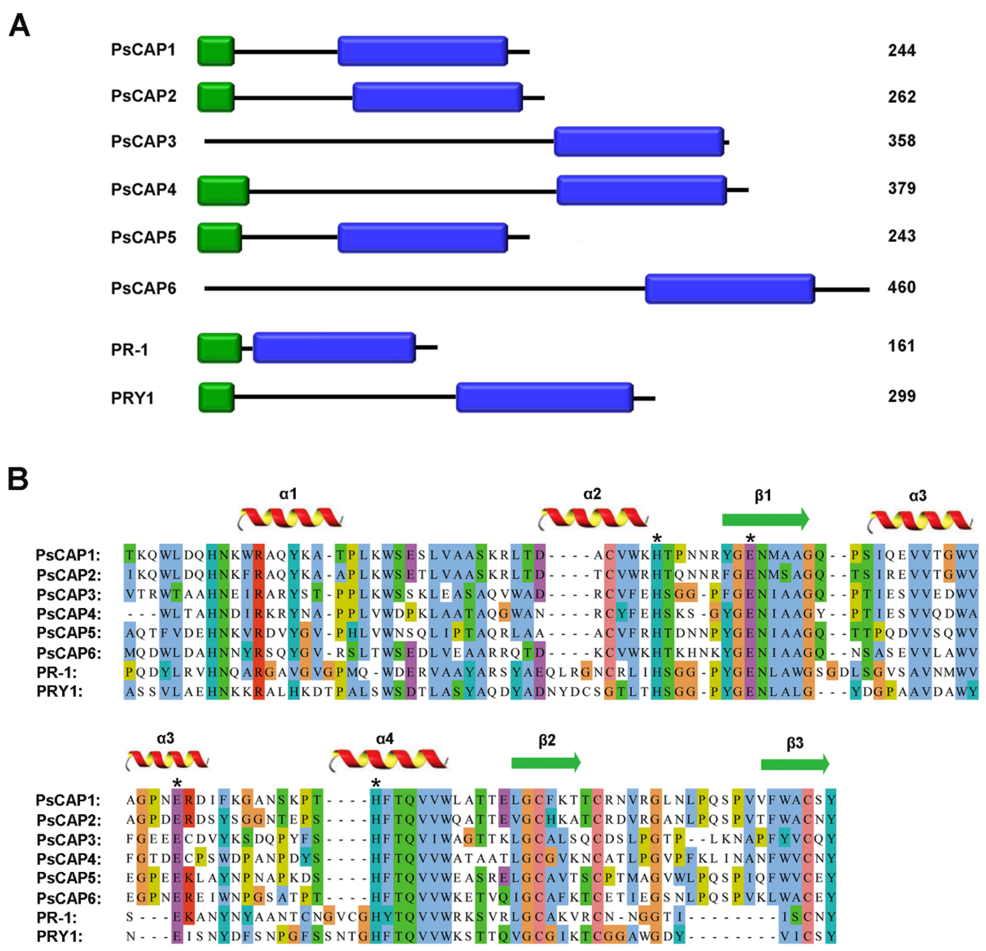
Figure 1. Domain architectures and sequences alignment of Pst CAP proteins. (A) Domain architectures of PsCAP1–PsCAP6. Signal peptides are represented by green blocks, and CAP domains are represented by blue blocks. The number on the right represents the size per PsCAP protein. (B) Sequences alignment of six CAP proteins and other known CAP proteins. The different colors of the amino acids are taken from Clustal X Colour Scheme in Jalview. The conserved secondary structural elements of CAP proteins are labeled above the sequences. The red helix represents the alpha-helix, and the green arrow represents the beta-strand. Asterisks represent the conserved residues of the CAP protein tetrad and the coordinate metals in the CAP domain [29]. PR-1: Fusarium oxysporum PR-1 protein (GenBank: ACV31371.1); PRY-1: Saccharomyces cerevisiae PRY-1 protein (GenBank: NP_012456.1).
Jof 09 00734 g001
Figure 2. Phylogenetic tree of Pst CAP proteins and CAP proteins from other organisms. The phylogenetic tree was constructed based on the neighbor-joining method. The amino acid sequences displayed in the phylogenetic tree were downloaded from the NCBI database. The scale in the figure represents the evolutionary distance. The species origin of genes and GenBank accession numbers are shown as follows. Fpr1, F. oxysporum (GenBank: ACV31371.1); RBT4, Candida albicans (GenBank: AAG09789); PRY1, S. cerevisiae (GenBank: NP_012456.1); PRY2, S. cerevisiae (GenBank: NP_012938.3); PRY3, S. cerevisiae (GenBank: NP_012457.1); SlP14c, Solanum lycopersicum (GenBank: NP_001234358.1); NtPR-1a, Nicotiana tabacum (GenBank: BAA14220); AtPR1, Arabidopsis thaliana (GenBank: NP_179068.1); GliPR-1, human glioma PR-1 protein (GenBank P48060); CRISP2, Homo sapiens (GenBank: AAI07708.1); Tex31, Conus textile (GenBank: CAD36507); Antigen 5, Dolichovespula maculate (GenBank: AAA28301.1); FG05, Fusarium graminearum (GenBank: GCA_000599445); GE21DRAFT, Neurospora crassa (GenBank: GCA_000786625); MGG, Magnaporthe oryzae; UMAG, Ustilago maydis; SETTUDRAFT, Setosphaeria turcica Et28A; ALT, Aspergillus lentulus; SCHCODRAFT, Schizophyllum commune H4-8; CC1G, Coprinopsis cinerea okayama7#130; MELLADRAFT, Melampsora larici-populina.
Jof 09 00734 g002
Figure 3. Secretion clarification of Pst CAP proteins. The predicted signal peptide sequences of PsCAP1, PsCAP2, PsCAP4, and PsCAP5, as well as the first 30 amino acids of PsCAP3 and PsCAP6, were fused to the pSUC2T7M13ORI vector (hereinafter referred to as pSUC2), and then the constructed vectors were transformed into yeast strain YTK12, respectively. The strains that successfully transformed with specified signal peptides:pSUC2 fusion vectors could grow on both a CMD-W medium and YPRAA medium because the signal peptides allow invertase to be secreted out of the yeast cell. Avr1b, the secreted protein from Phytophthora sojae, was the positive control. Mg87, a non-secreted Mg87 protein from Magnaporthe oryzae, was the negative control.
Jof 09 00734 g003
Figure 4. Transcription levels of PsCAP1-PsCAP6 (AF) during Pst infection stages. Wheat seedlings inoculated with CYR32 were sampled, and different stages of the relative transcript levels of CAP genes were calculated relative to that of the urediniospores by the comparative threshold (2−ΔΔCT) method. The data were normalized to the reference gene of PsEF1. Data are from three independent experiments ± S. E. U: urediniospores; GU: in vitro germinated urediniospores; B, infected Berberis shensiana (the alternate host of Pst).
Jof 09 00734 g004
Figure 5. PsCAP genes are required for Pst infection. (A) Chlorotic mosaic symptoms on wheat seedlings inoculated with BSMV: 00 and BSMV: PsCAPs. BSMV: TaPDS (positive control)-inoculated seedlings showed severe symptoms of chlorophyll bleaching. MOCK: wheat seedlings inoculated with 1× FES buffer; (B) Relative transcript levels of PsCAPs in knockdown wheat seedlings. RNA samples were isolated from the fourth leaves of wheat seedlings pre-infected with BSMV and then inoculated with the virulent Pst isolate of CYR32. The data were normalized to the reference gene of PsEF1, with BSMV:00 standardized at 1. Data are from three independent experiments ± S. E. Differences were assessed using Student’s t-tests. Single/double asterisks indicate p < 0.05 and p < 0.01, respectively. (C) Phenotypes of the fourth leaves of virus pre-inoculated wheat seedlings infected with the isolate of CYR32. (D) Pustules statistics of the fourth leaves of wheat seedlings pre-inoculated with BSMV and then infected with the CYR32 isolate. Data are from three independent experiments ± S. E. Differences were assessed using Student’s t-tests. Double asterisks indicate p < 0.01.
Jof 09 00734 g005
Table 1. SNP analysis of PsCAP genes among seven Pst isolates.
Single Nucleotide Polymorphisms (SNPs) aCompute Overall Mean Distance Estimation e
Gene NameNS bNNS cInsertion
/Deletin d
dNdSdN/dS
PsCAP1310.00050.00330.15
PsCAP2630.00140.00450.31
PsCAP31220.00070.01850.04
PsCAP431150.00060.00290.21
PsCAP51030.00190.01680.11
PsCAP6920.00090.00920.10
a SNPs were done in seven different Pst isolates including one Chinese isolate (CYR32), four US isolates (PST-21, PST-43, PST-78 and PST-130) and two UK isolates (PST-08/21 and PST-87/7); b Number of total nucleotide substitutions (NS); c Number of nonsynonymous nucleotide substitutions (NNS); d Number of nucleotide insertion/deletion; e Compute overall mean distance estimation was done by MEGA5.
Disclaimer/Publisher’s Note: The statements, opinions and data contained in all publications are solely those of the individual author(s) and contributor(s) and not of MDPI and/or the editor(s). MDPI and/or the editor(s) disclaim responsibility for any injury to people or property resulting from any ideas, methods, instructions or products referred to in the content.