Article

Ubiquitin Degradation of the AICAR Transformylase/IMP Cyclohydrolase Ade16 Regulates the Sexual Reproduction of Cryptococcus neoformans

1
State Key Laboratory of Resource Insects, Southwest University Medical Research Institute, Chongqing 400715, China
2
Jinfeng Laboratory, Chongqing 401329, China
*
Author to whom correspondence should be addressed.
J. Fungi 2023, 9(7), 699; https://doi.org/10.3390/jof9070699
Submission received: 3 May 2023 / Revised: 17 June 2023 / Accepted: 21 June 2023 / Published: 24 June 2023

Abstract

:
F-box protein is a key protein of the SCF E3 ubiquitin ligase complex, responsible for substrate recognition and degradation through specific interactions. Previous studies have shown that F-box proteins play crucial roles in Cryptococcus sexual reproduction. However, the molecular mechanism by which F-box proteins regulate sexual reproduction in C. neoformans is unclear. In the study, we discovered the AICAR transformylase/IMP cyclohydrolase Ade16 as a substrate of Fbp1. Through protein interaction and stability experiments, we demonstrated that Ade16 is a substrate for Fbp1. To examine the role of ADE16 in C. neoformans, we constructed the iADE16 strains and ADE16OE strains to analyze the function of Ade16. Our results revealed that the iADE16 strains had a smaller capsule and showed growth defects under NaCl, while the ADE16OE strains were sensitive to SDS but not to Congo red, which is consistent with the stress phenotype of the fbp1Δ strains, indicating that the intracellular protein expression level after ADE16 overexpression was similar to that after FBP1 deletion. Interestingly, although iADE16 strains can produce basidiospores normally, ADE16OE strains can produce mating mycelia but not basidiospores after mating, which is consistent with the fbp1Δmutant strains, suggesting that Fbp1 is likely to regulate the sexual reproduction of C. neoformans through the modulation of Ade16. A fungal nuclei development assay showed that the nuclei of the ADE16OE strains failed to fuse in the bilateral mating, indicating that Ade16 plays a crucial role in the regulation of meiosis during mating. In summary, our findings have revealed a new determinant factor involved in fungal development related to the post-translational regulation of AICAR transformylase/IMP cyclohydrolase.

1. Introduction

Cryptococcus neoformans is a yeast-like pathogenic fungus that is widely found in natural environments such as soil, plant surfaces, and bird feces [1]. The basidiospores or dried yeast cells of C. neoformans can be inhaled into the host’s lungs through the respiratory tract where they are eliminated or stay in a state of latent infection in immunocompetent hosts [2]. When the host’s immunity is impaired, or the hosts are immunocompromised individuals, C. neoformans can proliferate in the lungs of the host and cause cryptococcal pneumonia, penetrating the blood–brain barrier (BBB) to cause deadly cryptococcal meningitis [3]. Statistically,19% of the deaths related to acquired immune deficiency syndrome (AIDS) are caused by cryptococcal meningitis, and about 112,000 cases of cryptococcal meningitis are caused yearly [4].
As a basidiomycete, C. neoformans has two mating types, α and a, which can reproduce sexually or asexually [5]. Typically, C. neoformans grows as budding yeast and can switch from yeast growth to filamentous growth by mating and monokaryotic fruiting [6,7]. The transition of morphotypes from yeast to hyphae marks the beginning of the sexual development of C. neoformans [8]. During the mating of C. neoformans, the yeast cells of different mating types fuse and form dikaryotic mating hyphae, and eventually, a basidium, the specialized sporulation structure, is produced at the tip of the hyphae. Then, after completing meiosis inside the basidium, four chains of basidiospores are generated at the top of the basidium [7,8]. Similar environmental factors also affect the monokaryotic fruiting of C. neoformans but occur between the haploid yeast cells of the same mating type, such as α cells [9,10]. Compared with mating, the hyphal cells produced by monokaryotic fruiting are mononucleate diploid cells with unfused clamp connections [9].
Sexual reproduction of C. neoformans is regulated by many environmental factors and genetic circuits. Light, darkness, ambient temperature, nutrition deficiency, pheromone, and metal ions regulate cell–cell fusion and mating hypha growth during the sexual reproduction of C. neoformans [6,10], and the mechanisms of these environmental factors affecting the sexual cycle are usually rooted in gene regulatory circuits. The genetic circuits, such as the Cpk1 MAPK pathway and the pheromone response pathway, also regulate the sexual reproduction of C. neoformans, as the mutation of components in these pathways blocks the sexual reproduction of C. neoformans [6,11,12,13,14]. Besides these pheromone response pathways, other factors, such as zinc finger proteins, have also been identified as common regulators for sexual reproduction in C. neoformans [13,15,16].
The ubiquitin-proteasome system (UPS) is the main protein degradation system in eukaryotic cells, responsible for the degeneration of more than 80% of protein and maintaining intracellular protein homeostasis [17,18]. The Cullin1, Skp1, and F-box protein (SCF) E3 ligases are the most prominent E3 ubiquitin ligases, in which the F-box protein specifically recognizes downstream substrate proteins and meditates ubiquitination degradation of various substrate proteins [18]. Our previous studies showed that SCF E3 ligases play a vital role in the sexual reproduction of C. neoformans, as disruption of the key proteins of SCF E3 ligases such as Fbp1 and Cdc4 block basidiospore production in C. neoformans [19,20]. However, the molecular mechanism by which the F-box protein regulates C. neoformans sexual reproduction remained unclear. AICAR transformylase/IMP cyclohydrolase (ATIC) is a bifunctional, rate limiting enzyme that catalyzes the last two steps of the de novo purine biosynthesis pathway and plays an important role in cell proliferation and metabolism [21]. C. neoformans mutants lacking the ATIC-encoding ADE16 gene are adenine and histidine auxotrophic and are unable to infect mice [22]. Here, we show that the ATIC Ade16 of C. neoformans is a downstream target of Fbp1. Overexpression of the ADE16 gene blocks basidiospore formation by affecting the nuclear fusion of the meiosis process during mating. Our findings uncover a new determinant of fungal development involving the post-translational regulation of an AICAR transformylase/IMP cyclohydrolase.

2. Materials and Methods

2.1. Strains and Growth Conditions

Wild-type strains of C. neoformans, H99, and KN99a, and their derived strains were preserved routinely in our laboratory and cultured on a YPD agar or liquid medium at 30 °C. C. neoformans strains expressing the genes controlled by the CTR4 promoter were cultured on a YPD medium supplemented with 200 μM bathocuproinedisulfonic acid (BCS) or 25 μM CuSO4 with 1 mM ascorbic acid [23]. The yeast strain used in the yeast two-hybrid assay was Y187, whose transformants were cultured on a synthetic defined medium without leucine, tryptophan, histidine, or adenine. The C. neoformans and yeast strains used in the study are shown in Table S1. All other media used in the study were prepared as described previously [19].

2.2. Quantitative Real-Time PCR

To detect the ADE16 gene expression under different conditions, quantitative real-time PCR (qRT-PCR) was used to measure the mRNA expression levels of ADE16 as previously described [24]. Briefly, yeast cells or mating mixtures of each cryptococcal strain were collected and washed with distilled water (dH2O); total RNA was extracted and purified using an Omega total RNA kit (Omega Bio-tek, Norcross, GA, USA). The purified RNAs were quantified using a Nanodrop spectrometer (DeNovix, Wilmington, DE, USA), and first-strand cDNA synthesis was synthesized with a Hifair® II 1st Strand cDNA Synthesis Kit (Yeasen, Shanghai, China), as described by the manufacturer. The analysis of ADE16 gene expression was performed using FastStart Essential DNA Green Master (Roche, Mannhein, Germany), and the gene expression levels were normalized using the endogenous control gene GAPDH. Relative gene expression levels were calculated using the previously described comparative threshold cycle (CT) method [25]. The qRT-PCRs were carried out using a LightCycler®96 QPCR system (Roche) as described previously [26].

2.3. Generation of Fluorescence Strains

To detect the ADE16 gene expression in C. neoformans at different developmental stages, we amplified a 1996-bp ADE16 gene promoter sequence using H99 genomic DNA as a template with primers TL1166/1167 (see Table S2 for primer sequences) and inserted it into the pTBL5 [16] plasmid to construct the plasmid pTBL196. Then, the pTBL196 plasmid was linearized by SalI, concentrated, and biolistically introduced into the wild-type strains, H99 and KN99a, respectively, as previously described [27]. Stable transformants were further screened by growing on YPD plates with nourseothricin sulfate (100 mg/L) and fluorescence examination under an Olympus inverted confocal microscope (Olympus, FV1200). Finally, two mating types of the PADE16-mCherry fluorescence fusion expression strains (α and a) were obtained and named TBL310 and TBL378, respectively.
To determine Ade16 sub-cellular localization, we amplified the ADE16 gene coding sequence from the H99 genomic DNA using primers TL1164/TL1165 and inserted it into the pCN19 vector to construct the GFP and ADE16 fusion gene expression vector pTBL186. The XbaI-linearized pTBL186 was biolistically introduced into the H99 or KN99a strains, respectively. Stable transformants were further confirmed on YPD plates with nourseothricin sulfate (100 mg/L) and named TBL308 and TBL309, respectively. The fluorescence of the transformants was examined via confocal microscopy using a 100× objective lens (Olympus, FV1200, Tokyo, Japan).

2.4. Yeast Two-Hybrid Assays

We first carried out a yeast two-hybrid interaction assay to detect the interaction between Ade16 and Fbp1 proteins as described previously [19,28]. The full-length cDNAs of the ADE16 and FBP1 genes were amplified with primers TL879/880 and 588/589 and cloned into the bait vector pGBKT7 to fuse with the BD domain (pTBL106 and pTBL100), respectively. Meanwhile, the cDNAs of ADE16 and FBP1 were also amplified with primers TL883/884 and TL572/573 and inserted into the pGADT7 prey vector (pTBL145 and pTBL142), respectively, fusing with the AD domain. The inserted cDNAs were verified by sequencing to ensure the proteins were translated correctly. Both prey and bait constructs were co-transformed into the yeast strain Y187. After transformation, the yeast cells were transferred to an SD-Leu-Trp plate and incubated for 2–3 days at 30 °C. The transformants growing on SD-Leu-Trp-His or SD-Leu-Trp-His-Ade plates were considered positive interactions. The transformants carrying pGADT7-T7/pGBKT7-LAM and pGADT7-T7/pGBKT7-53 served as negative and positive controls, respectively (Clontech, Dalian, China).

2.5. Generation of Tagged Protein Strains

To obtain C. neoformans strains expressing the Ade16-HA protein, we first amplified the ADE16 cDNA with primers TL893/894. We then cloned it into the BamHI site of the vector pCTR4-2 [23] to generate the Ade16:HA fusion plasmid pTBL149 using the In-Fusion HD cloning kit (Clontech, Dalian, China). The C. neoformans strain (TBL81) expressing the Fbp1-Flag fusion protein was generated by our previous studies [29]. To detect the interaction between Ade16 and Fbp1 in C. neoformans, we biolistically introduced the EcoRI-linearized pTBL149 into the Fbp1-Flag strain TBL81 and the wild-type H99, generating the C. neoformans strains TBL248 and TBL264, respectively, which express Fbp1:Flag/Ade16:HA protein and Ade16:HA protein.
Total proteins were extracted and examined using Western blotting with anti-HA or anti-Flag antibodies to ensure the correct tagged strains were obtained. Protein pull-down was performed using SureBeads™ anti-HA or anti-Flag Magnetic Beads (Bio-RAD) as described previously [24] to collect the tagged proteins, and immunoblotting was used to detect the Ade16-HA or Fbp1-Flag proteins with anti-HA or anti-Flag antibodies, respectively.
To detect the Ade16 stability in the H99 and fbp1Δ mutant strains’ backgrounds, we introduced the EcoRI-linearized pTBL149 into the fbp1Δ mutant biolistically to construct the Ade16:HA strain TBL264 and TBL265. Then, the yeast cells of TBL264 and TBL265 strain were grown in liquid YPD to mid-logarithmic phase, transferred to YPD containing 25 μM CuSO4 and 1 mM ascorbic acid, and incubated for an additional 1, 2, 4, and 6 h. Yeast cells were harvested, and protein extracts were prepared as described above. The signals of Ade16:HA was detected via Western blotting using a monoclonal anti-HA antibody (Sigma, Saint Louis, MO, USA).

2.6. Generation of ADE16 Interference and Overexpression Strains

To generate the ADE16 promoter replacement strain, we used a split marker strategy to replace the ADE16 promoter with a copper-repressible CTR4 promoter. In the first round of PCR, as shown in Figure 3A, the 5′ fragment, the NEO marker, and the PCTR4-ADE16 fusion fragment were amplified with primers TL1036/TL1037, TL17/TL18, and TL1034/TL1038 using the H99 genomic DNA, pJAF1, and pTBL149 as templates, respectively. In the next round of PCR, the fusion fragment of the 5′ fragment and NEO marker was amplified with primers TL1036/TL20 using the mixture of the 5′ fragment and NEO marker as templates. Similarly, using the mixture of the NEO marker and PCTR4-ADE16 fragment as templates, the fusion fragment of the NEO marker and the PCTR4-ADE16 fragment were amplified with primers TL19/TL1038. The above two fusion fragments were mixed, concentrated, and biolistically introduced into the H99 strain to construct the PCTR4-ADE16 strain TBL270. The TBL270 strain was further confirmed via PCR with primers TL373/TL59 and Southern blotting. To verify whether ADE16 is an essential gene for C. neoformans, we inoculated the H99 and TBL270 strain onto YPD, YPD + 200 μM BCS, or YPD + 25 μM CuSO4 + 1 mM ascorbic acid plates, respectively, after serial dilution. The growth of each cryptococcal strain was observed after 2–3 days of incubation at 30 °C.
To generate the ADE16 RNAi-interference strains, we first constructed plasmid PiADE16 for RNA interference of the ADE16 gene in three steps. Step I: a 200-bp intron of the ADE16 gene was amplified using primers TL1334/1335 and inserted into the BamHI/PacI sites of pTBL5 to generate the plasmid pTBL226. Step II: a 480-bp ADE16 5′–3′ fragment of the ADE16 RNA interference sequence was amplified using primers TL1336/1337 and inserted into the BamHI site of pTBL226 to generate pTBL227. Step III: a reversed 480-bp ADE16 RNA interference sequence was amplified using primers TL1467/1468 and inserted into the PacI/SpeI sites of pTBL227 to generate pTBL237. After verification by sequencing, SalI-linearized pTBL237 was biolistically introduced into the wild-type strains H99 and KN99a. After PCR verification with primers TL1336/1340, the expression level of the ADE16 gene was quantified using quantitative real-time PCR in each transformant, and two mating types of ADE16 interference strains were named TBL414 and TBL415, respectively.
To obtain the ADE16 gene overexpression strain in C. neoformans, we first amplified the ADE16 gene using primers TL1092/1093 and inserted it into the BamHI site of the pTBL153 plasmid to generate pTBL174. Then, the ApaI-linearized pTBL174 was biolistically introduced into the wild-type H99 and KN99a strains. Stable transformants were further confirmed on a YPD medium containing 100 mg/L nourseothricin sulfate. The ADE16 gene overexpression (TBL288 and TBL302) was verified via qRT-PCR. To monitor the nuclear positioning in the ADE16 overexpression strains during mating, pTBL59, containing a NOP1-mCherry-NEO cassette, was biolistically transformed into the ADE16 overexpression strain of both α and a mating types to generate TBL445 and TBL446.

2.7. Analysis of Melanin and Capsule

To examine the role of Ade16 in C. neoformans melanin production, we grew the wild-type H99 strain and the Ade16-related strains in a YPD liquid medium overnight at 30 °C. One hundred microliters of each culture was inoculated on Niger seed medium to evaluate the melanin production; Niger seed agar plates were incubated for 2–3 days at 30 °C or 37 °C. The pigmentation of each cryptococcal strain was evaluated and photographed.
To assess the role of Ade16 in C. neoformans capsule production, overnight cultures of each cryptococcal strain were washed with 1× PBS buffer three times and incubated overnight in diluted Sabouraud medium (DSM) or MM with mannitol at 37 °C [30,31]. The quantification of the capsule size was performed as previously described [19].

2.8. Mating Assay

To investigate the role of Ade16 in C. neoformans mating, equal amounts of the α and a mating type yeast cells were mixed and grown on MS or V8 agar plates in the dark at 25 °C. After 14 days of incubation, the formation of mating hyphae and basidiospores was visualized and recorded using an Olympus CX41 light microscope.

2.9. Statistical Analysis

All statistical analyses were performed using Prism8.0 (GraphPad Software Inc., San Diego, CA, USA). Experiments consisting of only two groups were assessed with the Mann–Whitney test. Experiments involving a time series were compared using a Wilcoxon signed-rank test. Experiments involving multiple groups were analyzed with the nonparametric Kruskal–Wallis test. All data are represented with mean ± SD, unless specified in the figure legend. p-values of < 0.05 were considered statistically significant.

3. Results

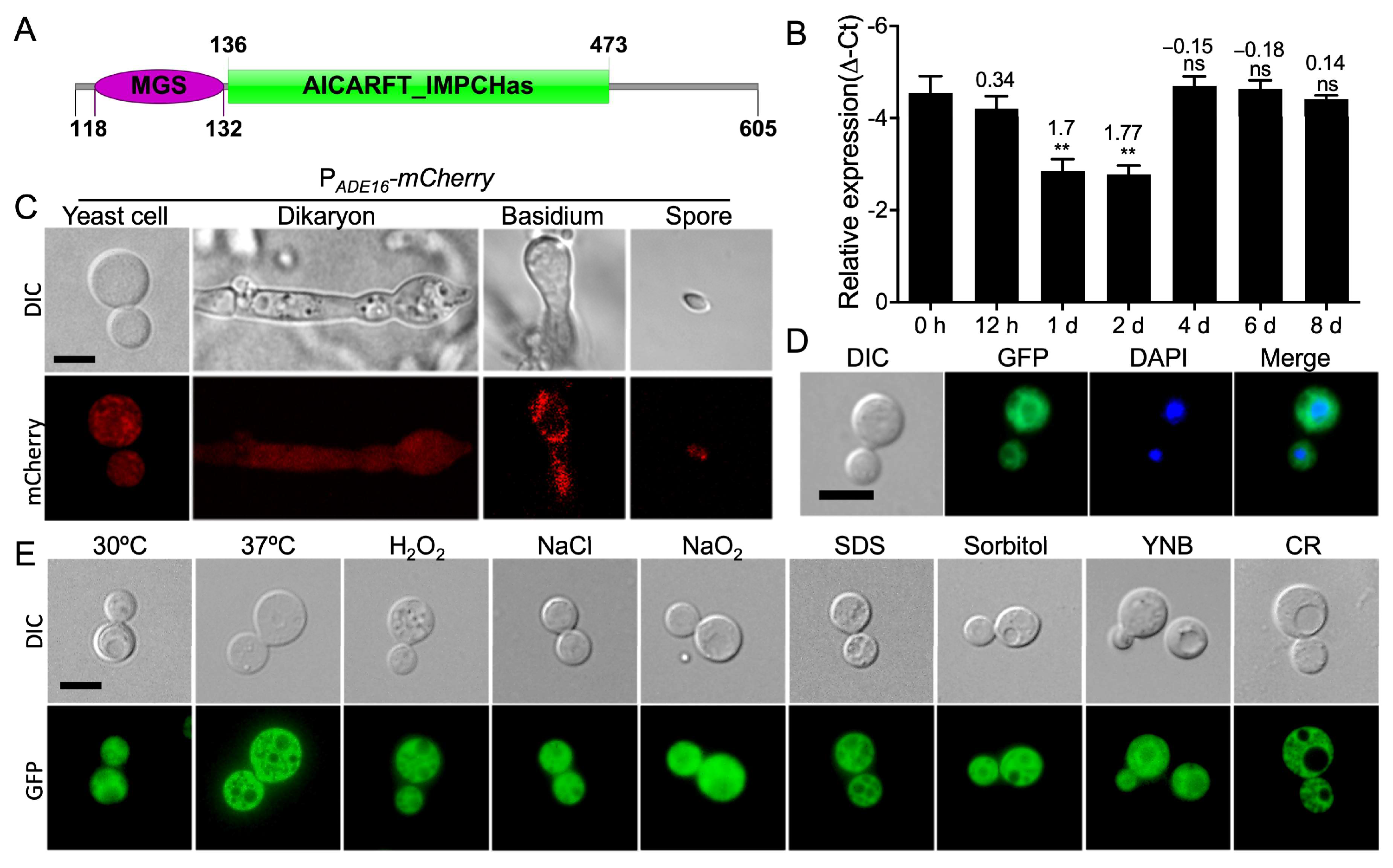
3.1. Interaction Profile and Expression Pattern of ADE16 during Mating of C. neoformans

Our previous study revealed that the F-box protein Fbp1, a key protein of the E3 ligase family, plays a crucial role in C. neoformans sexual reproduction, as in the bilateral mating of fbp1Δ mutants; basidiospore production was blocked despite the observation of normal dikaryotic hyphae during mating [19]. To identify downstream targets of Fbp1 in C. neoformans, we conducted an iTRAQ analysis coupled with LC-MS/MS in a previous study searching for enriched proteins in fbp1Δ mutants [24]. The partially enriched proteins are shown in Table 1 and have been selected as candidates for further study.
One of the candidate genes, CNAG_00700, was highly enriched in fbp1Δ mutants. Recently, Wizrah et al. identified the CNAG_00700 gene in C. neoformans and named it ADE16 [22]. Sequence analysis revealed that the Ade16 protein contains two domains; one is the MGS (methylglyoxal synthase) domain, and the other is the AICARFT_IMPCHas (AICARFT/IMPCHase bienzyme) domain (Figure 1A). To investigate the function of C. neoformans Ade16 during fungal development, we detected ADE16 gene expression H99 and KN99a strains using qRT-PCR. Expression of ADE16 was significantly upregulated during the mating process and reached its highest value at the 2 d time point, then returning to comparable expression to the 0 h timepoint, suggesting that Ade16 plays a role in the sexual reproduction of C. neoformans (Figure 1B).
To more precisely observe the ADE16 expression in C. neoformans at different developmental stages, we generated fluorescence strains (TBL310 and TBL378, see Table S2) expressing mCherry under the control of the native promoter of the ADE16 gene in both mating types. The mCherry strains (TBL310 and TBL378) were crossed on MS medium and the fluorescence at different developmental stages of mCherry was visualized using a confocal microscope (Olympus, FV1200). mCherry was expressed in yeast cells, dikaryons, basidia, and basidiospores (Figure 1C), suggesting that Ade16 may play a key role in the fungal development of C. neoformans.
To detect Ade16 localization in C. neoformans, the XbaI-linearized GFP-ADE16 plasmid was biolistically introduced into the wild-type H99 and KN99a strains to obtain TBL308 and TBL309. The expression of the GFP-Ade16 fusion protein was confirmed via Western blotting (Figure S1). The GFP-Ade16 fusion protein is localized in the cytoplasm of C. neoformans yeast cells and dikaryotic hyphae, except for the vacuoles (Figure 1D). Meanwhile, the subcellular localization of GFP-Ade16 in yeast cells was also examined under different stress conditions, and the results showed no difference among the GFP-Ade16 localizations under the above-tested stress conditions (Figure 1E).

3.2. Ade16 Interacts with Fbp1

Our previous proteomics data suggests that Ade16 is enriched in fbp1Δ mutants and might be a target for Fbp1-mediated ubiquitination (Table 1) [24]. We conducted a yeast two-hybrid protein–protein interaction assay to explore whether there is an interaction between Ade16 and Fbp1. Expression vectors containing Ade16 and Fbp1 fused to either the binding domain (BD) or the activation domain (AD) were co-transformed into the Y187 yeast strain, and transformants growing on SD-Leu-Trp-His or SD-Leu-Trp-His-Ade media were considered positive interactions. As shown in Figure 2A, Ade16 interacts with Fbp1 in a yeast two-hybrid protein–protein interaction system.
To further investigate the Ade16-Fbp1 interaction, we generated an Ade16:HA fusion expression vector (pTBL149) by cloning the ADE16 gene into the pCTR4-2 plasmid, in which the expression of ADE16 was regulated by the copper-repressible CTR4 promoter [23]. The resulting construct, pTBL149, linearized by EcoRI, was biolistically transformed into H99, the fbp1Δ mutants, and a previously constructed Fbp1:Flag overexpression strain [29]. The expression of Ade16:HA fusion proteins in each strain was confirmed via Western blot (Figure 2B). Total proteins from strains expressing Ade17:HA and Fbp1:Flag were purified with anti-HA Magnetic Beads (Bio-RAD), and Western blotted with anti-HA and anti-Flag antibodies. Both HA and Flag signals were detected from the co-immunoprecipitation (Co-IP) products, indicating that Ade16 interacts with Fbp1 in C. neoformans (Figure 2C).

3.3. The Ade16 Stability Depends on Fbp1 Function

To test our hypothesis that Ade16 is an Fbp1 downstream target, we investigated whether the Ade16 protein stability depends on the function of the SCF(Fbp1) E3 ligase. The Ade16:HA protein was expressed in the wild-type H99 strain (TBL264) and the fbp1Δ mutants (TBL265), and Ade16 protein stability was examined in the backgrounds of the above two strains. The Ade16:HA expressing strains were first cultured in a YPD medium containing 200 μM BCS to induce the CTR4 promoter. Then, the cultures were washed with PBS and transferred to a YPD medium containing 1 mM ascorbic acid and 25 μM CuSO4 to block the transcription of ADE16:HA. The yeast cells were harvested after incubation for another 0, 1, 2, 4, and 6 h, and the abundance of the Ade16:HA protein was evaluated via Western blot. Our results of the protein stability assay revealed that the Ade16:HA protein was hydrolyzed in the wild-type H99 background in a time-dependent manner during the examined period (0 to 6 h) but was relatively stable in the fbp1Δ mutants, suggesting that the Ade16:HA stability is dependent on the function of Fbp1 (Figure 2D).
Next, to detect the abundance of Ade16-HA protein in C. neoformans, we extracted the Ade16-HA proteins from the H99 or fbp1Δ mutants and examined them using Western blotting using the anti-HA antibody. As shown in Figure 2E, the relative amount of Ade16-HA in fbp1Δ mutants increased compared to that of the H99 strain, suggesting that the degradation of Ade16 depends on the Fbp1 function in C. neoformans.

3.4. ADE16 Is Required for Normal Growth of C. neoformans

Wizrah et al. revealed that the ADE16 null mutants are adenine and histidine auxotrophs and show severe growth defects on YPD [22]. To further confirm this phenotype, we decided to substitute the native promoter of the ADE16 gene with the copper-ion-suppressing CTR4 promoter to construct a PCTR4-ADE16 strain (TBL270) and test its growth on YPD. The strategy of the PCTR4-ADE16 strain construction is shown in Figure 3A.
Overnight cultures of the wild-type H99 strain and PCTR4-ADE16 strain (TBL270) were inoculated on YPD, YPD with 200 μM BCS, and YPD with 25 μM CuSO4 and 1 mM ascorbic acid and incubated at 30 °C for 2–3 days after series dilution. The growth of the PCTR4-ADE16 strain was comparable to that of the wild-type strain when grown on YPD or YPD with 200 μM BCS. However, when grown on YPD with 25 μM CuSO4 and 1 mM ascorbic acid, the PCTR4-ADE16 strain shows severe growth defects, while the growth capacity of the wild-type strain was not affected (Figure 3B), indicating that ADE16 is required for the growth of C. neoformans.

3.5. ADE16 Overexpression Increases Capsule Size in C. neoformans

We constructed an ADE16 RNA interference vector pTBL237 and introduced it into the C. neoformans wild-type strains to obtain the iADE16 interference strains (TBL414 and TBL415) in both α and a mating type. The construction of the ADE16 interference strains and the ADE16 gene interference efficiency was verified with PCR and qRT-PCR, respectively (Figure 3C,D). Meanwhile, the ADE16OE overexpression strains (TBL288 and TBL302) were also constructed by introducing the ADE16OE overexpression vector pTBL237 into the H99 and KN99a strains. The overexpression of ADE16 in both mating types was confirmed via Western blot and qRT-PCR (Figure 3E,F).
To confirm the role of Ade16 in C. neoformans virulence, we first examined the virulence factor production of iADE16 and ADE16OE strains in vitro. Interestingly, RNA interference of the ADE16 gene led to a significant decrease in capsule size, while overexpression led to a significant increase in capsule production in C. neoformans (Figure 4A,B). The average relative size of the capsule produced by the iADE16 strain or the ADE16OE strain was reduced by 35% or increased by 111%, respectively, compared with the H99 strain or fbp1Δ mutants (Figure 4B). This decrease or increase in capsule production is statistically significant (p < 0.0001) based on a one-way ANOVA analysis, indicating that Ade16 plays an important role in C. neoformans capsule production. We also examined the melanin formation of each C. neoformans strain and found that neither ADE16 interference nor overexpression affected melanin formation in C. neoformans (Figure S2).
Meanwhile, we also investigated the growth of the iADE16 strain and the ADE16OE strain under different stress conditions. The results showed that the iADE16 strain was sensitive to 1.5 M NaCl but not sensitive to cell integrity-targeting chemicals, such as SDS and Congo red, suggesting that Ade16 may play a role in maintaining the osmotic pressure in C. neoformans cells (Figure 4C).

3.6. Ade16 Regulates Sexual Reproduction of C. neoformans

Our previous study showed that knockout of the FBP1 gene blocked the basidiospore production in bilateral mating between fbp1Δ mutants. Since Ade16 is a downstream substrate of Fbp1, we speculated that Fbp1 might affect Cryptococcus sexual reproduction by regulating Ade16. To investigate the role of Ade16 in fungal mating, we constructed the iADE16 interference strains and ADE16OE overexpression strains in both H99 and KN99a strain backgrounds. The development of dikaryotic hyphae and basidiospores was examined in bilateral mating in iADE16 interference strains or ADE16OE overexpression strains. As with the wild-type strains, the iADE16 interference strains generated normal mating hyphae and morphologically normal-looking basidiospores (Figure 5). However, the bilateral mating between ADE16OE overexpression strains generated normal mating hyphae but failed to produce spores, consisting with the phenotype of the fbp1Δ mutants (Figure 5), suggesting Fbp1 may regulate the sexual reproduction by regulating the Ade16.
To explore the mechanism of the failure of ADE16OE overexpression strains to produce basidiospores, we constructed a nuclear-located Nop1-mCherry fusion expression vector. We introduced it into both α and a mating types of the wild type and ADE16OE overexpression strains to monitor the fungal nuclei positioning at different stages of mating in C. neoformans. The wild types (TBL101 and TBL102) or the ADE16OE overexpression strains (TBL445 and TBL446) expressing the Nop1-mCherry were crossed, respectively, and their nuclear positioning was monitored during the mating. As shown in Figure 6A, a single nucleus was visualized in the yeast cells of both the wild types and the ADE16OE overexpression strains, while two separated nuclei were found in each dikaryon produced after cell fusion. During mating, the wild-type strains had a single fused nucleus in the young basidium and four nuclei in each mature basidium, consistent with our previous findings [16]. However, the nuclei of the ADE16OE overexpression strains failed to fuse in the bilateral mating, and only two separated nuclei were observed in both the young and the mature basidia (Figure 6A,B). These findings suggested that Ade16 influences meiosis during mating, which may help explain why the ADE16OE overexpression strains could not produce basidiospores when crossed with a partner in bilateral mating. However, when grown in rich media, the growth rate and nuclear division of the ADE16OE overexpression strains were normal, suggesting that Ade16 is not involved in the cell cycle during mitosis.
Taken together, we identified a downstream substrate of Fbp1, the AICAR transformylase/IMP cyclohydrolase Ade16, and found that Fbp1 regulates the sexual reproduction of C. neoformans by regulating ubiquitination degradation of its downstream substrate, Ade16.

4. Discussion

The F-box proteins are a critical component of the SCF E3 ligases and play a vital role in fungal virulence and development. Our previous studies have shown that the F-box protein Fbp1 is required for the sexual reproduction of the human fungal pathogen C. neoformans, as basidiospore formation is blocked in bilateral mating of fbp1Δ mutants [19]. However, the mechanism by which Fbp1 regulates sexual reproduction in C. neoformans remains unclear. To identify the potential downstream targets in Fbp1, we carried out an iTRAQ analysis coupled with LC-MS/MS in a previous study to investigate the enriched target proteins in fbp1Δ mutants [24]. In this study, we identified the AICAR transformylase/IMP cyclohydrolase Ade16 and demonstrated that Ade16 interacts directly with Fbp1, and that the degradation and ubiquitination of Ade16 depend on the function of Fbp1. The gene expression analysis revealed that the ADE16 gene was expressed at all developmental stages, and the Ade16 protein was localized in the cytoplasm of C. neoformans cells. RNA interference of ADE16 led to a significant decrease in capsule size, while overexpression led to a significant increase in capsule size (confirming Wizrah and colleagues’ results [22]) in C. neoformans. Overexpression of ADE16 resulted in the production of normal mating hyphae but a failure of basidiospore formation, which is consistent with the phenotype of the fbp1Δ mutant (Figure 6), suggesting Fbp1 may regulate sexual reproduction by regulating Ade16. Our findings suggest that the SCF(Fbp1) E3 ligase-mediated UPS pathway regulates C. neoformans sexual reproduction by regulating Ade16.
We first analyzed the ADE16 gene expression and found that the ADE16 was expressed at all development stages of C. neoformans, and the Ade16 protein was localized in the cytoplasm of C. neoformans cells, consisting with the Ade17p localization in S. cerevisiae [32]. Since Ade16 was highly enriched in the fbp1Δ mutant and could be a downstream target of Fbp1, we then performed protein interaction, stability, and ubiquitination assays. We found that Ade16 interacts with Fbp1, and its stability and ubiquitination is dependent on the function of Fbp1. These findings suggest that the SCF(Fbp1) E3 ligase-mediated UPS pathway might regulate the purine de novo biosynthesis pathway in C. neoformans by regulating the ATIC Ade16. So far, to our knowledge, there have been no reports on the regulation of Ade16 protein degradation, so our findings may establish the regulatory pathway for Ade16 protein degradation.
The polysaccharide capsule is a virulence factor that plays a key role in the interaction between C. neoformans cells and the immune system, protecting C. neoformans cells from being phagocytosed by immune cells [33].In this study, we found that loss of ADE16 expression decreased the capsule size of C. neoformans, while ADE16 overexpression enlarged the capsule, indicating that formation of C. neoformans capsule is related to the expression level of ADE16. In the study by Wizrah et al., the ade16Δ strain also displayed reduced capsule size at 30 °C and 37 °C [22]. The results from both groups suggested that Ade16 plays an important role in the capsule formation of C. neoformans.
Purines are ubiquitous life-sustaining biomolecules and can be generated by the de novo purine biosynthesis pathway [34,35]. Inosine monophosphate (IMP) is the common intermediate production in purine biosynthesis and can be converted into the purine nucleotides AMP or GMP using additional GTP or ATP [35]. Therefore, as a core enzyme in the de novo purine biosynthesis pathway, changes in the abundance of Ade16 affect the production of IMP, which in turn affects the formation of AMP and, thus, ATP. Nucleotide sugars are essential for polysaccharide synthesis [36], which requires a large amount of energy, meaning that any AMP or ATP depletion will negatively affect polysaccharide formation. Additionally, cAMP, synthesized from ATP through a cyclization reaction catalyzed by adenylate cyclase, is an important intracellular second messenger molecule regulated in many physiological processes [37]. In C. neoformans, studies reveal that the Gpa1-cAMP pathway regulates capsule induction in response to environmental stimuli [38]. Thus, interference of ADE16 leads to a decrease in AMP, resulting in a significant decrease in capsule size, while overexpression leads to a significant increase in AMP and capsule size in C. neoformans. In Wizrah et al.’s study, due to adenine supplementation, the capsule defect observed in the ade16Δ mutant is much milder than the one we observed.
Sexual reproduction allows genetic material from two parents to recombine, resulting in recombinant offspring with the potential for variable adaptation, and allows natural selection to more effectively remove harmful mutations that accumulate in the parental genomes [39]. In C. neoformans, sexual reproduction links to virulence, antifungal drug resistance, and rapid adaptive evolution [40,41]. Many environmental factors and genetic circuits regulate sexual reproduction in C. neoformans [10]. Our previous study showed that the SCF(Fbp1) E3 ligase-mediated UPS regulates sexual reproduction in C. neoformans [19]. However, the molecular mechanism by which Fbp1 regulates sexual reproduction in C. neoformans remains unclear. In this study, we identified the AICAR transformylase/IMP cyclohydrolase (ATIC) Ade16 as a substrate of Fbp1 and demonstrated that Ade16 is involved in the sexual reproduction of C. neoformans. Ade16 is involved in the sexual reproductive process of C. neoformans, which is truly interesting. How Ade16 is involved in the sexual reproductive process of C. neoformans is unknown. There are few studies on Ade16 in fungi, mainly in S. cerevisiae [32,42,43,44]. In C. neoformans, Ade16 was identified by Wizrah et al. and proved to be essential for de novo purine biosynthesis [22]. The ADE16 gene was only mentioned in research on a new high-throughput screening procedure for the detection of sporulation defects in 624 non-lethal homozygous deletion mutants created by the European Joint research project EUROFAN [45]. So far, the mechanism of Ade16’s involvement in the regulation of fungal sexual reproduction remains unclear and will be the subject of future studies.
In conclusion, our study discovered a novel substrate of Fbp1 in C. neoformans and unveiled a novel sexual reproduction regulatory pathway involving the SCF(Fbp1) E3 ligase-mediated UPS and its regulation of the AICAR transformylase/IMP cyclohydrolase Ade16 in C. neoformans. Given the importance of Ade16 in purine metabolism, it will be interesting to investigate how this gene regulates sexual reproduction in C. neoformans.

Supplementary Materials

The following supporting information can be downloaded at: https://www.mdpi.com/article/10.3390/jof9070699/s1, Figure S1: Detection of the GFP-Ade16 fusion proteins; Figure S2: Ade16 is not involved in melanin production of C. neoformans. Table S1: Strains and plasmids used in this study; Table S2: Primers used in this study.

Author Contributions

Conceptualization, L.H. and T.L.; methodology, L.H. and Y.W.; software, L.H. and Y.W.; validation, L.H., Y.W. and S.X.; formal analysis, L.H. and Y.W.; investigation, L.H.; resources, L.H. and Y.W.; data curation, L.H. and Y.W.; writing—original draft preparation, T.L.; writing—review and editing, T.L.; visualization, L.H. and Y.W.; supervision, T.L.; project administration, T.L.; funding acquisition, T.L. All authors have read and agreed to the published version of the manuscript.

Funding

This research was funded by the National Natural Science Foundation of China (32170203 and 31970145) and Natural Science Foundation of Chongqing, China (cstc2021jcyj-msxmX1077).

Institutional Review Board Statement

Not applicable.

Informed Consent Statement

Not applicable.

Data Availability Statement

All relevant data are within the manuscript.

Acknowledgments

We acknowledge the use of the C. neoformans genome sequences at the Broad Institute.

Conflicts of Interest

The authors declare no conflict of interest. The funders had no role in the design of the study; in the collection, analyses, or interpretation of data; in the writing of the manuscript; or in the decision to publish the results.

References

  1. Idnurm, A.; Bahn, Y.S.; Nielsen, K.; Lin, X.; Fraser, J.A.; Heitman, J. Deciphering the model pathogenic fungus Cryptococcus neoformans. Nat. Rev. Microbiol. 2005, 3, 753–764. [Google Scholar] [CrossRef]
  2. Beardsley, J.; Sorrell, T.C.; Chen, S.C. Central Nervous System Cryptococcal Infections in Non-HIV Infected Patients. J. Fungi 2019, 5, 71. [Google Scholar] [CrossRef] [PubMed] [Green Version]
  3. Rodrigues, M.L.; Alviano, C.S.; Travassos, L.R. Pathogenicity of Cryptococcus neoformans: Virulence factors and immunological mechanisms. Microbes Infect. 1999, 1, 293–301. [Google Scholar] [CrossRef] [PubMed]
  4. Rajasingham, R.; Govender, N.P.; Jordan, A.; Loyse, A.; Shroufi, A.; Denning, D.W.; Meya, D.B.; Chiller, T.M.; Boulware, D.R. The global burden of HIV-associated cryptococcal infection in adults in 2020: A modelling analysis. Lancet Infect. Dis. 2022, 22, 1748–1755. [Google Scholar] [CrossRef] [PubMed]
  5. Alspaugh, J.A.; Davidson, R.C.; Heitman, J. Morphogenesis of Cryptococcus neoformans. Contrib. Microbiol. 2000, 5, 217–238. [Google Scholar] [CrossRef]
  6. Kozubowski, L.; Heitman, J. Profiling a killer, the development of Cryptococcus neoformans. FEMS Microbiol. Rev. 2012, 36, 78–94. [Google Scholar] [CrossRef] [PubMed] [Green Version]
  7. Lin, X.; Heitman, J. The biology of the Cryptococcus neoformans species complex. Annu. Rev. Microbiol. 2006, 60, 69–105. [Google Scholar] [CrossRef]
  8. Zhao, Y.; Lin, J.; Fan, Y.; Lin, X. Life Cycle of Cryptococcus neoformans. Annu. Rev. Microbiol. 2019, 73, 17–42. [Google Scholar] [CrossRef]
  9. Lin, X.; Hull, C.M.; Heitman, J. Sexual reproduction between partners of the same mating type in Cryptococcus neoformans. Nature 2005, 434, 1017–1021. [Google Scholar] [CrossRef] [PubMed]
  10. Fu, C.; Sun, S.; Billmyre, R.B.; Roach, K.C.; Heitman, J. Unisexual versus bisexual mating in Cryptococcus neoformans: Consequences and biological impacts. Fungal Genet. Biol. 2015, 78, 65–75. [Google Scholar] [CrossRef] [Green Version]
  11. Wang, L.; Lin, X. Mechanisms of unisexual mating in Cryptococcus neoformans. Fungal Genet. Biol. 2011, 48, 651–660. [Google Scholar] [CrossRef] [PubMed]
  12. Davidson, R.C.; Nichols, C.B.; Cox, G.M.; Perfect, J.R.; Heitman, J. A MAP kinase cascade composed of cell type specific and non-specific elements controls mating and differentiation of the fungal pathogen Cryptococcus neoformans. Mol. Microbiol. 2003, 49, 469–485. [Google Scholar] [CrossRef]
  13. Lin, X.; Jackson, J.C.; Feretzaki, M.; Xue, C.; Heitman, J. Transcription factors Mat2 and Znf2 operate cellular circuits orchestrating opposite- and same-sex mating in Cryptococcus neoformans. PLoS Genet. 2010, 6, e1000953. [Google Scholar] [CrossRef] [PubMed] [Green Version]
  14. Shen, W.C.; Davidson, R.C.; Cox, G.M.; Heitman, J. Pheromones stimulate mating and differentiation via paracrine and autocrine signaling in Cryptococcus neoformans. Eukaryot. Cell 2002, 1, 366–377. [Google Scholar] [CrossRef] [Green Version]
  15. Feretzaki, M.; Heitman, J. Genetic Circuits that Govern Bisexual and Unisexual Reproduction in Cryptococcus neoformans. PLoS Genet. 2013, 9, e1003688. [Google Scholar] [CrossRef] [PubMed] [Green Version]
  16. Fan, C.L.; Han, L.T.; Jiang, S.T.; Chang, A.N.; Zhou, Z.Y.; Liu, T.B. The Cys2His2 zinc finger protein Zfp1 regulates sexual reproduction and virulence in Cryptococcus neoformans. Fungal Genet. Biol. 2019, 124, 59–72. [Google Scholar] [CrossRef]
  17. Nandi, D.; Tahiliani, P.; Kumar, A.; Chandu, D. The ubiquitin-proteasome system. J. Biosci. 2006, 31, 137–155. [Google Scholar] [CrossRef]
  18. Liu, T.B.; Xue, C. The Ubiquitin-Proteasome System and F-box Proteins in Pathogenic Fungi. Mycobiology 2011, 39, 243–248. [Google Scholar] [CrossRef] [Green Version]
  19. Liu, T.B.; Wang, Y.; Stukes, S.; Chen, Q.; Casadevall, A.; Xue, C. The F-Box protein Fbp1 regulates sexual reproduction and virulence in Cryptococcus neoformans. Eukaryot. Cell 2011, 10, 791–802. [Google Scholar] [CrossRef] [Green Version]
  20. Wu, T.; Fan, C.L.; Han, L.T.; Guo, Y.B.; Liu, T.B. Role of F-box Protein Cdc4 in Fungal Virulence and Sexual Reproduction of Cryptococcus neoformans. Front. Cell. Infect. Microbiol. 2021, 11, 806465. [Google Scholar] [CrossRef]
  21. Brown, A.M.; Hoopes, S.L.; White, R.H.; Sarisky, C.A. Purine biosynthesis in archaea: Variations on a theme. Biol. Direct. 2011, 6, 63. [Google Scholar] [CrossRef] [Green Version]
  22. Wizrah, M.S.I.; Chua, S.M.H.; Luo, Z.; Manik, M.K.; Pan, M.; Whyte, J.M.L.; Robertson, A.A.B.; Kappler, U.; Kobe, B.; Fraser, J.A. AICAR transformylase/IMP cyclohydrolase (ATIC) is essential for de novo purine biosynthesis and infection by Cryptococcus neoformans. J. Biol. Chem. 2022, 298, 102453. [Google Scholar] [CrossRef] [PubMed]
  23. Ory, J.J.; Griffith, C.L.; Doering, T.L. An efficiently regulated promoter system for Cryptococcus neoformans utilizing the CTR4 promoter. Yeast 2004, 21, 919–926. [Google Scholar] [CrossRef] [PubMed]
  24. Han, L.T.; Wu, Y.J.; Liu, T.B. The F-Box Protein Fbp1 Regulates Virulence of Cryptococcus neoformans Through the Putative Zinc-Binding Protein Zbp1. Front. Cell. Infect. Microbiol. 2021, 11, 794661. [Google Scholar] [CrossRef] [PubMed]
  25. Livak, K.J.; Schmittgen, T.D. Analysis of relative gene expression data using real-time quantitative PCR and the 2(T)(-Delta Delta C) method. Methods 2001, 25, 402–408. [Google Scholar] [CrossRef] [PubMed]
  26. Xue, C.; Liu, T.; Chen, L.; Li, W.; Liu, I.; Kronstad, J.W.; Seyfang, A.; Heitman, J. Role of an expanded inositol transporter repertoire in Cryptococcus neoformans sexual reproduction and virulence. mBio 2010, 1, e00084-10. [Google Scholar] [CrossRef] [Green Version]
  27. Davidson, R.C.; Cruz, M.C.; Sia, R.A.; Allen, B.; Alspaugh, J.A.; Heitman, J. Gene disruption by biolistic transformation in serotype D strains of Cryptococcus neoformans. Fungal Genet. Biol. 2000, 29, 38–48. [Google Scholar] [CrossRef] [PubMed]
  28. Osman, A. Yeast two-hybrid assay for studying protein-protein interactions. Methods Mol. Biol. 2004, 270, 403–422. [Google Scholar] [CrossRef]
  29. Liu, T.B.; Xue, C. Fbp1-mediated ubiquitin-proteasome pathway controls Cryptococcus neoformans virulence by regulating fungal intracellular growth in macrophages. Infect. Immun. 2014, 82, 557–568. [Google Scholar] [CrossRef] [Green Version]
  30. Zaragoza, O.; Casadevall, A. Experimental modulation of capsule size in Cryptococcus neoformans. Biol. Proced. Online 2004, 6, 10–15. [Google Scholar] [CrossRef] [Green Version]
  31. Guimaraes, A.J.; Frases, S.; Cordero, R.J.; Nimrichter, L.; Casadevall, A.; Nosanchuk, J.D. Cryptococcus neoformans responds to mannitol by increasing capsule size in vitro and in vivo. Cell Microbiol. 2010, 12, 740–753. [Google Scholar] [CrossRef] [Green Version]
  32. Tibbetts, A.S.; Appling, D.R. Characterization of two 5-aminoimidazole-4-carboxamide ribonucleotide transformylase/inosine monophosphate cyclohydrolase isozymes from Saccharomyces cerevisiae. J. Biol. Chem. 2000, 275, 20920–20927. [Google Scholar] [CrossRef] [PubMed] [Green Version]
  33. Campuzano, A.; Wormley, F.L. Innate Immunity against Cryptococcus, from Recognition to Elimination. J. Fungi 2018, 4, 33. [Google Scholar] [CrossRef] [Green Version]
  34. Pareek, V.; Pedley, A.M.; Benkovic, S.J. Human de novo purine biosynthesis. Crit. Rev. Biochem. Mol. Biol. 2021, 56, 1–16. [Google Scholar] [CrossRef]
  35. Lane, A.N.; Fan, T.W. Regulation of mammalian nucleotide metabolism and biosynthesis. Nucleic Acids Res. 2015, 43, 2466–2485. [Google Scholar] [CrossRef] [PubMed] [Green Version]
  36. Frohnmeyer, H.; Elling, L. Enzyme cascades for the synthesis of nucleotide sugars: Updates to recent production strategies. Carbohydr. Res. 2023, 523, 108727. [Google Scholar] [CrossRef]
  37. Serezani, C.H.; Ballinger, M.N.; Aronoff, D.M.; Peters-Golden, M. Cyclic AMP: Master regulator of innate immune cell function. Am. J. Respir. Cell. Mol. Biol. 2008, 39, 127–132. [Google Scholar] [CrossRef]
  38. Pukkila-Worley, R.; Gerrald, Q.D.; Kraus, P.R.; Boily, M.J.; Davis, M.J.; Giles, S.S.; Cox, G.M.; Heitman, J.; Alspaugh, J.A. Transcriptional network of multiple capsule and melanin genes governed by the Cryptococcus neoformans cyclic AMP cascade. Eukaryot. Cell 2005, 4, 190–201. [Google Scholar] [CrossRef] [Green Version]
  39. Sun, S.; Coelho, M.A.; David-Palma, M.; Priest, S.J.; Heitman, J. The Evolution of Sexual Reproduction and the Mating-Type Locus: Links to Pathogenesis of Cryptococcus Human Pathogenic Fungi. Annu. Rev. Genet. 2019, 53, 417–444. [Google Scholar] [CrossRef] [PubMed]
  40. Heitman, J.; Carter, D.A.; Dyer, P.S.; Soll, D.R. Sexual reproduction of human fungal pathogens. Cold Spring Harb. Perspect. Med. 2014, 4, a019281. [Google Scholar] [CrossRef] [Green Version]
  41. Ni, M.; Feretzaki, M.; Li, W.; Floyd-Averette, A.; Mieczkowski, P.; Dietrich, F.S.; Heitman, J. Unisexual and heterosexual meiotic reproduction generate aneuploidy and phenotypic diversity de novo in the yeast Cryptococcus neoformans. PLoS Biol. 2013, 11, e1001653. [Google Scholar] [CrossRef] [PubMed] [Green Version]
  42. Tibbetts, A.S.; Appling, D.R. Saccharomyces cerevisiae expresses two genes encoding isozymes of 5-aminoimidazole-4-carboxamide ribonucleotide transformylase. Arch. Biochem. Biophys. 1997, 340, 195–200. [Google Scholar] [CrossRef] [PubMed]
  43. Rebora, K.; Laloo, B.; Daignan-Fornier, B. Revisiting purine-histidine cross-pathway regulation in Saccharomyces cerevisiae: A central role for a small molecule. Genetics 2005, 170, 61–70. [Google Scholar] [CrossRef] [PubMed] [Green Version]
  44. Holmes, W.B.; Appling, D.R. Cloning and characterization of methenyltetrahydrofolate synthetase from Saccharomyces cerevisiae. J. Biol. Chem. 2002, 277, 20205–20213. [Google Scholar] [CrossRef] [PubMed] [Green Version]
  45. Briza, P.; Bogengruber, E.; Thur, A.; Rutzler, M.; Munsterkotter, M.; Dawes, I.W.; Breitenbach, M. Systematic analysis of sporulation phenotypes in 624 non-lethal homozygous deletion strains of Saccharomyces cerevisiae. Yeast 2002, 19, 403–422. [Google Scholar] [CrossRef] [PubMed]
Figure 1. Identification of C. neoformans Ade16. Schematic illustration of the Ade16 protein (A) in C. neoformans. MGS: methylglyoxal synthase domain; AICARFT_IMPCHas: AICARFT/IMPCHase bienzyme domain. (B) The ADE16 expression during bilateral mating on the V8 medium was qualified using qRT-PCR. The error bars represent the standard deviations of three independent repeats. ns, not significant; ** p < 0.01 (determined using Wilcoxon signed-rank test). Expression of mCherry (C) under the control of ADE16 promoter in C. neoformans different development stages. Representative fluorescence and bright-field images of the yeast cells, dikaryons, basidia, and basidiospores are shown. Bar, 5 µm. (D) Subcellular localization of the GFP-Ade16 fusion protein in yeast cells of C. neoformans. DAPI staining was performed as previously described [19]. Bar, 5 µm. (E) Subcellular localization of the GFP-Ade16 fusion protein in yeast cells of C. neoformans under different stressors. DIC, differential interference contrast; Bar, 5 µm.
Jof 09 00699 g001
Figure 2. Ade16 interacts with Fbp1 and is a downstream target of Fbp1. (A) Ade16 interacts with Fbp1 in a yeast two-hybrid assay. The ADE16 and FBP1 full-length cDNAs were fused with the activation domain (AD) of the pGADT7 vector or the binding domain (BD) of the pGBKT7 vector. The two fusion vectors were transformed into the Y187 yeast host strain, and yeast colonies that grew on SD-Leu-Trp medium were further tested on SD-Leu-Trp-His and SD-Leu-Trp-His-Ade media. Yeast cells expressing AD-T7 and BD-53 fusion proteins were used as the positive controls, while that expressing AD-T7 and BD-LAM were used as the negative controls. (B) The expression of the Ade16-HA fusion protein in the wild-type H99, fbp1Δ mutants, or the Fbp1-Flag strains (TBL264, TBL265, and TBL248) was confirmed via Western blotting with anti-HA antibodies. (C) Ade16 interacts with Fbp1 in a Co-IP assay. Proteins from C. neoformans strains expressing Fbp1-Flag (TBL81), Ade16-HA (TBL270), or both Ade16-HA and Fbp1-Flag (TBL248) were extracted and purified. The potential interaction between Ade16 and Fbp1 was analyzed with Co-IP using anti-Flag (top panel) or anti-HA antibodies (bottom panel) and examined via Western blotting. (D) The Ade16 stability depends on the Fbp1 function. The PCTR4-ADE16:HA construct (pTBL149) was expressed in H99 (TBL264) or fbp1Δ mutants (TBL265). Yeast cells of TBL264 and TBL265 growing to the mid-logarithmic phase in YPD were collected at the indicated times after CuSO4 addition to stopping ADE16 transcription, and the Ade16 abundance was detected with Western blotting with HA antibody. Tubulin was used as a loading control. (E) Detection of the abundance of Ade16 protein. Proteins from the yeast cells of the wild-type H99, the wild-type H99 expressing, or the fbp1Δ mutants expressing Ade16-HA were extracted and examined via Western blotting using an anti-HA antibody. The expression of the tubulin gene was used as a loading control.
Jof 09 00699 g002
Figure 3. Generation of the ADE16 interference strains and overexpression strains in C. neoformans. (A) The strategy of the ADE16 native promoter substitution. 5′, ~1 kb DNA fragment upstream of ADE16 gene promoter; PADE16, ADE16 native promoter; ADE16 ORF, ADE16 open reading frame; NEO, NEO marker gene; PCTR4, the copper-ion-suppressing CTR4 promoter. (B) The growth of the PCTR4-ADE16 strains on YPD with 200 μM BCS and YPD with 25 μM CuSO4 and 1 mM ascorbic acid. Yeast cells grown overnight of the PCTR4-ADE16 strains were diluted to an OD600 value of 2.0, followed by ten-fold dilutions. 5-µL of each diluent was plated on YPD agar plates with different stressors and incubated at 30 °C for 2 days. The C. neoformans strains are indicated on the left, and the incubation conditions are on the top. (C) PCR verification of the iADE16 interference strains (TBL414 and TBL415) with primers TL1336/1340. (D) Determination of the interference efficiency of the ADE16 gene using qRT-PCR. The ADE16 gene expression levels in the H99 strain grown on YPD were measured, and the GAPDH gene expression was used as an internal control. The relative expression of the ADE16 was analyzed using ΔC(t) method. The data shown are the mean ± standard deviation from three repeats. ***, p < 0.001 (determined using the nonparametric Kruskal–Wallis test). (E) The expression of the Ade16-HA fusion protein in ADE16OE overexpression strains (TBL288 and TBL302) was confirmed via Western blotting. (F) Overexpression of ADE16-HA TBL288 and TBL302 was also measured via relative qRT-PCR analysis. ***, p < 0.001; ****, p < 0.0001 (determined using the nonparametric Kruskal–Wallis test).
Jof 09 00699 g003
Figure 4. Ade16 is involved in the formation of Cryptococcus capsules. (A) Capsule formation of Ade16-related strains. Capsule formation was examined in a diluted SAB medium. Each C. neoformans strain was grown in a diluted SAB medium for 24 h at 37 °C, and the capsule formation was examined using India ink staining. Bars = 5 μm. (B) Statistical analysis of the capsule formation in each C. neoformans strain in diluted SAB medium. The capsule sizes of more than 100 yeast cells were measured, and the data shown are medians, quartile lines and ranges of the three replicates. ns: not significant; **** p < 0.0001 (determined using one-way ANOVA analysis). (C) The growth of the C. neoformans strains under different stress conditions. Yeast cells grown overnight were diluted to an OD600 value of 2.0, followed by ten-fold dilutions. 5-µL of each diluent was plated on YPD agar plates with different stressors and incubated at 30 °C for 2 days. The C. neoformans strains are indicated on the left, and the incubation conditions are on the top.
Jof 09 00699 g004
Figure 5. Ade16 is involved in the sexual reproduction of C. neoformans. The wild types, iADE16 interference strains, and ADE16OE strains were crossed on MS medium for bilateral or unilateral mating assays. Mating hyphae and basidiospores at ×40 magnification (top panel, bar = 100 μm), ×200 magnification (middle panel, bar = 50 μm), and ×400 magnification (bottom panel, bar = 10 μm) were imaged after 2 weeks of incubation at 25 °C in the dark.
Jof 09 00699 g005
Figure 6. Fungal nuclei development in ADE16OE overexpression strains. (A) Yeast cells, dikaryons, and basidia of the wild types or ADE16OE overexpression strains were isolated and observed by a confocal microscope after incubation on MS medium for 1 or 2 weeks in the dark at 25 °C. DIC, differential interference contrast; WT, wild-type strains H99 and KN99a. Bars = 5 µm. (B) Statistical results of nuclei in basidium of wild types or ADE16OE overexpression strains.
Jof 09 00699 g006
Table 1. The partial enriched proteins identified in the fbp1Δ mutant of C. neoformans.
AccessionDescriptionAverage fbp1Δ/H99No. of PEST Domain
CNAG_05514Uncharacterized protein1.8990380
CNAG_01974Ribosomal protein1.6317072
CNAG_06195Uncharacterized protein1.5952740
CNAG_00700ATIC Ade161.5443540
CNAG_05498Uncharacterized protein1.5417691
CNAG_02860Endo-1,3(4)-beta-glucanase1.5221012
CNAG_01019Superoxide dismutase1.4705080
CNAG_02344Uracil phosphoribosyltransferase1.4310791
CNAG_02738Uncharacterized protein1.4009340
CNAG_01109Uncharacterized protein1.3842261
Disclaimer/Publisher’s Note: The statements, opinions and data contained in all publications are solely those of the individual author(s) and contributor(s) and not of MDPI and/or the editor(s). MDPI and/or the editor(s) disclaim responsibility for any injury to people or property resulting from any ideas, methods, instructions or products referred to in the content.

