Article

Transcriptional Profiles Elucidate Differential Host Responses to Infection with Cryptococcus neoformans and Cryptococcus gattii

by
Zachary E. Holcomb
1,
Julie M. Steinbrink
2,*,
Aimee K. Zaas
2,
Marisol Betancourt
2,
Jennifer L. Tenor
2,
Dena L. Toffaletti
2,
J. Andrew Alspaugh
2,3,
John R. Perfect
2 and
Micah T. McClain
2,4
1
Harvard Combined Dermatology Residency Program, Department of Dermatology, Massachusetts General Hospital, Boston, MA 02114, USA
2
Division of Infectious Diseases and International Health, Department of Medicine, Duke University Medical Center, Durham, NC 27710, USA
3
Department of Molecular Genetics and Microbiology, Duke University School of Medicine, Durham, NC 27710, USA
4
Infectious Diseases Section, Medical Service, Durham Veteran’s Affairs Medical Center, Durham, NC 27705, USA
*
Author to whom correspondence should be addressed.
J. Fungi 2022, 8(5), 430; https://doi.org/10.3390/jof8050430
Submission received: 6 April 2022 / Revised: 19 April 2022 / Accepted: 20 April 2022 / Published: 22 April 2022
(This article belongs to the Special Issue Novel Technologies for Diagnosis of Fungal Infection)

Abstract

:
Many aspects of the host response to invasive cryptococcal infections remain poorly understood. In order to explore the pathobiology of infection with common clinical strains, we infected BALB/cJ mice with Cryptococcus neoformans, Cryptococcus gattii, or sham control, and assayed host transcriptomic responses in peripheral blood. Infection with C. neoformans resulted in markedly greater fungal burden in the CNS than C. gattii, as well as slightly higher fungal burden in the lungs. A total of 389 genes were significantly differentially expressed in response to C. neoformans infection, which mainly clustered into pathways driving immune function, including complement activation and TH2-skewed immune responses. C. neoformans infection demonstrated dramatic up-regulation of complement-driven genes and greater up-regulation of alternatively activated macrophage activity than seen with C gattii. A 27-gene classifier was built, capable of distinguishing cryptococcal infection from animals with bacterial infection due to Staphylococcus aureus with 94% sensitivity and 89% specificity. Top genes from the murine classifiers were also differentially expressed in human PBMCs following infection, suggesting cross-species relevance of these findings. The host response, as manifested in transcriptional profiles, informs our understanding of the pathophysiology of cryptococcal infection and demonstrates promise for contributing to development of novel diagnostic approaches.

1. Introduction

Cryptococcal species trigger invasive fungal infections that often result in life-threatening disease. The vast majority of illness from cryptococcal species is due to Cryptococcus neoformans (C. neoformans), but the closely related species Cryptococcus gattii (C. gattii) is an emerging pathogen in some geographic areas. The most devastating manifestation of cryptococcal infection is cryptococcal meningitis, with over 200,000 cases occurring worldwide each year [1,2]. The clinical manifestations of cryptococcosis often overlap with many other infectious pathogens, which can make clinical diagnosis of cryptococcal infection challenging.
C. neoformans and C. gattii trigger disease states that share many similarities. Both species are capable of causing pulmonary cryptococcosis as well as cryptococcal meningitis and meningoencephalitis. However, C. neoformans is generally considered to be an opportunistic pathogen that infects primarily immunocompromised hosts, while C. gattii is more commonly isolated from immunocompetent hosts [3]. Clinically, C. gattii is more likely to cause pulmonary infection and less likely to infect the central nervous system than C. neoformans [4]. In terms of the host side of the infection equation, both intact innate and adaptive immune systems are necessary for optimal response to cryptococcal infection [5]. Studies have shown roles for complement components and anti-capsular antibodies in the phagocytosis of cryptococcal organisms and outlined the critical nature of TH1 polarization in successful immune responses [6,7,8,9,10,11,12,13]. Classically activated M1 macrophages that result from TH1 immune profiles are important for clearance of Cryptococcus, while alternatively activated M2 macrophages resulting from TH2 cytokine stimulation are associated with increased pathogenicity [14,15]. Natural killer cells are also important in the clearance of Cryptococcus, both through direct cryptococcal clearance and through promotion of a TH1-mediated immune response [16,17,18].
The gold standard for diagnosis is culture of cryptococcal organisms, but low sensitivity and prolonged time to results can delay important treatment decisions. Other diagnostic tests, including India ink staining and cryptococcal antigen detection, may sacrifice accuracy for low turnaround time [19,20,21]. Despite these available tests, diagnosis of this devastating disease in a timely manner often remains a challenge, and improved diagnostics would be a welcome addition to our armamentarium. One promising diagnostic niche that is rapidly evolving involves the identification of pathogen-specific, host-based gene expression changes in circulating white blood cells responding to infection [22,23,24,25,26,27,28]. There are data suggesting that fungal organisms trigger unique pathogen class-specific transcriptomic responses during Candida albicans infection [29], but, as yet, the utility of this approach for cryptococcosis has not been explored.
In order to further elucidate the nature of the host response to these organisms, we examined peripheral blood transcriptional changes in response to infection with C. gattii and C. neoformans in a mouse model. This has permitted exploration of the biology of the host response to cryptococcal challenge, the differentiation of the responses to two important cryptococcal species, and the development of a gene-expression based classifier of acute cryptococcal infection.

2. Materials and Methods

2.1. Inoculum Preparation

C. gattii R265 and C. neoformans H99 strains were used in this study. Each strain was incubated in yeast extract peptone dextrose (YPD) medium for 48 h in a shaking incubator (220 rpm) with adequate aeration at 30 °C. An amount of 10 milliliters of the culture was pelleted (3000 rpm) in 50-milliliter conical tubes for 5 min and washed twice with 10 milliliters of PBS at a pH of 7.4. The cells were resuspended in 1 milliliter of phosphate-buffered saline (PBS) and counted with a hemacytometer. The cell concentration was adjusted to 6 × 10⁵ cells/milliliter with PBS and mice were inoculated with a total of 1.5 × 10⁴ cells in a volume of 25 microliters.

2.2. C. gattii and C. neoformans Infection

This study was carried out in strict accordance with the recommendations in the Guide for the Care and Use of Laboratory Animals of the National Institutes of Health. All protocols for murine work (A178-14-07) were approved by the Duke Institutional Animal Care and Use Committee and the Duke Office of Animal Welfare Assurance (OAWA). A total of 45 mice (BALB/cJ, female, weighing 17 to 21 g, age 8 weeks, The Jackson Laboratory) were separated into three experimental groups. One group of 15 mice received intranasal inoculation of 1.0 × 10⁴–1.5 × 10⁴ C. neoformans cells suspended in 25 microliters of PBS. A second group of 15 mice received intranasal inoculation of 1.0 × 10⁴–1.5 × 10⁴ C. gattii cells suspended in 25 microliters of PBS. The control group of 15 mice received intranasal inoculation of 25 µL of the vehicle (PBS). The mice were weighed at the time of initial inoculation and daily during the second week of the experiment to assess clinical status. After 14 days post-infection, the mice were sacrificed for collection of whole blood via cardiac puncture. Blood (500 microliters) was placed in RNAlater tubes provided in the Mouse RiboPure RNA Isolation kit per the manufacturer’s (Ambion, Austin, TX, USA) instructions. WBC counts and manual cell differentials (using 100 cells) were performed on the blood from cardiac puncture. The lungs, brain, and spleen tissues of infected mice were collected into sterile PBS, weighed, homogenized, serially diluted, and plated on YPD agar for colony count.

2.3. RNA Preparation

Whole blood RNA isolation and β-globin reduction were carried out on 30 samples (10 from each experimental group) using the manufacturer’s protocol (Mouse RiboPure and GLOBINclear, Ambion). The amount and purity of RNA yield was analyzed using the NanoDrop spectrophotometer (Thermo Fischer Scientific, Waltham, MA, USA) and the integrity was analyzed using the Agilent Bioanalyzer. RNA from 28 of the 30 samples that met quality control checks (260/280 ratio > 1.8, 260/230 ratio > 1.0, and RNA integrity number > 7) were used for microarray analysis. RNA was amplified and biotin-labeled using MessageAmp Premier RNA Amplification kit (Ambion) according to standard protocols at the Duke University Microarray Core facility. The Duke University Microarray Core performed amplification and hybridization onto Affymetrix murine 430A2.0 microarrays. Probe intensities were detected using Axon GenePix 4000B Scanner (Molecular Devices, San Jose, CA, USA). Image files were generated using Affymetrix GeneChip Command Console software.

2.4. Statistical Analysis

Weight change and WBC counts were compared between the three experimental groups using Kruskal–Wallis one-way ANOVA. Pairwise comparisons of fungal tissue burden and white blood cell differentials between experimental groups were performed utilizing the Wilcoxon signed-rank test. For gene expression quantification, Affymetrix microarray data were initially processed, underwent quality control checks, and were normalized with the robust multi-array average method using the affy [30] Bioconductor [31] package from the R statistical programming environment [32]. Differential expression was carried out using a moderated t-statistic from the limma package [33]. The false discovery rate was used to control for multiple hypothesis testing.

2.5. Pathway Analysis

Differentially regulated pathways and gene ontology terms were identified using Ingenuity Pathway Analysis (IPA) [34] and the Database for Annotation, Visualization, and Integrated Discovery (DAVID) [35,36]. The significance of association between the differentially expressed genes in the data set and the canonical pathway was measured by Fisher’s exact test and by the ratio of genes from the data set that mapped to the pathway divided by the total number of genes in the pathway. Significance was reported as a p-value.
In order to determine up- or down-regulation of an individual pathway, Fisher’s exact test was used to compare the proportion of genes that were up-regulated and the proportion of genes that were down-regulated compared to baseline expression levels in healthy control mice. Similarly, to compare preferential activation of one pathway over another, Fisher’s exact test was used to compare the proportion of genes that were appropriately up- or down-regulated in each pathway compared to baseline expression levels in healthy control mice.

2.6. Classifier Development

A Lasso logistic regression model was employed on the normalized expression microarray data using the glmnet [37] package from the R statistical programming environment [32]. All probesets that matched that either didn’t match to a gene or matched to a gene on a sex chromosome were eliminated prior to the regression. We tested our algorithm for generating predictive models of infection with Cryptococcus species using 100 repetitions of 10-fold cross-validation, resulting in a range of possible predictions for each sample depending on which of the remaining samples were used to build the predictive model (probability).

2.7. Validation of Findings in Human PBMCs

Additional validation was performed with an in vitro PBMC microarray dataset consisting of viral (influenza), bacterial (Escherichia coli and Streptococcus pneumoniae), and fungal (Candida albicans, and Cryptococcus neoformans and gattii) infections of healthy human PBMCs. Whole blood was drawn from six healthy individuals (3 males, 3 females: ages 25–35) through the Duke Healthy Donor Research Protocol, and PBMCs were isolated via a standard Ficoll gradient procedure. Cells were then re-suspended in RPMI 5 and plated in duplicate at a concentration of 6 × 10⁶ cells per well into 24-well plates. Relevant pathogens or controls were then added at different concentrations (influenza viruses at a final concentration of 103TCID50, LPS 1 ug/mL, Poly I:C 5 ug/mL, Streptococcus pneumoniae and Escherichia coli at 105 per well, Candida albicans, Cryptococcus neoformans, and Cryptococcus gattii at 106 per well). Bacteria and fungi were heat-killed prior to exposure to human cells to prevent microbe-induced killing of the PBMCs. Cells were then incubated at 37 °C with 5% CO2 for 24 h, at which time cells were harvested and underwent centrifuge purification from culture media. Cells were washed and placed in Qiagen RLT lysis buffer per the manufacturer’s instructions. RNA was then extracted and hybridized, and microarray data collection was performed at Expression Analysis (Durham, NC, USA) using the GeneChip® Human Genome U133A 2.0 Array (Affymetrix, Santa Clara, CA, USA).

3. Results

A total of 45 BALB/cJ mice were divided into three experimental groups: C. gattii (R265) infection, C. neoformans (H99) infection, and healthy controls. In prior experience with this model, the mice typically succumb to lethal effects of infection between three and four weeks. Based on this timeline, an intermediate timepoint was selected for the current study (mice were sacrificed at 14 days post-inoculation), when mice would have developed disseminated infection but not yet be presenting with severely morbid disease. After intranasal inoculation, there was no significant difference in weight among the groups (p = 0.07), overall level of clinical disease was moderate, and none of the mice met preset criteria for early euthanasia during the course of the experiment.

3.1. Mice Infected with C. neoformans Develop More Severe Fungal Burden

Fungal burden in lung, brain, and spleen tissues was measured for the two cohorts of infected mice at the time of sacrifice 14 days after inoculation (Figure 1). C. neoformans infection resulted in increased burden of disease in the lungs compared to C. gattii (median 1.43 × 10⁷ CFU/g in H99 vs. 1.05 × 10⁷ CFU/g in R265; p = 0.01) and markedly increased tissue burden in the brain as well (median of 2.22 × 10³ CFU/g in H99 vs. 8.98 × 10² CFU/g in R265; p = 0.02). For splenic tissue, only three samples had fungal burden above the lower limit of detection (<10 CFU/g), all of which contained C. neoformans organisms. Although tissue collected from mice infected with both C. gattii and C. neoformans showed strong evidence of infection and fungal dissemination, the tissue burden was consistently heavier for mice infected with C. neoformans.
Peripheral blood drawn by cardiac puncture at the time of sacrifice (14 days) was also examined for evidence of fungal organisms. Interestingly, 10 of the 15 blood cultures in the C. gattii cohort grew fungal organisms, while only 5 of the 15 cultures in the C. neoformans cohort grew fungi. Therefore, despite the finding that fungal tissue burden was greater in the lungs, brain, and spleen in C. neoformans infection, C. gattii infection was associated with a greater degree of fungemia at the time of sampling. Interestingly, Ngamskulrungroj et al., similarly found that C. neoformans produced more central nervous system invasive disease than C. gattii. However, in contrast, C. gattii was not recovered from blood and did not grow well in serum in that model. The reason for our differences is not clear but may reside in variation present between the two studies in timing of collection, mouse background, or inoculum [38].

3.2. Peripheral White Blood Cell Differentials of Mice Infected with Both C. gattii and C. neoformans Differ from Control Mice

Despite minimal clinical symptoms of infection due to the relatively brief 14-day course of the experiment, a number of significant perturbations in laboratory variables, such as peripheral white blood cell (WBC) counts, were observed. Mean total peripheral WBC counts were not significantly different between the groups (p = 0.6). WBC subset analysis, however, demonstrated marked perturbations compared to healthy controls (Figure 2 and Table 1). Mice infected with Cryptococcus had significantly higher levels of circulating atypical lymphocytes (10.1% in C. neoformans infection, 15.4% in C. gattii infection, and 1.0% in controls) and eosinophils (8.3% in C. neoformans infection, 7.1% in C. gattii infection, and 1.9% in controls) in peripheral blood, while percentages of neutrophils and typical lymphocytes were significantly lower in infected animals. The cohort infected with C. neoformans showed increased circulating monocytes compared to healthy controls, while mice infected with C. gattii had fewer monocytes circulating in peripheral blood than healthy controls.

3.3. Mice Infected with Cryptococcus Exhibit a Powerful Transcriptomic Response to Infection with Many Broadly Conserved Components Regardless of Fungal Species

Transcriptomic responses of peripheral blood immune cells exhibit marked changes in response to Cryptococcus infection. When grouped together and compared to healthy controls, mice infected with C. gattii or C. neoformans had 3426 significant differentially expressed genes (p < 0.05), and 87 of these genes had a 2-fold change or greater from baseline expression levels in healthy mice. After correcting for multiple testing, 68 genes were found to be significantly differentially expressed (p < 0.05) between infected mice and controls, with 25 of these genes exhibiting at least a 2-fold change from baseline. Of the 68 significant differentially expressed genes, 40 were up-regulated in the infected mice, while 28 were down-regulated in response to infection (Figure 3). While there was some variability in the expression of each of these 68 genes within the infected group, the overall similarity in the response to infection permits strong clustering by infection status.
The most differentially expressed genes in mice infected with Cryptococcus, in terms of fold change as compared to healthy mice, are shown in Table 2. The majority of these differentially expressed genes function in known pathways relating to immunologic responses. In particular, of the top 10 genes with the largest change in response to cryptococcal infection, three are components of the classical complement activation pathway: C1qb, C1qa, and C1qc. Additionally, Rnase2a, Retnla, and Chil3 are components of TH2-mediated immune pathways, and Serpinb2 has been shown to suppress the TH1 arm of the immune response [39,40,41,42,43,44,45,46]. Therefore, the most strongly up-regulated genes provide evidence of complement-mediated immunity and an adaptive, TH2-skewed immune response to infection with Cryptococcus.
Pathway analysis of the most significantly up-regulated genes in response to cryptococcal infection also primarily revealed clustering of these genes into pathways related to immune function (Table 3), including “immune response,” “acute inflammatory response”, and “complement activation”. In addition, several of the most significant pathways relating to the up-regulated genes represented B cell immune function, including “humoral immune response”, “immunoglobulin-mediated immune response”, and “B cell-mediated immunity”. However, the significant genes that mapped to these B cell pathways were mostly complement components, including C1qa, C1qb, C1qc, and Cfp. Therefore, the significance of B cell pathway enrichment likely represents antibody-mediated complement activation, rather than other aspects of B cell activity.
As previous studies have demonstrated, our data show that complement plays a major role in the immune response to infection by both C. gattii and C. neoformans [47,48]. Examination of quantitative changes in expression of the genes of the classical, alternative, and lectin complement activation pathways shows that genes from all three pathways are highly up-regulated in mice infected with C. neoformans (Figure 4). For mice with C. gattii infection, certain components of the complement pathway are heavily up-regulated, primarily within the classical complement activation pathway, but these are notably muted compared to the up-regulation seen in C. neoformans.
Although many of the most significantly up-regulated host genes and functional pathways in response to infection with Cryptococcus are related to complement activation, we also see evidence of skewing of the adaptive immune response. Transcriptomic evidence of a TH2-mediated immune response is shown above in Table 2, where 3 of the 10 most up-regulated genes in infected mice have known roles in TH2 immunity (Rnase2a, Retnla, and Chil3). In addition, Serpinb2, another of the most heavily up-regulated genes, has been shown to actively suppress a TH1 immune response. Therefore, genes that were most differentially expressed from baseline provide strong evidence of a TH2 mediated immune response.
In addition to finding differentially expressed genes that are directly involved in TH2-mediated immunity, up-regulation of certain macrophage-related genes also pointed toward the presence of a TH2-skewed response. Alternatively activated M2 macrophages promote TH2 immune responses, and products of a TH2 immune response down-regulate M1 macrophage activity [49]. Certain genes that are up- and down-regulated in classically activated (M1) and alternatively activated (M2) macrophages have been identified [50].

3.4. Differences in the Responses to C. gattii and C. neoformans Are Reflected in the Host Transcriptome

The most powerful gene expression changes in response to infection with Cryptococcus, including up-regulation of complement activity and evidence of a TH2 immune response, are shared between mice infected with C. gattii and mice infected with C. neoformans. However, subtle differences in the transcriptomic response were also identified between infection with the two species. The most noticeable difference between the murine responses to C. gattii and C. neoformans infection was the magnitude of gene expression change observed. Compared to uninfected controls, mice infected with C. gattii exhibited 39 genes that were differentially expressed in response to infection, including 10 with at least a 2-fold change from baseline. In comparison, C. neoformans infection induced significant differential expression of 389 genes, with 67 of those showing at least a 2-fold change compared to control mice. Furthermore, the most differentially expressed gene in mice infected with C. gattii was Serpinb2 with a +5.5-fold change from baseline, while the most differentially expressed gene in response to C. neoformans infection, Rnase2a, had a +41-fold change from baseline. Overall, these data support a more robust immune response, as measured at the transcriptomic level, in response to C. neoformans infection. These notable differences in the transcriptome following C. neoformans infection suggest that this more robust immune response is necessary to combat a species more known for its dissemination and neuroinvasive tendencies.
Differences in the magnitude of the transcriptomic response between species are further evident in complement activation pathways. As shown in Figure 4, there was a larger degree of induction of complement-related genes in response to C. neoformans infection across all three complement activation pathways. Despite up-regulation of certain genes within each pathway in response to C. gattii infection, comparison of the proportion of up-regulated genes between the two infected cohorts revealed significantly greater transcriptional activity of the classical, alternative, and lectin pathways for mice infected with C. neoformans (p-values of 0.001, 0.004, and 0.002, respectively).
In addition to more transcriptionally active complement pathways, mice infected with C. neoformans also showed greater up-regulation of genes involved in M2 macrophage activation and M1 macrophage suppression [50]. For C. gattii infection, the degree of M2 activation on the gene expression level was not significantly greater than the degree of M1 activation (p = 0.1781). On the other hand, there were significant changes in C. neoformans (p = 0.0002), which favored M2 activation, demonstrating more powerful M2 polarization and therefore a more skewed TH2 immune response.
Aside from differences in complement activation and the degree of TH2-mediated immunity through M2 macrophage activation, there were other, less prominent fungal species-specific differences noted as well. Out of the 28 genes found to be significantly down-regulated in response to C. neoformans infection, eight probes corresponding to six unique genes (Klrc1, Pak1, Ighg, Klrk1, Fasl, and Klra7) were associated with natural killer cell-mediated cytotoxicity (p = 0.006). Previous studies have indicated the importance of natural killer cells in the successful eradication of C. neoformans [51,52,53,54], suggesting that suppression of natural killer cell function may contribute to fungal dissemination in the host as seen in our model.

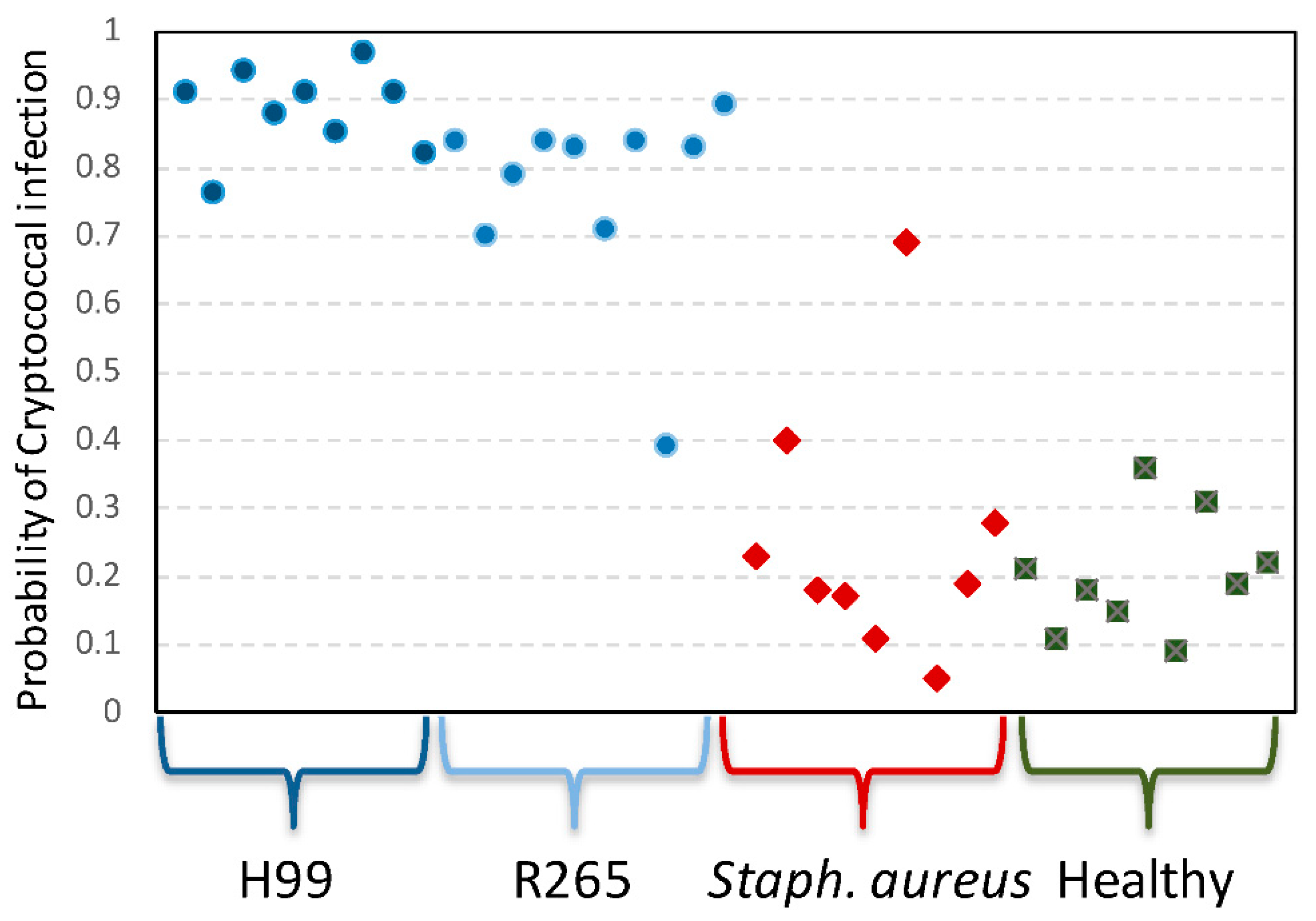
3.5. A Transcriptomic Classifier Accurately Identifies Cryptococcal Infection

In addition to elucidating the pathobiology of infectious processes, patterns of differentially expressed genes in the host can also be utilized to identify the presence of infection, and, potentially, to differentiate types of infectious agents. Based on differentially expressed genes in our experimental model, a binomial classifier that differentiated infected and healthy mice was produced. The classifier consisted of 28 probes representing 27 unique genes. Genes contained in the classifier included: Cmc2, Ear1, Il1rl1, Klk1, Slc16a3, Dedd2, Dnajb1, Ccl6, Camk1, Zfp704, Dffb, Prkg2, P2ry14, Prrx1, Klra19, Hspa8, Rab3c, Cwf19l1, Arhgap29, D6Ertd160e, Neu1, Rsrp1, Cxcr4, Retnla, Ccdc117, Arap2, and Mela (Supplemental Table S1). When applied to all samples from the experiment, the 27-gene binomial classifier had 100% accuracy in differentiating infected mice from healthy controls. We next examined the performance of this classifier in an existing microarray dataset from our prior work [29], and the cryptococcal classifier was also capable of differentiating cryptococcal infection from murine bacterial infection (with Staphylococcus aureus) with 94% sensitivity and 89% specificity (Figure 5).

3.6. Translation to Human Applications

To better understand how transcriptional responses identified in these data translate across species, the top genes differentially expressed in the murine response to Cryptococcus (as compared to controls) in this study were compared to the top differentially expressed genes seen when human PBMCs were subjected to yeast exposure in vitro (Candida albicans, Cryptococcus neoformans, and Cryptococcus gattii). When examining overlapping responses in the top differentially expressed genes, 73 genes were noted to be significantly expressed (p < 0.05) in both systems (Supplemental Table S2). Additionally, when specifically examining human orthologs of the 27 genes of the murine cryptococcal classifier, 6 of the 14 genes (43%) in which data were available were also significantly expressed in human cells upon exposure to these pathogens. These data suggest that the observed gene expression changes are not isolated to the murine species. This degree of species overlap is similar to our previously published Candida data [29,55], where an overlap of less than 50% of the differentially expressed genes between mice and humans still allowed for highly accurate performance of the classifier in human hosts.

4. Discussion

Using an intranasal inoculation model in mice, we have, for the first time, examined specifically the host response to C. neoformans and C. gattii infection as manifest through patterns of gene expression in circulating white blood cells. Conserved changes in host gene expression were evident in response to C. gattii and C. neoformans infection, although the magnitude of these conserved host responses was much greater in animals infected with C. neoformans. Furthermore, transcriptional analysis allows for description of unique differences between the biological responses elicited by these two species, and discriminatory models based on this differential expression can be used to develop novel classifiers that offer the potential to diagnose this challenging disease.
Alterations at the transcriptomic level reflect the pathophysiology of the way the host responds to these invading organisms. Early on in infection, elements of the complement cascade have been demonstrated to play a significant role in the host response to both C. gattii and C. neoformans [6,48,56,57]. This has even been demonstrated at the transcriptional level in other tissues and cell culture models, although not directly in circulating leukocytes [58,59,60]. This response was observed in our model as well, with genes involved in the classical, alternative, and lectin activation pathways up-regulated in response to both C. neoformans and C. gattii infection. However, all three complement pathways were much more significantly up-regulated in response to C. neoformans infection than to C. gattii infection, again reflecting the stronger response to C. neoformans in our experimental model. Previous work has demonstrated that mice deficient in C1q show overwhelming pulmonary fungal burden with C. gattii [48]. In our model, C1qa and C1qb were two of the top 10 most differentially up-regulated genes in response to C. gattii infection, providing additional evidence of their important role in the immune response to this fungal species.
Many of the most significantly up-regulated genes in response to Cryptococcus infection were either products of a TH2 immune response (Rnase2a, Retnla, Chil3) or negative regulators of a TH1 immune response (Serpinb2). Furthermore, there was evidence of alternatively activated M2 macrophage responses on the transcriptomic level, which are associated with a TH2-type immune response. When comparing the two infected cohorts, the host response to C. neoformans was significantly more skewed toward TH2-mediated immunity than the host response to C. gattii. Since a TH2-skewed immune response has been linked to greater fungal burden and extra-pulmonary dissemination [7,8,9,10], the stronger TH2 transcriptomic polarization seen in C. neoformans infection may partially explain the greater fungal burden found in the lungs, brain, and splenic tissue with this species. This may also explain some of the propensity of C. neoformans to infect HIV-positive patients, whose immune profiles tend to change from TH1 to being TH2-biased over time [61]. Additionally, mechanisms postulated to promote survival and extra-pulmonary dissemination of C. neoformans include eicosanoid production via arachidonic acid acquired from the host [62] and suppression of natural killer cell activity [51,52,53,54]. As described above, we discovered transcriptomic changes in the C. neoformans cohort that supported activation of both of these processes, linking their occurrence to fungal dissemination in the host.
C. gattii has a greater propensity for causing clinical disease in immunocompetent hosts. One suggested mechanism for this is the ability of C. gattii to suppress adaptive immune system recognition through suppression of antigen presentation on antigen-presenting cells and subsequent T lymphocyte proliferation [63]. We observed transcriptomic evidence of this mechanism of immune evasion occurring in the C. gattii cohort, as reflected by the down-regulation of genes in antigen processing and presentation pathways. Down-regulation of similar pathways were not observed in response to C. neoformans infection, raising the possibility that this absence of active immune evasion partially contributes to the more robust immune response to C. neoformans infection on the gene expression level.
In addition to providing insight into the pathophysiology and mechanistic responses to cryptococcal infection, analysis of differentially expressed genes in the infected state also shows promise for the development of pathogen class-specific diagnostics. By building a classifier composed of a small number of unique differentially expressed genes, infected and healthy mice could be differentiated with 100% accuracy. This same classifier was capable of accurately discriminating Cryptococcus-infected animals from animals with acute bacterial infection due to Staphylococcus aureus. Furthermore, we observed many of these same genes and pathways were activated (or suppressed) when human PBMCs were infected with the same species of Cryptococcus, which also overlaps with signals described during cryptococcal infection of human monocytes in vitro [64]. These preliminary findings are limited by the need to use heat-killed microbial strains for co-culture with the human PBMCs to prevent overt host cell toxicity by proliferating fungi and bacteria. However, despite these limitations, these studies suggest that findings from our murine studies may also be applicable to human disease [64]. As we have shown in prior work, sparse gene-based signatures such as these also lend themselves well to migration to more clinic-ready RT-PCR platforms, some of which are already commonly present in clinical microbiology laboratories [65].
Although peripheral blood gene expression analysis provides a wealth of information into the host response to infectious challenge, this approach has clear limitations. Peripheral blood immune effector cells are only one component of the broader immune response, and gene expression changes in sampled cells do not reflect the entirety of the host response. In terms of biological significance, genomic studies are a critical component of our armamentarium, but they are ultimately hypothesis-generating in scope, and future directed biological experiments are necessary to confirm preliminary conclusions about functional immune mechanisms. The experimental model utilized is limited by the collection of peripheral blood at only one time point, thereby precluding analysis of the transcriptomic response in later, more clinically severe infection. We chose the time point of 14 days after initial inoculation to allow for establishment of infection and dissemination within the host, while simultaneously avoiding clinical deterioration and gene expression changes associated with overwhelming illness and death, which tend to occur between three and four weeks in this infection model. While in our prior work early transcriptomic signatures offer even better diagnostic performance at the time of maximal clinical disease [25,66], we did not have the opportunity to definitively assess this in the current work. Future studies of how these types of responses are manifest in patients with clinical disease and under varying levels of immunosuppression will be critical to proving relevance of these data, and any cryptococcal disease classifier will require validation in other populations (clinical syndromic mimics, immunosuppressed hosts, etc.) in order to determine true clinical relevance.
We have demonstrated robust blood-based transcriptomic responses in the host during cryptococcal infection, including those which are conserved across species, as well as genomic pathways that differ in both character and magnitude between C. gattii and C. neoformans. This study highlights the ability of host peripheral blood gene expression analysis to elucidate the underlying biology of infection. Additionally, given the widening availability of clinic-ready RT-PCR based platforms capable of measuring gene expression in clinical samples, these findings lay the groundwork for development of diagnostic assays based on changes in host-derived biomarkers of cryptococcal infection.

Supplementary Materials

The following supporting information can be downloaded at: https://www.mdpi.com/article/10.3390/jof8050430/s1, Table S1: Genes of the Cryptococcus Classifier; Table S2: Genes significantly expressed (p < 0.05) in both PBMC (Candida and Cryptococcus) and Murine (Cryptococcus) Datasets.

Author Contributions

Conceptualization, M.T.M.; methodology, J.A.A., J.R.P., Z.E.H. and M.T.M.; data curation, M.B., J.L.T. and D.L.T.; formal analysis, Z.E.H.; validation, J.M.S.; writing—original draft preparation, Z.E.H. and M.T.M.; writing—review and editing, J.M.S. and M.T.M.; supervision, J.A.A., A.K.Z., J.R.P. and M.T.M. All authors have read and agreed to the published version of the manuscript.

Funding

This work was supported by funding from the Veteran’s Affairs Medical Center, number 1IK2CX000611 (M.T.M.), Public Service Grants AI 73896 and AI 93257 (J.R.P.) and AI 074677 (J.A.A.), and funds from the Eugene A. Stead Research Endowment at Duke University (Z.E.H.).

Institutional Review Board Statement

The study was conducted in accordance with the Declaration of Helsinki and approved by the Institutional Review Board (or Ethics Committee) of Duke University (Pro00022849). The animal experiments were approved by the Duke University Institutional Animal Care and Use Committee under an approved protocol number A102-20-05.

Data Availability Statement

Primary microarray datasets analyzed in this work will be uploaded to the National Center of Biotechnology Information’s Gene Expression Omnibus upon acceptance for publication.

Acknowledgments

The authors would like to thank David Corcoran and colleagues at the Duke Center for Genomic and Computational Biology for analytical assistance and direction.

Conflicts of Interest

The authors declare no conflict of interest. The funders had no role in the design of the study; in the collection, analyses, or interpretation of data; in the writing of the manuscript; or in the decision to publish the results.

References

  1. Desalermos, A.; Kourkoumpetis, T.K.; Mylonakis, E. Update on the epidemiology and management of cryptococcal meningitis. Expert Opin. Pharmacother. 2012, 13, 783–789. [Google Scholar] [CrossRef] [PubMed] [Green Version]
  2. Rajasingham, R.; Smith, R.M.; Park, B.J.; Jarvis, J.N.; Govender, N.P.; Chiller, T.M.; Denning, D.W.; Loyse, A.; Boulware, D.R. Global burden of disease of HIV-associated cryptococcal meningitis: An updated analysis. Lancet Infect. Dis. 2017, 17, 873–881. [Google Scholar] [CrossRef] [Green Version]
  3. Lewis, J.L.; Rabinovich, S. The wide spectrum of cryptococcal infections. Am. J. Med. 1972, 53, 315–322. [Google Scholar] [CrossRef]
  4. Galanis, E.; Macdougall, L.; Kidd, S.; Morshed, M. Epidemiology of cryptococcus gattii, British Columbia, Canada, 1999–2007. Emerg. Infect. Dis. 2010, 16, 251–257. [Google Scholar] [CrossRef]
  5. Gibson, J.F.; Johnston, S.A. Immunity to cryptococcus neoformans and C. gattii during cryptococcosis. Fungal Genet. Biol. FG B 2015, 78, 76–86. [Google Scholar] [CrossRef] [Green Version]
  6. Dromer, F.; Perronne, C.; Barge, J.; Vilde, J.L.; Yeni, P. Role of IgG and complement component C5 in the initial course of experimental cryptococcosis. Clin. Exp. Immunol. 1989, 78, 412–417. [Google Scholar]
  7. Zhang, Y.; Wang, F.; Tompkins, K.C.; McNamara, A.; Jain, A.V.; Moore, B.B.; Toews, G.B.; Huffnagle, G.B.; Olszewski, M.A. Robust Th1 and Th17 immunity supports pulmonary clearance but cannot prevent systemic dissemination of highly virulent cryptococcus neoformans H99. Am. J. Pathol. 2009, 175, 2489–2500. [Google Scholar] [CrossRef] [Green Version]
  8. Flaczyk, A.; Duerr, C.U.; Shourian, M.; Lafferty, E.I.; Fritz, J.H.; Qureshi, S.T. IL-33 signaling regulates innate and adaptive immunity to Cryptococcus neoformans. J. Immunol. 2013, 191, 2503–2513. [Google Scholar] [CrossRef] [Green Version]
  9. Muller, U.; Stenzel, W.; Kohler, G.; Werner, C.; Polte, T.; Hansen, G.; Schutze, N.; Straubinger, R.K.; Blessing, M.; McKenzie, A.N.; et al. IL-13 induces disease-promoting type 2 cytokines, alternatively activated macrophages and allergic inflammation during pulmonary infection of mice with cryptococcus neoformans. J. Immunol. 2007, 179, 5367–5377. [Google Scholar] [CrossRef] [Green Version]
  10. Jain, A.V.; Zhang, Y.; Fields, W.B.; McNamara, D.A.; Choe, M.Y.; Chen, G.H.; Erb-Downward, J.; Osterholzer, J.J.; Toews, G.B.; Huffnagle, G.B.; et al. Th2 but not Th1 immune bias results in altered lung functions in a murine model of pulmonary cryptococcus neoformans infection. Infect. Immun. 2009, 77, 5389–5399. [Google Scholar] [CrossRef] [Green Version]
  11. Wozniak, K.L.; Hardison, S.E.; Kolls, J.K.; Wormley, F.L. Role of IL-17A on resolution of pulmonary C. neoformans infection. PLoS ONE 2011, 6, e17204. [Google Scholar] [CrossRef] [PubMed] [Green Version]
  12. Jarvis, J.N.; Casazza, J.P.; Stone, H.H.; Meintjes, G.; Lawn, S.D.; Levitz, S.M.; Harrison, T.S.; Koup, R.A. The phenotype of the cryptococcus-specific CD4+ memory T-cell response is associated with disease severity and outcome in HIV-associated cryptococcal meningitis. J. Infect. Dis. 2013, 207, 1817–1828. [Google Scholar] [CrossRef] [PubMed]
  13. Voelz, K.; Lammas, D.A.; May, R.C. Cytokine signaling regulates the outcome of intracellular macrophage parasitism by cryptococcus neoformans. Infect. Immun. 2009, 77, 3450–3457. [Google Scholar] [CrossRef] [PubMed] [Green Version]
  14. Arora, S.; Olszewski, M.A.; Tsang, T.M.; McDonald, R.A.; Toews, G.B.; Huffnagle, G.B. Effect of cytokine interplay on macrophage polarization during chronic pulmonary infection with cryptococcus neoformans. Infect. Immun. 2011, 79, 1915–1926. [Google Scholar] [CrossRef] [Green Version]
  15. Hardison, S.E.; Ravi, S.; Wozniak, K.L.; Young, M.L.; Olszewski, M.A.; Wormley, F.L., Jr. Pulmonary infection with an interferon-gamma-producing cryptococcus neoformans strain results in classical macrophage activation and protection. Am. J. Pathol. 2010, 176, 774–785. [Google Scholar] [CrossRef] [PubMed]
  16. Islam, A.; Li, S.S.; Oykhman, P.; Timm-McCann, M.; Huston, S.M.; Stack, D.; Xiang, R.F.; Kelly, M.M.; Mody, C.H. An acidic microenvironment increases NK cell killing of cryptococcus neoformans and cryptococcus gattii by enhancing perforin degranulation. PLoS Pathog. 2013, 9, e1003439. [Google Scholar] [CrossRef]
  17. Kawakami, K.; Kinjo, Y.; Uezu, K.; Yara, S.; Miyagi, K.; Koguchi, Y.; Nakayama, T.; Taniguchi, M.; Saito, A. Monocyte chemoattractant protein-1-dependent increase of V alpha 14 NKT cells in lungs and their roles in Th1 response and host defense in cryptococcal infection. J. Immunol. 2001, 167, 6525–6532. [Google Scholar] [CrossRef]
  18. Kawakami, K.; Kinjo, Y.; Yara, S.; Koguchi, Y.; Uezu, K.; Nakayama, T.; Taniguchi, M.; Saito, A. Activation of Valpha14(+) natural killer T cells by alpha-galactosylceramide results in development of Th1 response and local host resistance in mice infected with cryptococcus neoformans. Infect. Immun. 2001, 69, 213–220. [Google Scholar] [CrossRef] [Green Version]
  19. Saha, D.C.; Xess, I.; Biswas, A.; Bhowmik, D.M.; Padma, M.V. Detection of cryptococcus by conventional, serological and molecular methods. J. Med. Microbiol. 2009, 58, 1098–1105. [Google Scholar] [CrossRef]
  20. Jarvis, J.N.; Percival, A.; Bauman, S.; Pelfrey, J.; Meintjes, G.; Williams, G.N.; Longley, N.; Harrison, T.S.; Kozel, T.R. Evaluation of a novel point-of-care cryptococcal antigen test on serum, plasma, and urine from patients with HIV-associated cryptococcal meningitis. Clin. Infect.Dis. 2011, 53, 1019–1023. [Google Scholar] [CrossRef]
  21. Boulware, D.R.; Rolfes, M.A.; Rajasingham, R.; von Hohenberg, M.; Qin, Z.; Taseera, K.; Schutz, C.; Kwizera, R.; Butler, E.K.; Meintjes, G.; et al. Multisite validation of cryptococcal antigen lateral flow assay and quantification by laser thermal contrast. Emerg. Infect. Dis. 2014, 20, 45–53. [Google Scholar] [CrossRef] [PubMed]
  22. Ramilo, O.; Allman, W.; Chung, W.; Mejias, A.; Ardura, M.; Glaser, C.; Wittkowski, K.M.; Piqueras, B.; Banchereau, J.; Palucka, A.K.; et al. Gene expression patterns in blood leukocytes discriminate patients with acute infections. Blood 2007, 109, 2066–2077. [Google Scholar] [CrossRef] [PubMed] [Green Version]
  23. Zaas, A.K.; Chen, M.; Varkey, J.; Veldman, T.; Hero, A.O., 3rd; Lucas, J.; Huang, Y.; Turner, R.; Gilbert, A.; Lambkin-Williams, R.; et al. Gene expression signatures diagnose influenza and other symptomatic respiratory viral infections in humans. Cell Host Microbe 2009, 6, 207–217. [Google Scholar] [CrossRef] [PubMed] [Green Version]
  24. Mejias, A.; Dimo, B.; Suarez, N.M.; Garcia, C.; Suarez-Arrabal, M.C.; Jartti, T.; Blankenship, D.; Jordan-Villegas, A.; Ardura, M.I.; Xu, Z.; et al. Whole blood gene expression profiles to assess pathogenesis and disease severity in infants with respiratory syncytial virus infection. PLoS Med. 2013, 10, e1001549. [Google Scholar] [CrossRef] [PubMed] [Green Version]
  25. Woods, C.W.; McClain, M.T.; Chen, M.; Zaas, A.K.; Nicholson, B.P.; Varkey, J.; Veldman, T.; Kingsmore, S.F.; Huang, Y.; Lambkin-Williams, R.; et al. A host transcriptional signature for presymptomatic detection of infection in humans exposed to influenza H1N1 or H3N2. PLoS ONE 2013, 8, e52198. [Google Scholar] [CrossRef] [Green Version]
  26. Berry, M.P.; Graham, C.M.; McNab, F.W.; Xu, Z.; Bloch, S.A.; Oni, T.; Wilkinson, K.A.; Banchereau, R.; Skinner, J.; Wilkinson, R.J.; et al. An interferon-inducible neutrophil-driven blood transcriptional signature in human tuberculosis. Nature 2010, 466, 973–977. [Google Scholar] [CrossRef] [Green Version]
  27. Zak, D.E.; Penn-Nicholson, A.; Scriba, T.J.; Thompson, E.; Suliman, S.; Amon, L.M.; Mahomed, H.; Erasmus, M.; Whatney, W.; Hussey, G.D.; et al. A blood RNA signature for tuberculosis disease risk: A prospective cohort study. Lancet 2016, 387, 2312–2322. [Google Scholar] [CrossRef] [Green Version]
  28. Holcomb, Z.E.; Tsalik, E.L.; Woods, C.W.; McClain, M.T. Host-based peripheral blood gene expression analysis for diagnosis of infectious diseases. J. Clin. Microbiol. 2017, 55, 360–368. [Google Scholar] [CrossRef] [Green Version]
  29. Zaas, A.K.; Aziz, H.; Lucas, J.; Perfect, J.R.; Ginsburg, G.S. Blood gene expression signatures predict invasive candidiasis. Sci. Transl. Med. 2010, 2, 21ra17. [Google Scholar] [CrossRef]
  30. Gautier, L.; Cope, L.; Bolstad, B.M.; Irizarry, R.A. Affy–analysis of Affymetrix GeneChip data at the probe level. Bioinformatics 2004, 20, 307–315. [Google Scholar] [CrossRef]
  31. Gentleman, R.C.; Carey, V.J.; Bates, D.M.; Bolstad, B.; Dettling, M.; Dudoit, S.; Ellis, B.; Gautier, L.; Ge, Y.; Gentry, J.; et al. Bioconductor: Open software development for computational biology and bioinformatics. Genome Biol. 2004, 5, R80. [Google Scholar] [CrossRef] [PubMed] [Green Version]
  32. R Development Core Team. R: A Language and Environment for Statistical Computing; R Foundation for Statistical Computing: Vienna, Austria, 2016; Available online: http://www.R-project.org (accessed on 5 April 2022).
  33. Ritchie, M.E.; Phipson, B.; Wu, D.; Hu, Y.; Law, C.W.; Shi, W.; Smyth, G.K. Limma powers differential expression analyses for RNA-sequencing and microarray studies. Nucleic Acids Res. 2015, 43, e47. [Google Scholar] [CrossRef] [PubMed]
  34. Kramer, A.; Green, J.; Pollard, J., Jr.; Tugendreich, S. Causal analysis approaches in ingenuity pathway analysis. Bioinformatics 2014, 30, 523–530. [Google Scholar] [CrossRef] [PubMed]
  35. Huang, D.W.; Sherman, B.T.; Lempicki, R.A. Systematic and integrative analysis of large gene lists using DAVID bioinformatics resources. Nat. Protoc. 2009, 4, 44–57. [Google Scholar] [CrossRef]
  36. Huang, D.W.; Sherman, B.T.; Lempicki, R.A. Bioinformatics enrichment tools: Paths toward the comprehensive functional analysis of large gene lists. Nucleic Acids Res. 2009, 37, 1–13. [Google Scholar] [CrossRef] [Green Version]
  37. Friedman, J.; Hastie, T.; Tibshirani, R. Regularization paths for generalized linear models via coordinate descent. J. Stat. Softw. 2010, 33, 1–22. [Google Scholar] [CrossRef] [Green Version]
  38. Ngamskulrungroj, P.; Chang, Y.; Sionov, E.; Kwon-Chung, K.J. The primary target organ of cryptococcus gattii is different from that of cryptococcus neoformans in a murine model. mBio 2012, 3, e00103-12. [Google Scholar] [CrossRef] [Green Version]
  39. Cormier, S.A.; Yuan, S.; Crosby, J.R.; Protheroe, C.A.; Dimina, D.M.; Hines, E.M.; Lee, N.A.; Lee, J.J. T(H)2-mediated pulmonary inflammation leads to the differential expression of ribonuclease genes by alveolar macrophages. Am. J. Respir. Cell Mol. Biol. 2002, 27, 678–687. [Google Scholar] [CrossRef] [Green Version]
  40. Ivanov, I.; Kuhn, H.; Heydeck, D. Structural and functional biology of arachidonic acid 15-lipoxygenase-1 (ALOX15). Gene 2015, 573, 1–32. [Google Scholar] [CrossRef]
  41. Nair, M.G.; Cochrane, D.W.; Allen, J.E. Macrophages in chronic type 2 inflammation have a novel phenotype characterized by the abundant expression of Ym1 and Fizz1 that can be partly replicated in vitro. Immunol. Lett. 2003, 85, 173–180. [Google Scholar] [CrossRef]
  42. Nair, M.G.; Du, Y.; Perrigoue, J.G.; Zaph, C.; Taylor, J.J.; Goldschmidt, M.; Swain, G.P.; Yancopoulos, G.D.; Valenzuela, D.M.; Murphy, A.; et al. Alternatively activated macrophage-derived RELM-{alpha} is a negative regulator of type 2 inflammation in the lung. J. Exp. Med. 2009, 206, 937–952. [Google Scholar] [CrossRef] [PubMed]
  43. Novince, C.M.; Koh, A.J.; Michalski, M.N.; Marchesan, J.T.; Wang, J.; Jung, Y.; Berry, J.E.; Eber, M.R.; Rosol, T.J.; Taichman, R.S.; et al. Proteoglycan 4, a novel immunomodulatory factor, regulates parathyroid hormone actions on hematopoietic cells. Am. J. Pathol. 2011, 179, 2431–2442. [Google Scholar] [CrossRef] [PubMed]
  44. O’Reilly, M.A.; Yee, M.; Buczynski, B.W.; Vitiello, P.F.; Keng, P.C.; Welle, S.L.; Finkelstein, J.N.; Dean, D.A.; Lawrence, B.P. Neonatal oxygen increases sensitivity to influenza A virus infection in adult mice by suppressing epithelial expression of Ear1. Am. J. Pathol. 2012, 181, 441–451. [Google Scholar] [CrossRef] [PubMed] [Green Version]
  45. Schroder, W.A.; Le, T.T.; Major, L.; Street, S.; Gardner, J.; Lambley, E.; Markey, K.; MacDonald, K.P.; Fish, R.J.; Thomas, R.; et al. A physiological function of inflammation-associated SerpinB2 is regulation of adaptive immunity. J. Immunol. 2010, 184, 2663–2670. [Google Scholar] [CrossRef] [Green Version]
  46. Yamada, K.J.; Barker, T.; Dyer, K.D.; Rice, T.A.; Percopo, C.M.; Garcia-Crespo, K.E.; Cho, S.; Lee, J.J.; Druey, K.M.; Rosenberg, H.F. Eosinophil-associated ribonuclease 11 is a macrophage chemoattractant. J. Biol. Chem. 2015, 290, 8863–8875. [Google Scholar] [CrossRef] [Green Version]
  47. Diamond, R.D.; May, J.E.; Kane, M.A.; Frank, M.M.; Bennett, J.E. The role of the classical and alternate complement pathways in host defenses against cryptococcus neoformans infection. J. Immunol. 1974, 112, 2260–2270. [Google Scholar]
  48. Mershon, K.L.; Vasuthasawat, A.; Lawson, G.W.; Morrison, S.L.; Beenhouwer, D.O. Role of complement in protection against cryptococcus gattii infection. Infect. Immun. 2009, 77, 1061–1070. [Google Scholar] [CrossRef] [Green Version]
  49. Mills, C.D. M1 and M2 macrophages: Oracles of health and disease. Crit. Rev. Immunol. 2012, 32, 463–488. [Google Scholar] [CrossRef] [Green Version]
  50. Jablonski, K.A.; Amici, S.A.; Webb, L.M.; Ruiz-Rosado Jde, D.; Popovich, P.G.; Partida-Sanchez, S.; Guerau-de-Arellano, M. Novel markers to delineate murine M1 and M2 macrophages. PLoS ONE 2015, 10, e0145342. [Google Scholar] [CrossRef] [Green Version]
  51. Hidore, M.R.; Nabavi, N.; Sonleitner, F.; Murphy, J.W. Murine natural killer cells are fungicidal to cryptococcus neoformans. Infect. Immun. 1991, 59, 1747–1754. [Google Scholar] [CrossRef] [Green Version]
  52. Ma, L.L.; Wang, C.L.; Neely, G.G.; Epelman, S.; Krensky, A.M.; Mody, C.H. NK cells use perforin rather than granulysin for anticryptococcal activity. J. Immunol. 2004, 173, 3357–3365. [Google Scholar] [CrossRef] [PubMed] [Green Version]
  53. Wiseman, J.C.; Ma, L.L.; Marr, K.J.; Jones, G.J.; Mody, C.H. Perforin-dependent cryptococcal microbicidal activity in NK cells requires PI3K-dependent ERK1/2 signaling. J. Immunol. 2007, 178, 6456–6464. [Google Scholar] [CrossRef] [PubMed] [Green Version]
  54. Marr, K.J.; Jones, G.J.; Zheng, C.; Huston, S.M.; Timm-McCann, M.; Islam, A.; Berenger, B.M.; Ma, L.L.; Wiseman, J.C.; Mody, C.H. Cryptococcus neoformans directly stimulates perforin production and rearms NK cells for enhanced anticryptococcal microbicidal activity. Infect. Immun. 2009, 77, 2436–2446. [Google Scholar] [CrossRef] [PubMed] [Green Version]
  55. Steinbrink, J.M.; Myers, R.A.; Hua, K.; Johnson, M.D.; Seidelman, J.L.; Tsalik, E.L.; Henao, R.; Ginsburg, G.S.; Woods, C.W.; Alexander, B.D.; et al. The host transcriptional response to Candidemia is dominated by neutrophil activation and heme biosynthesis and supports novel diagnostic approaches. Genome Med. 2021, 13, 108. [Google Scholar] [CrossRef] [PubMed]
  56. Diamond, R.D.; May, J.E.; Kane, M.; Frank, M.M.; Bennett, J.E. The role of late complement components and the alternate complement pathway in experimental cryptococcosis. Proc. Soc. Exp. Biol. Med. 1973, 144, 312–315. [Google Scholar] [CrossRef]
  57. Shapiro, S.; Beenhouwer, D.O.; Feldmesser, M.; Taborda, C.; Carroll, M.C.; Casadevall, A.; Scharff, M.D. Immunoglobulin G monoclonal antibodies to cryptococcus neoformans protect mice deficient in complement component C3. Infect. Immun. 2002, 70, 2598–2604. [Google Scholar] [CrossRef] [Green Version]
  58. Li, H.; Li, Y.; Sun, T.; Du, W.; Li, C.; Suo, C.; Meng, Y.; Liang, Q.; Lan, T.; Zhong, M.; et al. Unveil the transcriptional landscape at the cryptococcus-host axis in mice and nonhuman primates. PLoS Negl. Trop. Dis. 2019, 13, e0007566. [Google Scholar] [CrossRef] [Green Version]
  59. Farrer, R.A.; Ford, C.B.; Rhodes, J.; Delorey, T.; May, R.C.; Fisher, M.C.; Cloutman-Green, E.; Balloux, F.; Cuomo, C.A. Transcriptional heterogeneity of cryptococcus gattii VGII compared with non-VGII Lineages underpins key pathogenicity pathways. mSphere 2018, 3, e00445-18. [Google Scholar] [CrossRef] [Green Version]
  60. Movahed, E.; Munusamy, K.; Tan, G.M.; Looi, C.Y.; Tay, S.T.; Wong, W.F. Genome-wide transcription study of cryptococcus neoformans H99 clinical strain versus environmental strains. PLoS ONE 2015, 10, e0137457. [Google Scholar] [CrossRef]
  61. Altfeld, M.; Addo, M.M.; Kreuzer, K.A.; Rockstroh, J.K.; Dumoulin, F.L.; Schliefer, K.; Leifeld, L.; Sauerbruch, T.; Spengler, U. T(H)1 to T(H)2 shift of cytokines in peripheral blood of HIV-infected patients is detectable by reverse transcriptase polymerase chain reaction but not by enzyme-linked immunosorbent assay under nonstimulated conditions. J. Acquir. Immune Defic. Syndr. 2000, 23, 287–294. [Google Scholar] [CrossRef]
  62. Wright, L.C.; Santangelo, R.M.; Ganendren, R.; Payne, J.; Djordjevic, J.T.; Sorrell, T.C. Cryptococcal lipid metabolism: Phospholipase B1 is implicated in transcellular metabolism of macrophage-derived lipids. Eukaryot. Cell 2007, 6, 37–47. [Google Scholar] [CrossRef] [PubMed] [Green Version]
  63. Huston, S.M.; Li, S.S.; Stack, D.; Timm-McCann, M.; Jones, G.J.; Islam, A.; Berenger, B.M.; Xiang, R.F.; Colarusso, P.; Mody, C.H. Cryptococcus gattii is killed by dendritic cells, but evades adaptive immunity by failing to induce dendritic cell maturation. J. Immunol. 2013, 191, 249–261. [Google Scholar] [CrossRef] [PubMed] [Green Version]
  64. Chen, S.; Yan, H.; Zhang, L.; Kong, W.; Sun, Y.; Zhang, W.; Chen, Y.; Deng, A. Cryptococcus neoformans infection and immune cell regulation in human monocytes. Cell. Physiol. Biochem. 2015, 37, 537–547. [Google Scholar] [CrossRef] [PubMed]
  65. Tsalik, E.L.; Henao, R.; Montgomery, J.L.; Nawrocki, J.W.; Aydin, M.; Lydon, E.C.; Ko, E.R.; Petzold, E.; Nicholson, B.P.; Cairns, C.B.; et al. Discriminating bacterial and viral infection using a rapid host gene expression test. Crit. Care Med. 2021, 49, 1651–1663. [Google Scholar] [CrossRef] [PubMed]
  66. McClain, M.T.; Nicholson, B.P.; Park, L.P.; Liu, T.Y.; Hero, A.O., 3rd; Tsalik, E.L.; Zaas, A.K.; Veldman, T.; Hudson, L.L.; Lambkin-Williams, R.; et al. A genomic signature of influenza infection shows potential for presymptomatic detection, guiding early therapy, and monitoring clinical responses. Open Forum Infect. Dis. 2016, 3, ofw007. [Google Scholar] [CrossRef]
Figure 1. Fungal tissue burden in lungs, brain, and spleen. Fungal burden in lung, brain, and spleen tissues was measured for the two cohorts of infected mice at the time of sacrifice 14 days after inoculation.
Jof 08 00430 g001
Figure 2. Variation in circulating white blood cells. Mean levels of each cell type for C. neoformans (H99), C gattii (R265), and controls in peripheral blood samples taken at day 14 post-inoculation. Error bars indicate standard deviation. p values denote significant differences between groups.
Jof 08 00430 g002
Figure 3. Heat map of genes differentially expressed between infected and healthy mice. Solid red indicates a Z-score of 2.0 or greater, while solid blue indicates a Z-score of −2.0 or lower. Gene names are listed on the left, while clustering is indicated at the top of the figure.
Jof 08 00430 g003
Figure 4. Alteration in expression of genes involved in the classical, alternative, and lectin complement activation pathways. Relative fold change for each gene due to infection with C. gattii R265 and C. neoformans H99 in comparison to healthy control mice. Genes listed are from BIOCARTA-defined pathways, with solid red representing 2-fold up-regulation and solid blue representing 2-fold down-regulation. Genes with no fold change listed did not have probes on the microarray panel.
Jof 08 00430 g004
Figure 5. Performance of a 27-gene transcriptomic signature of cryptococcal infection. Depicted are calculated probabilities of cryptococcal infection for individual mice infected with C. neoformans (“H99”), C gattii (“R265”), Staphylococcus aureus, or uninfected control animals (“Healthy”).
Jof 08 00430 g005
Table 1. Differences in peripheral white blood cell counts.
Leukocyte SubtypeH99R265Controlsp-Values (** = Statistically Significant)
Neutrophils10.5%11.3%19.2%H99 vs. R265 (p = 0.8786)
H99 vs. Controls (p = 0.0055) **
R265 vs. Controls (p = 0.0014) **
Lymphocytes67.4%65.1%75.5%H99 vs. R265 (p = 0.2552)
H99 vs. Controls (p = 0.0636)
R265 vs. Controls (p = 0.0025) **
Atypical Lymphocytes10.1%15.4%1.0%H99 vs. R265 (p = 0.1033)
H99 vs. Controls (p = 0.0119) **
R265 vs. Controls (p = 0.0001) **
Monocytes3.7%1.1%2.4%H99 v. R265 (p = 0.0296) **
H99 v. Control (p = 0.0116) **
R265 v. Control (p = 0.0004) **
Eosinophils8.3%7.1%1.9%H99 vs. R265 (p = 0.3803)
H99 vs. Controls (p = 0.0003) **
R265 vs. Controls (p = 0.0004) **
Table 2. Top 10 up-regulated genes in mice infected with Cryptococcus compared to controls.
GeneFold-Change (Infected vs. Control)Function
Rnase2a+14.15Produced in response to TH2 cytokine stimulation and serves as a macrophage chemoattractant [39,46].
Retnla+6.85Produced by macrophages in response to IL-4 and TH2-mediated inflammation [41]. Inhibits TH2-mediated immunity and inflammation [42].
Serpinb2+5.77Expressed by myeloid antigen-presenting cells and plays a role in suppression of the TH1 immune response [45].
C1qb+5.37Component of the classical pathway of complement activation.
Chil3+5.35Produced by macrophages in response to IL-4 and TH2-mediated inflammation [41].
Alox15+5.26Involved in arachidonic acid metabolism [40]
C1qa+4.76Component of the classical pathway of complement activation.
Prg4+4.65Produces a glycoprotein for articular joint protection and PTH-responsive hematopoiesis and megakaryopoiesis [43].
C1qc+4.48Component of the classical pathway of complement activation.
Ear1+4.33Produces a host defense protein secreted by eosinophils, macrophages, and neutrophils [44].
Table 3. The 10 most significant biological process GO terms that correspond to the 40 genes up-regulated in cryptococcal infection.
GO TermBonferroni-Corrected p-Value
Immune response (GO:0006955)0.00002
Acute inflammatory response (GO:0002526)0.00389
Complement activation (GO:0006956)0.01135
Activation of plasma proteins involved in acute inflammatory response (GO:0002541)0.01135
Innate immune response (GO:0045087)0.01166
Immune effector process (GO:0002252)0.02201
Defense response (GO:0006952)0.03399
Humoral immune response (GO:0006959)0.03808
Immunoglobulin mediated immune response (GO:0016064)0.05967
B cell mediated immunity (GO:0019724)0.06529
Publisher’s Note: MDPI stays neutral with regard to jurisdictional claims in published maps and institutional affiliations.

