Article

MaAts, an Alkylsulfatase, Contributes to Fungal Tolerances against UV-B Irradiation and Heat-Shock in Metarhizium acridum

1
Genetic Engineering Research Center, School of Life Sciences, Chongqing University, Chongqing 401331, China
2
Chongqing Engineering Research Center for Fungal Insecticide, Chongqing 401331, China
3
Key Laboratory of Gene Function and Regulation Technologies under Chongqing Municipal Education Commission, Chongqing 401331, China
*
Authors to whom correspondence should be addressed.
These authors contributed equally to this work.
J. Fungi 2022, 8(3), 270; https://doi.org/10.3390/jof8030270
Submission received: 11 February 2022 / Revised: 25 February 2022 / Accepted: 1 March 2022 / Published: 8 March 2022

Abstract

:
Sulfatases are commonly divided into three classes: type I, type II, and type III sulfatases. The type III sulfatase, alkylsulfatase, could hydrolyze the primary alkyl sulfates, such as sodium dodecyl sulfate (SDS) and sodium octyl sulfate. Thus, it has the potential application of SDS biodegradation. However, the roles of alkylsulfatase in biological control fungus remain unclear. In this study, an alkylsulfatase gene MaAts was identified from Metarhizium acridum. The deletion strain (ΔMaAts) and the complemented strain (CP) were constructed to reveal their functions in M. acridum. The activity of alkylsulfatase in ΔMaAts was dramatically reduced compared to the wild-type (WT) strain. The loss of MaAts delayed conidial germination, conidiation, and significantly declined the fungal tolerances to UV-B irradiation and heat-shock, while the fungal conidial yield and virulence were unaffected in M. acridum. The transcription levels of stress resistance-related genes were significantly changed after MaAts inactivation. Furthermore, digital gene expression profiling showed that 512 differential expression genes (DEGs), including 177 up-regulated genes and 335 down-regulated genes in ΔMaAts, were identified. Of these DEGs, some genes were involved in melanin synthesis, cell wall integrity, and tolerances to various stresses. These results indicate that MaAts and the DEGs involved in fungal stress tolerances may be candidate genes to be adopted to improve the stress tolerances of mycopesticides.

1. Introduction

The use of biological pesticides to protect plants is becoming increasingly widespread in various agricultural crops [1]. Entomopathogenic fungi have shown a great potential to reduce the application of chemical pesticides due to their advantages, such as low possibility to induce insect resistance, environmental friendliness, and safety [1,2,3,4]. To date, about 80 companies worldwide have developed more than 170 kinds of pesticide products based on entomopathogenic fungi [5]. For instance, entomopathogenic fungi Metarhizium spp. have been successfully applied to control locust [6,7,8], fruit fly [9], and grasshoppers [10]. The available genome sequences have made the Metarhizium genera become one of the important model fungi for exploring some questions about insect fungal conidiation, stress tolerances, and pathogenesis [11].
Conidia are the asexual propagules in many entomopathogenic fungi and usually the infective unit of mycopesticides [12,13]. Thus, the conidial pathogenicity, conidial yield, as well as sensitivities of conidia to various adverse conditions are all critical for the production cost and the application efficiency of the mycopesticides [14,15]. Among the adverse factors, the high-temperature and the ultraviolet (UV) irradiation from sunlight can lead to the decrease of the conidial vitality and are closely related to the application efficiency of the mycopesticides [16,17,18,19]. The UV irradiation usually results in DNA mutation and damage, as well as protein denaturation, and it also damages a variety of intracellular substances [17,20,21]. In addition, CPDs, 6-4PPs, and its isomer transformed from DNA can cause cell mutation and even death by bending DNA double helix to make different angles [22,23,24]. High temperatures can cause protein denaturation and DNA damage through base loss, leading to depurination and membrane disorganization [25,26,27,28]. However, 20 to 30 °C is a suitable temperature for conidial germination, growth, and the conidiation of most entomopathogenic fungi [29]. Therefore, exploring the mechanisms of the conidial tolerances to UV irradiation and heat-shock will be helpful to improve the quality of conidia, which is important for the effectiveness and sustainability of the mycopesticides in pest control.
As a widespread substance in nature, sulfate esters can be used by many microorganisms as a sulfur source for growth by hydrolyzing the sulfate esters using sulfatases [30]. At present, three classes of sulfatases, including type I, type II, and type III sulfatases, have been identified [31]. Type I sulfatases can utilize one equivalent of water to cleave the RO–SO3 in the process, which is the largest group among three classes of sulfatases [31]. Type I sulfatases contain a highly conserved sulfatase motif, C/S-X-P-X-A-XXXX-T-G [32], in the N-terminal region and a unique active-site aldehyde residue (the left C/S), α-formylglycine (FGly), which is installed post-translationally [33]. So far, fifteen of the type I sulfatases have been identified in humans [33]. The research found it related to the disease and the regulation of embryonic development [33,34]. Type II sulfatases release inorganic sulfates and the aldehyde by cleave sulfate esters and require α-ketoglutarate as a cosubstrate in this process [35,36,37]. Type III sulfatases contain a metallo-β-lactamases catalytic domain in the N-terminal, the C-terminal, and the central domain related to recruit substrates and the resistance to SDS [35]. The type III sulfatases usually activate a nucleophilic water molecule by a Zn2+ cofactor [31,38]. Mutations of residues Tyr246 and Gly263 of SdsAP, a type III sulfatase in Pseudomonas sp. S9, show that the mutants abolish the enzyme activity for SDS degradation, indicating that these residues are important for the functions of type III sulfatases [39].
To date, type III sulfatases were found and identified mainly in microorganisms, with a focus on the identification of the crystal structure [39] and the activities in degrading a surfactant [40]. In Pseudomonas sp. ATCC19151, alkylsulfatase could cleave alkyl-sulfate, such as SDS, and was named as SdsA accordingly [41]. In Pseudomonas sp. C12B, it could secrete up to five different alkylsulfatases, and these exert hydrolysis of many kinds of alkyl-sulfate substrate [42]. Alkylsulfatase from P. aeruginosa PAO1 has a wide substrate specificity; it could not only degrade long chain alkyl-sulfate but also short chain ones, such as decyl-sulfate, octyl-sulfate, and hexyl-sulfate [35]. The disruption of the alkylsulfatase gene causes the inactivation of that enzyme, rendering it unable to degrade SDS [35]. Beyond that, some bacteria isolated from wastewater showed the capacity to biodegrade alkylsulfatase [43]. In eukaryon, research related to alkylsulfatase is limited. Alkylsulfatases from Saccharomyces cerevisiae surprisingly degrade certain ary-sulfates, and this strain could utilize SDS regarded as a sulfur source to grow [44,45,46]. However, the functions of the type III sulfatases in filamentous fungi are still a mystery.
In this study, we characterized a type III sulfatase gene MaAts and presented the functional analysis of alkylsulfatase in M. acridum. We have found, unexpectedly, that the loss of MaAts delayed conidial germination and conidiation, but it had no effect on conidial yield and fungal virulence. In addition, the fungal tolerances to UV-B irradiation and heat-shock were significantly decreased after the deletion of MaAts. Furthermore, digital gene expression (DGE) profiling results showed the disruption of MaAts affected the expression levels of some genes related to melanin synthesis, cell wall integrity, and tolerances to various stresses.

2. Materials and Methods

2.1. Strains and Cultivation

M. acridum CQMa102 (wild type) has been deposited in the Genetic Engineering Center of Chongqing University. Nutrient-rich one-quarter SDAY medium was used for fungal cultivation. It contains 2.5 g peptone, 5 g yeast extract, 10 g dextrose, and 18 g agar per liter. Nutrient-poor Czapek-Dox medium was used to screen fungal transformants, and it contains 2 g NaNO3, 0.01 g FeSO4·7H2O, 0.5 g MgSO4·7H2O, 1 g KH2PO4, 1 g KCl, 30 g sucrose, and 15 g agar, per liter. Escherichia coli DH5α was prepared to propagate plasmid and grown on LB medium, which contained 5 g yeast extract, 10 g tryptone, 10 g NaCl, and 18 g agar per liter. Agrobacterium tumefaciens strain AGL-1 was adopted in fungal transformation [47].

2.2. Construction of MaAts Mutants

We used pK2-PB with phosphinothricin resistance gene and pK2-sur chlorimuron ethyl resistance gene to construct knockout and complementation strains, respectively [48]. For the deletion of MaAts, the upstream and downstream sequences of the MaAts gene were amplified with primer pairs MaAts-LF/MaAts-LR and MaAts-RF/MaAts-RR. The confirmed fragments were ligated to the pK2-PB vector to build pK2-PB-MaAts-LR (the targeted gene disruption vector). Then, the resulting vector was transformed into WT by A. tumefaciens mediated transformation. Nutrient-poor Czapek-Dox medium with phosphinothricin (500 μg/mL) was prepared to select fungal transformants. For the complementation of MaAts, the full length of MaAts (containing promoter 2.0-kb) was cloned with primer pair MaAts-HF/MaAts-HR. The fragment was ligated into pK2-sur vector to generate pK2-sur-MaAts. Then, it was transformed into ΔMaAts by A. tumefaciens mediated transformation. Nutrient-poor Czapek-Dox medium with chlorimuron ethyl (20 μg/mL) (Sigma-Aldrich, Bellefonte, PA, USA) was prepared to screen fungal transformants. All transformants were verified by PCR and Southern blotting. Primers used in this study are shown in Table S1.

2.3. Southern Blotting

About 5 μg of genomic DNA from various fungal strains was digested with Nru I and Xho I, respectively. The digested fragments were separated, then transferred into a nylon membrane. The probe was prepared by cloning 500 bp fragment from genomic sequence of MaAts with primers MaAts-PF/MaAts-PR (Table S1). DIG High Prime DNA Labeling, and Detection Starter Kit I (Roche, Mannheim, Germany) was used to label the probe.

2.4. Assessments of Conidial Germination and Conidiation Capacity

The conidial germination assays were conducted as described previously [49]. For the conidial yield assays, aliquots of 2 μL 107 conidia suspensions of each strain were vertically spotted into 24-well plates supplied with 1 mL 1/4 SDAY medium per well, which were incubated at 28 °C for 3, 5, 7, 9, 11, 13, and 15 days, respectively. The conidia in each well were collected and then suspended in ddH2O containing 0.05% Tween-80. The total number of conidia in each well was counted by a haemocytometer. All experiments were repeated three times.

2.5. Assessments for Fungal Stress Tolerances

The assessments for fungal stress tolerances were performed as described previously [50]. For the fungal tolerances to UV-B irradiation, 50 μL conidial suspensions (1 × 10⁷ conidia/mL) of each strain were spread on nutrient-rich 1/4 SDAY plates, which were treated by 1350 mW/m2 UV-B irradiation for 1.5, 3.0, 4.5, and 6.0 h, respectively, then cultivated in darkness at 28 °C for 20 h. For the fungal tolerances to heat-shock, sterile 1.5-mL centrifuge tubes containing 100-μL aliquot of conidial suspensions (1 × 10⁷ conidia/mL) of different fungal strains were exposed in a water bath at 45 °C for 3.0, 6.0, 9.0, and 12.0 h. After treatment, 50 μL conidial suspensions of WT, ΔMaAts, and CP strains were spread onto 1/4 SDAY plates, then incubated at 28 °C for 20 h. The conidial germination rate of each strain was measured by microscopic examination and the time for 50% inhibition time (IT50) was determined.

2.6. Bioassays

The bioassays were conducted by topical inoculation against Locusta migratoria manilensis (5th-instar nymphs) according to the method described previously [51]. In brief, aliquots of 3 μL 1 × 10⁷ conidia/mL conidial suspension of WT, ΔMaAts, and CP strains, and 3 μL paraffin oil were dipped on the locust pronotums. After the treatments, the locusts were fed on fresh corn leaves daily and kept at 28 ± 2 °C with 75% relative humidity and a 16:8 h (light–dark) photoperiod. The survival was recorded every 12 h until all locusts died. The experiment was carried out with three replicates with 30 locusts in each group, and the experiment was repeated three times. The mean median lethal time (LT50) was estimated for the WT, ΔMaAts, and CP strains.

2.7. Quantitative Reverse Transcription (qRT) PCR

We spread 100 μL conidial suspensions (1 × 10⁸ conidia/mL) on nutrient-rich 1/4 SDAY medium and collected the fungal cultures after 3-d cultivation at 28 °C. The total RNA of fungal samples were extracted using RNA Kit (CoWin Biosciences, Beijing, China). PrimeScriptTM RT reagent kit (Takara, Dalian, China) was used for the reverse transcription to generate cDNA with oligo-dT primer. The SYBR-Green PCR Master Mix kit (Bio-Rad, Foster City, CA, USA) was applied for qRT-PCR in the iCycler system (Bio-Rad, Hercules, CA, USA). The Magpd (GenBank accession No. XM_007817733.1) gene was used as the internal standard. Using the 2−ΔΔCT method, we assessed the transcript levels of target genes [52]. The experiment was replicated three times. All the primers for qRT-PCR are presented in Table S1.

2.8. Alkylsulfatase Activity Assays

The conidia of each strain were harvested from nutrient-rich 1/4 SDAY medium after 3 d of growth at 28 °C and washed with ddH2O. The conidia were quick-frozen in liquid nitrogen and fully grinded (70 Hz for 3 min) using Tissue lyser-24 (Jingxin, Shanghai, China). Then, we transferred 100 mg conidial powder of each strain in a new 2 mL centrifuge tube. We added 1 mL of the extraction buffer (50 mM Tris-HCl buffer, 1 mM DTT, 1 mM EDTA, pH 7.5) and mixed well; we followed that with centrifugation (16,000× g for 10 min) to obtain the enzyme stock solution. The alkylsulfatase activities in different fungal strains were measured by the barium chloride-gelatin method [30]. In brief, 200 μL of enzyme stock solution and 200 μL substrate (sodium octyl sulfate) were mixed in reaction buffer (0.1 M Tris-HCl, pH 7.5). The final concentration of the substrate was 15 mM. The reaction was carried out at 30 °C for 10 min and stopped by adding trichloroacetic acid (50 μL of 15%, wt/vol). After centrifugation (800× g for 1 min), aliquots of 200 μL supernatant were added into the barium chloride-gelatin and mixed thoroughly. The blank control was 200 μL of the extraction buffer instead of 200 μL of the enzyme stock solution. The resultant mixtures were detected by measuring maximum absorbance at 360 nm. One unit of the alkylsulfatase activity was defined as ΔA360 = 0.001 after 10 min.

2.9. DGE Profiling

The raw data of DGE profiling based on biological triplicates have been deposited in the NCBI BioProject database (accession number: PRJNA753293). The RNA sequencing was conducted on the BGISEQ-500 platform by Beijing Genomics Institution (Wuhan, China). The differentially expressed genes (DEG) were identified with a fold change ≥ 2 and q value < 0.05. The DEG annotation was based on the NCBI protein databases and subjected to gene ontology (GO) analysis (http://www.geneontology.org/, accessed on 10 January 2022) for the enrichments of GO terms to three function classes (p < 0.05).

2.10. Data Analysis

All data obtained from the repeated experiments in each test were expressed as means ± standard deviations. The one-factor analysis of variance was adopted to analyze the experimental data based on three replicates. SPSS 17.0 (IBM, Armonk, NY, USA) was used to analyze the datasets in this study.

3. Results

3.1. MaAts Belongs to the Type III Sulfatase

In M. acridum, a type III sulfatase gene, MaAts (MAC_08440) was cloned. The MaAts comprised an ORF of 1890 bp encoding a protein consisting of 629-amino acid with predicted isoelectric point of 5.45 and molecular mass of 68.6 kD. This protein has no signal peptide and no transmembrane domain. The MaAts contains a β-lactamase domain at the N terminus, followed by an alkylsulfatase dimerization, and an alkylsulfatase C terminal region (Figure 1A). The phylogenetic analysis showed that MaAts belongs to the type III of sulfatase (Figure 1A). To examine the activity of the MaAts in M. acridum, we successfully generated the MaAts-disruption mutants as well as the complementation strains (Figure S1). The alkylsulfatase activity assays showed that the samples from the WT and CP strains exhibited significantly higher alkylsulfatase activities than those from ΔMaAts strain, and the alkylsulfatase activities were hardly detected in all the samples (WT, ΔMaAts, CP) with heat-shock treatment (Figure 1B).

3.2. Disruption of MaAts Delays Conidial Germination and Conidiation but Does Not Affect Fungal Virulence and Conidial Yield

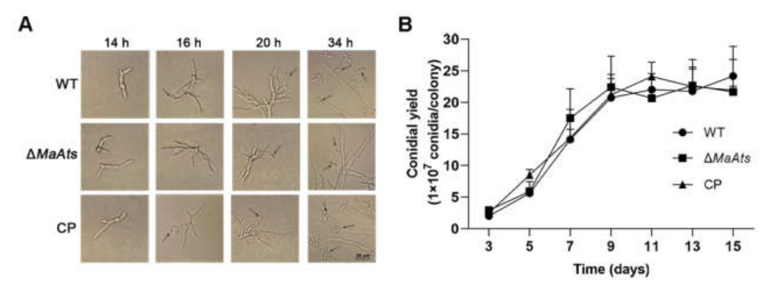
To identify the biological function of MaAts on growth, we observed the colonies of each strain on nutrient-rich 1/4 SDAY medium by microscopy. Our results showed no significant difference between the fungal strains in growth rates (Figure S1). Interestingly, the conidial germination rate of ΔMaAts was significantly lower than the WT and CP strains (Figure 2A). The GT50 of the ΔMaAts (5.50 ± 0.06 h) strain was significantly longer compared to the WT (5.10 ± 0.02 h) and CP (4.98 ± 0.08 h) strains (p < 0.05; Figure 2B). In addition, we observed the whole conidiation process of each strain by microscopy. The results showed that the WT and CP strains had formed conidia at 16 h, while the ΔMaAts strain did not form conidia until 20 h (Figure 3A). At 34 h, the knockout strain produced a small amount of conidia, while the control strains (WT and CP) had produced numerous conidia. (Figure 3A). However, there was no significant difference in the conidial yield of each strain from 3 days to 15 days (Figure 3B).
To explore the virulence of fungal strains, we conducted bioassays by topical inoculation onto the pronotum of L. migratoria manilensis with conidial suspensions of each strain. As a result, there were insignificant differences in LT50 among each strain (p > 0.05), indicating that the MaAts-disruption did not affect the virulence of M. acridum (Figure S2).

3.3. Disruption of MaAts Reduced the Tolerances to UV-B Irradiation and Heat-Shock

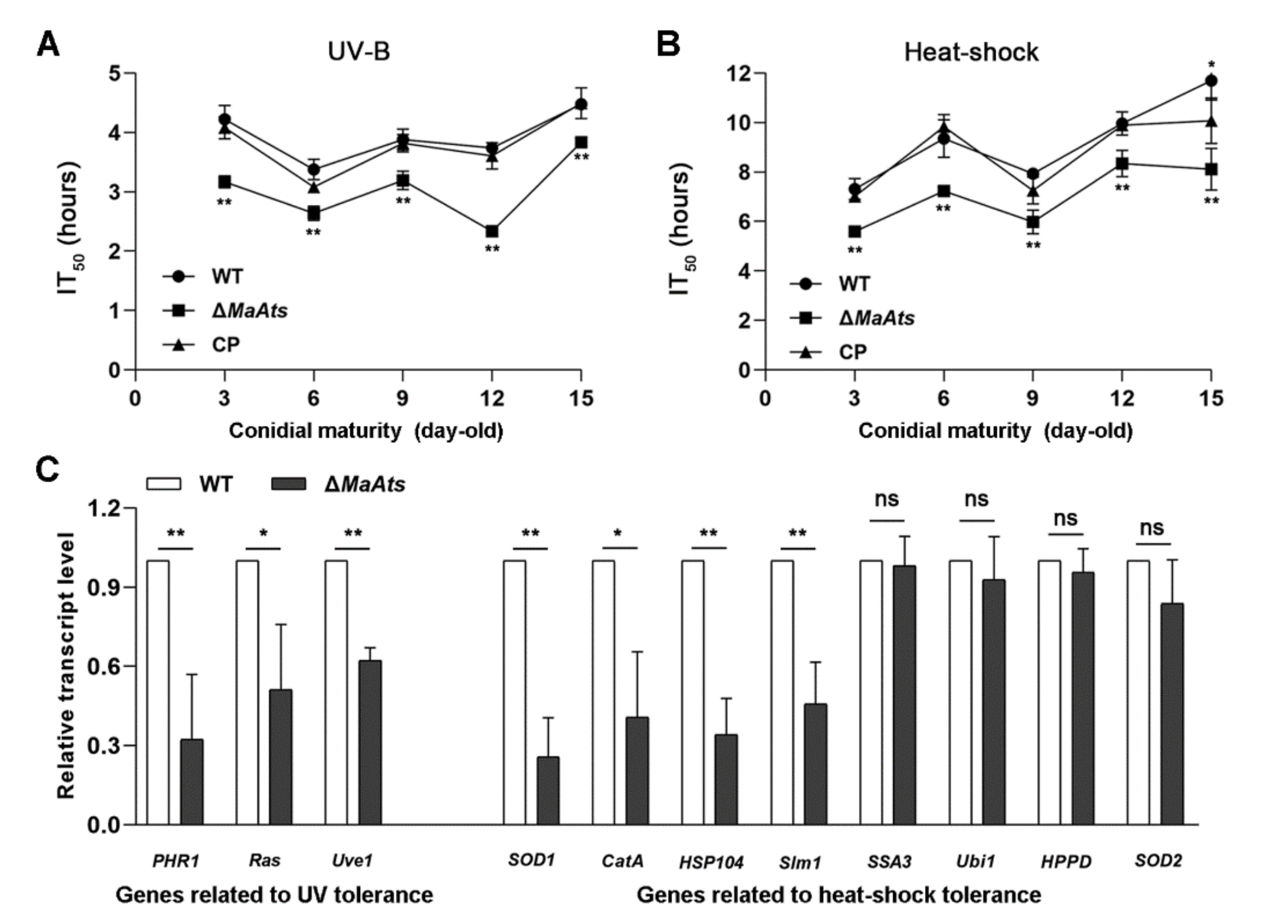
To reveal the contributions of the MaAts gene to stress tolerances, the mature conidia (15-day-old conidia) of tolerances to UV-B irradiation and heat-shock were evaluated. The results showed that there is a dramatic difference in the conidial stress tolerances between the ΔMaAts and the control strains (Figure 4A). The inhibition time of 50% (IT50) of ΔMaAts (3.8 ± 0.09 h) was significantly reduced compared to control strains with the IT50s of 4.5 ± 0.1 h (WT) and 4.5 ± 0.2 h (CP), respectively (Figure 4B, p < 0.01). After the heat-treatment in time-course, the conidial germination rates of the control strains were significantly higher than those of ΔMaAts (Figure 4C, p < 0.01). The inhibition time of 50% (IT50) of ΔMaAts (8.1 ± 0.9 h) was significantly reduced compared to control strains with the IT50s of 11.7 ± 0.9 h (WT) and 10.1 ± 1.0 h (CP), respectively (Figure 4D, p < 0.01). The above results indicated that the deletion of the MaAts reduced conidial tolerances to UV-B irradiation and heat-shock.
Furthermore, the conidia with different maturity (3, 6, 9, 12, and 15-day-old) were respectively collected to determine their tolerances to UV-B irradiation and heat-shock. The results showed that the stress tolerances of all stages of the conidia from the ΔMaAts strain were severely compromised (Figure 4A,B, p < 0.01). Furthermore, the transcription levels of some genes related to fungal stress tolerances were detected by qRT-PCR in the 3-day-old conidia of the WT and ΔMaAts strains. The results showed that the transcription levels of seven genes were significantly reduced in ΔMaAts (Figure 5C).

3.4. Identification of DEGs Influenced by the MaAts

To further reveal the mechanism of the disruption of MaAts reducing the fungal tolerances to UV-B irradiation and heat-shock, the total RNA was isolated from the 3-day-old conidia of the ΔMaAts and WT strains to identify the DEGs between the ΔMaAts and WT strains via DGE profiling. As a result, 512 DEGs (fold change ≥ 2 and p ≤ 0.05) were identified from the DGE data. Of the DEGs, 335 DEGs were down-regulated and 177 DEGs were commonly up-regulated (Figure 6A). The details of DEGs are given in Table S2. To further verify the reliability of the DGE data, we randomly selected 15 genes, including 5 up-regulated genes (MAC_06945, MAC_00193, MAC_04050, MAC_00228, MAC_03018) and 10 down-regulated genes (MAC_01777, MAC_07727, MAC_08480, MAC_07363, MAC_02161, MAC_01480, MAC_01513, MAC_06626, MAC_08198, MAC_06979), and detected their expression by qRT-PCR. For all the selected genes, the expression patterns obtained by qRT-PCR showed similar patterns of down-regulation and up-regulation, indicating that the DGE data were reliable (Figure S3). By gene ontology (GO) annotation, all DEGs (p < 0.05) were classified as cellular component, molecular function, and biological process, which were 160, 292, and 242, respectively (Figure 6B).
From the DGE data, dozens of DEGs involved in cell wall biosynthesis and stress tolerances were strongly affected by the disruption of MaAts. Seven DEGs related to plasma membrane and membrane fusion are down-regulated in ΔMaAts, including a rab GDP-dissociation inhibitor gene (MAC_00732), cytochrome P450 gene (MAC_01212), ABC multidrug transporter gene (MAC_01512), glucose-methanol-choline oxidoreductase gene (MAC_02934), VHS domain protein gene (MAC_03504), phosphate permease gene (MAC_05171), and tetraspanin gene (MAC_00860). Ten DEGs involved in cell wall biosynthesis were down-regulated, such as phospholipase A gene (MAC_06774), a putative maltase gene (MAC_00567), a putative oxidoreductase gene (MAC_01213), and so on (Table S3). It is worth noting that two DEGs, the beta-glucosidase gene (MAC_00623) and a putative C2H2 finger domain protein gene (MAC_07727) related to fungal tolerances to heat were remarkably down-regulated in ΔMaAts (Table S3). The PQQ-repeat-containing protein gene (MAC_07325), which could inhibit the melanin synthesis, was dramatically up-regulated in ΔMaAts (Table S3). In addition, eight DEGs involved in oxidative stress were remarkably down-regulated in ΔMaAts, such as the phospholipase A-2-activating protein gene (MAC_00293), quinone oxidoreductase gene (MAC_02039), 6-phosphogluconate dehydrogenase, NAD-binding protein gene (MAC_01012), a putative ankyrin-repeat-containing protein gene (MAC_01774), and so on. Two DEGs, the guanyl-specific ribonuclease F1 gene (MAC_01777) and sugar transporter family protein gene (MAC_04267) related to salt stress were down-regulated in ΔMaAts. Furthermore, eleven DEGs related to other stresses in ΔMaAts, such as the cysteine synthase K/M:Cysteine synthase B gene (MAC_00603) and the transporter-like protein gene (MAC_04282) involved in aluminum tolerance in Arabidopsis. Thus, the absence of MaAts reduced the tolerances to UV-B irradiation and heat-shock, owing to the alteration of the transcription levels of genes involved in melanin synthesis, cell wall integrity, and tolerances to various stresses.

4. Discussion

Sulfatases are commonly divided into three classes: type I, type II, and type III sulfatases [31]. They can also hydrolyze the sulfate esters, which are widespread in nature, to generate a source of sulfur for growth in many microorganisms [30]. To date, the research of sulfatase largely focuses on type I sulfatase in bacteria and humans [33,40,53], while there is a little information about type III sulfatase. Ats, an alkylsulfatase, belongs to type III sulfatases and can hydrolyze the primary alkyl sulfate [31]. However, the roles of alkylsulfatase in a biological control fungus are still unclear. In this study, the roles of the alkylsulfatase gene MaAts were characterized in the model entomopathogenic fungus M. acridum. The results showed that the disruption of MaAts delayed the germination and the conidiation, while the conidial yield and fungal virulence were not affected. Interestingly, the conidial tolerances to UV-B irradiation and heat-shock were significantly impaired, which has not been reported before in other fungi. According to the DGE data, MaAts might affect the conidial tolerances to UV-B irradiation and heat-shock by influencing the transcriptional levels of some genes related to melanin synthesis, cell wall integrity, and tolerances to various stresses in M. acridum.
During the applications of mycopesticides in the field, many natural abiotic factors, especially UV irradiation and heat-shock, directly affect the effectiveness of fungal biocontrol agents [15,18]. In addition, the conidial tolerances to UV-B irradiation and high temperature are also served as the important indicators of the conidial quality during the industrial production of mycopesticides [54]. In this study, the results showed the loss of MaAts resulted in significantly reduced conidial tolerances to UV-B irradiation and heat-shock. Accordingly, the DGE data showed that the expression levels of some genes involved in fungal tolerances to different stresses were significantly changed in ΔMaAts compared to WT. The gene for C2H2 finger domain protein (MAC_07727), which was directly related to the response to cold, drought, and heat stresses in rice [55], was significantly down-regulated in ΔMaAts. The ROS levels in the fungal cell would rise after treatment with UV irradiation [16]. Among the DEGs, the ThiJ/PfpI family protein gene (MAC_08673), which was involved in the tolerance to ROS and specifically H2O2 and superoxide radicals in Candida albicans [56], were significantly down-regulated in ΔMaAts, suggesting that the ΔMaAts strain was more vulnerable to UV irradiation than the WT strain. Furthermore, a β-glucosidase gene (MAC_00623), which was involved in the thermotolerance of Pyrococcus furiosus [57], was significantly changed in ΔMaAts. Many reports have demonstrated that melanin synthesis and cell wall organization were both closely involved in fungal stress tolerances [58,59,60,61]. The PQQ-repeat-containing protein gene (MAC_07325), which could inhibit the melanin synthesis in murine [62], was dramatically up-regulated in ΔMaAts. In addition, ten cell wall-associated protein genes (MAC_06774 [63], MAC_00567 [64], MAC_01213 [65], MAC_01513 [66], MAC_02366 [67], MAC_02951 [68], MAC_04850 [69], MAC_06773 [70], MAC_00235 [71], and MAC_04881 [69]) were significantly down-regulated in ΔMaAts, which possibly resulted in a decrease in stress tolerances of M. acridum. The gene for feruloyl esterase B precursor (MAC_05632), which was involved in the cell wall composition and structure of plants [72], was significantly up-regulated in ΔMaAts. The vacuolar protein-sorting protein BRO1 gene (MAC_06773) was dramatically down-regulated in ΔMaAts, which was related to the regulation of secondary wall biosynthesis in Arabidopsis [70].
The loss of MaAts delayed germination and conidiation compared to the control strains. Consistently, some genes involved in germination, conidiation and fungal growth were down-regulated in ΔMaAts from the DGE data, such as the GmcA gene for the glucose-methanol-choline oxidoreductase (MAC_02934), which was involved in conidial germination and conidiation in Aspergillus nidulans [73]. Meanwhile, the fungal specific transcription factor gene StuA (MAC_06675) related to the growth of A. nidulans [74] was also down-regulated. In addition, the gene for the kelch repeat protein (MAC_09450), which could regulate cell apoptosis processes in Drosophila [75], was also down-regulated.
In summary, our results showed that the deletion of MaAts significantly decreased the conidial tolerances to UV-B irradiation and heat-shock. MaAts and some DEGs involved in fungal stress tolerances may be candidate genes to be adopted to improve the stress tolerances of mycopesticides. In the future, clarifying the roles of the DEGs identified in this study may be conducive to further reveal the molecular mechanisms of the fungal tolerances to UV-B irradiation and heat-shock.

Supplementary Materials

The following are available online at https://www.mdpi.com/article/10.3390/jof8030270/s1, Figure S1: Deletion and complementation of MaAts in M. acridum. (A) Strategies of the MaAts deletion and complementation in M. acridum. (B) Southern blotting. The genomic DNAs from different fungal strains were cut with Nru I and Xho I. The probe was cloned from 5′ genomic sequence of MaAts by PCR using the primers of MaAts-PF/MaAts-PR. (C) Colonies of each strain on 1/4 SDAY plates at 28℃ for 6 days. Figure S2: Bioassays. (A) Survival of locusts after topical inoculation; paraffin oil was used as control. (B) The mean 50% lethal time (LT50) of fungal strains for topical inoculation; ns indicates p > 0.05. Figure S3: Verification of the DGE data via qRT -PCR. Table S1: Primers used in this study. Table S2: DGE data of ΔMaAts vs. WT. Table S3: DEGs of ΔMaAts vs. WT involved in stress tolerances and cell wall components.

Author Contributions

Conceptualization, Y.X. and K.J.; methodology, Y.X. and K.J.; project administration, Y.X. and K.J.; investigation, L.S., X.X., S.W. and J.L.; data curation, L.S., X.X., S.W. and J.L.; writing—original draft preparation, L.S. and X.X.; writing—review and editing, L.S. and K.J.; funding acquisition, Y.X. and K.J. All authors have read and agreed to the published version of the manuscript.

Funding

This work was supported by the Natural Science Foundation of China (31972330), Chongqing talent program of China (cstc2021ycjh-bgzxm0313), the Venture and Innovation Support Program for Chongqing Overseas Returnees (cx2019035).

Institutional Review Board Statement

Not applicable.

Informed Consent Statement

Not applicable.

Data Availability Statement

Not applicable.

Conflicts of Interest

The authors declare no conflict of interest.

References

  1. Gerhardson, B. Biological substitutes for pesticides. Trends Biotechnol. 2002, 20, 338–343. [Google Scholar] [CrossRef]
  2. Arthurs, S.; Thomas, M.B.; Langewald, J. Field observations of the effects of fenitrothion and Metarhizium anisopliae var. acridum on non-target ground dwelling arthropods in the Sahel. Biol. Control. 2003, 26, 333–340. [Google Scholar] [CrossRef]
  3. Guerrero-Guerra, C.; Reyes-Montes, M.; Toriello, C.; Hernandez-Velazquez, V.; Santiago-Lopez, I.; Mora-Palomino, L.; Calderon-Segura, M.E.; Fernandez, S.D.; Calderon-Ezquerro, C. Study of the persistence and viability of Metarhizium acridum in Mexico’s agricultural area. Aerobiologia 2013, 29, 249–261. [Google Scholar] [CrossRef]
  4. Roberts, D.W.; St Leger, R.J. Metarhizium spp. cosmopolitan insect-pathogenic fungi: Mycological aspects. Adv. Appl. Microbiol. 2004, 54, 1–70. [Google Scholar] [PubMed]
  5. Chandler, D.; Bailey, A.S.; Tatchell, G.M.; Davidson, G.; Greaves, J.; Grant, W.P. The development, regulation and use of biopesticides for integrated pest management. Philos. Trans. R. Soc. Lond. B Biol. Sci. 2011, 366, 1987–1998. [Google Scholar] [CrossRef] [PubMed]
  6. Hunter, D.M.; Milner, R.J.; Scanlan, J.C.; Spurgin, P.A. Aerial treatment of the migratory locust, Locusta migratoria (L.) (Orthoptera: Acrididae) with Metarhizium anisopliae (Deuteromycotina: Hyphomycetes) in Australia. Crop Prot. 1999, 18, 699–704. [Google Scholar] [CrossRef]
  7. Peng, G.; Wang, Z.; Yin, Y.; Zeng, D.; Xia, Y. Field trials of Metarhizium anisopliae var. acridum (Ascomycota: Hypocreales) against oriental migratory locusts, Locusta migratoria manilensis (Meyen) in Northern China. Crop Prot. 2008, 27, 1244–1250. [Google Scholar] [CrossRef]
  8. Niassy, S.; Diarra, K. Pathogenicity of local Metarhizium anisopliae var. acridum strains on Locusta migratoria migratorioides Reiche and Farmaire and Zonocerus variegatus Linnaeus in Senegal. Afr. J. Biotechnol. 2011, 10, 28–33. [Google Scholar]
  9. Yousef, M.; Garrido-Jurado, I.; Ruíz-Torres, M.; Quesada-Moraga, E. Reduction of adult olive fruit fly populations by targeting preimaginals in the soil with the entomopathogenic fungus Metarhizium brunneum. J. Pest Sci. 2016, 90, 345–354. [Google Scholar] [CrossRef]
  10. Milner, R.J.; Lozano, L.B.; Driver, F.; Hunter, D. A comparative study of two Mexican isolates with an Australian isolate of Metarhizium anisopliae var. acridum—Strain characterisation, temperature profile and virulence for wingless grasshopper, Phaulacridium vittatum. BioControl 2003, 48, 335–348. [Google Scholar] [CrossRef]
  11. Gao, Q.; Jin, K.; Ying, S.H.; Zhang, Y.J.; Xiao, G.H.; Shang, Y.F.; Duan, Z.B.; Hu, X.; Xie, X.Q.; Zhou, G.; et al. Genome sequencing and comparative transcriptomics of the model entomopathogenic fungi Metarhizium anisopliae and M. acridum. PLoS Genet. 2011, 7, e1001264. [Google Scholar] [CrossRef] [PubMed] [Green Version]
  12. Adams, T.H.; Wieser, J.K.; Yu, J.H. Asexual sporulation in Aspergillus nidulans. Microbiol. Mol. Biol. Rev. 1998, 62, 35–54. [Google Scholar] [CrossRef] [PubMed] [Green Version]
  13. Papagianni, M. Fungal morphology and metabolite production in submerged mycelial processes. Biotechnol. Adv. 2004, 22, 189–259. [Google Scholar] [CrossRef] [PubMed]
  14. Zimmermann, G. Effect of high temperatures and artificial sunlight on the viability of conidia of Metarhizium anisopliae. J. Invertebr. Pathol. 1982, 40, 36–40. [Google Scholar] [CrossRef]
  15. Li, J.; Guo, M.; Cao, Y.; Xia, Y. Disruption of a C69-family cysteine dipeptidase gene enhances heat shock and UV-B tolerances in Metarhizium acridum. Front. Microbiol. 2020, 11, 849. [Google Scholar] [CrossRef]
  16. Rangel, D.E.; Anderson, A.J.; Roberts, D.W. Evaluating physical and nutritional stress during mycelial growth as inducers of tolerance to heat and UV-B radiation in Metarhizium anisopliae conidia. Mycol. Res. 2008, 112, 1362–1372. [Google Scholar] [CrossRef]
  17. Braga, G.U.; Rangel, D.E.; Fernandes, É.K.; Flint, S.D.; Roberts, D.W. Molecular and physiological effects of environmental UV radiation on fungal conidia. Curr. Genet. 2015, 61, 405–425. [Google Scholar] [CrossRef]
  18. Fernandes, É.K.; Rangel, D.E.; Braga, G.U.; Roberts, D.W. Tolerance of entomopathogenic fungi to ultraviolet radiation: A review on screening of strains and their formulation. Curr. Genet. 2015, 61, 427–440. [Google Scholar] [CrossRef]
  19. Ortiz-Urquiza, A.; Keyhani, N.O. Stress response signaling and virulence: Insights from entomopathogenic fungi. Curr. Genet. 2015, 61, 239–249. [Google Scholar] [CrossRef]
  20. Yao, S.L.; Ying, S.H.; Feng, M.G.; Hatting, J.L. In vitro and in vivo responses of fungal biocontrol agents to gradient doses of UV-B and UV-A irradiation. BioControl 2010, 55, 413–422. [Google Scholar] [CrossRef]
  21. Griffiths, H.R.; Mistry, P.; Herbert, K.E.; Lunec, J. Molecular and cellular effects of ultraviolet light-induced genotoxicity. Crit. Rev. Clin. Lab. Sci. 1998, 35, 189–237. [Google Scholar] [CrossRef]
  22. Kim, J.i.; Patel, D.; Choi, B.S. Contrasting structural impacts induced by cis-syn cyclobutane dimer and (6-4) adduct in DNA duplex decamers: Implication in mutagenesis and repair activity. Photochem. Photobiol. 1995, 62, 44–50. [Google Scholar] [CrossRef] [PubMed]
  23. Sinha, R.P.; Häder, D.P. UV-induced DNA damage and repair: A review. Photochem. Photobiol. Sci. 2002, 1, 225–236. [Google Scholar] [CrossRef]
  24. Wang, C.I.; Taylor, J.S. Site-specific effect of thymine dimer formation on dAn.dTn tract bending and its biological implications. Proc. Natl. Acad. Sci. USA 1991, 88, 9072–9076. [Google Scholar] [CrossRef] [PubMed] [Green Version]
  25. Crisan, E.V. Current concepts of thermophilism and the thermophilic fungi. Mycologia 1973, 65, 1171–1198. [Google Scholar] [CrossRef] [PubMed]
  26. Setlow, B.; Setlow, P. Heat killing of Bacillus subtilis spores in water is not due to oxidative damage. Appl. Environ. Microbiol. 1998, 64, 4109–4112. [Google Scholar] [CrossRef] [Green Version]
  27. Rangel, D.E.; Braga, G.U.; Anderson, A.J.; Roberts, D.W. Variability in conidial thermotolerance of Metarhizium anisopliae isolates from different geographic origins. J. Invertebr. Pathol. 2005, 88, 116–125. [Google Scholar] [CrossRef]
  28. Nicholson, W.L.; Munakata, N.; Horneck, G.; Melosh, H.J.; Setlow, P. Resistance of Bacillus endospores to extreme terrestrial and extraterrestrial environments. Microbiol. Mol. Biol. Rev. 2000, 64, 548–572. [Google Scholar] [CrossRef] [Green Version]
  29. Rangel, D.E.; Fernandes, E.K.; Dettenmaier, S.J.; Roberts, D.W. Thermotolerance of germlings and mycelium of the insect-pathogenic fungus Metarhizium spp. and mycelial recovery after heat stress. J. Basic Microbiol. 2010, 50, 344–350. [Google Scholar] [CrossRef]
  30. Pogorevc, M.; Faber, K. Purification and characterization of an inverting stereo- and enantioselective sec-alkylsulfatase from the gram-positive bacterium Rhodococcus ruber DSM 44541. Appl. Environ. Microbiol. 2003, 69, 2810–2815. [Google Scholar] [CrossRef] [Green Version]
  31. Sogi, K.M.; Gartner, Z.J.; Breidenbach, M.A.; Appel, M.J.; Schelle, M.W.; Bertozzi, C.R. Mycobacterium tuberculosis Rv3406 is a type II alkyl sulfatase capable of sulfate scavenging. PLoS ONE 2013, 8, e65080. [Google Scholar] [CrossRef]
  32. Kertesz, M.A. Riding the sulfur cycle—Metabolism of sulfonates and sulfate esters in gram-negative bacteria. FEMS Microbiol. Rev. 2000, 24, 135–175. [Google Scholar] [PubMed] [Green Version]
  33. Adang, L.A.; Schlotawa, L.; Groeschel, S.; Kehrer, C.; Harzer, K.; Staretz-Chacham, O.; Silva, T.O.; Schwartz, I.V.D.; Gärtner, J.; De Castro, M.; et al. Natural history of multiple sulfatase deficiency: Retrospective phenotyping and functional variant analysis to characterize an ultra-rare disease. J. Inherit. Metab. Dis. 2020, 43, 1298–1309. [Google Scholar] [CrossRef] [PubMed]
  34. Dhoot, G.K.; Gustafsson, M.K.; Ai, X.; Sun, W.; Standiford, D.M.; Emerson, C.P., Jr. Regulation of Wnt signaling and embryo patterning by an extracellular sulfatase. Science 2001, 293, 1663–1666. [Google Scholar] [CrossRef] [PubMed]
  35. Hagelueken, G.; Adams, T.M.; Wiehlmann, L.; Widow, U.; Kolmar, H.; Tümmler, B.; Heinz, D.W.; Schubert, W.D. The crystal structure of SdsA1, an alkylsulfatase from Pseudomonas aeruginosa, defines a third class of sulfatases. Proc. Natl. Acad. Sci. USA 2006, 103, 7631–7636. [Google Scholar] [CrossRef] [Green Version]
  36. Müller, I.; Kahnert, A.; Pape, T.; Sheldrick, G.M.; Usón, I. Crystal structure of the alkylsulfatase AtsK: Insights into the catalytic mechanism of the Fe(II) α-ketoglutarate-dependent dioxygenase superfamily. Biochemistry 2004, 43, 3075–3088. [Google Scholar] [CrossRef] [Green Version]
  37. Kahnert, A.; Kertesz, M.A. Characterization of a sulfur-regulated oxygenative alkylsulfatase from Pseudomonas putida S-313. J. Biol. Chem. 2000, 275, 31661–31667. [Google Scholar] [CrossRef] [Green Version]
  38. Bebrone, C. Metallo-β-lactamases (classification, activity, genetic organization, structure, zinc coordination) and their superfamily. Biochem. Pharmacol. 2007, 74, 1686–1701. [Google Scholar] [CrossRef]
  39. Sun, L.; Chen, P.; Su, Y.; Cai, Z.; Ruan, L.; Xu, X.; Wu, Y. Crystal structure of thermostable alkylsulfatase SdsAP from Pseudomonas sp. S9. Biosci. Rep. 2017, 37, BSR20170001. [Google Scholar] [CrossRef] [Green Version]
  40. Toesch, M.; Schober, M.; Faber, K. Microbial alkyl- and aryl-sulfatases: Mechanism, occurrence, screening and stereoselectivities. Appl. Microbiol. Biotechnol. 2014, 98, 1485–1496. [Google Scholar] [CrossRef] [Green Version]
  41. Davison, J.; Brunel, F.; Phanopoulos, A.; Prozzi, D.; Terpstra, P. Cloning and sequencing of Pseudomonas genes determining sodium dodecyl sulfate biodegradation. Gene 1992, 114, 19–24. [Google Scholar] [CrossRef]
  42. Long, M.; Ruan, L.; Li, F.; Yu, Z.; Xu, X. Heterologous expression and characterization of a recombinant thermostable alkylsulfatase (sdsAP). Extremophiles 2011, 15, 293–301. [Google Scholar] [CrossRef] [PubMed]
  43. Shahbazi, R.; Kasra-Kermanshahi, R.; Gharavi, S.; Moosavi-Nejad, Z.; Borzooee, F. Screening of SDS-degrading bacteria from car wash wastewater and study of the alkylsulfatase enzyme activity. Iran. J. Microbiol. 2013, 5, 153–158. [Google Scholar] [PubMed]
  44. Boer, V.M.; de Winde, J.H.; Pronk, J.T.; Piper, M.D. The genome-wide transcriptional responses of Saccharomyces cerevisiae grown on glucose in aerobic chemostat cultures limited for carbon, nitrogen, phosphorus, or sulfur. J. Biol. Chem. 2003, 78, 3265–3274. [Google Scholar] [CrossRef] [PubMed] [Green Version]
  45. Hall, C.; Brachat, S.; Dietrich, F.S. Contribution of horizontal gene transfer to the evolution of Saccharomyces cerevisiae. Eukaryot. Cell 2005, 4, 1102–1115. [Google Scholar] [CrossRef] [Green Version]
  46. Waddell, G.L.; Gilmer, C.R.; Taylor, N.G.; Reveral, J.R.S.; Forconi, M.; Fox, J.L. The eukaryotic enzyme Bds1 is an alkyl but not an aryl sulfohydrolase. Biochem. Biophys. Res. Commun. 2017, 491, 382–387. [Google Scholar] [CrossRef]
  47. Fang, W.; Pei, Y.; Bidochka, M.J. Transformation of Metarhizium anisopliae mediated by Agrobacterium tumefaciens. Can. J. Microbiol. 2006, 52, 623–626. [Google Scholar] [CrossRef]
  48. Jin, K.; Ming, Y.; Xia, Y.X. MaHog1, a Hog1-type mitogen-activated protein kinase gene, contributes to stress tolerance and virulence of the entomopathogenic fungus Metarhizium acridum. Microbiology 2012, 158, 2987–2996. [Google Scholar] [CrossRef] [Green Version]
  49. Zhang, J.; Jiang, H.; Du, Y.; Keyhani, N.O.; Xia, Y.; Jin, K. Members of chitin synthase family in Metarhizium acridum differentially affect fungal growth, stress tolerances, cell wall integrity and virulence. PLoS Pathog. 2019, 15, e1007964. [Google Scholar] [CrossRef]
  50. Zhang, J.; Wang, Z.; Keyhani, N.O.; Peng, G.X.; Jin, K.; Xia, Y. The protein phosphatase gene MaPpt1 acts as a programmer of microcycle conidiation and a negative regulator of UV-B tolerance in Metarhizium acridum. Appl. Microbiol. Biotechnol. 2019, 103, 1351–1362. [Google Scholar] [CrossRef]
  51. Du, Y.; Jin, K.; Xia, Y. Involvement of MaSom1, a downstream transcriptional factor of cAMP/PKA pathway, in conidial yield, stress tolerances, and virulence in Metarhizium acridum. Appl. Microbiol. Biotechnol. 2018, 102, 5611–5623. [Google Scholar] [CrossRef] [PubMed]
  52. Livak, K.J.; Schmittgen, T.D. Analysis of relative gene expression data using real-time quantitative PCR and the 2−ΔΔCT method. Methods 2001, 25, 402–408. [Google Scholar] [CrossRef] [PubMed]
  53. Agrawal, S.; Ransom, R.F.; Saraswathi, S.; Garciagonzalo, E.; Webb, A.; Fernandezmartinez, J.L.; Popovic, M.; Guess, A.J.; Kloczkowski, A.; Benndorf, R. Sulfatase 2 is associated with steroid resistance in childhood nephrotic syndrome. J. Clin. Med. 2021, 10, 523. [Google Scholar] [CrossRef] [PubMed]
  54. Xie, X.Q.; Li, F.; Ying, S.H.; Feng, M.G. Additive contributions of two manganese-cored superoxide dismutases (MnSODs) to antioxidation, UV tolerance and virulence of Beauveria bassiana. PLoS ONE 2012, 7, e30298. [Google Scholar] [CrossRef] [PubMed]
  55. Zhang, X.; Zhang, B.; Li, M.J.; Yin, X.M.; Huang, L.F.; Cui, Y.C.; Wang, M.L.; Xia, X.J. OsMSR15 encoding a rice C2H2-type zinc finger protein confers enhanced drought tolerance in transgenic Arabidopsis. J. Plant. Biol. 2016, 59, 271–281. [Google Scholar] [CrossRef]
  56. Jia, J.H.; Wang, Y.; Cao, Y.B.; Gao, P.H.; Jia, X.M.; Ma, Z.P.; Xu, Y.G.; Dai, B.D.; Jiang, Y.Y. CaIPF7817 is involved in the regulation of redox homeostasis in Candida albicans. Biochem. Biophys. Res. Commun. 2007, 359, 163–167. [Google Scholar] [CrossRef] [Green Version]
  57. Kengen, S.W.; Luesink, E.J.; Stams, A.J.; Zehnder, A.J. Purification and characterization of an extremely thermostable β-glucosidase from the hyperthermophilic archaeon Pyrococcus furiosus. Eur. J. Biochem. 2010, 213, 305–312. [Google Scholar] [CrossRef]
  58. Eisenman, H.C.; Casadevall, A. Synthesis and assembly of fungal melanin. Appl. Microbiol. Biotechnol. 2012, 93, 931–940. [Google Scholar] [CrossRef] [Green Version]
  59. Gessler, N.N.; Egorova, A.S.; Belozerskaya, T.A. Melanin pigments of fungi under extreme environmental conditions (review). Biokhim Mikrobiol. 2014, 50, 125–134. [Google Scholar]
  60. Jeong, R.D.; Chu, E.H.; Shin, E.J.; Park, H.J. Effects of ionizing radiation on postharvest fungal pathogens. Plant Pathol. J. 2015, 31, 176–180. [Google Scholar] [CrossRef] [Green Version]
  61. Kimura, T.; Fukuda, W.; Sanada, T.; Imanaka, T. Characterization of water-soluble dark-brown pigment from antarctic bacterium, Lysobacter oligotrophicus. J. Biosci. Bioeng. 2015, 120, 58–61. [Google Scholar] [CrossRef] [PubMed]
  62. Sato, K.; Toriyama, M. Effect of pyrroloquinoline quinone (PQQ) on melanogenic protein expression in murine B16 melanoma. J. Dermatol. Sci. 2009, 53, 140–145. [Google Scholar] [CrossRef] [PubMed]
  63. May, K.L.; Silhavy, T.J.; Gottesman, S. The Escherichia coli phospholipase PldA regulates outer membrane homeostasis via lipid signaling. mBio 2018, 9, e00379-18. [Google Scholar] [CrossRef] [PubMed] [Green Version]
  64. Wang, X.; Yang, Z.; Wang, M.; Meng, L.; Jiang, Y.; Han, Y. The BRANCHING ENZYME1 gene, encoding a glycoside hydrolase family 13 protein, is required for in vitro plant regeneration in Arabidopsis. Plant Cell Tiss. Org. 2014, 117, 279–291. [Google Scholar] [CrossRef]
  65. Fang, L.; Zhao, F.; Cong, Y.; Sang, X.; Du, Q.; Wang, D.; Li, Y.; Ling, Y.; Yang, Z.; He, G. Rolling-leaf14 is a 2OG-Fe (II) oxygenase family protein that modulates rice leaf rolling by affecting secondary cell wall formation in leaves. Plant Biotechnol. J. 2012, 10, 524–532. [Google Scholar] [CrossRef]
  66. Johnson, S.S.; Hanson, P.K.; Manoharlal, R.; Brice, S.E.; Cowart, L.A.; Moye-Rowley, W.S. Regulation of yeast nutrient permease endocytosis by ATP-binding cassette transporters and a seven-transmembrane protein, RSB1. J. Biol. Chem. 2010, 85, 35792–35802. [Google Scholar] [CrossRef] [Green Version]
  67. Hayashi, S. The glycerophosphoryl diester phosphodiesterase-like proteins SHV3 and its homologs play important roles in cell wall organization. Plant Cell Physiol. 2008, 49, 1522–1535. [Google Scholar] [CrossRef] [Green Version]
  68. Song, X.; Zhang, B.; Zhou, Y. Golgi-localized UDP-glucose transporter is required for cell wall integrity in rice. Plant Signal. Behav. 2011, 6, 1097–1100. [Google Scholar] [CrossRef] [Green Version]
  69. Zhong, R.; Ye, R.Z.H. The MYB46 transcription factor is a direct target of SND1 and regulates secondary wall biosynthesis in Arabidopsis. Plant Cell 2007, 19, 2776–2792. [Google Scholar] [CrossRef] [Green Version]
  70. Nickas, M.E.; Yaffe, M.P. BRO1, a novel gene that interacts with components of the Pkc1p-mitogen-activated protein kinase pathway in Saccharomyces cerevisiae. Mol. Cell. Biol. 1996, 16, 2585–2593. [Google Scholar] [CrossRef] [Green Version]
  71. Zhong, R. Mutation of SAC1, an Arabidopsis SAC domain phosphoinositide phosphatase, causes alterations in cell morphogenesis, cell wall synthesis, and actin organization. Plant Cell 2005, 17, 1449–1466. [Google Scholar] [CrossRef] [PubMed] [Green Version]
  72. Mathew, S.; Abraham, T.E. Ferulic acid: An antioxidant found naturally in plant cell walls and feruloyl esterases involved in its release and their applications. Crit. Rev. Biotechnol. 2004, 24, 59–83. [Google Scholar] [CrossRef] [PubMed]
  73. Etxebeste, O.; Herrero-García, E.; Cortese, M.S.; Garzia, A.; Oiartzabal-Arano, E.; de los Ríos, V.; Ugalde, U.; Espeso, E.A. GmcA is a putative glucose-methanol-choline oxidoreductase required for the induction of asexual development in Aspergillus nidulans. PLoS ONE 2012, 7, e40292. [Google Scholar] [CrossRef] [PubMed] [Green Version]
  74. Dutton, J.R. StuAp is a sequence-specific transcription factor that regulates developmental complexity in Aspergillus nidulans. EMBO J. 2014, 16, 5710–5721. [Google Scholar] [CrossRef] [Green Version]
  75. Sekine, Y.; Hatanaka, R.; Watanabe, T.; Sono, N.; Iemura, S.; Natsume, T.; Kuranaga, E.; Miura, M.; Takeda, K.; Ichijo, H. The Kelch repeat protein KLHDC10 regulates oxidative stress-induced ASK1 activation by suppressing PP5. Mol. Cell. 2012, 48, 692–704. [Google Scholar] [CrossRef] [Green Version]
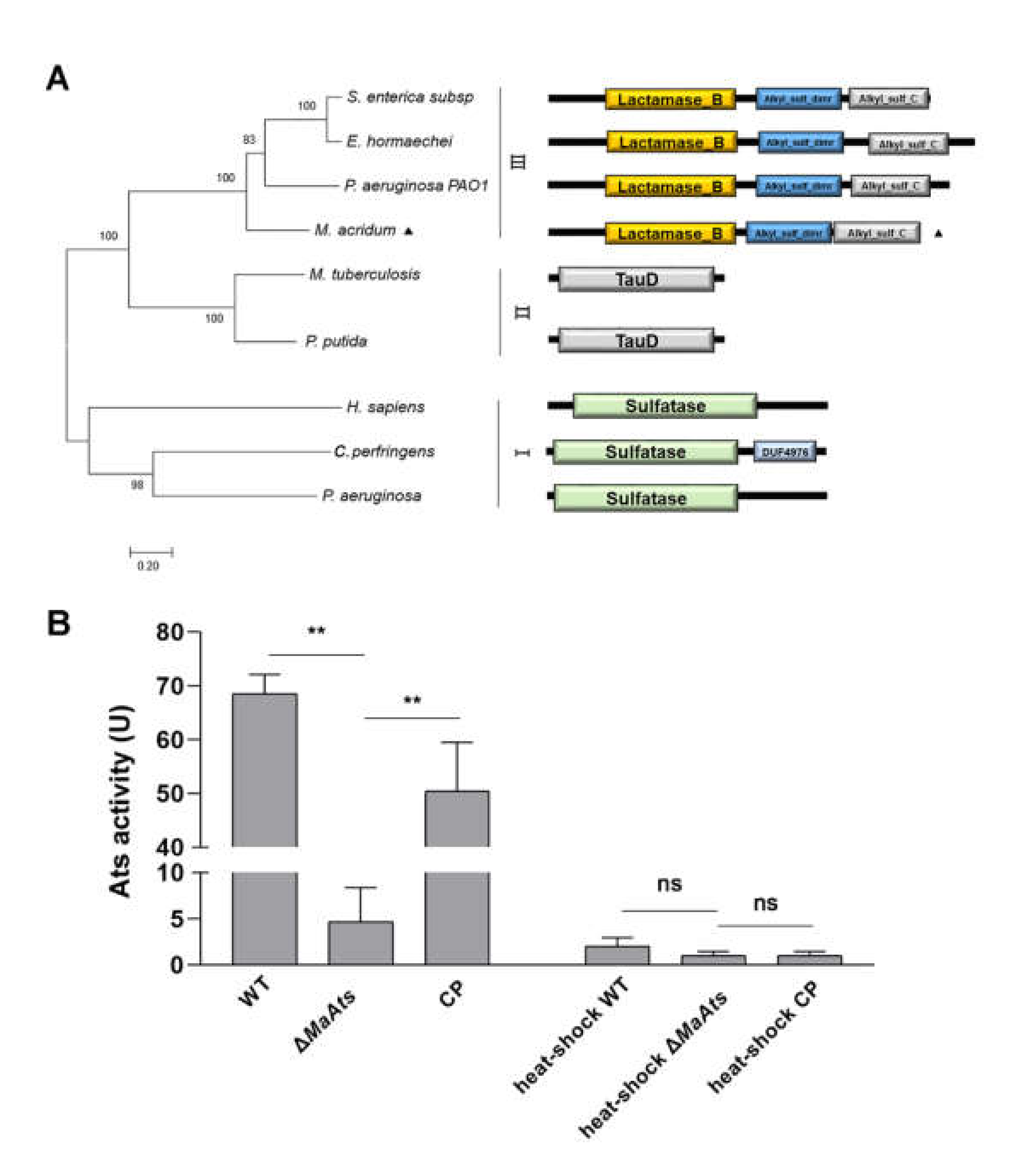
Figure 1. Features of MaAts in M. acridum. (A) Phylogenetic relationships of sulfatase proteins from bacteria, fungi, human. S. enterica subsp: Salmonella enterica subsp (EDZ09460.1); E. hormaechei: Enterobacter hormaechei (CBK85007.1); P. aeruginosa PAO1: Pseudomonas aeruginosa PAO1 (SKC13380.1); M. acridum: Metarhizium acridum (EFY85493.1); M. tuberculosis: Mycobacterium tuberculosis (NP_217923.1); P. putida: Pseudomonas putida (SUD73147.1); H. sapiens: Homo sapiens (NP_001012301.1); C. perfringens: Clostridium perfringens (WP_131337334.1); P. aeruginosa: Pseudomonas aeruginosa (SKC13380.1).I, II and III represent different types of sulfatases. (B) Alkylsulfatase activities in the 3-day-old conidia from nutrient-rich 1/4 SDAY medium. The heat-shock WT, heat-shock ΔMaAts, and heat-shock CP denotes that the samples were treated at 100 °C for 10 min. One unit of the alkylsulfatase activity was defined as ΔA360 = 0.001 after 10 min. Asterisk indicates significant difference at (**) p < 0.01, (ns) p > 0.05.
Jof 08 00270 g001
Figure 2. The deletion of MaAts delayed the conidial germination. (A) The germination rates of conidia of each strain on nutrient-rich 1/4 SDAY medium. (B) The mean 50% germination time (GT50) of each strain. Asterisk indicates significant difference at (*) p < 0.05, (**) p < 0.01, (ns) p > 0.05.
Jof 08 00270 g002
Figure 3. The deletion of MaAts delayed conidiation. (A) Conidiation of each strain cultured on nutrient-rich 1/4 SDAY medium. Black arrows indicate the typical conidiophores and conidia. (B) Conidial yield of WT, ΔMaAts and CP strains on nutrient-rich 1/4 SDAY medium at 28 °C.
Jof 08 00270 g003
Figure 4. The deletion of MaAts impaired the UV-B irradiation and heat-shock tolerances of the mature (15-day-old) conidia. (A) Conidial germination after exposure of UV-B irradiation (1350 mW/m2) in a 1.5 h interval. (B) The half-inhibition time (IT50) under UV-B irradiation. (C) Germination rates after heat-shock treatment (45 °C) in a 3 h interval. (D) IT50 under high temperature (45 °C). After treatments, WT, ΔMaAts and CP strains were cultured on nutrient-rich 1/4 SDAY medium at 28 °C for 20 h. Asterisk indicates significant difference at (**) p < 0.01, (*) p < 0.05, (ns) p > 0.05.
Jof 08 00270 g004
Figure 5. The deletion of MaAts diminished the UV-B irradiation and heat-shock tolerances of the conidia with different maturity. (A) The IT50 of conidia with different maturity (3, 6, 9, 12 and 15-day-old) under UV-B irradiation. (B) The IT50 of conidia with different maturity (3, 6, 9, 12 and 15-day-old) under heat-shock. (C) Relative transcript level of genes related to conidia tolerances to UV or heat-shock in the 3-day-old conidia. MaPHR1 (MAC_05772); MaRas (MAC_07622); MaUve1 (MAC_07337); MaSOD1 (MAC_03040); MaCatA (MAC_09645); MaHsp104 (MAC_05034); Maslm1 (MAC_04714); MaSSA3 (MAC_02927); MaUbi1 (MAC_01946); MaHPPD (MAC_02015); MaSOD2 (MAC_01660). Asterisk indicates significant difference at (**) p < 0.01, (*) p < 0.05, (ns) p > 0.05.
Jof 08 00270 g005
Figure 6. Identification and GO annotation of DEGs. (A) Identification of DEGs in the 3-day-old conidia from the ΔMaAts vs. WT. Green dots, down-regulated genes (log2 ratio ≤ 1). Red dots, up-regulated genes (log2 ratio ≥ 1). Grey dots, not differentially regulated (−1< log2 ratio < 1). (B) GO annotation of DEGs in the 3-day-old conidia from the ΔMaAts vs. WT.
Jof 08 00270 g006
Publisher’s Note: MDPI stays neutral with regard to jurisdictional claims in published maps and institutional affiliations.