Article

Natural Product Citronellal can Significantly Disturb Chitin Synthesis and Cell Wall Integrity in Magnaporthe oryzae

1
Institute of Crop Protection, Guizhou University, Guiyang 550025, China
2
College of Agriculture, Guizhou University, Guiyang 550025, China
3
The Provincial Key Laboratory for Agricultural Pest Management in Mountainous Region, Guiyang 550025, China
*
Author to whom correspondence should be addressed.
J. Fungi 2022, 8(12), 1310; https://doi.org/10.3390/jof8121310
Submission received: 24 October 2022 / Revised: 10 December 2022 / Accepted: 15 December 2022 / Published: 16 December 2022
(This article belongs to the Section Fungal Cell Biology, Metabolism and Physiology)

Abstract

:
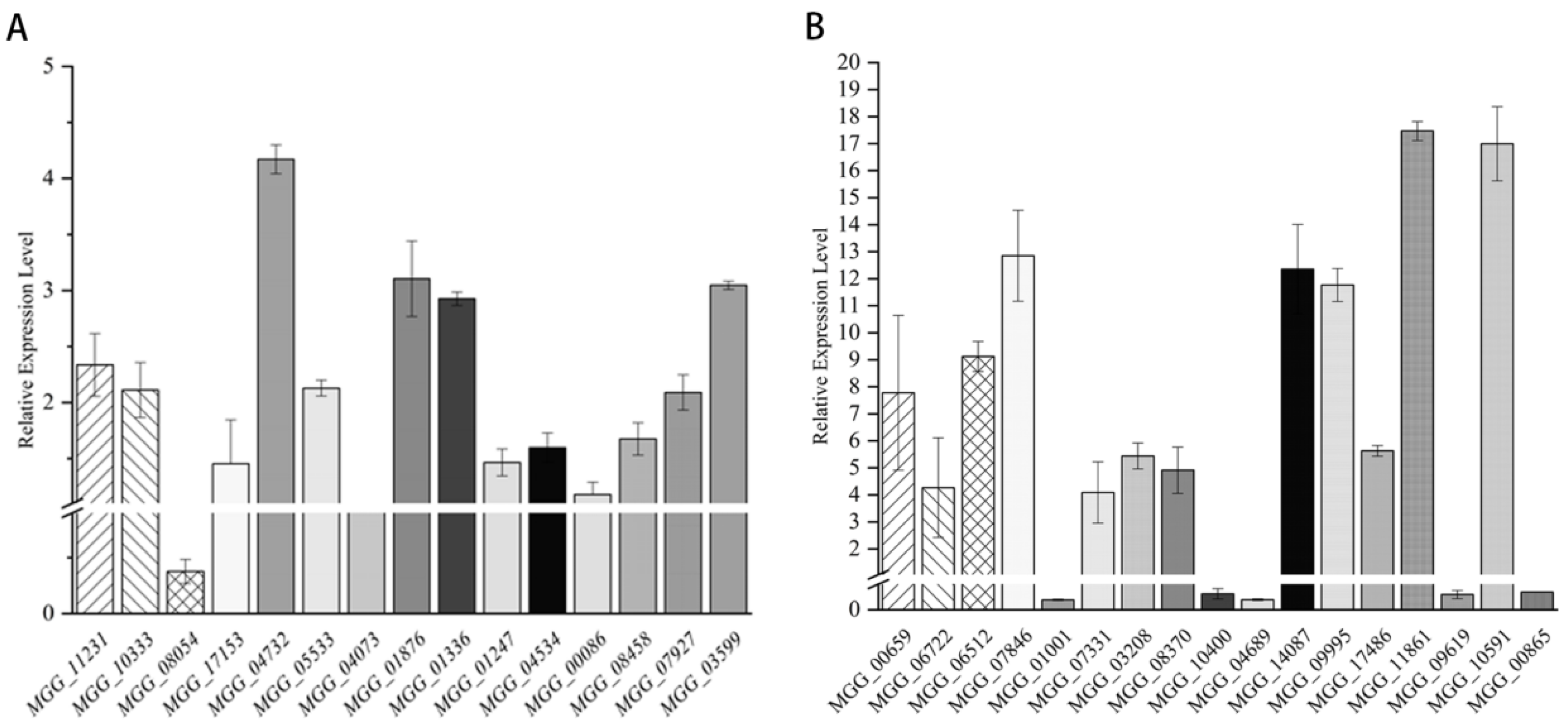
Background: Natural products are often favored in the study of crop pests and diseases. Previous studies have shown that citronellal has a strong inhibition effect on Magnaporthe oryzae. The objective of this study was to clarify its mechanism of action against M. oryzae. Results: Firstly, the biological activity of citronellal against M. oryzae was determined by direct and indirect methods, and the results show that citronellal had a strong inhibition effect on M. oryzae with EC50 values of 134.00 mg/L and 70.48 μL/L air, respectively. Additionally, a preliminary study on its mechanism of action was studied. After citronellal treatment, electron microscopy revealed that the mycelium became thin and broken; scanning electron microscopy revealed that the mycelium was wrinkled and distorted; and transmission electron microscopy revealed that the mycelium cell wall was invaginated, the mass wall of mycelium was separated, and the organelles were blurred. The mycelium was further stained with CFW, and the nodes were blurred, while the mycelium was almost non-fluorescent after PI staining, and there was no significant difference in the relative conductivity of mycelium. In addition, chitinase was significantly enhanced, and the expression of chitin synthesis-related genes was 17.47-fold upregulated. Finally, we found that the efficacy of citronellal against the rice blast was as high as 82.14% according to indoor efficacy tests. Conclusion: These results indicate that citronellal can affect the synthesis of chitin in M. oryzae and damage its cell wall, thereby inhibiting the growth of mycelium and effectively protecting rice from rice blasts.

1. Introduction

Rice is a famous high-yielding crop providing people with basic daily dietary needs [1]. However, diseases and pests are important factors determining the yield and quality of rice [2,3]. Rice blasts caused by Magnaporthe oryzae (M. oryzae) can occur in all growth stages of rice and reduce rice yield by up to 50% in severe cases [4]. Currently, chemical control is the most common solution to this problem, and commonly used chemical fungicides include isoprothiolane, tricyclazole, pyraclostrobin, etc. [5,6,7]. Therefore, as an emphasis on biological control, many researchers have turned their attention to natural products and the development of organic agriculture has also increased the demand for biological pesticides [8,9,10,11].
At present, many natural products with antifungal activity have been studied, such as citral, camptothecin, and citronellal, which can effectively inhibit M. oryzae [12,13,14,15]. Magnolol and eugenol also have excellent antifungal activity against Rhizoctonia solani [16,17]. Citronellal (Figure 1) is a volatile colorless-to-yellowish liquid, with lemon, citronella and rose aroma; soluble in ethanol and the most non-volatile oil; slightly soluble in volatile oil and propylene glycol; and insoluble in glycerin and water. It is naturally present in citronella oil and eucalyptus oil, which is widely used in medicine, edible spices, and agriculture [18]. For example, citronellal can inhibit the expression of leukotrienes, reduce inflammation, reduce edema, anti-inflammatory and analgesic [19,20], and it is an effective mosquito repellent [21]. It can also be used to add flavor to food and beverages and enrich the aroma of products, such as synthetic menthone, menthol, citronellol, hydroxycitronellal, and other edible incense materials [22]. Due to its antifungal properties, citronellal can improve the hypersensitivity of fungi to membrane interferents, reduce ergosterol levels, and damage cell membrane stability, especially against Candida albicans [23]. Citronellal has a dual antifungal mechanism, including cell membrane destruction, in addition to mitochondrial and DNA oxidative damage [24]. Our previous studies found that citronellal has a good inhibitory effect on M. oryzae. In order to make its use in agricultural production more widespread, our main objective is to clarify the action mechanism of citronellal on M. oryzae. Thus far, the mechanism of citronellal against M. oryzae has not been researched.
The structure of this study is as follows. We observed the effects of citronellal on mycelial morphology, cell microstructure, and cell wall membrane in M. oryzae. The changes in membrane permeability and various biochemical substrate levels were detected from a physiological point of view, and expression levels of related genes regulating chitin synthesis were determined on a molecular level. The control effect of citronellal on rice blast was determined through laboratory experiments. The ultimate goal of understanding these mechanisms is not only to clarify the action mechanism of citronellal on M. oryzae and its application prospects, but also lay a theoretical foundation for studying the application of citronellal in production and better designing and synthesizing new compounds against M. oryzae.

2. Materials and Methods

2.1. Magnaporthe oryzae, Medium, Rice Leaves and Citronellal

M. oryzae was isolated from rice blast disease, provided by Institute of Crop Protection, Guizhou University. Preparation of agarized potato medium: wash and peel the potato, weigh 200 g and cut into pieces, add 1000 mL of water and boil for 20 min, filter, fix the volume to 1000 mL, add 20 g of glucose and 20 g of agar to dissolve and divide, autoclave at 121 °C for 20 min, and use. The non-agarized potato medium was based on the agarose potato medium without agar. Leaves of 5-instar rice collected from field susceptible varieties. The temperature range for rice growth was from 22 to 32 °C and the humidity range was from 50 to 90%. Citronellal with a purity of 96% was purchased from Shanghai Aladdin Biochemical Technology Co., Ltd. (Shanghai, China) and stored at 4 °C.

2.2. Antifungal Activity of Citronellal on Mycelial Growth

The mycelial growth rate method suggested by Mo et al. [16] was followed. Citronellal was dissolved with N, N-dimethylformamide, diluted with water to 5 concentrations, and evenly mixed with PDA. M. oryzae colonies with a diameter of 5 mm were placed in the medium and cultured at 28 °C for 9 d under dark conditions. The EC50 (concentration for 50% of maximal effect) of citronellal was calculated by SPSS 25.0 [25].
The indirect activity method was slightly modified with reference to Soylu et al. [26]: Melted PDA medium was poured into the plates: 10 mL per plate. M. oryzae colonies with diameters of 5 mm were placed in the center of the plate, and the plate was turned upside down. Aspirated 0.64, 1.92, 3.20, 4.48, 5.76, 7.04, 8.32, 73.6 μL of citronellal essence were dropped in the center of each plate lid with no drops of fungicide, and then used as a control group (CK). Plates were immediately sealed with parafilm, incubated at 28 °C under dark conditions, and colony diameter was measured after 9 d.

2.3. Determination of the Mycelial Quantity of M. oryzae

The method suggested by Mo et al. [16] was followed. M. oryzae colonies with a diameter of 5 mm were placed into the non-agarized potato media, and shaken at 28 °C and 150 r/min for 72 h, and 5 g of mycelium was added to 100 mL of citronellal-containing the non-agarized potato media. Culturing was continued under the same conditions: mycelia were filtered out, washed with distilled water 3 times, and vacuum filtered for 10 min, and the remaining weights of mycelia and loss rates were calculated according to the following Equation (1).
Loss   rate   ( % ) = Initial   weight   of   mycelia Final   weight   of   mycelia Initial   weight   of   mycelia

2.4. Observation of Mycelial Morphology

The method was slightly modified with reference to He et al. [27] Citronellal was dissolved with N, N-dimethylformamide, then diluted with water to three concentrations of EC25, EC50, and EC75, and evenly mixed with PDA. M. oryzae colonies with diameter of 5 mm were placed in the medium. Aseptic cover slides were placed around the colony and cultured at 28 °C under dark conditions. The morphologies of the mycelia were observed under a light microscope after 36, 48, 72, 96, and 120 h.

2.5. Mycelia Morphology and Ultrastructure of Mycelial Cell

The method of SEM and TEM suggested by Mo et al. [16] and Li et al. [12,13] was followed. The mycelia were cultured according to the method described in Section 2.4. A small amount of mycelium was taken and placed in 2.5% glutaraldehyde–0.2 M phosphate-buffered solution, and stored at 4 °C for 12 h. The fixative solution was poured out, and mycelia were rinsed 3 times with 0.1 M, pH 7.0 phosphate-buffered solution for 15 min each time, and then fixed with 1% osmic acid solution for 2 h. The mycelia were removed and rinsed 3 times with 0.1 M, pH 7.0 phosphate buffer for 15 min each time; dehydrated with 30%, 50%, 70%, 80%, 90%, and 95% ethanol solution for 15 min at each concentration; and then treated with anhydrous ethanol for 2 times for 20 min each time. The mycelia were removed for further treatment under scanning electron microscopy (SEM) and transmission electron microscopy (TEM).
SEM: The mycelia were treated with a mixture of ethanol and isoamyl acetate (v/v = 1/1) for 30 min, followed by 1 h treatment with pure isoamyl acetate. The treated samples were subjected to critical point drying and coating before observations.
TEM: The mycelia were placed in acetone for 20 min, and then treated with a mixture of embedding agent and acetone (v/v = 1/1) for 1 h, followed by a mixture of embedding agent and acetone (v/v = 3/1) for 3 h, and then pure embedded agent for 12 h. The permeabilized mycelia were embedded at 70 °C for 12 h and sectioned at 70~90 nm using a Leica EMUC7 ultrathin microtome. The slices were stained with lead citrate solution and uranium oxyacetate 50% ethanol saturated solution for 5~10 min, respectively. The slices were observed under TEM after drying.

2.6. Scanning Cell Membrane and Cell Wall Integrity

Propidium iodide (PI) staining and Calcofluor white (CFW) staining followed the methods of Li et al. [12,13] and Mo et al. [16] The fluorescent dye PI (propidium iodide) is an analogue of ethidium bromide that releases a red fluorescence when embedded in double-stranded DNA. It can reflect the damage of the cell membrane [28]. CFW is a non-specific brightening dye that binds to β-linked glycosides to produce fluorescence. Staining can reflect damage to the cell wall [29]. The mycelia were cultured according to the method described in Section 2.4.
Propidium iodide (PI) staining: Mycelia were removed and washed 3 times with distilled water. A small number of mycelia were taken, and 500 μL of PBS buffer solution (50 mmol/L pH 7.0) was added. Then, 5 μL of PI (produced by Beijing Solaibao Technology Co., Ltd., Beijing, China) was added and shaken well; the specimen was placed in a water bath at a constant temperature of 30 °C for 15 min, washed 3 times with a 50 mmol/L pH 7.0 PBS buffer solution, placed on a glass slide, and observed under a fluorescence microscope with an excitation wavelength of 535 nm.
Calcofluor white (CFW) staining: The mycelia were washed 3 times with distilled water. A small number of mycelia were collected, and 5 μL of HEPENGBIO staining solution (Hepeng Biological Technology Co., Ltd., Shanghai, China) was added, followed by full shaking and incubation in the dark for 5 min at room temperature. Next, 500 μL of HEPENGBIO cleaning solution was added, and this step was repeated 5 to 7 times. Then, 100 μL of HEPENGBIO cleaning solution was added, mixed gently and 10 μL of mycelium suspension was removed. The solution was placed on a glass slide, covered with a coverslip, and observed under a fluorescence microscope with an excitation wavelength of 535 nm and emission wavelength of 440 nm. The cell wall had a strong blue fluorescence.

2.7. Determination of Mycelial Cell Membrane Permeability

The relative conductivity is an indicator of cell membrane permeability [30]. M. oryzae colonies with a diameter of 5 mm were placed in the PDB medium and incubated in a shaker at a temperature of 28 °C and a speed of 150 r/min for 72 h. Citronellal was added to mycelia suspensions at a ratio of 9:1 (concentration of citronellal in the medium reached EC25, EC50, and EC75), and sterile water was used as a control, then mixed thoroughly. The electrical conductivity of the mycelia was measured at 30, 60, 90, 120, 240, 360, 520, and 680 min with a DDS-307 conductivity meter. After 680 min, the samples were placed in a boiling water bath for 10 min, cooled, and the measured conductivity was the same a the absolute conductivity. Each treatment was repeated three times, and the average value was taken to calculate the relative permeability for each period time, according to the following Equation (2):
Relative   conductivity   at   each   time   ( % ) = Electrical   conductivity   at   each   time Absoluteel   ectrical   conductivity × 100

2.8. Determination of Mycelial Chitinase Activity

Chitinase is an enzyme that catalyzes the hydrolysis of chitin [31]. The hydrolysis of titin by chitinase produced N-acetylglucosamine, which further produced a red compound with p-dimethylaminobenzaldehyde, with a characteristic absorption peak at 585 nm, and the rate of increase in the absorption value reflected the activity of chitinase. The mycelia were cultured according to the method described in Section 2.7. After continuous cultivation for 9, 24, 48, 72, 96, 120, and 144 h, mycelia were washed with distilled water 3 times, filtered for 10 min. Then, 0.15 g was added to 1.5 mL chitinase extract solution (produced by Beijing Solaibao Technology Co., Ltd., Beijing, China) and homogenized in an ice bath with centrifuged at 10,000 g and 4 °C for 20 min. Then, the supernatant was put on ice for measurement. Chitinase standard curve was drawn, and chitinase activity was measured (chitinase was obtained from Beijing Solaibao Technology Co., Ltd., Beijing, China).

2.9. Determination of Mycelial Β-1,3-Glucanase Activity

β-glucanase can effectively break down β-glucan in the cell wall [32]. Kombucha polysaccharide was hydrolyzed by β-1,3-GA with endo-cutting of β-1,3-glucosidic bonds to produce reducing ends, and its enzymatic activity was calculated by measuring the rate of reducing sugar production. Mycelia were cultured according to the method described in Section 2.7. After continuous cultivation for 9, 24, 48, 72, 96, 120, and 144 h, the mycelia were washed with distilled water 3 times, filtered for 10 min. Next, 0.10 g was added to 1.0 mL β-1,3-glucanase extract solution (produced by Beijing Solaibao Technology Co., Ltd., Beijing, China) and homogenized in an ice bath with centrifuge at 12,000 g and 4 °C for 10 min. Then, the supernatant was put on ice for measurement. The β-1,3-glucanase standard curve was drawn, and β-1,3-glucanase activity was measured (produced by Beijing Solaibao Technology Co., Ltd., Beijing, China).

2.10. Detection of Expression of Mycelial Chitinase Gene

The method suggested by Song et al. [33] was followed. The mycelia were cultured according to the method of step 2.7. After 72 h continuous cultivation, mycelia were washed with distilled water 3 times, filtered for 10 min, and 0.1 g mycelia was weighed for subsequent experiments.
Total RNA extraction: The total RNA for RT-qPCR analysis was extracted using RNAprep Pure Plant Kit. (Beijing Tiangen Biochemical Technology Co., Ltd., Beijing, China). The yield and purity of RNA samples were quantified using NanoDrop ND-1000 (NanoDrop, Wilmington, DE, USA). Additionally, a Gel Electrophoresis Imaging System was used to detect the quality of RNA.
cDNA synthesis: 2.0 µg of RNA was taken for reverse transcription with a FastKing gDNA Dispelling RT SuperMix (Beijing Tiangen Biochemical Technology Co., Ltd., Beijing, China) in a 20 µL reaction volume according to the manufacturer’s instructions.
Quantitative real-time PCR analysis: The chitinase-related genes of the M. oryzae were queried, and 15 gene names were clarified: MGG_05533, MGG_04732, MGG_01247, MGG_01336, MGG_01876, MGG_08054, MGG_03599, MGG_00086, MGG_07927, MGG_04073, MGG_10333, MGG_04534, MGG_06594, MGG_11231, and MGG_08458. Through the Rice Blast Genome Library (http://fungi.ensembl.org/Magnaporthe_oryzae/Info/Index, accessed on 16 December 2022), the sequences of these 15 genes were found. The actin gene was used as an internal control. Gene-specific primer pairs were designed using the Sangon primer design and synthesis online platform (https://www.sangon.com/newPrimerDesign, accessed on 16 December 2022) on the basis of the transcript sequence and synthesized by Sangon Biotech Co., Ltd., Shanghai, China) (Table 1). The PCR was performed using SYBR Green’s method and a Bio-Rad CFX96 real-time PCR system (Bio-Rad, Berkeley, CA, USA) with a total of 96-well plates; each sample was set up with three biological replicates. The final volume for each reaction was 20 µL with the following components: 0.8 µL of cDNA template (1 ng/µL), 10 µL of 2× SG Fast qPCR Master Mix (Sangon, Shanghai, China), 0.6 µL of forward primer (200 nM), 0.6 µL of reverse primer (200 nM), and 8 µL of PCR-grade water. The reaction was conducted under the following conditions: 95 °C for 1 min, followed by 40 cycles of denaturation at 95 °C for 5 s, annealing at 56 °C for 10 s, and extension at 72 °C for 15 s. The melting curve was obtained by heating the amplicon from 65 °C to 95 °C at increments of 0.5 °C per 5 s. The relative quantification of gene expression was computed using the 2−∆∆Ct method.

2.11. Detection of Expression of Mycelial Β-1,3-Glucanase Gene

The mycelia were cultured according to the method of step 2.7. Total RNA extraction, cDNA synthesis, quantitative real-time PCR analysis, Gene sequence search, primer design (Table 2), and synthesis were carried out according to the method described in Section 2.10. The genes related to β-1,3-glucanase of M. oryzae were queried, and 17 gene names were identified: MGG_00659, MGG_06722, MGG_06512, MGG_07846, MGG_01001, MGG_07331, MGG_03208, MGG_08370, MGG_10400, MGG_04689, MGG_14087, MGG_09995, MGG_17486, MGG_11861, MGG_09619, MGG_10591, and MGG_00865.

2.12. Evaluation of Indoor Effectiveness

To test whether citronellal protects rice plants against rice blast disease, we used the laboratory leaf test method [34]. Healthy leaves of the same leaf age were selected. The middle part of the leaf, about 10 cm, was cut and soaked in 75% alcohol for 10 s, and then washed with sterile water 3 times. Citronellal was prepared with concentrations of EC25, EC50 and EC75, and rice leaves were soaked in citronellal for 10 s. Then, three small wounds were gently cut at equal intervals on the leaf surface with a sterilized needle and placed in an inoculation box covered with moisturizing cotton. Mycelium blocks of 5 mm diameter were inserted into each small wound. Inoculated plants were kept in a growth chamber at 28 °C with 80% humidity and darkness for the first 24 h, followed by a 12/12 h light (simulating natural sunlight in 5000 LUX)/dark cycle. The incidence of rice leaves was counted after 14 d. A rating scale was used to measure the disease incidence and severity [35] 0, no lesions; 1, small brown spots the size of a pinhead; 2, large brown spots; 3, oval necrotic gray spots with brown margins about 1–2 mm in diameter; 4, typical spots, oval, 1–2 cm long, usually confined to the main veins of the leaf, covering less than 2% of the entire leaf area; 5, typical spots, with spots covering 10% of the whole leaf area; 6, typical spot, spot area of 10–25% of the whole leaf area; 7, typical spot, spot area of 26–50% of the whole leaf area; 8, typical spot, spot area of 51–75% of the whole leaf area, many leaves die; 9, all leaves die. The disease index was calculated according to the following Equations (3) and (4). These and other data were subjected to analysis of variance (ANOVA) using SPSS 25.0 software (SPSS Inc., Chicago, IL, USA).
Disease   index   ( % ) = ( Rice   blast   rating   × Number   of   leavesat   that   rating ) ( Total   rice   leaves   × 9 ) × 100
Control   effect   ( % ) =   ( control   treatment ) control × 100

3. Results

3.1. The Inhibition of Citronellal on Mycelial Growth of M. oryzae

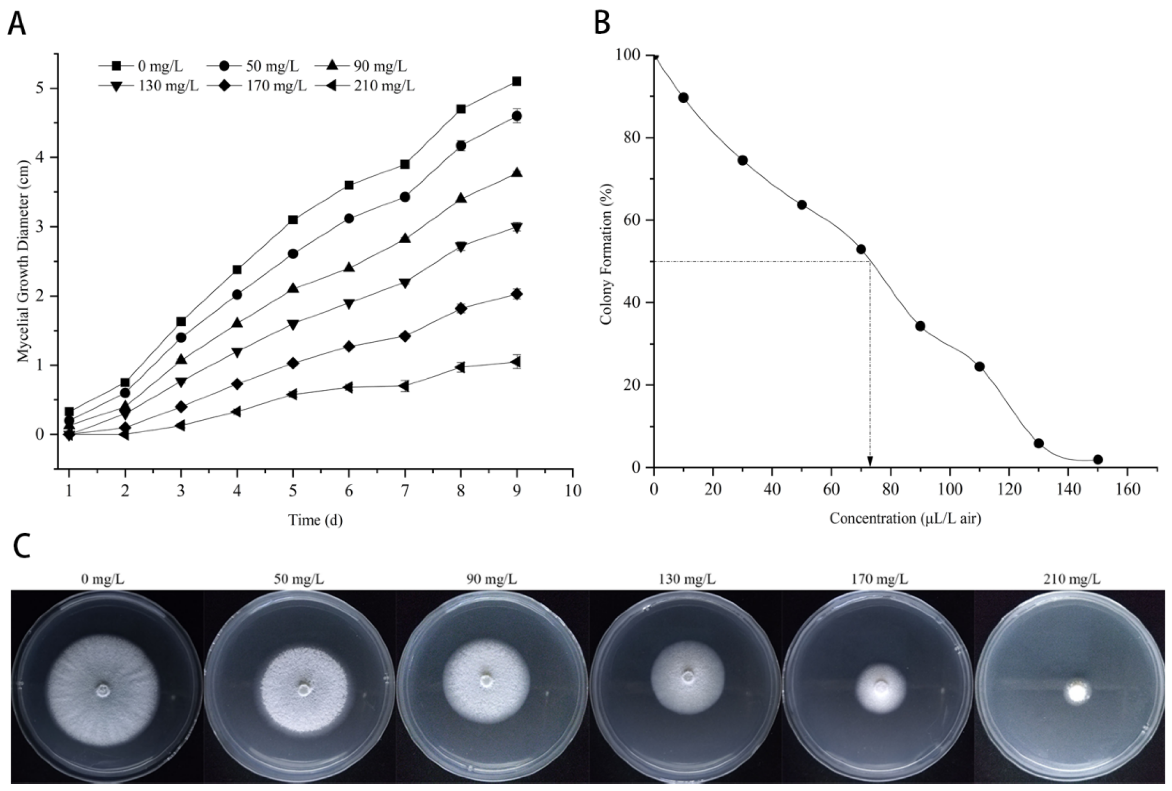
The inhibitory effect of citronellal against M. oryzae was determined by contact and indirect activity methods (Figure 2). The results show that citronellal has a good inhibition effect on M. oryzae with EC25, EC50, and EC75 values of 85 mg/L, 134 mg/L, and 218 mg/L, respectively. The experimental results of the indirect activity method (Figure 2B) show that the colony diameter of M. oryzae became smaller and smaller with increasing concentrations of citronellal. When the concentration of citronellal reached 150 μL/L, there was almost no trend of mycelial growth, with an EC50 value of 70.48 μL/L. It was shown that citronellal also had a better indirect activity inhibition effect on M. oryzae.

3.2. Effect of Citronellal on Mycelial Quantity of M. oryzae

As shown in Figure 3, after 72 h, the mycelium weight of the control groups was increased by 35.83%, while the mycelium weights of citronellal groups (85 and 134 mg/L) were significantly different from the control group with an increase of 1.15% and a decrease of 5.89%, respectively. However, the weight loss rate of mycelia significantly increased after treatment with a high content of citronellal with a loss rate of 88.59%. The results show that citronellal could inhibit the growth of mycelia in M. oryzae and reduce the weight of M. oryzae.

3.3. Effect of Citronellal on Mycelial Morphology of M. oryzae

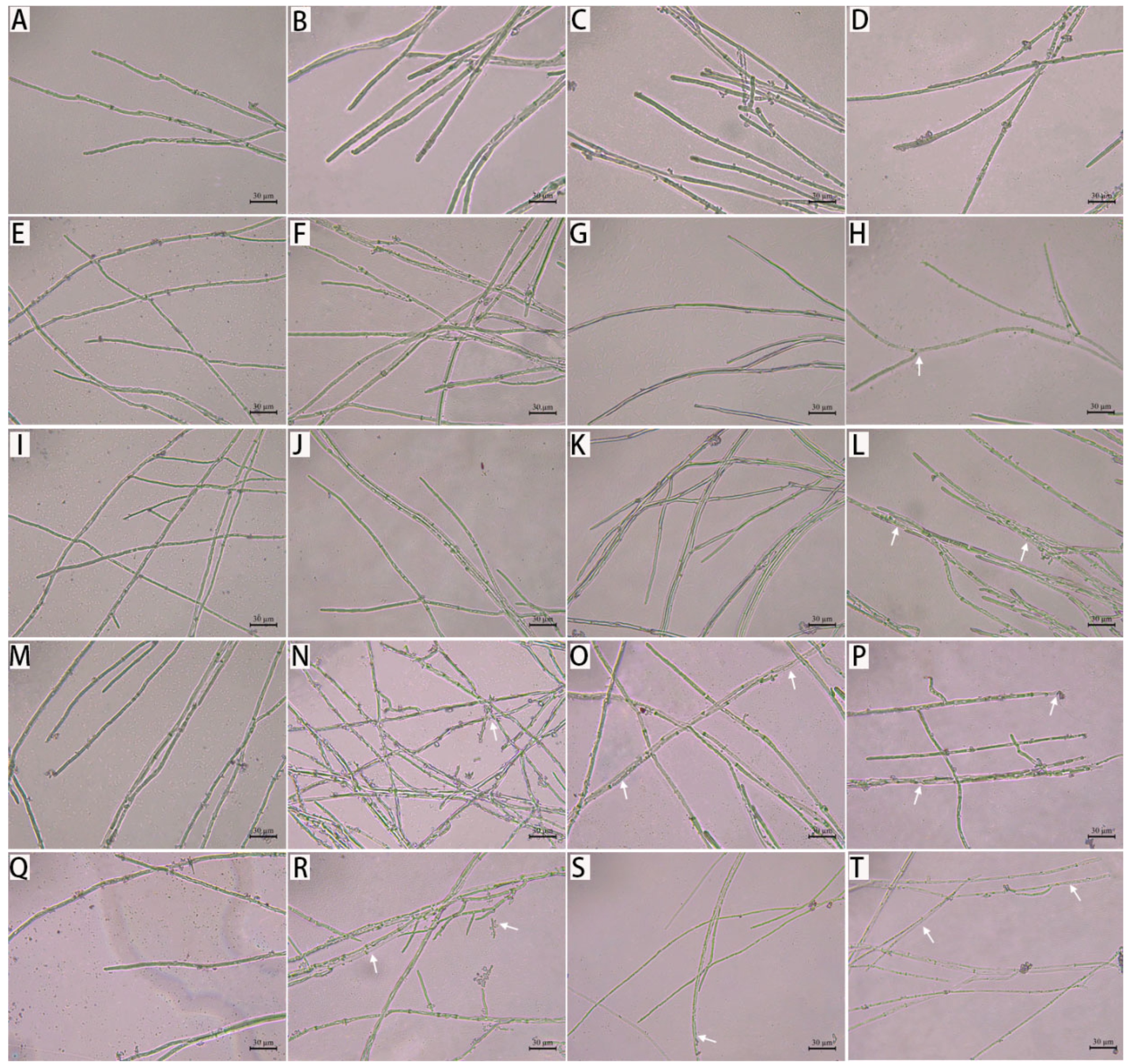
As shown in Figure 4, the mycelia of M. oryzae were damaged to varying degrees after treatment with different concentrations of citronellal at different times. The mycelium morphology of the control groups did not significantly change, with longer mycelia and a clear separation at different times. The mycelium morphology showed almost no difference between treatment groups before 36 h, and then, mycelium began to thin and deform over time. In particular, the mycelium of M. oryzae appeared vacuolated, broken, and shattered into pieces, and twisted after 72 h of treatment with citronellal. This phenomenon became increasingly evident as the citronellal increased. It suggested that citronellal may affect the growth of the pathogen.

3.4. Effect of Citronellal on Mycelial Morphology and Ultrastructure of M. oryzae

Under a scanning electron microscope, in the control groups (Figure 5A,B), the mycelia of M. oryzae were uniform in thickness. The mycelial surfaces of M. oryzae were smooth, plump, and clearly separated. In the citronellal groups (85 mg/L, Figure 5C,D), the mycelial surfaces of M. oryzae were slightly shrunken, swelled, and uneven in thickness. In Figure 5E,F, the mycelium surface of M. oryzae was partially deformed, partially ruptured, and wrinkled. As shown in Figure 5G,H, the mycelial surfaces of M. oryzae were abnormally enlarged, distorted, and even broken. It can be seen that different concentrations of citronellal damaged mycelial shape and structural integrity.
The ultrastructures of M. oryzae under different treatments, as observed under a transmission electron microscope, are shown in Figure 6. The mycelium ultrastructures of M. oryzae in the control group had regular cell morphologies, uniform cell wall textures, uniform thicknesses closely linked with the cell membrane, organelles evenly distributed in the cytoplasm, and clear and complete structures (Figure 6A,B). In the citronellal groups (Figure 6C,D), cells were slightly deformed and slightly vacuolated, and the distribution of intracellular substances was uneven. Following the exposure to 134 mg/L citronellal (Figure 6E,F), cells were invaginated and deformed. Obvious swelling, cytoplasmic wall separation extravasation, and many vesicular structures were observed in the cells, and there was even no complete organelle structure. When hyphae were exposed to 218 mg/L citronellal (Figure 6G,H), the cell wall of M. oryzae was severely lysed, while the cell membrane was invaginated. The plasmon of M. oryzae was clearly separated, and the intracellular structure was blurred with content extravasation. This indicated that the action site of citronellal against M. oryzae might be the cell wall of mycelium.

3.5. Mycelial PI and CFW Fluorescence Reaction of M. oryzae

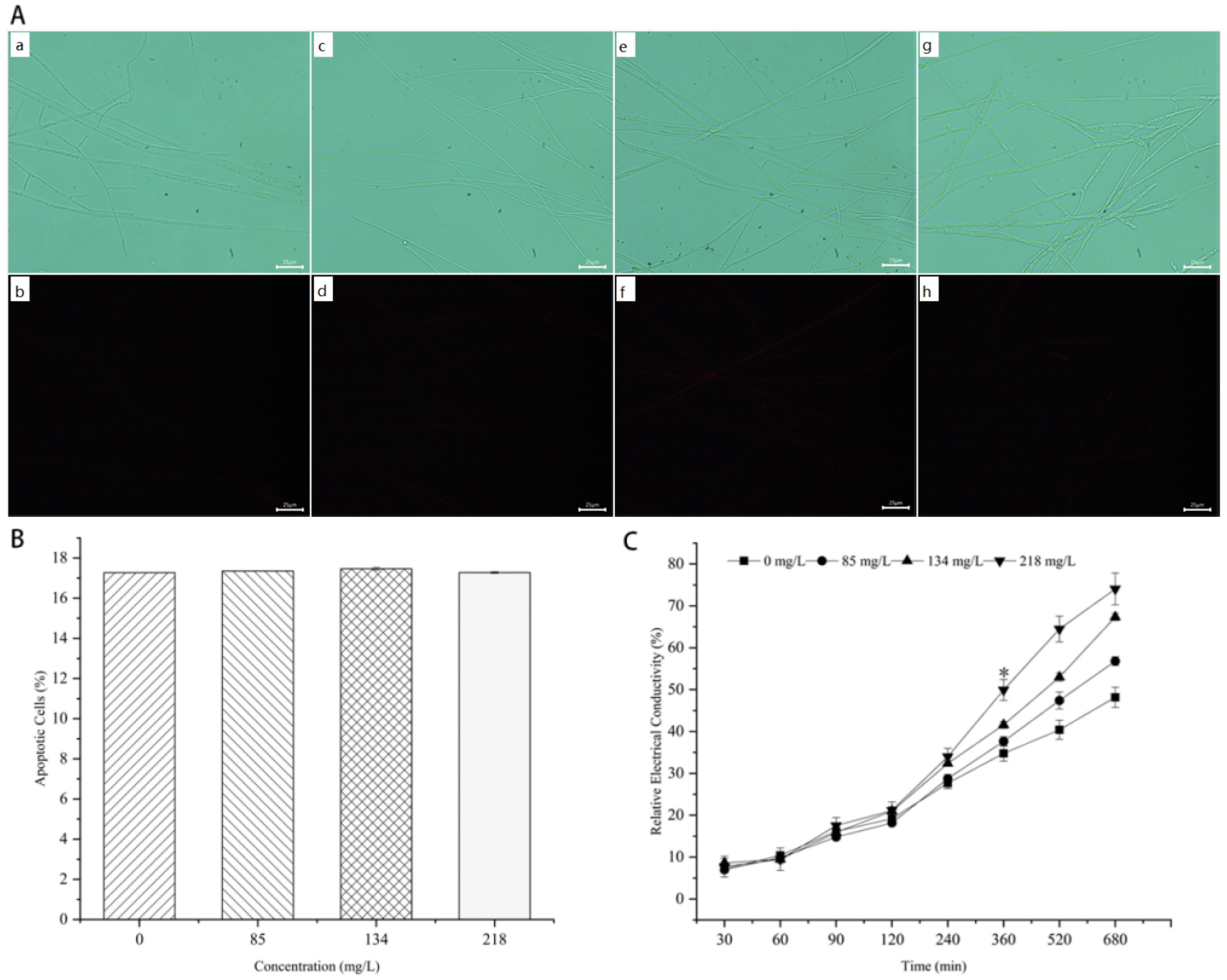
PI is a nucleic acid dye that cannot penetrate intact cell membranes. Necrotic cells have a strong fluorescence due to loss of membrane integrity, where the dye enters the cell and binds to DNA, while intact cell membranes are not damaged since the dye cannot penetrate into the cell and bind to DNA, and thus, it does not show fluorescence. Based on this feature, dead cells can be identified using PI staining, and the more significant the fluorescence, the more serious the cell membrane damage. As shown in Figure 7A,B, fluorescence could be barely observed in all the treatments, and there was no significant difference in the percentage of dead cells after fluorescence quantification. The results show that the fluorescence of mycelia hardly changes with the increase in citronellal concentration, indicating that citronellal did not damage the membrane integrity of M. oryzae.
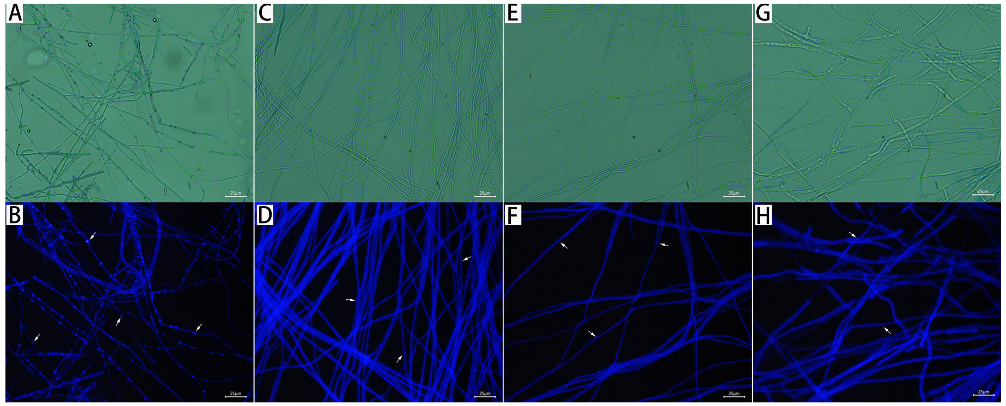
CFW is a non-specific fluorescent whitening dye that binds to cellulose and chitin at the cell wall to generate fluorescence, detecting changes in the formation of the cell wall, which has a strong blue fluorescence. As shown in Figure 8, all treated mycelia of M. oryzae had a blue fluorescence after 72 h. The fluorescence intensity of treated mycelium in nodes separation became increasingly blurred with the increase in citronellal content, while control mycelial nodes emitted a strong, bright and clear fluorescence, indicating that citronellol could destroy the integrity of the cell wall in M. oryzae.

3.6. Effect of Citronellal on Mycelial Cell Membrane Permeability of M. oryzae

Relative conductivity is an important index for measuring the permeability of the cell membrane. The relative conductivity of the mycelia in all the treatments steadily increased to 30–120 min (Figure 7C). After 120 min, the relative conductivities of the mycelia in low, medium, and high concentrations of citronellal treatments and control groups all increased from about 20%. The relative conductivity of the mycelia in all treatments significantly increased until reaching 360 min. There was a significant difference between the citronellal treatment group (218 mg/L) and other treatment groups, the relative conductivity of the treatment group (218 mg/L) at this time was 49.93%, and there were no significant differences between the other three groups (the relative conductivities are 34.78%, 37.65%, and 41.61%, respectively). These results further confirm that citronellal did not directly damage the cell membrane of the mycelium.

3.7. Effect of Citronellal on Chitinase Activity in M. oryzae

Chitin is an important component of the cell wall, and chitinase is an important indicator to evaluate its integrity. Absorbance was measured at 540 nm using a visible spectrophotometer, and there was a good linear relationship between the chitinase concentration of standard solvent and the area of the absorbed peak. The regression equation was Y = 0.33X − 0.2074, and the correlation coefficient of the standard curve was 0.9994 (Figure 9A). As shown in Figure 9C, the chitinase activity of mycelium in treatments with citronellal began to gradually increase after 24 h, reached a peak at 72 h, and then began to decline. While the chitinase activity of the mycelium in the control group did not significantly change with time, it fluctuated below 5 U/g, and remained lower than the overall level of treatment groups. Especially, the chitinase activities of the mycelia treated with 85 mg/L, 134 mg/L, and 218 mg/L citronellal were 42.43, 54.57, and 55.28 times higher than those of the control group after 72 h, respectively. This enhanced chitinase activity resulted in accelerated hydrolysis of chitin in the cell wall of M. oryzae, indicating that citronellal indirectly acted at the cell wall of M. oryzae and accelerated the death of M. oryzae cells.

3.8. Effect of Citronellal on Β-1,3-Glucanase Activity of M. oryzae

Glucan is an important component of the cell wall. β-1,3-glucanase can catalyze its hydrolysis. The absorbance was measured at 540 nm using a visible spectrophotometer, and standard curve R2 = 0.9991, Y = 0.5322X − 0.0635 was obtained by using β-1,3-glucanase concentration as a horizontal coordinate and the absorbance value as the vertical coordinate (Figure 9B). As shown in Figure 9D, for the control group mycelia and those treated with a medium concentration (134 mg/L) for 9 h, enzyme activity was higher than at low-concentration (85 mg/L) and high-concentration (218 mg/L) citronellal treatments. It first showed a decreasing trend, and then started to increase after 48 h, whereas the enzyme activity of mycelium had an increasing trend from a lower value at 9 h treatment with a low and high concentration of citronellal. The enzyme activity of mycelium in all the treatments reached a peak at 72 h, decreasing and stabilizing after 96 h. The difference in enzyme activity of mycelium in all the treatments was greatest after 48 h. The β-1,3-glucanase activity of the mycelium treated with citronellal (218 mg/L) was only 1.96 times higher than that in the control group. The overall trend of the treated groups was higher than the control group, but the difference was not significant, indicating that citronellal had no significant effect on β-1,3-glucan of M. oryzae.

3.9. Effect of Citronellal on Chitinase Gene Expression in M. oryzae

To further verify the regulatory effect of citronellal on the chitin synthesis pathway of M. oryzae, we selected 15 chitinase genes of M. oryzae for quantitative real-time PCR analysis. As shown in Figure 10A, the results show that expression levels of MGG_08054 and MGG_04073 were lower than those of the control group after treatment with citronellal for 72 h, and gene expressions were downregulated by 1.41 and 0.09 times, respectively. Meanwhile, the expression levels of the remaining 13 genes were higher than those of the control group and had an upregulation from 1.1790- to 4.1709-fold. Especially, expression levels of MGG_04732, MGG_01876, and MGG_03599 were upregulated by more than three-fold.

3.10. Effect of Citronellal on Β-1,3-Glucanase Gene Expression of M. oryzae

In order to further verify the effect of citronellal on the β-1,3-glucanase gene expression of M. oryzae, 17 related genes were screened for Quantitative Real-Time PCR analysis. The results show that the expression levels of five genes (MGG_01001, MGG_10400, MGG_04689, MGG_09619, and MGG_00865) were lower than those of the control treated with citronellal for 72 h and were downregulated by 0.61–1.44-fold (Figure 10B), while expression levels of the other 12 genes were upregulated by 4.09–17.47-fold, which were significantly higher than that of control. Therefore, most β-1,3-glucanase genes are positively regulated when they are transcribed into mRNA, which might affect the subsequent synthesis of β-1,3-glucan.

3.11. Indoor Control Effect of Citronellal on Rice Blast

In order to clarify the control effect of citronellal on a rice blast, a laboratory leaf test method was used for determination. The lesions on inoculated leaves gradually decreased, and the symptoms of the yellowing of leaves were alleviated with the increase in citronellal content after 14 d (Figure 11A). After quantifying symptoms, statistical results show that the control effects of treatment groups were significantly different: 14.29%, 25.00% and 82.14%, respectively (Figure 11B), indicating that citronellal had a good control effect on rice blast.

4. Discussion

M. oryzae can cause rice blast, which poses a serious threat to rice yields worldwide [36,37]. As a result, natural products with a low toxicity to non-target organisms have attracted a widespread attention of researchers [38]. Citronellal, an aromatic component present in essential oils of various plants, such as citronella oil and eucalyptus oil, can be extracted by various routes [39]. It is not only highly effective in mosquitoes, but also has strong antifungal properties against a variety of pathogenic fungi [40,41,42]. Studying the antifungal mechanism of citronellal can provide an important reference for subsequent research and applications and play an important role in agricultural applications.
In this study, the inhibition effect of citronellal was measured from the perspective of contact and indirect activity, and the inhibition mechanism of citronellal against M. oryzae was systematically studied from the perspective of morphological structures, physiology and molecular levels. The results show that citronellal had a strong inhibition effect on M. oryzae, increased the activity of chitinase, significantly up-regulated the expression level of related genes, and thus accelerated the hydrolysis of chitin. β-1,3-glucanase-related gene expression was significantly upregulated, while glucanase activity was not significantly different from the control group. This may be due to the presence of other related genes regulating the expression of glucanase. This expression inhibited the growth of mycelia and led to the destruction of the cell wall, which was consistent with the distortion and rupture of mycelia observed by SEM and the serious dissolution of cell walls observed by TEM. It was also found that citronellal had no significant effect on the cell membrane permeability of mycelium in M. oryzae by relative conductivity measurements, which was consistent with a lack of clear fluorescence in PI staining, and might be related to the fact that citronellal did not directly affect the cell membrane. Therefore, the main pathway of citronellal against M. oryzae might be related to genes regulating chitin synthesis, destroying cell walls, damaging cell function, and inhibiting mycelium growth. Thus, citronellal treatment has a significant protective effect on rice leaves. At present, the pharmacodynamic mechanism of citronellal had been widely studied in Penicillium and Candida albicans. For example, citronellal could reduce ergosterol synthesis in Penicillium digitorum spores by inhibiting the expression of ERG3, thus affecting spore germination. This impaired the cell membrane integrity of P. fingertip by downregulating the ERG gene responsible for converting lanolin to ergosterol [43,44], and exerted antifungal properties by inhibiting the biofilm formation of C. albicans [23].
The reported studies suggest that citronellal might exert its inhibition effect by affecting the synthesis of ergosterol and biofilm [45,46]. These results are inconsistent with the findings of this study. The possible factors for discrepancy included different pathogenic species or citronellal having multiple sites of action on the fungal cell wall and cell membrane. However, citral, which is also a natural product of aldehydes, was shown to inhibit Trichoderma viride [47] and M. oryzae [14] by disrupting the cell wall in studies of antifungal mechanisms. Cinnamaldehyde could also exhibit antifungal activity against Citrus sour rot and Fusarium by interfering with the construction of cell walls [48,49]. When used alone or in combination with cinnamaldehyde, citronellal could inhibit mycelial growth by accelerating the destruction of cell membranes and cell walls. It could also be used to prevent mold and prolong the shelf life of citrus, strawberry and other fruits [50,51,52]. These results support the conclusion of this study: citronellal has the potential for effective applications in the prevention and control of rice blasts.
This study screened genes that regulate chitin synthesis and confirmed the strong inhibitory effect of citronellal on M. oryzae and a better control effect on rice blast, exerting an antifungal effect via the synthesis of chitin and disruption of the cell wall. These conclusions could provide a reference for the selection of drugs for the prevention and control of rice blasts and lay the foundation for further study of molecular mechanisms, field applications, and the design and synthesis of target drugs. In particular, the indirect activity of citronellal brings more possibilities to its application. It could also be combined with related technologies to improve the biological activity of plant extracts [53,54,55] to improve its stability and achieve a slow release of the drug. Although this study reveals the antifungal mechanism of citronellal against M. oryzae and preliminarily elucidated its control effect on rice blast, its molecular regulation mechanism and field application technology still require further study.

5. Conclusions

Currently, rice blast is dependent on several types of chemicals, and thus, the development and application of natural products are extremely urgent. Citronellal has good antifungal activity against M. oryzae, and reveals its potential in the prevention and control of rice blasts. In this study, it was found that citronellal could affect the mycelia morphology of M. oryzae, destroy the cell wall, and inhibit mycelia growth. To further clarify the action mechanism of citronellal, the relevant indexes were determined to verify. The results show that citronellal up-regulated the expression of chitinase- and β-1,3-glucanase-related genes, and significantly increased chitinase activity. These results suggest that citronellal can upregulate the expression of genes related to chitin synthesis, and interfere with this synthesis, destroying the cell wall, inhibiting mycelia growth, and providing effective protection against rice blast. This study preliminarily clarifies the action mechanism of citronellal on M. oryzae and provides an effective method for the green prevention and control of rice blasts and the safe production of rice. However, the key genes in regulatory pathways and their specific regulatory relationships are not yet clear. The instability of citronellal limits its application in this field. Therefore, in the next stage, we aim to pay attention to key genes in the regulatory pathway and clarify their regulatory mechanism. Additionally, thorough research should be combined with related technologies to improve the biological activity of plant extracts and their potential for applications in the field of agriculture.

Author Contributions

Conceptualization, F.-X.M.; methodology, X.G. and K.H.; software, R.-T.L.; validation, Y.D.; formal analysis, A.-A.Z.; investigation, A.-A.Z.; resources, R.-Y.L.; data curation, A.-A.Z.; writing—original draft preparation, A.-A.Z.; writing—review and editing, R.-Y.L.; visualization, R.-Y.L.; supervision, M.L.; project administration, R.-Y.L.; funding acquisition, R.-Y.L. All authors have read and agreed to the published version of the manuscript.

Funding

This work was supported by the National Natural Science Foundation of China (No. 32160657, 32160658), and the Cultivation Project of Guizhou University (No. (2019) 43). Demonstration base for transfer and transformation of scientific and technological achievements in colleges and universities to serve rural revitalization, Guizhou provincial department of education (No. (2022) 060).

Institutional Review Board Statement

Not applicable.

Informed Consent Statement

This study did not involve humans.

Data Availability Statement

This study did not report any data.

Acknowledgments

The authors thank Li, R.-Y. for her help with the statistical analyses and Li, M. for her help with the methodology. All individuals included in this section have consented to the acknowledgement.

Conflicts of Interest

The authors declare no conflict of interest.

References

  1. Ito, S. Contemporary Global Rice Economies: Structural Changes of Rice Production/Consumption and Trade. J. Nutr. Sci. Vitaminol. 2019, 65, S23–S25. [Google Scholar] [CrossRef] [PubMed] [Green Version]
  2. Datta, K.; Baisakh, N.; Thet, K.M.; Tu, J.; Datta, S. Pyramiding transgenes for multiple resistance in rice against bacterial blight, yellow stem borer and sheath blight. Theor. Appl. Genet. 2002, 106, 1–8. [Google Scholar] [CrossRef] [PubMed]
  3. Gong, J.-T.; Li, Y.; Li, T.-P.; Liang, Y.; Hu, L.; Zhang, D.; Zhou, C.-Y.; Yang, C.; Zhang, X.; Zha, S.-S.; et al. Stable Introduction of Plant-Virus-Inhibiting Wolbachia into Planthoppers for Rice Protection. Curr. Biol. 2020, 30, 4837–4845.e5. [Google Scholar] [CrossRef] [PubMed]
  4. Jeon, J.; Kim, K.-T.; Choi, J.; Cheong, K.; Ko, J.; Choi, G.; Lee, H.; Lee, G.-W.; Park, S.-Y.; Kim, S.; et al. Alternative splicing diversifies the transcriptome and proteome of the rice blast fungus during host infection. RNA Biol. 2022, 19, 373–386. [Google Scholar] [CrossRef] [PubMed]
  5. Wang, Z.-Q.; Meng, F.-Z.; Yin, L.-F.; Yin, W.-X.; Lv, L.; Yang, X.-L.; Chang, X.-Q.; Zhang, S.; Luo, C.-X. Transcriptomic Analysis of Resistant and Wild-Type Isolates Revealed Fludioxonil as a Candidate for Controlling the Emerging Isoprothiolane Resistant Populations of Magnaporthe oryzae. Front. Microbiol. 2022, 13, 874497. [Google Scholar] [CrossRef] [PubMed]
  6. Bezerra, G.D.A.; Chaibub, A.A.; Oliveira, M.I.D.S.; Mizubuti, E.S.G.; de Filippi, M.C.C. Evidence of Pyricularia oryzae adaptability to tricyclazole. J. Environ. Sci. Health Part B 2021, 56, 869–876. [Google Scholar] [CrossRef]
  7. Ruan, H.; Tian, P.; Shi, N.; Du, Y.; Chen, F.; Chen, F. Characterization of pyraclostrobin-resistant Magnaporthe oryzae. J. Phytopathol. 2022, 170, 233–241. [Google Scholar] [CrossRef]
  8. Naranjo, S.E.; Ellsworth, P.C.; Frisvold, G.B. Economic Value of Biological Control in Integrated Pest Management of Managed Plant Systems. Annu. Rev. Èntomol. 2015, 60, 621–645. [Google Scholar] [CrossRef] [Green Version]
  9. Angane, M.; Swift, S.; Huang, K.; Butts, C.A.; Quek, S.Y. Essential Oils and Their Major Components: An Updated Review on Antimicrobial Activities, Mechanism of Action and Their Potential Application in the Food Industry. Foods 2022, 11, 464. [Google Scholar] [CrossRef]
  10. Chang, Y.; Harmon, P.F.; Treadwell, D.D.; Carrillo, D.; Sarkhosh, A.; Brecht, J.K. Biocontrol Potential of Essential Oils in Organic Horticulture Systems: From Farm to Fork. Front. Nutr. 2022, 8, 5138. [Google Scholar] [CrossRef]
  11. Zhang, X.; Guo, J.; Cheng, F.; Li, S. Cytochrome P450 enzymes in fungal natural product biosynthesis. Nat. Prod. Rep. 2021, 38, 1072–1099. [Google Scholar] [CrossRef] [PubMed]
  12. Li, R.-Y.; Wu, X.-M.; Yin, X.-H.; Liang, J.-N.; Li, M. The Natural Product Citral Can Cause Significant Damage to the Hyphal Cell Walls of Magnaporthe grisea. Molecules 2014, 19, 10279–10290. [Google Scholar] [CrossRef] [PubMed] [Green Version]
  13. Li, R.-Y.; Wu, X.-M.; Yin, X.-H.; Long, Y.-H.; Li, M. Naturally produced citral can significantly inhibit normal physiology and induce cytotoxicity on Magnaporthe grisea. Pestic. Biochem. Physiol. 2015, 118, 19–25. [Google Scholar] [CrossRef] [PubMed]
  14. Zhao, Q.; Ding, Y.; Song, X.; Liu, S.; Li, M.; Li, R.; Ruan, H. Proteomic analysis reveals that naturally produced citral can significantly disturb physiological and metabolic processes in the rice blast fungus Magnaporthe oryzae. Pestic. Biochem. Physiol. 2021, 175, 104835. [Google Scholar] [CrossRef] [PubMed]
  15. Xu, S.; Wang, B.; Li, L.; Zhou, Q.; Tian, M.; Zhao, X.; Peng, J.; Liu, F.; Chen, Y.; Xu, Y.; et al. Effects of camptothecin on the rice blast fungus Magnaporthe oryzae. Pestic. Biochem. Physiol. 2019, 163, 108–116. [Google Scholar] [CrossRef]
  16. Mo, F.; Hu, X.; Ding, Y.; Li, R.; Long, Y.; Wu, X.; Li, M. Naturally produced magnolol can significantly damage the plasma membrane of Rhizoctonia solani. Pestic. Biochem. Physiol. 2021, 178, 104942. [Google Scholar] [CrossRef]
  17. Zhao, Y.; Wang, Q.; Wu, X.; Jiang, M.; Jin, H.; Tao, K.; Hou, T. Unraveling the polypharmacology of a natural antifungal product, eugenol, against Rhizoctonia solani. Pest Manag. Sci. 2021, 77, 3469–3483. [Google Scholar] [CrossRef]
  18. Tibenda, J.J.; Yi, Q.; Wang, X.; Zhao, Q. Review of phytomedicine, phytochemistry, ethnopharmacology, toxicology, and pharmacological activities of Cymbopogon genus. Front. Pharmacol. 2022, 13, 997918. [Google Scholar] [CrossRef]
  19. Avoseh, O.N.; Ogunwande, I.A.; Lawal, O.A.; Atabo, J.; Ascrizzi, R.; Guido, F. Anti-inflammatory and anti-nociceptive activities of essential oil of Waltheria indica. Boletín Latinoam. Y Del Caribe Plantas Med. Y Aromáticas 2019, 18, 566–576. [Google Scholar] [CrossRef]
  20. Abbas, M.W.; Hussain, M.; Qamar, M.; Ali, S.; Shafiq, Z.; Wilairatana, P.; Mubarak, M.S. Antioxidant and Anti-Inflammatory Effects of Peganum harmala Extracts: An In Vitro and In Vivo Study. Molecules 2021, 26, 6084. [Google Scholar] [CrossRef]
  21. Wu, W.J.; Li, S.S.; Yang, M.; Lin, Y.W.; Zheng, K.B.; Akutse, K.S. Citronellal perception and transmission by Anopheles gambiae s.s. (Diptera: Culicidae) females. Sci. Rep. 2020, 10, 18615. [Google Scholar] [CrossRef] [PubMed]
  22. Tülek, Z.; Alaşalvar, H.; Başyiğit, B.; Berktas, S.; Salum, P.; Erbay, Z.; Telci, I.; Çam, M. Extraction optimization and microencapsulation of phenolic antioxidant compounds from lemon balm (Melissa officinalis L.): Instant soluble tea production. J. Food Process. Preserv. 2020, 45, 14995. [Google Scholar] [CrossRef]
  23. Singh, S.; Fatima, Z.; Hameed, S. Citronellal-induced disruption of membrane homeostasis in Candida albicans and attenuation of its virulence attributes. Rev. Soc. Bras. Med. Trop. 2016, 49, 465–472. [Google Scholar] [CrossRef] [PubMed] [Green Version]
  24. Saibabu, V.; Singh, S.; Ansari, M.; Fatima, Z.; Hameed, S. Insights into the intracellular mechanisms of citronellal in Candida albicans: Implications for reactive oxygen species-mediated necrosis, mitochondrial dysfunction, and DNA damage. Rev. Soc. Bras. Med. Trop. 2017, 50, 524–529. [Google Scholar] [CrossRef] [PubMed] [Green Version]
  25. Melo, R.; Armstrong, V.; Navarro, F.; Castro, P.; Mendoza, L.; Cotoras, M. Characterization of the Fungitoxic Activity on Botrytis cinerea of N-phenyl-driman-9-carboxamides. J. Fungi 2021, 7, 902. [Google Scholar] [CrossRef] [PubMed]
  26. Soylu, E.M.; Soylu, S.; Kurt, S. Antimicrobial Activities of the Essential Oils of Various Plants against Tomato Late Blight Disease Agent Phytophthora infestans. Mycopathologia 2006, 161, 119–128. [Google Scholar] [CrossRef] [PubMed]
  27. He, L.; Wang, M.; Wang, H.; Zhao, T.; Cui, K.; Zhou, L. iTRAQ proteomic analysis of the inhibitory effect of 1,6-O,O-diacetylbritannilactone on the plant pathogenic oomycete Phytophthora capsici. Pestic. Biochem. Physiol. 2022, 184, 105125. [Google Scholar] [CrossRef] [PubMed]
  28. Zhang, J.; Zhang, B.; Zhu, F.; Fu, Y. Baseline sensitivity and fungicidal action of propiconazole against Penicillium digitatum. Pestic. Biochem. Physiol. 2020, 172, 104752. [Google Scholar] [CrossRef]
  29. Wang, J.; Guo, X.; Ling, L.; Qiu, H.; Zhen, Z.; Wang, Y.; Sun, G. Application of the Fluorescent Dye BODIPY in the Study of Lipid Dynamics of the Rice Blast Fungus Magnaporthe oryzae. Molecules 2018, 23, 1594. [Google Scholar] [CrossRef] [Green Version]
  30. Elsherbiny, E.A.; Dawood, D.H.; Safwat, N.A. Antifungal action and induction of resistance by β-aminobutyric acid against Penicillium digitatum to control green mold in orange fruit. Pestic. Biochem. Physiol. 2020, 171, 104721. [Google Scholar] [CrossRef]
  31. Jiménez-Ortega, E.; Kidibule, P.E.; Fernández-Lobato, M.; Sanz-Aparicio, J. Structural inspection and protein motions modelling of a fungal glycoside hydrolase family 18 chitinase by crystallography depicts a dynamic enzymatic mechanism. Comput. Struct. Biotechnol. J. 2021, 19, 5466–5478. [Google Scholar] [CrossRef]
  32. Li, Q.; Zhao, Y.; Xie, Y. Paeonol Disrupts the Integrity of Aspergillus flavus Cell Walls via Releasing Surface Proteins, Inhibiting the Biosynthesis of β-1,3-Glucan and Promoting the Degradation of Chitin, and an Identification of Cell Surface Proteins. Foods 2021, 10, 2951. [Google Scholar] [CrossRef] [PubMed]
  33. Song, X.; Zhao, Q.; Zhou, A.; Wen, X.; Li, M.; Li, R.; Liao, X.; Xu, T. The Antifungal Effects of Citral on Magnaporthe oryzae Occur via Modulation of Chitin Content as Revealed by RNA-Seq Analysis. J. Fungi 2021, 7, 1023. [Google Scholar] [CrossRef] [PubMed]
  34. Xin, W.; Mao, Y.; Lu, F.; Li, T.; Wang, J.; Duan, Y.; Zhou, M. In vitro fungicidal activity and in planta control efficacy of coumoxystrobin against Magnaporthe oryzae. Pestic. Biochem. Physiol. 2019, 162, 78–85. [Google Scholar] [CrossRef] [PubMed]
  35. Li, Q.; Jiang, Y.; Ning, P.; Zheng, L.; Huang, J.; Li, G.; Jiang, D.; Hsiang, T. Suppression of Magnaporthe oryzae by culture filtrates of Streptomyces globisporus JK-1. Biol. Control 2011, 58, 139–148. [Google Scholar] [CrossRef]
  36. Chung, H.; Jeong, D.G.; Lee, J.-H.; Kang, I.J.; Shim, H.-K.; An, C.J.; Kim, J.Y.; Yang, J.-W. Outbreak of Rice Blast Disease at Yeoju of Korea in 2020. Plant Pathol. J. 2022, 38, 46–51. [Google Scholar] [CrossRef]
  37. Fernandez, J.; Orth, K. Rise of a Cereal Killer: The Biology of Magnaporthe oryzae Biotrophic Growth. Trends Microbiol. 2018, 26, 582–597. [Google Scholar] [CrossRef]
  38. Pavela, R.; Benelli, G. Essential Oils as Ecofriendly Biopesticides? Challenges and Constraints. Trends Plant Sci. 2016, 21, 1000–1007. [Google Scholar] [CrossRef]
  39. Yang, G.-Z.; Zhu, J.-K.; Yin, X.-D.; Yan, Y.-F.; Wang, Y.-L.; Shang, X.-F.; Liu, Y.-Q.; Zhao, Z.-M.; Peng, J.-W.; Liu, H. Design, Synthesis, and Antifungal Evaluation of Novel Quinoline Derivatives Inspired from Natural Quinine Alkaloids. J. Agric. Food Chem. 2019, 67, 11340–11353. [Google Scholar] [CrossRef]
  40. Kaur, H.; Bhardwaj, U.; Kaur, R.; Kaur, H. Chemical Composition and Antifungal Potential of Citronella (Cymbopogon nardus) Leaves Essential Oil and its Major Compounds. J. Essent. Oil Bear. Plants 2021, 24, 571–581. [Google Scholar] [CrossRef]
  41. Kaur, H.; Bhardwaj, U.; Kaur, R. Cymbopogon nardus essential oil: A comprehensive review on its chemistry and bioactivity. J. Essent. Oil Res. 2021, 33, 205–220. [Google Scholar] [CrossRef]
  42. Pontes, E.K.U.; Melo, H.M.; Nogueira, J.W.A.; Firmino, N.C.S.; de Carvalho, M.G.; Júnior, F.E.A.C.; Cavalcante, T.T.A. Antibiofilm activity of the essential oil of citronella (Cymbopogon nardus) and its major component, geraniol, on the bacterial biofilms of Staphylococcus aureus. Food Sci. Biotechnol. 2018, 28, 633–639. [Google Scholar] [CrossRef] [PubMed]
  43. Liu, J.; Zong, Y.; Qin, G.; Li, B.; Tian, S. Plasma Membrane Damage Contributes to Antifungal Activity of Silicon Against Penicillium digitatum. Curr. Microbiol. 2010, 61, 274–279. [Google Scholar] [CrossRef] [PubMed]
  44. Wu, Y.; OuYang, Q.; Tao, N. Plasma membrane damage contributes to antifungal activity of citronellal against Penicillium digitatum. J. Food Sci. Technol. 2016, 53, 3853–3858. [Google Scholar] [CrossRef] [PubMed] [Green Version]
  45. Timper, P.; Wilson, J.P.; Johnson, A.W.; Hanna, W.W. Evaluation of Pearl Millet Grain Hybrids for Resistance to Meloidogyne spp. and Leaf Blight Caused by Pyricularia grisea. Plant Dis. 2002, 86, 909–914. [Google Scholar] [CrossRef] [PubMed] [Green Version]
  46. Trindade, L.A.; Cordeiro, L.V.; Silva, D.D.F.; Figueiredo, P.T.R.; de Pontes, M.L.C.; Lima, E.D.O.; Carvalho, A.D.A.T. The antifungal and antibiofilm activity of Cymbopogon nardus essential oil and citronellal on clinical strains of Candida albicans. Braz. J. Microbiol. 2022, 53, 1231–1240. [Google Scholar] [CrossRef] [PubMed]
  47. Zhang, J.; Du, C.; Li, Q.; Hu, A.; Peng, R.; Sun, F.; Zhang, W. Inhibition mechanism and antibacterial activity of natural antibacterial agent citral on bamboo mould and its anti-mildew effect on bamboo. R. Soc. Open Sci. 2021, 8, 202244. [Google Scholar] [CrossRef]
  48. Tan, X.; Long, C.; Meng, K.; Shen, X.; Wang, Z.; Li, L.; Tao, N. Transcriptome sequencing reveals an inhibitory mechanism of Penicillium digitatum by sodium dehydroacetate on citrus fruit. Postharvest Biol. Technol. 2022, 188, 111898. [Google Scholar] [CrossRef]
  49. Wang, Z.-Q.; Meng, F.-Z.; Zhang, M.-M.; Yin, L.-F.; Yin, W.-X.; Lin, Y.; Hsiang, T.; Peng, Y.-L.; Wang, Z.-H.; Luo, C.-X. A Putative Zn2Cys6 Transcription Factor Is Associated With Isoprothiolane Resistance in Magnaporthe oryzae. Front. Microbiol. 2018, 9, 2608. [Google Scholar] [CrossRef] [Green Version]
  50. OuYang, Q.; Okwong, R.O.; Chen, Y.; Tao, N. Synergistic activity of cinnamaldehyde and citronellal against green mold in citrus fruit. Postharvest Biol. Technol. 2019, 162, 111095. [Google Scholar] [CrossRef]
  51. Xu, Y.; Tong, Z.; Zhang, X.; Wang, Y.; Fang, W.; Li, L.; Luo, Z. Unveiling the Mechanisms for the Plant Volatile Organic Compound Linalool To Control Gray Mold on Strawberry Fruits. J. Agric. Food Chem. 2019, 67, 9265–9276. [Google Scholar] [CrossRef] [PubMed]
  52. OuYang, Q.; Liu, Y.; Oketch, O.; Zhang, M.; Shao, X.; Tao, N. Citronellal Exerts Its Antifungal Activity by Targeting Ergosterol Biosynthesis in Penicillium digitatum. J. Fungi 2021, 7, 432. [Google Scholar] [CrossRef] [PubMed]
  53. Mouhoub, A.; Guendouz, A.; Belkamel, A.; Talibi, Z.E.A.; Koraichi, S.I.; El Modafar, C.; Delattre, C. Assessment of the antioxidant, antimicrobial and antibiofilm activities of essential oils for potential application of active chitosan films in food preservation. World J. Microbiol. Biotechnol. 2022, 38, 1–16. [Google Scholar] [CrossRef] [PubMed]
  54. Hosseini, S.F.; Ghaderi, J.; Gómez-Guillén, M.C. Tailoring physico-mechanical and antimicrobial/antioxidant properties of biopolymeric films by cinnamaldehyde-loaded chitosan nanoparticles and their application in packaging of fresh rainbow trout fillets. Food Hydrocoll. 2021, 124, 107249. [Google Scholar] [CrossRef]
  55. Duarte, L.G.; Picone, C.S. Antimicrobial activity of lactoferrin-chitosan-gellan nanoparticles and their influence on strawberry preservation. Food Res. Int. 2022, 159, 111586. [Google Scholar] [CrossRef]
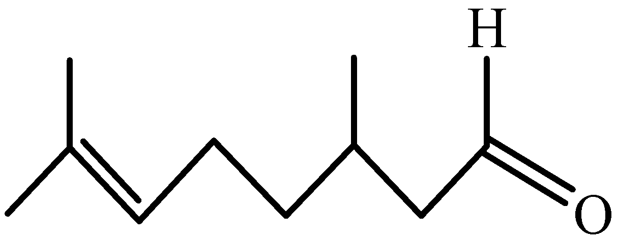
Figure 1. Chemical structure of citronellal.
Jof 08 01310 g001
Figure 2. Inhibitory effect of citronellal on M. oryzae. Error bars denote standard error of mean for three independent experiments, and below is the same. (A) Mycelial growth rate method, (B) Indirect activity method, (C) The colony formation of M. oryzae by mycelial growth velocity method.
Jof 08 01310 g002
Figure 3. The loss rate of mycelium. Different lowercase letters denote statistically significant differences at α = 0.05; below is the same.
Jof 08 01310 g003
Figure 4. Effects of citronellal on morphology of M. oryzae: 36 h: (A) (CK), (B) (85 mg/L), (C) (134 mg/L), (D) (218 mg/L); 48 h: (E) (CK), (F) (85 mg/L), (G) (134 mg/L), (H) (218 mg/L); 72 h: (I) (CK), (J) (85 mg/L), (K) (134 mg/L), (L) (218 mg/L); 96 h: (M) (CK), (N) (85 mg/L), (O) (134 mg/L), (P) (218 mg/L); 120 h: (Q) (CK), (R) (85 mg/L), (S) (134 mg/L), (T) (218 mg/L). The scale bar is 30 μm.
Jof 08 01310 g004
Figure 5. Morphology of mycelium after 72 h treatment with different concentrations of citronellal. (A,B): CK; (C,D): 85 mg/L; (E,F): 134 mg/L; (G,H): 218 mg/L.
Jof 08 01310 g005
Figure 6. Ultrastructure of mycelial cells after 72 h treatment with different concentrations of citronellal: (A,B): CK; (C,D): 85 mg/L; (E,F): 134 mg/L; (G,H): 218 mg/L. The arrows in the figure indicate cell wall (cw) and vacuole (v).
Jof 08 01310 g006
Figure 7. Effect of citronellal on the cell membrane of M. oryzae. (A,B): PI fluorescence and nonfluorescent effects of mycelia after treatment with citronellal for 72 h: (a,b): CK; (c,d): 85 mg/L; (e,f): 134 mg/L; (g,h): 218 mg/L. (C): Effect of citronellal on mycelial cell membrane permeability of M. oryzae.
Jof 08 01310 g007
Figure 8. CFW fluorescence effect and nonfluorescent of mycelia after treatment with citronellal for 72 h. (A,B): CK; (C,D): 85 mg/L; (E,F): 134 mg/L; (G,H): 218 mg/L.
Jof 08 01310 g008
Figure 9. Enzyme activity of mycelium in M. oryzae treating with citronellal. Standard curve of chitinase (A) and β-1,3-glucanase (B); Change trend of chitinase (C) and β-1,3-glucanase (D) activity.
Jof 08 01310 g009
Figure 10. Relative expression level of gene of chitinase (A) and β-1,3-glucanase (B).
Jof 08 01310 g010
Figure 11. Indoor control effect of citronellal on rice blast. (A) Rice leaves inoculated with M. oryzae. (B) Control effect. Error bars indicate means ± SD (n = 3).
Jof 08 01310 g011
Table 1. Primer design for RT-qPCR.
GenePrimer NameForward Primer (5′–3′)Reverse Primer (5′–3′)
actinMGG_03982CTGGCACCGTCGTCGATGAAGAAGGTCCGCTCTCGTCGTACTC
42 kDa endochitinaseMGG_11231GGCAACTCCAGCGAGATATTTCCCGGTATTCCCAGTCAACGTCAATCCC
class III chitinaseMGG_10333CCTCGCCTGTGCCTTCCATTTGGTGACCAACAAAGAAGACGCCAAAC
chitinase 1MGG_08054CTCATCCCAACAAACTCGGCGTCAAGCCACTCATAT
putative uncharacterized proteinMGG_17153ACCCGCTGGATGGTGTACTACGCGACGAGTTGAGGAACAAGTCTGAC
acidic mammalian chitinaseMGG_04732AAGGGCAAGTACATCACCGACAACCATGGCATCGACGACAATGTTCTTG
chitinase 1MGG_05533CGTCGCCTATGTGACCAACTGGGGCAAGAATATCCGCAAACGCATAC
class III chitinaseMGG_04073CATCACGCTCAACGACCACCACCCCTCCCAGCATCCCGAGAATC
chitinase 3MGG_01876CGTCGCTCTCCTAGCAACAACAGGAGTGCGTCGTCTGGAAGTAGATG
bacteriodes thetaiotaomicron symbiotic chitinaseMGG_01336CGGAGGAAACGTGACCACAGACTTGACAGCCAGTGCCACAATGAG
chitinase 1MGG_01247AGAACCGTTACTGTCACCGTTGATGGGACTTGTGCCAGAGGGTATGATG
chitinaseMGG_04534CCACCTGTCACTACAAGAGCGAATGAAGCCAAACTCTGAGCAGCACAC
endochitinaseMGG_00086CACCCATCTGCTTTATGCGTTTGCTCTGTGGGGTAGTGCTTGGAGAC
chitinase 1 precursorMGG_08458CCCTCACCTCCCAGAAACATTGAGTAGGCTGCGATGGATA
endochitinase 1MGG_07927GTCGTTTCCTTCGCCCTGATAGTCGTGGAGAAGATTGTGGTGGAGTGTC
acidic endochitinase SE2MGG_03599TTAGGCTGCTGACGGCTAGTCCGTCAAACCTCACCGCCCGAATC
Table 2. Primer design for RT-qPCR.
GenePrimer NameForward Primer (5′–3′)Reverse Primer (5′–3′)
glucan 1,3-beta-glucosidaseMGG_00659CGACAAACATCCCACCTCAGACTCAGAAACCGCCAGACCCAGACTC
1,3-beta-glucanosyltransferase gel2MGG_06722ATTTCAAGAAGGTTCAGGGCGTCTCCGGGCAGCGTAAAGTTGTTGTTG
glucan 1,3-beta-glucosidase 2MGG_06512CCAGGACCGTTACCGCAACATCACCATTCTTCCGCACCAAGTCATAC
endoglucanase family 5 glycoside hydrolaseMGG_07846CACCACTTCTTCACAGAGGCAGACGCTTCTTGATGACGGACGGGTTG
endo-1,3(4)-beta-glucanase 1MGG_01001ACCGAGGACTTGCCATCAATATGCGTGAGGATGCCGATGTTGGTGTC
1,3-beta-glucanosyltransferase gel1MGG_07331CCGCCAAGATTGAGGATGCTGAGTTGTTGCTGCCCTGGTTGCTAG
glycolipid-anchored surface protein 5MGG_03208TGGATGGGATGGAACTGGGATGGTAAAGTGCCCGTGACCTCTCCTG
1,3-beta-glucanosyltransferase gel3MGG_08370GGGTCGTTGGTTAGGTGTTGGATACCCGCACCACTCGTAGATGTTGTAG
GPI-anchored cell wall beta-1,3-endoglucanase EglCMGG_10400GCAATCCAGGGCTTCAACTACGGTACAGGCGGGCAGAGTTCCATC
glucan 1,3-beta-glucosidaseMGG_04689CATCACCAAGAACCTGTCCACCAAGTGTTGCTACCACCAGTGCTGTTTC
beta 1,3 exoglucanaseMGG_14087GCAAGGCAGACGACACAGTAGCAGGAGTGGTGGTGGTGGAGTTG
glucan 1,3-beta-glucosidaseMGG_09995TTACCTTGTCAGCGGCACGATTCGATGACTCCCAGCCCAAGAAACG
glucan 1,3-beta-glucosidaseMGG_17486ACTTCTTCCGCTCAACTCGCAACGGTGTTCTCCTCAGTACGCATCTTG
1,3-beta-glucanosyltransferase gel4MGG_11861CGCAGTACGACCAGCACATCAAGAGCACCGCCAATACCGTCAATG
GPI-anchored cell wall beta-1,3-endoglucanase EglCMGG_09619AGGCGAGCGGTCAGGATAATGGACCCAGTCTCCGTGACCCAAAC
endo-beta-1,3-glucanaseMGG_10591CACTACGACGACCGCAGTCAATACGTCCTCCTGTTGTTGGTGGTGATG
1,3-beta-glucan synthase component FKS1MGG_00865CGACGCCAACCAGGACAACTATCGACGGCATTCTTGACACCAGGAG
Publisher’s Note: MDPI stays neutral with regard to jurisdictional claims in published maps and institutional affiliations.