Article

Participation of the ABC Transporter CDR1 in Azole Resistance of Candida lusitaniae

by
Valentin Borgeat
1,
Danielle Brandalise
1,
Frédéric Grenouillet
2,3 and
Dominique Sanglard
1,*
1
Institute of Microbiology, University of Lausanne and University Hospital, CH-1011 Lausanne, Switzerland
2
Pole de Biologie Médicale, Centre Hospitalier Universitaire (CHU) Besançon, F-25000 Besançon, France
3
Chrono-Environnement Unité Mixte de Recherche (UMR) 6249, Centre National de la Recherche Scientifique (CNRS), Université Bourgogne Franche-Comté, F-25000 Besançon, France
*
Author to whom correspondence should be addressed.
J. Fungi 2021, 7(9), 760; https://doi.org/10.3390/jof7090760
Submission received: 25 August 2021 / Revised: 10 September 2021 / Accepted: 13 September 2021 / Published: 15 September 2021
(This article belongs to the Special Issue Clinical Resistance to Antifungal Mechanism)

Abstract

:
Candida lusitaniae is an opportunistic pathogen in humans that causes infrequent but difficult-to-treat diseases. Antifungal drugs are used in the clinic to treat C. lusitaniae infections, however, this fungus can rapidly acquire antifungal resistance to all known antifungal drugs (multidrug resistance). C. lusitaniae acquires azole resistance by gain-of-function (GOF) mutations in the transcriptional regulator MRR1. MRR1 controls the expression of a major facilitator transporter (MFS7) that is important for fluconazole resistance. Here, we addressed the role of the ATP Binding Cassette (ABC) transporter CDR1 as additional mediator of azole resistance in C. lusitaniae. CDR1 expression in isolates with GOF MRR1 mutations was higher compared to wild types, which suggests that CDR1 is an additional (direct or indirect) target of MRR1. CDR1 deletion in the azole-resistant isolate P3 (V688G GOF) revealed that MICs of long-tailed azoles, itraconazole and posaconazole, were decreased compared to P3, which is consistent with the role of this ABC transporter in the efflux of these azoles. Fluconazole MIC was only decreased when CDR1 was deleted in the background of an mfs7Δ mutant from P3, which underpins the dominant role of MFS7 in the resistance of the short-tailed azole fluconazole. With R6G efflux readout as Cdr1 efflux capacity, our data showed that R6G efflux was increased in P3 compared to an azole-susceptible wild type parent, and diminished to background levels in mutant strains lacking CDR1. Milbemycin oxim A3, a known inhibitor of fungal ABC transporters, mimicked efflux phenotypes of cdr1Δ mutants. We therefore provided evidence that CDR1 is an additional mediator of azole resistance in C. lusitaniae, and that CDR1 regulation is dependent on MRR1 and associated GOF mutations.

1. Introduction

Candida spp. can cause invasive and opportunistic fungal diseases associated with variable mortality in immunocompromised patients as well as those with cancer and under hematopoietic cell transplantation [1]. Candida albicans is considered to be the most common species isolated from blood cultures, however, fungal infections caused by non-albicans Candida species are on the rise [2]. Among the non-albicans Candida species, Candida lusitaniae is an uncommon pathogen that accounts for approximately 1% of isolates in large datasets of adult and pediatric patients with candidemia and other forms of invasive candidiasis [3]. C. lusitaniae has the ability to rapidly acquire resistance to currently used antifungals including azoles, echinocandins, polyenes and pyrimidine analogs. While resistance to single agents has been reported in this species [4,5], resistance to multiple agents (multidrug resistance, MDR) has also been documented [6,7]. Candida auris, which is phylogenetically closely related to C. lusitaniae and is a recently emerging fungal pathogen, exhibits quite similar characteristics in terms of rapid drug resistance acquisition and multidrug resistance [8].
Antifungal drug resistance occurs by several molecular mechanisms involving specific genome mutations [9]. In C. albicans, at least two transcriptional regulators involved in drug resistance, TAC1 and MRR1, have been described. While TAC1 regulates ABC transporter genes such as CDR1 and CDR2, MRR1 regulates transporter genes of another family called major facilitators, among which is MDR1 [10,11]. In C. lusitaniae, an important mediator of fluconazole resistance is the transcription factor MRR1 (CLUG_00542). Mutations in this factor (gain-of-function mutations, GOF) result in the transcriptional activation of several genes, the Major Facilitator transporter MFS7 (CLUG_01938, also named MDR1, 56% identity with MDR1 of C. albicans) being one of these [5,7,12]. The deletion of both genes in C. lusitaniae is critical for azole resistance [5,7,12]. Interestingly, MRR1 and its target MFS7 are also involved in resistance against the pyrimidine analog 5-fluorocytosine (5-FC), probably by active efflux of the drug [7].
Transcriptional studies have revealed other potential mediators of fluconazole resistance in C. lusitaniae [5,7,12]. Inspecting the genes that were upregulated in azole-resistant isolates or those upregulated by the presence of MRR1 GOF mutations in C. lusitaniae, one interesting candidate was the homolog of the C. albicans ABC transporter CDR1 named CLUG_03113. This gene (about 90% similarity with CDR1 of C. albicans) was renamed CDR1 in the present study. Here, we addressed the role of CDR1 in the azole resistance of C. lusitaniae. We confirmed first that a novel MRR1 GOF from a clinical azole-resistant isolate was associated with CDR1 upregulation. We observed that CDR1 expression was under the control of MRR1. The inactivation of CDR1 in C. lusitaniae resulted in the decreased efflux capacity of the fluorophore rhodamine 6G, and was associated with decreased resistance against long-tailed azoles such as itraconazole and posaconazole, which are known ABC transporter substrates. Together our data highlight a novel regulatory association between the ABC transporter CDR1 and the transcriptional activator MRR1 in C. lusitaniae.

2. Materials and Methods

2.1. Strains, Media and Primers

C. lusitaniae isolates were grown in complete medium Yeast Extract Peptone Dextrose (YEPD) (1% Bacto peptone, Difco Laboratories, Basel, Switzerland), 0.5% yeast extract (Difco) and 2% glucose (Fluka, Buchs, Switzerland) at 30 °C under agitation. YEPD agar plate were used containing 2% agar (Difco). When required, YEPD was supplemented with 200 µg/mL nourseothricin (Werner BioAgents, Jena, Germany) or with 250 µg/mL hygromycin (Mediatech, Manassas, VA, USA). Strains and primers used in this study can be found in Tables S1 and S2. As earlier described, P3 isolate was recovered from a patient treated with multiple agents about 2 months after recovery of the initial susceptible isolate P1 [6].

2.2. Case Report of C. lusitaniae Infection

A female preterm neonate was born at 25.7 weeks of gestational age by spontaneous delivery after premature rupture of membranes. She was admitted to the Neonatal Intensive Care Unit (NICU, CHRU Besançon) and weighed 880 g. She was intubated at birth for poor respiratory effort. Apgar scores were 3 and 5 at 1 and 5 min, respectively. Central venous catheters were placed. Proteus mirabilis (>100 CFU, Colony Forming Unit) and C. lusitaniae (10 CFU) were isolated from vagina of the mother before delivery. Proteus mirabilis was also isolated in gastric aspirate of infant, and diagnosis of chorioamnionitis was retained. Cefotaxime and amikacine were administered intravenously for 8 and 2 days, respectively. Intravenously, fluconazole prophylaxis was a standard protocol in the NICU for premature infants with a birth weight of less than 1000 g, or a gestational age of less than 28 weeks, in agreement with international guidelines [13,14]. On day 2, fluconazole was given at a dose of 3 mg per kilogram of body weight every third day for the first two weeks. Sampling for fungal colonization detection (axillae, umbilicus, anus) allowed repeated isolation of few C. lusitaniae on axillae (day 2: 10 CFU, day 12: 40 CFU). Urine sampled on day 14 showed 700 CFU/mL C. lusitaniae. Fluconazole regimen were thus increased to 6 mg/kg every two days until day 24, and later continued at prophylactic dosage 3 mg/kg every three day until day 43. Infant received concomitantly new cure of cefotaxime (from day 29 to day 36) for pneumonia due to Haemophilus parainfluenzae. Systematic fungal screening (groin, axillae, nose, urine) performed on day 40 showed high colonization of groin with C. lusitaniae (>200 CFU, isolate later called DSY4941). E-test method revealed high minimal inhibitory concentration (MIC) to fluconazole (128 µg/mL; read at 48 h). On day 42, infant presented with fever of unexplained origin, leading to fluconazole withdrawal and liposomal amphotericin B implementation (5 mg/kg/d, from day 42 to day 55). Blood cultures remained negative. She was extubated at day 50. No other infectious complication was further suspected. She was discharged from NICU, then hospital, at day 63 and day 91, respectively. Long-term follow-up did not show any abnormality or neurodevelopmental impairment. C. lusitaniae isolated from infant before day 40 and from mother’s vagina were unfortunately not stored for further investigations.

2.3. Deletion of CDR1

In order to inactivate CDR1 (CLUG_03113) in different strain backgrounds, a CRISPR approach was used. First, a repair fragment was constructed by fusion PCR using the hygromycin resistance marker from pYM70 [15] and C. lusitaniae genomic DNA. The first PCR fragment amplified the 5′ UTR (untranslated region) of CDR1 from isolate P1 with primers ClCDR1-P1 and ClCDR1-TEF-5R (20-bp overlap with pYM70). The second PCR fragment amplified the selective marker HygR from pYM70 with primers CLCDR1-TEF-5f (20-bp overlap with CDR1) and ClCDR1-ACT1-3r (20-bp overlap with pYM70). The third PCR fragment amplified the 3′ UTR of CDR1 with primers ClCDR1-P2 and ClCDR1-ACT1-3f (20-bp overlap with CDR1). These PCRs were carried out with in a peqStar Instrument (Peqlab, Erlangen, Germany) with Taq DNA Polymerase (New England Biolabs, Ipswich, MA, USA). PCR fragments were purified with NucleoSpin Gel and PCR clean-up kit (Macherey-Nagel, Oensingen, Switzerland). The final PCR was performed with the three purified fragments and Phusion DNA Polymerase (New England Biolabs, Ipswich, MA, USA) in the presence of the nested primers ClCDR1-P3 and ClCDR1-P4 with the addition of 1 M betaine, which increases PCR yields by inhibiting secondary structure formation [16].
To target deletion of CDR1, the RNA-protein complexes (RNPs) approach was used that employs reconstituted purified Cas9 protein in complex with scaffold and gene-specific guide RNAs [17]. gRNAs specific for CDR1 (crRNAs: ClCDR1_5_Cas9 and ClCDR1_3_Cas9) were obtained from IDT (Integrated DNA Technologies, Inc., Coralville, IO, USA) as CRISPR guide RNA (crRNA), which contains 20-bp homologous to the target gene fused to the scaffold sequence. Gene-specific RNA guides were designed in silico using Geneious Prime (Biomatters, Ltd., Auckland, New Zealand). RNPs were created as following: briefly, crRNAs and tracrRNA (a universal transactivating CRISPR RNA) were dissolved in RNase-free distilled water (dH2O) at 100 µM and stored at −80 °C. Before use, each crRNA and the tracrRNA were diluted to 16 µM and the complete guide RNAs were generated by mixing equimolar concentrations of each crRNA and tracrRNA to obtain 8 µM solutions. These mixes were incubated at 95 °C for 5 min and cooled down to room temperature. The Cas9 nuclease 3NLS (60 µM stock from IDT) was diluted to 8 µM in dH2O. RNPs were assembled by mixing each complete guide RNA (3.6 µL of each gene-specific crRNA/tracrRNA) with 3 µL of diluted Cas9 protein, followed by incubation at room temperature for 5 min. Transformation of C. lusitaniae cells was carried out by electroporation with 3.3 µL of each gene-specific RNPs (6.6 µL total), 40 µL of C. lusitaniae cells and 1–2 µg of repair construct (up to 3.4 µL volume) as earlier described [7]. Selection of transformants was performed on hygromycin-containing agar plates after 1–2 days of incubation at 30 °C. Correct CDR1 deletion was verified by PCR with primer pairs ClCDR1-P1/Hygro_1949R and ClCDR1-3-verif/ACT1-pYM70 to obtain expected amplification fragments.

2.4. Restoration of MRR1 Alleles

In order to restore the MRR1 GOF mutation (V654A) of isolate DSY4941 in the background of a mrr1Δ isolate (DSY5416), a CRISPR approach was used as published [7]. An MRR1 template was first constructed which contained the GOF mutation, which was achieved by fusion PCR of two different fragments that used pDS2140 (containing the wild type MRR1 allele flanked by an SAT1 marker) [7]. The amplifications used primer pairs pDS1918-P1/V654A-R and V654A-F/ClMRR1-SacI with Phusion DNA Polymerase (New England Biolabs, Ipswich, MA, USA) in the presence of 1 M betaine. The resulting fragments were pooled in a final PCR using the nested primer pair ClMRR1-Apa/MRR1-3_rev_new with the same above-described conditions. The final PCR fragment was used to transform DSY5416 (P3 mrr1Δ) using the published CRISPR-based procedure [7]. Transformants were selected on nourseothricin agar plates and correct isolates exhibiting the MRR1 GOF mutation V654A were selected upon PCR verification with primer pair ClMRR1_F/Cl_MRR1_3377_R followed by Sanger sequencing.

2.5. Rhodamine 6G Efflux

Efflux of rhodamine 6G (R6G) was carried out as described [18]. Briefly, cells were grown overnight in YEPD and were diluted in 5 mL YEPD to grow at 30 °C under constant agitation until a density of 2 × 10⁷ cells/mL was obtained. Cells were centrifuged, washed, and resuspended in 2 mL PBS (pH 7). Energy deprivation was next achieved by 1 h incubation in PBS at 30 °C. R6G was then added at a concentration of 10 µg/mL and the incubation was continued for 1 h. After this incubation time, cells were centrifuged, washed with PBS at 4 °C, and resuspended in a final volume of 200 µL PBS. Fifty microliters of individual strains were diluted in 50 µL PBS and aliquoted in a 96-well microtiter plate, which was placed in a LUMIstar Omega microplate reader (BMG LABTECH, Ortenberg, Germany) with temperature control at 30 °C. Baseline fluorescence emission (excitation wavelength: 340 nm; emission wavelength: 555 nm) was recorded as relative fluorescence units (RFU) for 5 min and glucose (1% final concentration) was next injected to initiate R6G efflux. As a control, no glucose was added to separate aliquots of each strain. Data points were recorded in duplicates for 60 min at 1 min intervals and were plotted in Graph Prism software (Version 9.1.0, GraphPad Software, San Diego, CA, USA).

2.6. qPCR Assays

Total RNA was extracted from log phase cultures grown in YEPD at 30 °C under constant agitation as described [7]. Gene expression levels were determined by real-time quantitative reverse transcription-PCR (qRT-PCR) in a StepOne real-time PCR system (Applied Biosystems, Foster City, CA, USA). cDNA was prepared with a PrimeScript RT reagent kit (Perfect Real Time) (Takara). Subsequent qPCRs were performed with a 0.2 µM concentration of each primer and a 0.1 µM concentration of TaqMan probes (see Table S1) and iTaq Supermix with ROX (Amine-reactive carboxy-x-rhodamine) (Bio-Rad, Reinach, Switzerland) according to the manufacturer’s instructions. Assays were performed in biological triplicates and normalized to ACT1 (CLUG_03241).

2.7. Minimum Inhibitory Concentrations (MIC) Assays

To determine MICs of C. lusitaniae isolates to antifungal agents, we used Sensititre YeastOne (Thermo Fisher Scientific, Reinach, Switzerland) 96-wells plates. Overnight cultures were diluted in 11 mL of YeastOne inoculum broth (Thermo Fisher Scientific, Reinach, Switzerland) to reach a final concentration of 5 × 10³ cells/mL. Next, 100 µL of the inoculum was added in each well of the plates. The results were read after 24 h incubation at 35 °C. Serial dilution assays were performed as described earlier [7].

2.8. Sequencing

Sanger sequencing was carried out by Microsynth AG (Balgach, Switzerland).

3. Results

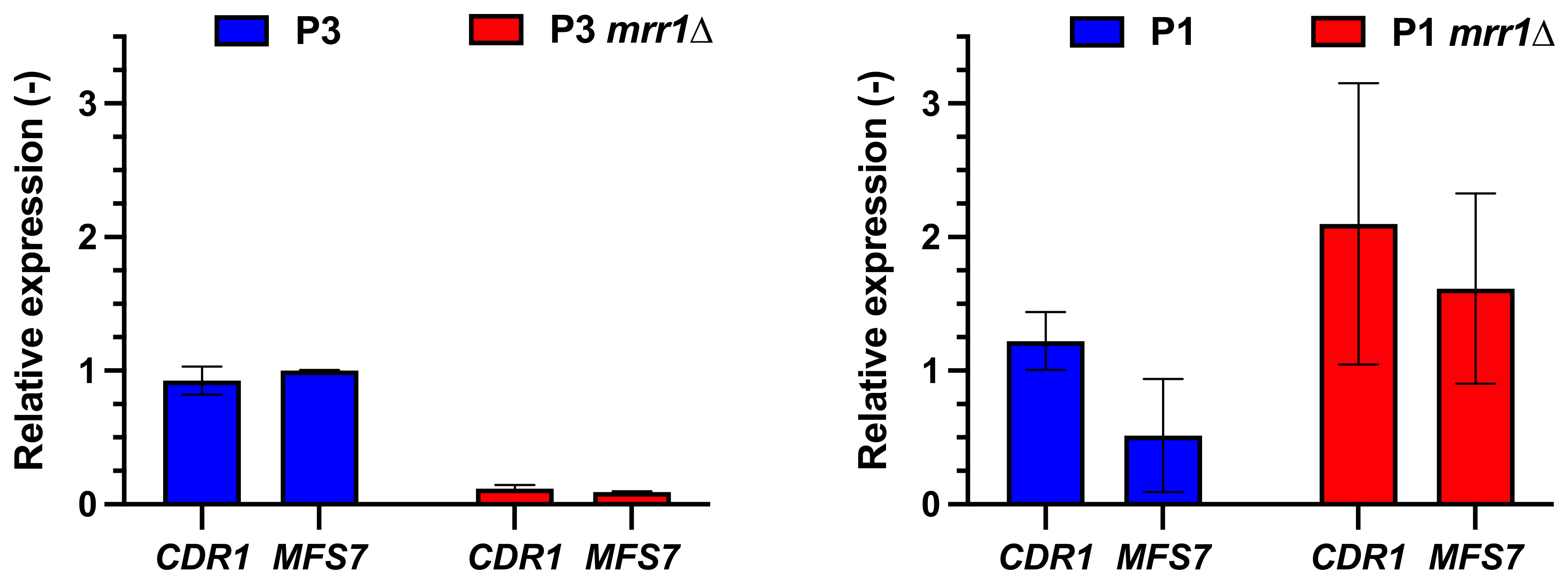
3.1. CDR1 Expression Patterns in C. lusitaniae

Previous studies performed in C. lusitaniae with azole-resistant isolates have reported that CDR1 expression is increased when compared to wild type isolates or isolates lacking MRR1 (Table 1). Isolates P3, P4 and U04 contain each of the MRR1 GOF mutations (V668G in P3 and P4; Y813C in U04). The presence of these mutations triggers not only the upregulation of MFS7 but also of CDR1. While screening a collection of recovered C. lusitaniae isolates from a French hospital, we recovered a specific azole-resistant isolate (DSY4941; MICs in Table S3) from an infected premature child (case report in Material and Methods). This isolate exhibited a novel MRR1 mutation (V654A). In order to evaluate the effect of this mutation not only on MFS7 but also on CDR1 expression, it was introduced on a wild type MRR1 allele. The results in Figure 1 show that the V654A GOF (DSY5639) elevated both MFS7 and CDR1 expression compared to a wild type MRR1 allele. These elevated expression levels were similar to those obtained with the known V668G mutation (DSY5439). Both GOF mutations resulted in an increase in azoles and 5-FC MICs compared to the wild type MRR1 (Table S4). Data in Figure 1 also confirms that in the absence of MRR1 (DSY5416, P3 mrr1Δ), both CDR1 and MFS7 were expressed at low levels compared to GOF isolates.
These data were confirmed by probing the expression levels of CDR1 and MFS7 in isolate P3 and its derived MRR1 mutant (DSY5416, P3 mrr1Δ): both CDR1 and MFS7 expression were decreased by about 10-fold when MRR1 was deleted in this isolate background (Figure 2). Expression of CDR1 and MFS7 in the MRR1 mutant from the parent azole-susceptible isolate P1 (DSY5658, P1 mrr1Δ) did not vary as extensively when compared to P1 (Figure 2). Taken together, our results, and those from others, illustrate that CDR1 expression is under the control, either directly or indirectly, of at least MRR1.

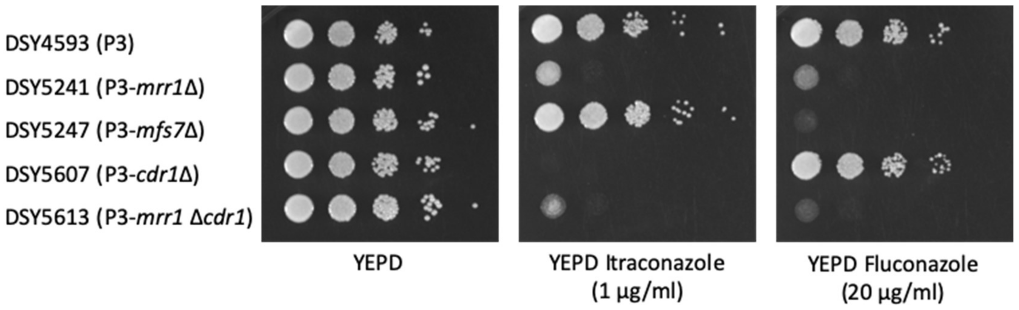
3.2. Effect of CDR1 on Antifungal Susceptibility

While it is known that MFS7 participates not only in fluconazole but also in 5-FC resistance of C. lusitaniae [7], the contribution of CDR1 in these phenotypes is not yet clear. CDR1 was, therefore, deleted in the background of different strain backgrounds and mutants (Table S1). We observed that in the absence of CDR1 in the isolate P3 (P3-cdr1Δ), the MICs of the long-tailed azoles itraconazole and posaconazole were decreased by 8-fold while not altering the MIC of the short-tailed azole fluconazole (Figure 3 and Table 2). Moreover, 5-FC resistance of isolate P3 was not dependent on CDR1 as judged by the measured 5-FC MICs (Table 2). These trends were similar when CDR1 was deleted in isolates complemented with MRR1 GOF alleles (Table S4). In the background of an mrr1Δ mutant in P3, CDR1 deletion has little additional effect on azole MICs, probably due to the presence of MFS7 and the low CDR1 expression of the mrr1Δ mutant. Serial dilutions on YEPD agar containing itraconazole or fluconazole confirmed these observations: deletion of CDR1 in isolate P3 decreased resistance to itraconazole but not to fluconazole. Deletion of MRR1 in P3 decreased resistance to both itraconazole or fluconazole as a consequence of decreased expression of both MFS7 and CDR1 (Figure 4).
As expected, the deletion of MFS7 in P3 decreased fluconazole MIC by 8-fold. However, the additional CDR1 deletion in the mfs7Δ mutant decreased fluconazole MIC by 256-fold compared to the initial isolate P3 (Figure 3 and Table 2). The effect of CDR1 on fluconazole MIC is, therefore, better distinguished in the background of an mfs7Δ mutant of isolate P3 since MFS7 is the major contributor of fluconazole resistance in C. lusitaniae [7]. These data also suggest that CDR1 covers a broad range of azoles, including short- and long-tailed azoles, which is known from other CDR1-like ABC transporters of other Candida spp. [19,20].
CDR1 was also deleted in the background of the azole-susceptible isolate P1, which is the parent of P3. In this isolate background, the effect of CDR1 deletion was again best observed in an mfs7Δ mutant, in which the fluconazole MIC was decreased by at least 4-fold compared to P1 (Table 3 and Figure S1).

3.3. Probing Cdr1 Activity by R6G Efflux

We next explored the efflux activity of Cdr1 in C. lusitaniae by using an efflux reporter system based on the fluorescence measurements of rhodamine 6G (R6G) with whole cells. This system was used previously to measure efflux activities of ABC transporters in C. albicans and C. glabrata [18,21]. As shown in Figure 5, R6G efflux activity was higher in P3 compared to P1 after glucose was added in the incubation buffer. Maximal slope in R6G efflux kinetics was 2.7-fold increased in P3 compared to P1, which is consistent with the higher expression of CDR1 in P3 compared to P1. Efflux activity was strongly diminished after glucose addition when CDR1 was deleted in the background of both isolates P3 and P1. We confirmed that R6G efflux was principally mediated by Cdr1 since MFS7 deletion in both P1 and P3 isolates did not alter efflux profiles compared to wild types. Interestingly, when MRR1 was deleted in P3, efflux activity after glucose supply was similar to isolate P1 (Figure 6). Given that CDR1 expression is down-regulated in mrr1Δ mutants compared to MRR1 GOF isolates (Figure 2), this result is consistent with the assumption that Cdr1 is responsible for R6G efflux in C. lusitaniae. The combined deletion of MRR1 and CDR1 in both P1 and P3 backgrounds and the associated decrease in R6G efflux to basal levels confirmed this hypothesis.
The importance of Cdr1 for efflux activity was also addressed by efflux inhibition with milbemycin oxim (AOx3), a known ABC transporter inhibitor in other Candida spp. [21]. The addition of AOx3 to P1 and P3 isolates reduced R6G efflux to basal activities (Figure 7). When the same experiments were carried out with cdr1Δ mutants from the same isolates, we noticed that AOx3 was still able to slightly decrease R6G efflux compared to mutant cells incubated with glucose only (Figure 7). This suggests the presence of additional transporter activity potentially mediated by other ABC transporters in this fungal species. Interestingly, in the presence of AOx3, the MICs of the long-tailed azoles posaconazole and itraconazole were strongly reduced in isolate P3, while MICs of the short-tailed azoles fluconazole and voriconazole were little affected (Table 4). This tendency was confirmed in isolate P1 for itraconazole MICs. These results are consistent with the AOx3-dependent inhibition of ABC transporters, which are known to exhibit efflux activity on long-tailed azoles [22]. The MICs of the short-tailed azoles fluconazole and voriconazole in isolate P3 are mostly attributed to MFS7, a major facilitator that is upregulated in P3. Major facilitators are not inhibited by milbemycins [23,24,25], which, therefore, preserved the short-tailed azole MICs of isolate P3 even in the presence of AOx3.

4. Discussion

Antifungal resistance is mediated by several mechanisms involving transcriptional changes, genome mutations and post-transcriptional modifications [9]. The upregulation of multidrug transporters and the resulting drug resistance is a very widespread mechanism in fungal pathogens [26]. In C. albicans, upregulation of ABC transporters and major facilitators results in resistance to azoles [22]. The increased expression of these transporters compared to drug-susceptible isolates relies on the transcriptional control of two major regulators, TAC1 and MRR1 [10,11]. In C. albicans, TAC1 regulates the ABC transporter genes CDR1 and CDR2, while MRR1 regulates the major facilitator MDR1 [10,18,27,28]. In the past few years, TAC1 and MRR1 homologs have been described in several other fungal pathogens including Candida parapsilosis, Candida tropicalis and C. auris [29,30,31]. A TAC1 homolog exists in C. lusitaniae (CLUG_02369) with about 55% similarity to the C. albicans TAC1. This gene has not been associated, up to now, with mutations in azole-resistant isolates (upregulating MFS7 and CDR1) compared to susceptible isolates [5,7,12]. Therefore, the role of CLUG_02369 in drug resistance is likely not to be relevant, although cannot be excluded. In C. auris, it has been established that TAC1b (a TAC1 homolog) can regulate the expression of at least CDR1 [32]. Among the three MRR1 homologs of C. auris, only the deletion of MRR1a resulted in decreased azole susceptibility of an isolate of Clade III [33]. The full repertoire of MRR1a targets in C. auris remains unknown still. In C. lusitaniae, only one MRR1 homolog has been investigated for its role in azole resistance [7,12]. MRR1 of C. lusitaniae regulates at least the major facilitator MFS7, an important mediator of fluconazole and 5-FC resistance in this species [7]. Here, we show with several approaches that CDR1 of C. lusitaniae may be considered as another direct or indirect target of MRR1. This is supported by (i) the upregulation of CDR1 in the presence of MRR1 GOF mutations; (ii) the loss of CDR1 upregulation when MRR1 is deleted. This is an unexpected result given that CDR1-like genes in other Candida spp. have been shown to be under the control of TAC1 homologs [32,34]. The C. lusitaniae MRR1 may have, therefore, accommodated different targets compared to other Candida spp. Transcriptional rewiring is a common feature of fungal pathogens [35]. Future studies should address the MRR1 regulon in C. lusitaniae by more elaborate approaches through promoter occupancy experiments.
Cdr1 activity was measured here by R6G efflux, which was conveniently recorded by a whole cell-based assay over time. R6G is considered as a specific substrate for ABC transporters [21,36]. This was indirectly confirmed here in efflux assays with transporter mutants. While R6G efflux activity was decreased in mutant P3 cdr1Δ compared to the parent P3, efflux activity was not changed in the P3 msf7Δ mutant, thus suggesting that Cdr1 was the major mediator of R6G efflux in C. lusitaniae. The C. lusitaniae genome contains several other ABC transporters [7,37], and it is possible that these transporters could also contribute to R6G efflux activity. The data presented in Figure 5 and Figure 7 are consistent with this hypothesis. CDR1 deletion in isolate P3 as well as milbemycin efflux inhibition could not reduce R6G efflux to background activity (e.g., efflux activity in absence of glucose), thus suggesting the participation of additional transporters. The C. lusitaniae genome contains a close relative to CDR1 (CLUG_03113), CLUG_02179 (or EJF14_60115 in isolate P1) with 95% similarity. This is reminiscent of the CDR1/CDR2 pair of ABC transporters in C. albicans [38]. These two transporters are co-regulated by TAC1 [10]. When both CDR1 and CDR2 are deleted in C. albicans, this results in increased azole susceptibility compared to a wild type and single deletion mutants [38]. However, EJF14_60115 is not regulated by MRR1 GOF mutations in C. lusitaniae [7,12] and, therefore, is not likely to participate in azole resistance of this fungal pathogen. Nevertheless, it is possible that EJF14_60115 could still contribute to the basal R6G activity that is detectable in cdr1Δ mutants. Future studies could address this possibility.

5. Conclusions

In conclusion, we showed here that the expression of the C. lusitaniae ABC transporter CDR1 is regulated by MRR1 and is participating in the resistance to long-tailed azoles. It is remarkable that C. lusitaniae MRR1 is able to regulate simultaneously two different types of multidrug transporters (ABC transporter and major facilitator) that are known to play major roles in the acquisition of antifungal resistance. This allows C. lusitaniae to enlarge its repertoire of defenses against drug stresses. This way, the combined CDR1 and MFS7 upregulation triggered by GOF mutations in MRR1 confer not only decreased susceptibility to long-tailed azoles but also to short-tailed azoles. Since MRR1 GOF mutations may arise even in azole-untreated patients, this regulatory relationship should not be underestimated in antifungal therapy.

Supplementary Materials

The following are available online at https://www.mdpi.com/article/10.3390/jof7090760/s1, Figure S1: Pairwise comparisons of MICs between isolate P1 and derived mutants, Table S1: list of strains used in this study, Table S2: primers used in this study, Table S3: MICs of clinical isolate DSY4941, Table S4: Antifungal MICs of C. lusitaniae isolates and mutant derivatives.

Author Contributions

Conceptualization, D.S.; methodology, D.S.; investigation, V.B. and D.B.; resources, F.G.; writing, D.S., V.B. and F.G. All authors have read and agreed to the published version of the manuscript.

Funding

This research was funded by a Swiss National Research Foundation grant 31003A_172958 to D.S.

Institutional Review Board Statement

Not applicable.

Informed Consent Statement

Routine screening of patients was conducted under the surveillance protocol of the participating institution (CHU Besançon).

Data Availability Statement

The datasets generated during and/or analyzed during the current study are available from the corresponding author upon request.

Acknowledgments

Authors are thankful to A. Coste for critical reading of the manuscript.

Conflicts of Interest

The authors declare no conflict of interest.

References

  1. McCarty, T.P.; White, C.M.; Pappas, P.G. Candidemia and Invasive Candidiasis. Infect. Dis. Clin. N. Am. 2021, 35, 389–413. [Google Scholar] [CrossRef]
  2. Pfaller, M.A.; Andes, D.R.; Diekema, D.J.; Horn, D.L.; Reboli, A.C.; Rotstein, C.; Franks, B.; Azie, N.E. Epidemiology and Outcomes of Invasive Candidiasis Due to Non-albicans Species of Candida in 2,496 Patients: Data from the Prospective Antifungal Therapy (PATH) Registry 2004–2008. PLoS ONE 2014, 9, e101510. [Google Scholar] [CrossRef] [Green Version]
  3. Andes, D.R.; Safdar, N.; Baddley, J.W.; Playford, G.; Reboli, A.C.; Rex, J.H.; Sobel, J.D.; Pappas, P.G.; Kullberg, B.J.; Mycoses Study Group. Impact of Treatment Strategy on Outcomes in Patients with Candidemia and Other Forms of Invasive Candidiasis: A Patient-Level Quantitative Review of Randomized Trials. Clin. Infect. Dis. 2012, 54, 1110–1122. [Google Scholar] [CrossRef]
  4. Desnos-Ollivier, M.; Moquet, O.; Chouaki, T.; Guerin, A.M.; Dromer, F. Development of Echinocandin Resistance in Clavispora Lusitaniae during Caspofungin Treatment. J. Clin. Microbiol. 2011, 49, 2304–2306. [Google Scholar] [CrossRef] [Green Version]
  5. Demers, E.G.; Biermann, A.R.; Masonjones, S.; Crocker, A.W.; Ashare, A.; Stajich, J.E.; Hogan, D.A. Evolution of Drug Resistance in an Antifungal-Naive Chronic Candida lusitaniae Infection. Proc. Natl. Acad. Sci. USA 2018, 32, 201807698. [Google Scholar] [CrossRef] [Green Version]
  6. Asner, S.A.; Giulieri, S.; Diezi, M.; Marchetti, O.; Sanglard, D. Acquired Multidrug Antifungal Resistance in Candida lusitaniae during Therapy. Antimicrob. Agents Chemother. 2015, 59, 7715–7722. [Google Scholar] [CrossRef]
  7. Kannan, A.; Asner, S.A.; Trachsel, E.; Kelly, S.; Parker, J.; Sanglard, D. Comparative Genomics for the Elucidation of Multidrug Resistance in Candida lusitaniae. mBio 2019, 10, e02512-19. [Google Scholar] [CrossRef] [Green Version]
  8. Ruiz, G.B.; Lorenz, A. What Do We Know about the Biology of the Emerging Fungal Pathogen of Humans Candida auris? Microbiol. Res. 2021, 242, 126621. [Google Scholar] [CrossRef]
  9. Sanglard, D. Emerging Threats in Antifungal-Resistant Fungal Pathogens. Front. Med. 2016, 3, 11. [Google Scholar] [CrossRef] [Green Version]
  10. Coste, A.T.; Karababa, M.; Ischer, F.; Bille, J.; Sanglard, D. TAC1, transcriptional activator of CDR genes, is a new transcription factor involved in the regulation of Candida albicans ABC transporters CDR1 and CDR2. Eukaryot. Cell 2004, 3, 1639–1652. [Google Scholar] [CrossRef] [Green Version]
  11. Morschhäuser, J.; Barker, K.S.; Liu, T.T.; BlaB-Warmuth, J.; Homayouni, R.; Rogers, P.D. The transcription factor Mrr1p controls expression of the MDR1 efflux pump and mediates multidrug resistance in Candida albicans. PLoS Pathog. 2007, 3, e164. [Google Scholar] [CrossRef] [Green Version]
  12. Demers, E.G.; Stajich, J.E.; Ashare, A.; Occhipinti, P.; Hogan, D.A. Balancing Positive and Negative Selection: In Vivo Evolution of Candida lusitaniae MRR1. Mbio 2021, 12. [Google Scholar] [CrossRef]
  13. Pappas, P.G.; Kauffman, C.A.; Andes, D.; Benjamin, D.K.; Calandra, T.F.; Edwards, J.E.; Filler, S.G.; Fisher, J.F.; Kullberg, B.-J.; Ostrosky-Zeichner, L.; et al. Clinical Practice Guidelines for the Management Candidiasis: 2009 Update by the Infectious Diseases Society of America. Clin. Infect. Dis. 2009, 48, 503–535. [Google Scholar] [CrossRef] [Green Version]
  14. Kaufman, D.; Boyle, R.; Hazen, K.C.; Patrie, J.T.; Robinson, M.; Donowitz, L.G. Fluconazole Prophylaxis against Fungal Colonization and Infection in Preterm Infants. N. Engl. J. Med. 2001, 345, 1660–1666. [Google Scholar] [CrossRef]
  15. Basso, L.R.; Bartiss, A.; Mao, Y.; Gast, C.E.; Coelho, P.S.R.; Snyder, M.; Wong, B. Transformation of Candida albicans with a synthetic hygromycin B resistance gene. Yeast 2010, 27, 1039–1048. [Google Scholar] [CrossRef] [Green Version]
  16. Jensen, M.A.; Fukushima, M.; Davis, R.W. DMSO and Betaine Greatly Improve Amplification of GC-Rich Constructs in De Novo Synthesis. PLoS ONE 2010, 5, e11024. [Google Scholar] [CrossRef] [Green Version]
  17. Grahl, N.; Demers, E.G.; Crocker, A.W.; Hogan, D.A. Use of RNA-Protein Complexes for Genome Editing in Non-albicans Candida Species. mSphere 2017, 2, e00218-17. [Google Scholar] [CrossRef] [Green Version]
  18. Coste, A.; Turner, V.; Ischer, F.; Morschhäuser, J.; Forche, A.; Selmecki, A.; Berman, J.; Bille, J.; Sanglard, D. A mutation in Tac1p, a transcription factor regulating CDR1 and CDR2, is coupled with loss of heterozygosity at chromosome 5 to mediate antifungal resistance in Candida albicans. Genetics 2006, 172, 2139–2156. [Google Scholar] [CrossRef] [Green Version]
  19. Nakamura, K.; Niimi, M.; Niimi, K.; Holmes, A.R.; Yates, J.E.; Decottignies, A.; Monk, B.C.; Goffeau, A.; Cannon, R.D. Functional expression of Candida albicans drug efflux pump Cdr1p in a Saccharomyces cerevisiae strain deficient in membrane transporters. Antimicrob. Agents Chemother. 2001, 45, 3366–3374. [Google Scholar] [CrossRef] [Green Version]
  20. Sanglard, D.; Ischer, F.; Bille, J. Role of ATP-binding-cassette transporter genes in high-frequency acquisition of resistance to azole antifungals in Candida glabrata. Antimicrob. Agents Chemother. 2001, 45, 1174–1183. [Google Scholar] [CrossRef] [Green Version]
  21. Silva, L.V.; Sanguinetti, M.; Vandeputte, P.; Torelli, R.; Rochat, B.; Sanglard, D. Milbemycins: More than Efflux Inhibitors for Fungal Pathogens. Antimicrob. Agents Chemother. 2013, 57, 873–886. [Google Scholar] [CrossRef] [Green Version]
  22. Sanglard, D.; Kuchler, K.; Ischer, F.; Pagani, J.L.; Monod, M.; Bille, J. Mechanisms of Resistance to Azole Antifungal Agents in Candida albicans Isolates from AIDS Patients Involve Specific Multidrug Transporters. Antimicrob. Agents Chemother. 1995, 39, 2378–2386. [Google Scholar] [CrossRef] [PubMed] [Green Version]
  23. Monod, M.; Feuermann, M.; Salamin, K.; Fratti, M.; Makino, M.; Alshahni, M.M.; Makimura, K.; Yamada, T. Trichophyton Rubrum Azole Resistance Mediated by a New ABC Transporter, TruMDR3. Antimicrob. Agents Chemother. 2019, 63. [Google Scholar] [CrossRef] [Green Version]
  24. Walker, B.; Izumikawa, K.; Tsai, H.-F.; Bennett, J.E. Milbemycin A4 Oxime as a Probe of Azole Transport in Candida glabrata. FEMS Yeast Res. 2014, 14, 755–761. [Google Scholar] [CrossRef] [PubMed] [Green Version]
  25. Cannon, R.D.; Lamping, E.; Holmes, A.R.; Niimi, K.; Baret, P.V.; Keniya, M.V.; Tanabe, K.; Niimi, M.; Goffeau, A.; Monk, B.C. Efflux-Mediated Antifungal Drug Resistance. Clin. Microbiol. Rev. 2009, 22, 291–321. [Google Scholar] [CrossRef] [PubMed] [Green Version]
  26. Cowen, L.E.; Sanglard, D.; Howard, S.J.; Rogers, P.D.; Perlin, D.S. Mechanisms of Antifungal Drug Resistance. Cold Spring Harb. Perspect. Med. 2015, 5, a019752. [Google Scholar] [CrossRef]
  27. Liu, T.T.; Znaidi, S.; Barker, K.S.; Xu, L.; Homayouni, R.; Saidane, S.; Morschhäuser, J.; Nantel, A.; Raymond, M.; Rogers, P.D. Genome-wide expression and location analyses of the Candida albicans Tac1p regulon. Eukaryot. Cell 2007, 6, 2122–2138. [Google Scholar] [CrossRef] [Green Version]
  28. Schubert, S.; Popp, C.; Rogers, P.D.; Morschhauser, J. Functional dissection of a Candida albicans zinc cluster transcription factor, the multidrug resistance regulator Mrr1. Eukaryot. Cell 2011, 10, 1110–1121. [Google Scholar] [CrossRef] [Green Version]
  29. Arastehfar, A.; Hilmioğlu-Polat, S.; Daneshnia, F.; Pan, W.; Hafez, A.; Fang, W.; Liao, W.; Şahbudak-Bal, Z.; Metin, D.Y.; de Almeida Júnior, J.N.; et al. Clonal Candidemia Outbreak by Candida parapsilosis Carrying Y132F in Turkey: Evolution of a Persisting Challenge. Front. Cell. Infect. Microbiol. 2021, 11, 676177. [Google Scholar] [CrossRef]
  30. Paul, S.; Singh, S.; Sharma, D.; Chakrabarti, A.; Rudramurthy, S.M.; Ghosh, A.K. Dynamics of in Vitro Development of Azole Resistance in Candida tropicalis. J. Glob. Antimicrob. Resist. 2020, 22, 553–561. [Google Scholar] [CrossRef]
  31. Li, J.; Coste, A.T.; Liechti, M.; Bachmann, D.; Sanglard, D.; Lamoth, F. Novel ERG11 and TAC1b Mutations Associated with Azole Resistance in Candida auris. Antimicrob. Agents Chemother. 2021. [Google Scholar] [CrossRef]
  32. Rybak, J.M.; Muñoz, J.F.; Barker, K.S.; Parker, J.E.; Esquivel, B.D.; Berkow, E.L.; Lockhart, S.R.; Gade, L.; Palmer, G.E.; White, T.C.; et al. Mutations in TAC1B: A Novel Genetic Determinant of Clinical Fluconazole Resistance in Candida auris. mBio 2020, 11, e00365-20. [Google Scholar] [CrossRef]
  33. Mayr, E.-M.; Ramírez-Zavala, B.; Krüger, I.; Morschhäuser, J. A Zinc Cluster Transcription Factor Contributes to the Intrinsic Fluconazole Resistance of Candida auris. mSphere 2020, 5, e00279-20. [Google Scholar] [CrossRef] [PubMed]
  34. Liu, Z.; Rossi, J.M.; Myers, L.C. Candida albicans Zn Cluster Transcription Factors Tac1 and Znc1 Are Activated by Farnesol To Upregulate a Transcriptional Program Including the Multidrug Efflux Pump CDR1. Antimicrob. Agents Chemother. 2018, 62, 1–18. [Google Scholar] [CrossRef] [Green Version]
  35. Coste, A.T.; Ramsdale, M.; Ischer, F.; Sanglard, D. Divergent functions of three Candida albicans zinc-cluster transcription factors (CTA4, ASG1 and CTF1) complementing pleiotropic drug resistance in Saccharomyces cerevisiae. Microbiology 2008, 154, 1491–1501. [Google Scholar] [CrossRef] [Green Version]
  36. Maesaki, S.; Marichal, P.; Bossche, H.V.; Sanglard, D.; Kohno, S. Rhodamine 6G efflux for the detection of CDR1-overexpressing azole-resistant Candida albicans strains. J. Antimicrob. Chemother. 1999, 44, 27–31. [Google Scholar] [CrossRef]
  37. Reboutier, D.; Piednoël, M.; Boisnard, S.; Conti, A.; Chevalier, V.; Florent, M.; Gibot-Leclerc, S.; Silva, B.D.; Chastin, C.; Fallague, K.; et al. Combination of Different Molecular Mechanisms Leading to Fluconazole Resistance in a Candida lusitaniae Clinical Isolate. Diagn. Microbiol. Infect. Dis. 2009, 63, 188–193. [Google Scholar] [CrossRef]
  38. Sanglard, D.; Ischer, F.; Monod, M.; Bille, J. Cloning of Candida albicans genes conferring resistance to azole antifungal agents: Characterization of CDR2, a new multidrug ABC transporter gene. Microbiology 1997, 143 Pt 2, 405–416. [Google Scholar] [CrossRef] [PubMed] [Green Version]
Figure 1. MRR1 GOF alleles trigger the upregulation of multidrug transporters CDR1 and MFS7. qPCRs were performed as described in Material and Methods. Expression fold-changes were reported to the parent isolate DSY5416 for CDR1 and MFS7 expression patterns. Blue and red colors indicate the effect of MRR1 GOF mutations V668G and G654A, respectively.
Jof 07 00760 g001
Figure 2. CDR1 and MSF7 are regulated by MRR1 in isolate P3. qPCRs were performed as described in Material and Methods. Expression fold-changes were reported to the parent isolates P1 and P3, respectively. The P3 isolate contains the MRR1 GOF mutation V668G, while P1 contains a wild type MRR1 allele.
Jof 07 00760 g002
Figure 3. Pairwise comparisons of MICs between isolate P3 and derived mutants. Fold-changes are given in log2 scale. Comparisons were made always starting from top row. Values > 0 indicate MIC decreases, while values <0 indicate MIC increases. Greyed colored boxes indicate comparisons between same isolates (Value = 0).
Jof 07 00760 g003
Figure 4. Serial dilution assays for itraconazole and fluconazole. Isolates were 10-fold serially diluted and spotted in the corresponding agar plates starting from approximately 107 cells/mL from inoculum cultures. Plates were incubated at 35 °C for 24 h.
Jof 07 00760 g004
Figure 5. Rhodamine 6G efflux activities are dependent on the presence of CDR1. Efflux activities were performed as described in Material and Methods. Means of duplicates are reported. Glucose was added 300 s after start of measurements (arrow). Maximal slopes were calculated with Graph Prism. Maximal slopes were derived from data points ranging between 740 and 1040 s and calculated by linear regression implemented in Graph Prism.
Jof 07 00760 g005
Figure 6. Rhodamine 6G efflux activities are dependent on the presence of CDR1 and MRR1. Efflux activities were performed as described in Material and Methods. Means of duplicates are reported. Glucose was added 300 s after start of measurements (arrow).
Jof 07 00760 g006
Figure 7. Milbemycin oxim 3 (AOx3) inhibits rhodamine 6G efflux. Efflux activities were performed as described in Material and Methods. Means of duplicates are reported. Glucose was added 300 s after start of measurements (arrow). Milbemycin AOx3 (5 µg/mL) was added to indicated samples at start of experiment.
Jof 07 00760 g007
Table 1. CDR1 expression in different isolate backgrounds.
IsolateFold-Change (vs. Azole-Susceptible Isolates)Experimental ConditionReference
DSY4593 (P3)2,4RNAseq[7]
DSY4661 (P4)4,7RNAseq[7]
U042,6 aRNAseq[12]
DSY4593 (P3)5,0qPCR[6]
DSY4661 (P4)3,5qPCR[6]
a fold-change was calculated by comparison with an mrr1Δ mutant from U04.
Table 2. Antifungal MICs of C. lusitaniae P3 and mutant derivatives.
Antifungal DrugsMIC (µg/mL)
P3P3-mfs7ΔP3-cdr1ΔP3-mrr1ΔP3-mfs7Δcdr1ΔP3-mrr1Δcdr1Δ
Anidulafungin0.120.250.120.120.120.12
Micafungin0.120.120.120.060.060.03
Caspofungin0.120.250.120.120.120.12
5-Flucytosine644320.50.50.5
Posaconazole0.250.250.030.0150.0150.015
Voriconazole0.50.120.250.015<0.0080.015
Itraconazole0.50.50.060.060.030.06
Fluconazole324321<0.121
Amphotericin B0.50.50.50.250.250.25
Table 3. Antifungal MICs of C. lusitaniae P1 and mutant derivatives.
Antifungal DrugsMIC (µg/mL)
P1P1-mfs7ΔP1-cdr1ΔP1-mrr1ΔP1-mfs7Δcdr1ΔP1-mrr1Δcdr1Δ
Anidulafungin0.120.060.120.120.060.12
Micafungin0.060.060.060.030.120.06
Caspofungin0.120.250.120.120.250.12
5-Flucytosine40.5410.51
Posaconazole0.030.0150.030.0150.0150.015
Voriconazole<0.008<0.008<0.008<0.008<0.008<0.008
Itraconazole0.060.060.060.060.03<0.008
Fluconazole0.50.250.251<0.121
Amphotericin B0.250.50.250.250.250.25
Table 4. Effect of milbemycin AOx3 (5 µg/mL) on antifungal MICs.
Antifungal DrugsMIC (µg/mL)
P1P1-AOx3P3P3-AOx3
Anidulafungin0.120.060.120.06
Micafungin0.060.060.060.06
Caspofungin0.120.250.120.06
5-Flucytosine40.56464
Posaconazole0.030.0150.25<0.008
Voriconazole<0.008<0.0080.50.25
Itraconazole0.060.060.5<0.008
Fluconazole0.50.253216
Amphotericin B0.250.50.50.5
Publisher’s Note: MDPI stays neutral with regard to jurisdictional claims in published maps and institutional affiliations.

