Article

Genetic Analyses of Saprolegnia Strains Isolated from Salmonid Fish of Different Geographic Origin Document the Connection between Pathogenicity and Molecular Diversity

1
Division of Biotechnology and Plant Health, Norwegian Institute of Bioeconomy Research (NIBIO), N-1431 Ås, Norway
2
Norwegian Veterinary Institute, Pb 64, N-1431 Ås, Norway
*
Author to whom correspondence should be addressed.
Co first authorship.
J. Fungi 2021, 7(9), 713; https://doi.org/10.3390/jof7090713
Submission received: 10 August 2021 / Revised: 27 August 2021 / Accepted: 29 August 2021 / Published: 30 August 2021
(This article belongs to the Special Issue Diversity and Classification of Environmental Fungi)

Abstract

:
Saprolegnia parasitica is recognized as one of the most important oomycetes pests of salmon and trout species. The amplified fragment length polymorphism (AFLP) and method sequence data of the internal transcribed spacer (ITS) were used to study the genetic diversity and relationships of Saprolegnia spp. collected from Canada, Chile, Japan, Norway and Scotland. AFLP analysis of 37 Saprolegnia spp. isolates using six primer combinations gave a total of 163 clear polymorphic bands. Bayesian cluster analysis using genetic similarity divided the isolates into three main groups, suggesting that there are genetic relationships among the isolates. The unweighted pair group method with arithmetic mean (UPGMA) and principal coordinate analysis (PCO) confirmed the pattern of the cluster analyses. ITS analyses of 48 Saprolegnia sequences resulted in five well-defined clades. Analysis of molecular variance (AMOVA) revealed greater variation within countries (91.01%) than among countries (8.99%). We were able to distinguish the Saprolegnia isolates according to their species, ability to produce oogonia with and without long spines on the cysts and their ability to or not to cause mortality in salmonids. AFLP markers and ITS sequencing data obtained in the study, were found to be an efficient tool to characterize the genetic diversity and relationships of Saprolegnia spp. The comparison of AFLP analysis and ITS sequence data using the Mantel test showed a very high and significant correlation (r2 = 0.8317).

1. Introduction

Saprolegniasis causes great damage and infection in fish in aquaculture and fish farms [1], and Saprolegnia parasitica is recognized as one of the most important oomycetes pests of salmon and trout species in Scandinavia, Chile, Japan, Canada and Scotland [2]. Thus, it causes losses of tens of millions of dollars in aquaculture businesses worldwide [1]. Saprolegnia spp. are generally termed “watermolds” and share common features with both fungi and algae [3]. All fish and ova in fresh water can possibly be infected by Saprolegnia spp., and the disease is termed saprolegniasis. Infected fish are easily recognized by the cotton-like white to greyish patches on the skin and gills [4]. During the last few decades there has been an increased focus on saprolegniasis in salmonid fish and a number of Saprolegnia outbreaks, and attempts to characterize Saprolegnia isolates have been reported [5,6]. Differences in pathogenicity have been proved between strains of a Saprolegnia species even within the same taxonomic grouping [7,8,9,10].
Traditionally, taxonomic characterization of Saprolegnia spp. has been based upon morphological and physiological characteristics [8,11], but in recent years, molecular methods have become useful tools when describing phylogeny, taxonomy and epidemiology [12,13,14,15,16]. Epidemiological studies of Saprolegnia spp. are particularly useful for identifying sources of infection, characterizing disease spread and improving disease management. However, molecular studies of Saprolegnia isolates are still limited [6], and an improved knowledge is important in order to reduce Saprolegnia outbreaks in the future.
Our aim was to investigate the genetic diversity and relationships of Saprolegnia spp. isolates collected from Canada, Chile, Japan, Norway and Scotland. Our hypotheses were that genetic diversity within and among these countries expresses broader genetic structures or, alternatively, that gene flow is high across all these countries, due to the trade and exchange of breeding materials. Furthermore, we tested if molecular markers (amplified fragment length polymorphism (AFLP) and sequence data of the internal transcribed spacer (ITS)) could distinguish the Saprolegnia isolates according to their species, their ability to produce oogonia with and without long spines on the cysts, and their ability to or not to cause mortality in salmonids. To achieve these goals, we investigated Saprolegnia spp. isolates (Table 1) using AFLP and ITS markers. We believe our study may provide novel insights into the genetics and biology of Saprolegnia spp., as well as indirect knowledge for disease management.

2. Materials and Methods

2.1. Saprolegnia Material

Thirty-seven Saprolegnia spp. strains were collected from salmonids in Canada (5), Chile (6), Norway (14), Scotland (9) and Japan (3) were investigated in this study (Table 1). The sampling procedure and all morphological, physiological and pathogenic characteristics were described by Stueland et al. [10]. Most strains were isolated from cultured Atlantic salmon (Salmo salar L.), ova, fry or brood stock suffering from saprolegniasis. However, the three Saprolegnia species from Japan were isolated from cultured sockeye salmon (Oncorhynchus nerka) and Coho salmon (Oncorhynchus kisutch) [7,9,17,18,19]. One of the Norwegian Saprolegnia strains was isolated from brown trout (Salmo trutta) and one strain from common whitefish (Coregonus lavaretus) (Table 1). Twenty-five of the strains have previously been analyzed with respect to their morphological and physiological characteristics [10], and 9 of these strains have also been previously characterized with regard to their pathogenicity to Atlantic salmon [10].

2.2. Amplified Fragment Length Polymorphism (AFLP) Analysis

DNA extraction of mycelia was performed by use of the Puregene Gentra DNA Isolation Kit (Gentra Systems, Minneapolis, MN, USA) according to the manufacturers’ description. The DNA pellets were dissolved in 50 μL of a TE buffer (pH 7.5) and stored at −20 °C. AFLP analysis was performed as previously described [20]. A total of 300 ng of genomic DNA was double-digested with EcoRI and the MseI isoschizomer Tru1I. Following ligation of the restriction fragments to the adaptors, pre-amplification PCR was carried out with non-selective primers in a total volume of 25 µL containing 5 µL of fivefold diluted ligation product. Ten primer combinations with two selective nucleotides were chosen for a pre-screening of eight randomly chosen isolates. Six highly polymorphic primer pairs were selected and used to generate AFLP fragments of all 37 isolates (Table 2). After the selective amplification using EcoRI primers end-labeled with γ-P33-ATP (3000 Ci/mmol−1), the PCR products were separated on 5% polyacrylamide gels. All primer combinations were repeated twice.

2.3. Data Analysis of the AFLP

Data were recorded manually, and only clear polymorphic bands were scored for presence (1) or absence (0). The genetic similarity (GS) was estimated using the Dice coefficient, calculated as GSxy = 2a/(2a + b + c), where a is the number of bands present in both isolates, b is the number of bands present only in isolate x and c is the number of bands present only in isolate y [21]. The genetic similarity among the clones, based on the presence or absence of amplified fragments, was also calculated by Jaccard coefficients [22]. Both analyses resulted in the same clusters, and only the results obtained by the Dice coefficient are presented.
We measured the percentage of polymorphic bands in four countries, calculated by dividing the number of polymorphic bands at the country level by the total number of bands scored. The estimates were based on the isolates from four of the countries. Isolates from Japan were excluded from the analyses, since this country was represented by fewer than five isolates.
The matrix of similarity data was analyzed using the unweighted pair group method with arithmetic mean (UPGMA), as suggested by Sneath and Sokal (1973) [23]. UPGMA clustering was also carried out for all the isolates according to their country of origin, their ability to produce oogonia with and without spines, and their pathogenicity. We performed principal coordinate (PCO) analysis to classify and detect the structure of the relationships between the isolates with different countries of origin, differing ability to produce oogonia with and without spines, and differing pathogenicity. Statistical analyses and construction of dendrograms were performed using NTSYS-pc software version 2.1 [24].
Analyses of molecular variance (AMOVA) [25] was carried out using Arlequin software, version 2.000 [26]. The analysis was estimated according to the country of origin. Isolates from Japan were eliminated from the study because they were represented by fewer than five individuals. The genetic distances among Saprolegnia spp. isolates from Canada, Chile, Norway and Scotland were calculated using the pairwise genetic distance method [27]. The mean FST was estimated in order to study genetic differentiation between countries. The significance of the FST values was tested by 1000 permutations. These analyses were performed using Arlequin software, version 2.000 [26]. Gene flow was estimated by assuming Nm = (1/FST − 1)/4 [28].

2.4. Sequencing of the ITS

The internal transcribed spacer 2 (ITS2) part of the nuclear ribosomal DNA was amplified with the primer pair ITS2/ITS4 according to White, Bruns, Lee and Taylor (1990) on a PTC-225 (Peltier Thermal Cycler, MJ Research, Waltham, MA, USA). PCR amplicons were purified with ExoSap IT (GE healthcare, Buckinghamshire, UK) according to the manufacturer’s procedure and visualized on standard agarose gel to ensure the presence of single-band products. Both strands of the PCR amplicons were sequenced with the PCR primers using DYEnamic ET dye terminator chemistry (Amersham Biosciences, Chicago, IL, USA), purified on AutoSeq96 (Amersham Biosciences) plates, diluted with 10 µL of MQ-water and subsequently analyzed on a MegaBace 1000 (Amersham Biosciences). Sequences were analyzed in Vector NTI Advanced 11 (Invitrogen, Waltham, MA, USA) and assembled in BioEdit 7.0.9.0 [29].

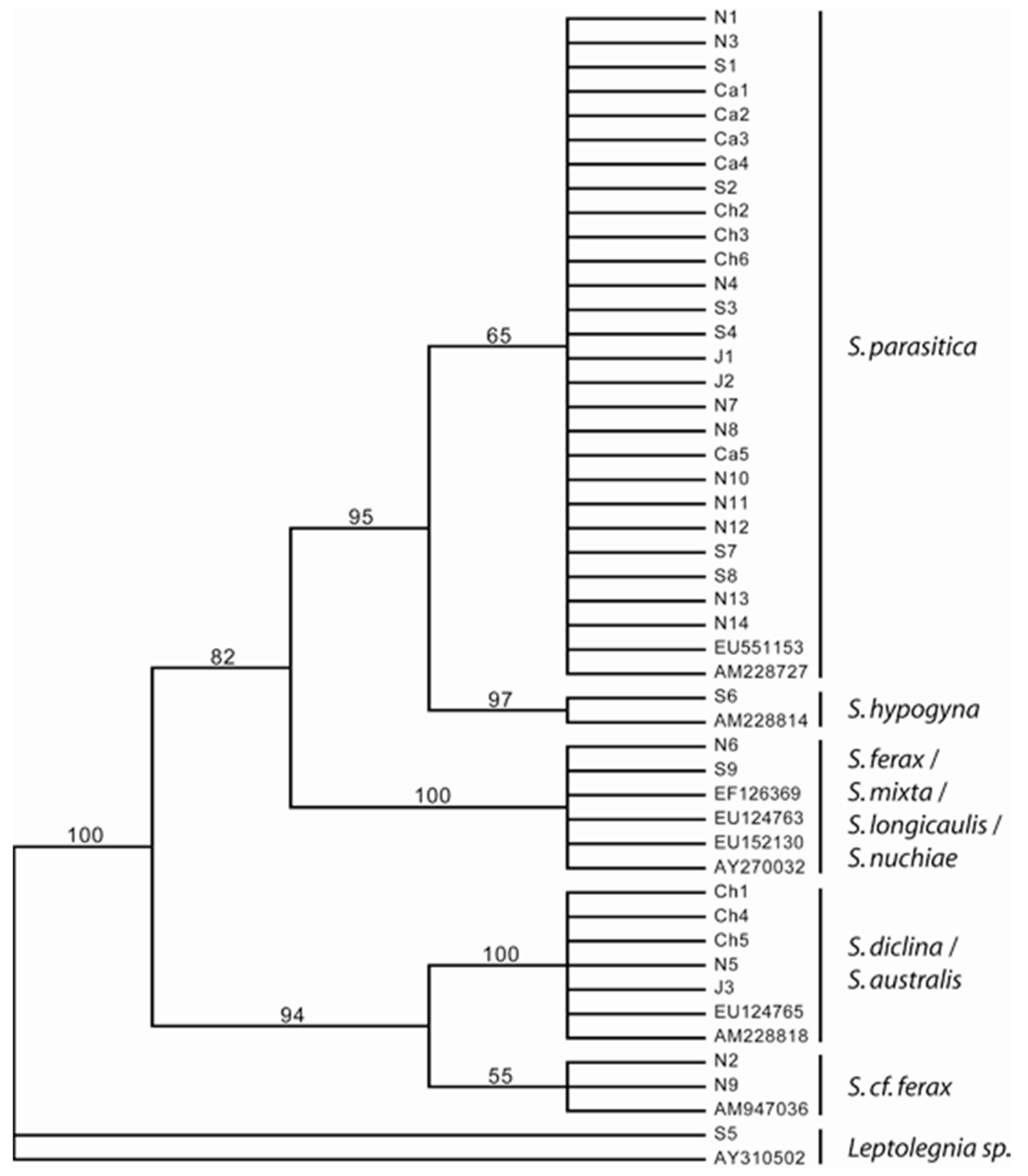
2.5. Phylogenetic Analyses

All new sequences are available from the EMBL/GenBank sequence database under accessions FN186015–FN186051 (Table 1). In addition, we downloaded and included the following sequence accessions: EU551153 (S. salmonis), AM228727 (S. parasitica), AM228814 (S. hypogyna), EF126339 (S. mixta), EU124763 (S. ferax), EU152130 (S. sp. nuchiae), AY270032 (S. longicaulis), EU124765 (S. diclina), AM228818 (S. australis), AM947036 (S. cf. ferax) and AY310502 (Leptolegnia sp.) based on similarity analyses against the EMBL/GenBank sequence database. The sequence alignment included a total of 48 isolates (46 Saprolegnia and 2 Leptolegnia), with ITS2 sequences 685–691 bp in length, representing at least 11 species and 35 haplotypes. Sequences were manually aligned; the data matrices are available from TreeBASE (Table 1). ITS sequences were selected from the correct data base published by Matthews et al. (2021) [30]. Phylogenetic analyses of the ITS were performed using maximum parsimony, using a heuristic search (2000 random replicates) in PAUP* v4.0b10 [31]. Gaps were treated as missing, and isolate N66, Leptolegnia sp., was selected as an outgroup based on previous phylogenetic studies of Saprolegnia [6]. To evaluate the support for the observed branching topologies for maximum parsimony, we performed bootstrap analysis [32], with 2000 bootstrap replicates. Bootstrapping was carried out with a heuristic algorithm and 2000 random additions of sequences per bootstrap replicate.

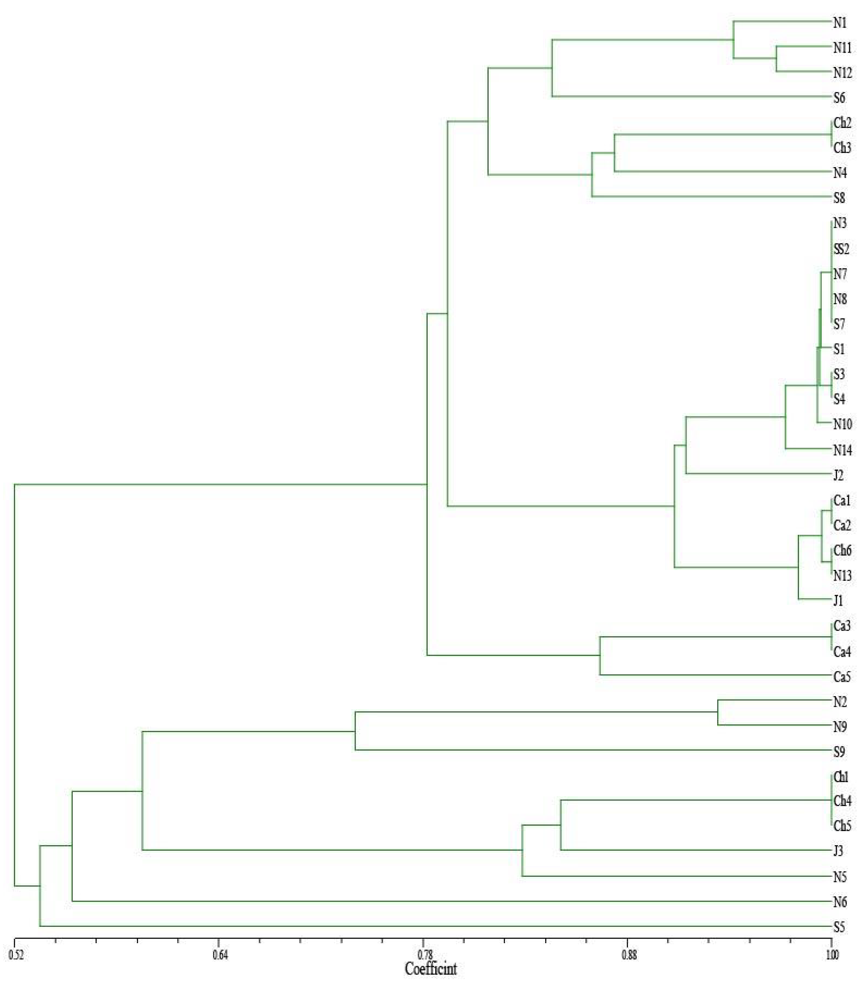
2.6. Cluster Analyses
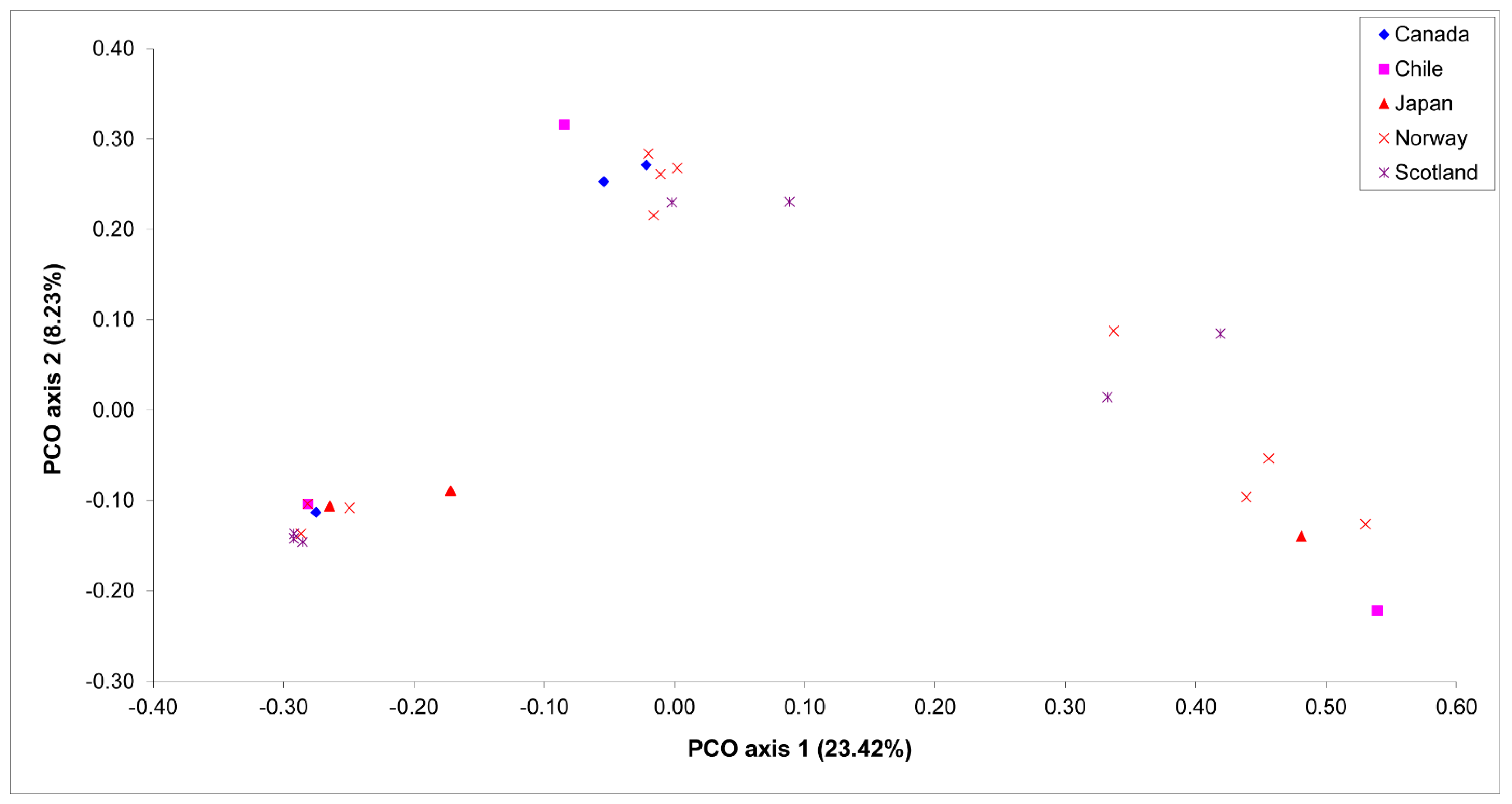
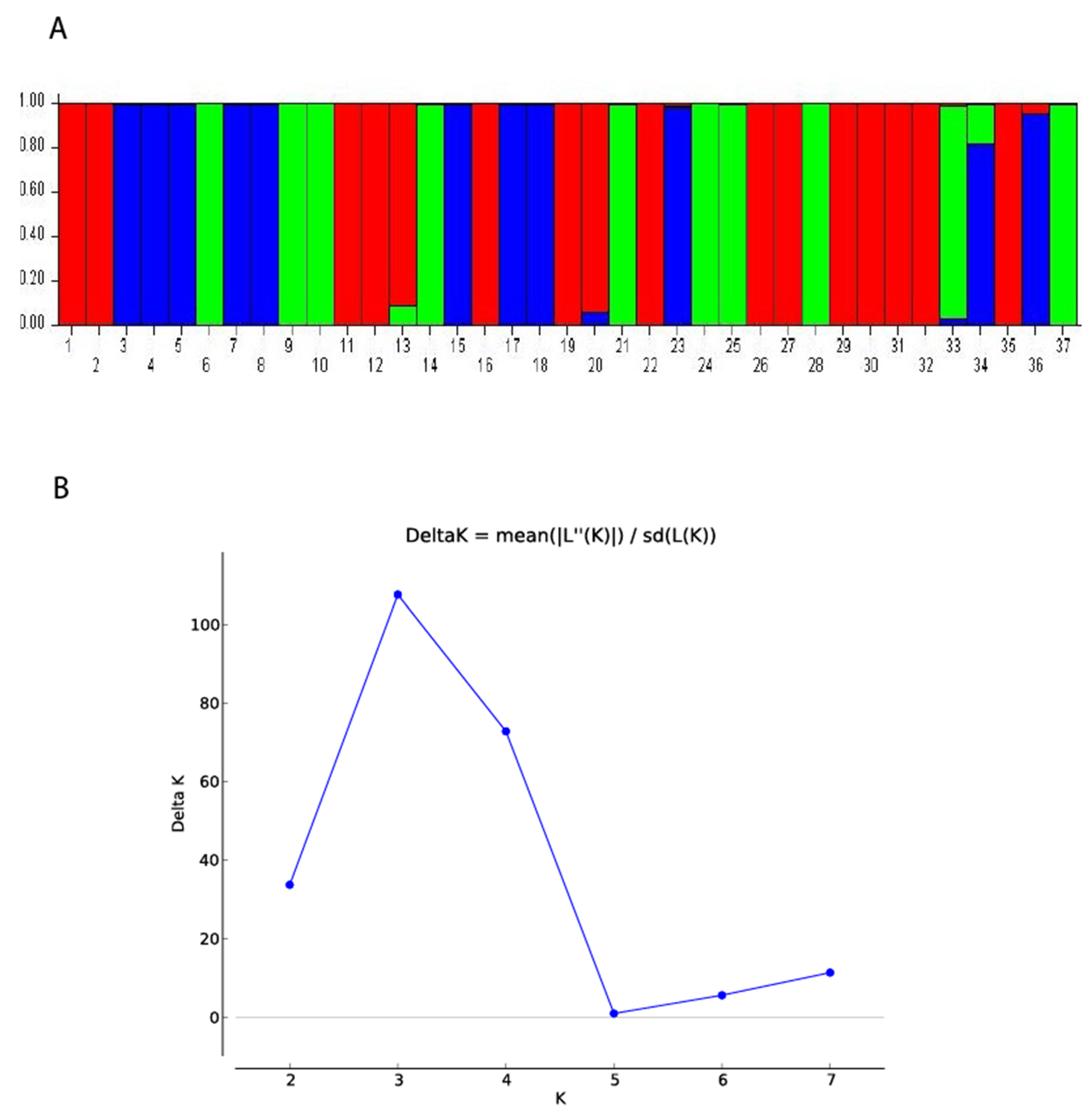
The genetic structure of the Saprolegnia isolates were also investigated using the model-based Bayesian clustering approach of genetic admixture analysis (Structure 2.3.4 software) [33]. Simulations were performed with a dataset from K = 1 to K = 7. The method developed by Evanno et al. (2005) [34] was used to identify the number of genetically homogeneous clusters (K). We used a burn-in period of 100,000 runs and 500,000 MCMC runs to compute the probability of the data for estimating K. Among the seven independent runs, the one with the highest Ln Pr (X|K) value (log probability) was chosen and represented as bar plots. Bar plots of likelihoods and ΔK values were made with STRUCTURE HARVESTER [35].

3. Results

AFLP analyses of all 37 isolates using six primer combinations resulted in a total of 163 clear polymorphic bands (Table 2). The number of polymorphic bands per primer combination ranged from 23 to 31 bands, with an average of 27.17 polymorphic bands per primer pair. With these six primer combinations, it was possible to uniquely distinguish among all 37 isolates. Genetic similarity for Saprolegnia isolates using Dice coefficients based on the AFLP data ranged from 0.0 to 1.0, with a mean of 0.41. The highest genetic similarity (1.0) was obtained between two isolates, one from Norway and the other from Scotland, while the lowest genetic similarity value (0.0) was observed between two isolates from Norway.
The percentage of polymorphic bands (PPB) in each group (i.e., country) was high, ranging from 29.45% to 84.05%, with a mean value of 55.52%. The Norwegian isolates showed the highest PPB and the Canadian showed the lowest PPB (Table 3).
UPGMA clustering of the Saprolegnia isolates, based on their countries of origin, did not consistently reflect the geographic origin of the Saprolegnia isolates. Some Saprolegnia isolates from Norway clustered together with isolates from Scotland (Figure 1). However, based on UPGMA, the Saprolegnia isolates were completely clustered according to their ability to produce oogonia with and without long spines on the cysts (Figure 1). We detected 15 specific markers that differentiated isolates that had the ability to produce oogonia with long spines from those without spines. UPGMA analysis of the Saprolegnia isolates according to their pathogenicity resulted in three clusters. All pathogenic isolates clustered together in one group, whereas the non-pathogenic isolates clustered together in two groups (Figure 1).
The results of the PCO analyses supported the results of the UPGMA analysis. There was no distinguishable clustering pattern of Saprolegnia isolates from a certain country, but the isolates were completely grouped according to their ability to produce oogonia with spines or without spines, and according to their ability to cause mortality or not in salmonids (Figure 2).
In general, the genetic distances between the regions measured by pairwise differences were low (Table 4), the highest genetic distance (0.25198) was between Canada and Chile, and the lowest was between Norway and Scotland (0.04209).
The AMOVA analyses based on the geographic origin of the isolates showed that most of the total genetic variability, i.e., 91.01%, was attributed to the variance within countries, while the among-country variance component only accounted for 8.99% (Table 5). A low proportion of the observed genetic differentiation can be explained by the level of the FST value (0.089), and the average estimated mean of gene flow (Nm) between the countries was relatively high (Nm = 2.559).
As regards sequencing of the ITS, maximum parsimony analyses with a heuristic search gave the 30 most parsimonious trees (MPTs) with a length of 117 steps. Figure 3 shows a bootstrap tree where all bootstrap values above 50 are indicated on the representative branches. The phylogenetic analyses conducted on 48 partial ITS sequences of Saprolegnia resulted in five well-supported clades. The majority of strains, namely 26, formed a clade with S. salmonis and S. parasitica sequences retrieved from EMBL/GenBank. All isolates within this complex either produced cysts with long hairs (Table 1) or were not tested. A well supported sister clade of this S. parasitica complex consisted of two strains, and one of these was identified as S. hypogyna. The third clade consisted of strains identified as S. ferax, S.mixta, S. longicaulis and S. sp. nuchiae. Some of these different species have identical ITS haplotypes. The last two clades were well-supported and formed sister clades. Five isolates grouped with two strains previously identified as S. diclina and S. australis. The last two isolates grouped with one strain previously identified as S. cf. ferax. Strain S5 was received as a Saprolegnia sp. strains, but both the phylogenetic analysis and the EMBL/GenBank Blast search revealed that this strain clearly belongs to Leptolegnia. Based on the phylogenetic analyses, no geographic grouping could be observed.
Bayesian clustering analysis with STRUCTURE software assigned the 37 Saprolegnia strains to three different clusters. Structure analysis showed the maximum likelihood distribution L(K) of the real number of three groups (K = 3). This value was obtained using the value of ad hoc quantity (ΔK) rather than maximum likelihood value L(K), as described by Evanno et al. (2005) [32]. Structure analysis clustered the Saprolegnia isolates into three main clusters (Figure 4) as the UPGMA and PCO groupings.

4. Discussion

The present study provides, for the first time, a good indication that the different pathogenic isolates of Saprolegnia strains clustered at a molecular level. Thereby, we provide documentation that there is a connection between pathogenicity to Atlantic salmon and the molecular diversity of Saprolegnia strains. The pathogenic Saprolegnia strains included in this study that formed one genetic cluster also shared the same morphological characteristics, i.e., long hooked hairs on the secondary zoospore cysts. Several reports have tried to prove that the variability among Saprolegnia isolates is correlated to infectivity [7,8]. We have previously shown significant differences in pathogenicity among seven of the strains included in the present study, and concluded that initial growth rate of germinating cysts in pure water, together with the presence of long hooked hairs on the secondary cysts, may be an indicator of the pathogenicity of Saprolegnia strains to Atlantic salmon [10]. In this study, we have extended this also to include characterization at the genetic level, reassuring our initial findings. However, the number of strains tested in vivo in the present study was limited for animal welfare reasons as well as the high cost of performing in vivo pathogenicity testing. Thus, these findings should be followed by further investigations including in vivo studies.

4.1. Geographical Origin

The present study proved there is more genetic variation among Saprolegnia strains within each country than among the countries included in the study. UPGMA clustering, phylogenetic analyses, PCO and STRUCTURE analyses consistently reflected the geographic origin of the Saprolegnia isolates. Actually, the AMOVA showed that most of the total genetic variability was attributed to the variance within countries. These results suggest that Saprolegnia from the countries included in this study share much of the same genetic material, which was also supported by the average estimated mean of gene flow (Nm) among the countries, which was relatively high (Nm = 2.559). This is in contrast to Bangyeekhun et al. (2003) [13], who reported the genetic dissimilarity of pathogenic Saprolegnia from different geographic locations (Northern Europe, Southern Europe and USA) in their study. Factors to consider are the industrialized nature of aquaculture and the transfer of fish among the countries included in the present study. This is an important risk factor for pathogen transfer in general. All the Saprolegnia strains included in the AFLP analysis were sampled from Atlantic salmon-farming countries, with fish/eggs being originally exported from Norway. In this context, it is not surprising that there is more genetic variation among Saprolegnia strains within each country than among the countries included in the study. The high gene flow detected in the study may be due to the repeated Saprolegnia introduction events among these countries and a low level of sexual recombination over time. These results are in agreement with the result detected by Paul et al. (2018) [36].

4.2. Morphology

The long hairs on the zoospore cysts seem to be a characteristic dividing the Saprolegnia strains into different genetic clusters. Wiilloughby (1985) [37] stated that this characteristic of Saprolegnia strains isolated from fish is typical for S. parasitica. Beakes et al. (1994) [38] suggested that Saprolegnia strains isolated from infected fish that had the distinctive clusters of long spines on their secondary cysts fulfilled an updated species concept of Saprolegnia parasitica in function, if not in taxonomic context. However, the taxonomic status of Saprolegnia species isolated from fish has still not been resolved. Dieguez-Uribeondo et al. (2007) [6] stated that the species-level identification of parasitic isolates of Saprolegnia has, at best, proven problematic and, at worst, impossible. In the present study, all Saprolegnia strains with long hooked hairs on the secondary zoospore cysts grouped together into a single major cluster, suggesting that they form a separate taxon.

4.3. Phylogeny

The phylogenetic analyses of 48 ITS Saprolegnia sequences resulted in five well-defined clades with Leptolegnia as an outgroup. These well-defined clades are largely congruent with previous studies [6]. All strains of S. parasitica formed a well-defined clade consistent with Clade I in a previous study [6]. In the present study, the term S. parasitica has been used for all strains within this clade. All strains tested herein within the S. parasitica complex produced cysts with long hairs. The S. hypogyna strains formed a very well-supported sister clade (97%) and corresponded to Clade Ia in [6]. The AFLP results were, for the main part, congruent with these groupings, with the exception of S. hypogyna, which grouped within the S. parasitica complex. The third clade consisted of at least four Saprolegnia species. This highly supported clade (100%) corresponds to Clade II proposed by Dièguez-Uribeondo et al. (2007) [6] but contains at least an additional three species. The grouping of the last two clades was very well supported (bootstrap: 94%). One of these clades consists of a combination of S. diclina and S. australis strains. The other clade consists of three strains, one identified as S. cf. ferax. These isolates are well separated from S. ferax and are therefore most likely a different species. This grouping into two clades is not congruent with earlier studies [6], which separated these species into three clades, designated III, IV and V. They included a broader range of species and a higher number of isolates for each species than the present study, which may explain the few observed differences between the two studies.
Most of the species included in the present study have ITS haplotypes in EMBL/GenBank that are identical to those of other species of Saprolegnia. This illustrates a common problem with ITS and fungal and fungal-like organisms: the lack of resolution to distinguish closely related species. In other well-studied fungi of fungal-like genera, there has been a shift from ITS to other genetic markers (e.g., β-tubulin, translation elongation factor 1-α). To reveal the true phylogeny of the genus Saprolegnia, other genetic markers should be used.

Author Contributions

Conceptualization, I.S., S.S. and A.E.; methodology, A.E.., S.S., R.F.F.; software, A.E. and S.S.; validation, A.E., S.S., and I.S.; formal analysis, A.E. and S.S.; investigation, A.E., S.S. and I.S.; resources, R.K., R.F.F. and T.V.; data curation, A.E., S.S. and I.S..; writing—original draft preparation, A.E. and S.S.; writing—review and editing, A.E., S.S., R.K., R.F.F., T.V. and I.S.; visualization, A.E., S.S., and I.S.; supervision, A.E. and I.S.; project administration, I.S.; funding acquisition, I.S. All authors have read and agreed to the published version of the manuscript.

Funding

Alpharma AS/Pharmaq AS, The Research Council of Norway (project number NFR: 150113/120), the Norwegian School of Veterinary Science, Nutreco ARC and the National Veterinary Institute – Norway have financed this work.

Institutional Review Board Statement

The study was conducted according to the guidelines of the Norwegian Food Safety Authority: https://www.mattilsynet.no/language/english/.

Data Availability Statement

Data supporting the reported results can be found at https://www.vetinst.no/en.

Acknowledgments

This work was supported by Alpharma AS/Pharmaq AS, The Research Council of Norway, the Norwegian School of Veterinary Science, Nutreco ARC and the National Veterinary Institute, Norway. We thank Øystein Evensen for valuable comments and critical reading of the manuscript. Procedures for DNA extraction and sequencing of the ITS were supported by Ann Kristin Knutsen and Astrid Løvseth of the National Veterinary Institute, Norway. The Saprolegnia strains from Japan were kindly provided by Kishio Hatai, Nippon Veterinary and Animal Science University, Japan.

Conflicts of Interest

The authors declare no conflict of interest.

References

  1. van den Berg, A.H.; McLaggan, D.; Dieguez-Uribeond, J.; van West, P. The impact of the water moulds Saprolegnia diclina and Saprolegnia parasitica on natural ecosystems and the aquaculture industry. Fungal Biol. Rev. 2013, 27, 33–42. [Google Scholar] [CrossRef]
  2. Torto-Alalibo, T.; Tian, M.Y.; Gajendran, K.; Waugh, M.E.; van West, P.; Kamoun, S. Expressed sequence tags from the oomycete fish pathogen Saprolegnia parasitica reveal putative virulence factors. BMC Microbiol. 2005, 5, 13. [Google Scholar] [CrossRef] [Green Version]
  3. Dick, M.W. Straminipilous Fungi: Systematics of the Peronosporomycetes Including Accounts of the Marine Straminipilous Protists, the Plasmodiophorids and Similar Organisms; Kluwer Academic, Springer Science & Business Media: Dordrecht, The Netherlands, 2001. [Google Scholar]
  4. Hughes, G. Saprolegniasis, Then and Now: A Retrospective. Salmon Saprolegniasis; Bonneville Power Administration: Portland, OR, USA, 1994; pp. 3–32. [Google Scholar]
  5. Mueller, G.J. Salmon Saprolegniasis; Prepared for US Department of Energy; Bonneville Power Administration: Portland, OR, USA, 1994. [Google Scholar]
  6. Diéguez-Uribeondo, J.; Fregeneda-Grandes, J.M.; Cerenius, L.; Pérez-Iniesta, E.; Aller-Gancedo, J.M.; Tellería, M.T.; Söderhäll, K.; Martín, M.P. Re-evaluation of the enigmatic species complex Saprolegnia diclina–Saprolegnia parasitica based on morphological, physiological and molecular data. Fungal Genet. Biol. 2007, 44, 585–601. [Google Scholar] [CrossRef]
  7. Yuasa, K.; Hatai, K. Relationship between pathogenicity of Saprolegnia spp. isolates to rainbow trout and their biological characteristics. Fish Pathol. 1995, 30, 101–106. [Google Scholar] [CrossRef]
  8. Grandes, J.M.F.; Diez, M.F.; Gancedo, J.M.A. Ultrastructural analysis of Saprolegnia secondary zoospore cyst ornamentation from infected wild brown trout, Salmo trutta L. and river water indicates two distinct morphotypes amongst long-spined isolates. J. Fish Dis. 2000, 23, 147–160. [Google Scholar] [CrossRef]
  9. Hussein, M.M.; Hatai, K. Pathogenicity of Saprolegnia species associated with outbreaks of salmonid saprolegniosis in Japan. Fish. Sci. 2002, 68, 1067–1072. [Google Scholar] [CrossRef]
  10. Stueland, S.; Hatai, K.; Skaar, I. Morphological and physiological characteristics of Saprolegnia spp. strains pathogenic to Atlantic salmon, Salmo salar L. J. Fish Dis. 2005, 28, 445–453. [Google Scholar] [CrossRef]
  11. Seymour, R.L. The genus Saprolegnia. Nova Hedwig. 1970, 19, 1–124. [Google Scholar]
  12. Molina, F.I.; Jong, S.-C.; Ma, G. Molecular characterization and identification of Saprolegnia by restriction analysis of genes coding for ribosomal RNA. Antonie Van Leeuwenhoek 1995, 68, 65–74. [Google Scholar] [CrossRef] [PubMed]
  13. Bangyeekhun, E.; Pylkkö, P.; Vennerström, P.; Kuronen, H.; Cerenius, L. Prevalence of a single fish-pathogenic Saprolegnia sp. clone in Finland and Sweden. Dis. Aquat. Org. 2003, 53, 47–53. [Google Scholar] [CrossRef] [PubMed] [Green Version]
  14. Hulvey, J.P.; Padgett, D.E.; Bailey, J.C. Species boundaries within Saprolegnia (Saprolegniales, Oomycota) based on morphological and DNA sequence data. Mycologia 2007, 99, 421–429. [Google Scholar] [CrossRef]
  15. Paul, Y.; Leung, W.L.; Hintz, W.E. Species composition of the genus Saprolegnia in fin fish aquaculture environments, as determined by nucleotide sequence analysis of the nuclear rDNA ITS regions. Fungal Biol. 2015, 119, 27–43. [Google Scholar]
  16. Sandoval-Sierra, J.V.; Martin, M.P.; Dieguez-Uribeondo, J. Species identification in the genus Saprolegnia (Oomycetes): Defining DNA-based molecular operational taxonomic units. Fungal Biol. 2014, 118, 559–578. [Google Scholar] [CrossRef]
  17. Hatai, K.; Hoshiai, G.-I. Characteristics of two Saprolegnia species isolated from coho salmon with saprolegniosis. J. Aquat. Anim. Health 1993, 5, 115–118. [Google Scholar] [CrossRef]
  18. Hussein, M.M.; Hatai, K.; Nomura, T. Saprolegniosis in salmonids and their eggs in Japan. J. Wildl. Dis. 2001, 37, 204–207. [Google Scholar] [CrossRef] [PubMed] [Green Version]
  19. Kitancharoen, N.; Hatai, K. Experimental infection of Saprolegnia spp. in rainbow trout eggs. Fish Pathol. 1996, 31, 49–50. [Google Scholar] [CrossRef] [Green Version]
  20. Elameen, A.; Fjellheim, S.; Larsen, A.; Rognli, O.A.; Sundheim, L.; Msolla, S.; Masumba, E.; Mtunda, K.; Klemsdal, S.S. Analysis of genetic diversity in a sweet potato (Ipomoea batatas L.) germplasm collection from Tanzania as revealed by AFLP. Genet. Resour. Crop Evol. 2008, 55, 397–408. [Google Scholar] [CrossRef]
  21. Dice, L.R. Measures of the amount of ecologic association between species. Ecology 1945, 26, 297–302. [Google Scholar] [CrossRef]
  22. Jaccard, P. Nouvelles Recherches sur la Distribution Florale; Bull. Soc. Vaud. Sci. Nat. 1908, 44, 223–270. [Google Scholar]
  23. Sneath, P.H.; Sokal, R.R. Numerical Taxonomy. The Principles and Practice of Numerical Classification; Taylor & Francis, Ltd.: Oxford, UK, 1973. [Google Scholar]
  24. Rohlf, F.J. NTSYS-pc: Numerical Taxonomy and Multivariate Analysis System; Applied Biostatistics: New York, NY, USA, 1992. [Google Scholar]
  25. Excoffier, L.; Smouse, P.E.; Quattro, J.M. Analysis of molecular variance inferred from metric distances among DNA haplotypes: Application to human mitochondrial DNA restriction data. Genetics 1992, 131, 479–491. [Google Scholar] [CrossRef]
  26. Schneider, S.; Roessli, D.; Excoffier, L. Arlequin: A software for population genetics data analysis. User Man. Ver 2000, 2, 2496–2497. [Google Scholar]
  27. Excoffier, L.; Smouse, P.E. Using allele frequencies and geographic subdivision to reconstruct gene trees within a species: Molecular variance parsimony. Genetics 1994, 136, 343–359. [Google Scholar] [CrossRef]
  28. Whitlock, M.C.; McCauley, D.E. Indirect measures of gene flow and migration: FST≠ 1/(4Nm+ 1). Heredity 1999, 82, 117–125. [Google Scholar] [CrossRef]
  29. Hall, T.A. BioEdit: A User-Friendly Biological Sequence Alignment Editor and Analysis Program for Windows 95/98/NT; Nucleic acids symposium series; ScienceOpen: Burlington, NJ, USA, 1999; pp. 95–98. [Google Scholar]
  30. Matthews, E.; Ellison, A.; Cable, J. Saprolegnia parasitica zoospore activity and host survival indicates isolate variation in host preference. Fungal Biol. 2021, 125, 260–268. [Google Scholar] [CrossRef] [PubMed]
  31. Swofford, D.L. Paup-a computer-program for phylogenetic inference using maximum parsimony. J. Gen. Physiol. 1993, 102, 9–10. [Google Scholar]
  32. Felsenstein, J. Confidence limits on phylogenies: An approach using the bootstrap. Evolution 1985, 39, 783–791. [Google Scholar] [CrossRef] [PubMed]
  33. Falush, D.; Stephens, M.; Pritchard, J.K. Inference of population structure using multilocus genotype data: Linked loci and correlated allele frequencies. Genetics 2003, 164, 1567–1587. [Google Scholar] [CrossRef]
  34. Evanno, G.; Regnaut, S.; Goudet, J. Detecting the number of clusters of individuals using the software STRUCTURE: A simulation study. Mol. Ecol. 2005, 14, 2611–2620. [Google Scholar] [CrossRef] [PubMed] [Green Version]
  35. Earl, D.A. STRUCTURE HARVESTER: A website and program for visualizing STRUCTURE output and implementing the Evanno method. Conserv. Genet. Resour. 2012, 4, 359–361. [Google Scholar] [CrossRef]
  36. Paul, Y.; Naumann, C.; Hintz, W.E. Assessment of intra-specific variability in Saprolegnia parasitica populations of aquaculture facilities in British Columbia, Canada. Dis. Aquat. Org. 2018, 128, 235–248. [Google Scholar]
  37. Willoughby, L. Rapid preliminary screening of Saprolegnia on fish. J. Fish Dis. 1985, 8, 473–476. [Google Scholar] [CrossRef]
  38. Beakes, G. Features which characterize Saprolegnia isolates from salmonid fish lesions-a review. Salmon Saprolegniasis 1994, 33–66. [Google Scholar]
Figure 1. Dendrogram of isolates of Saprolegnia spp. as revealed by UPGMA cluster analysis of AFLP-based genetic similarity (Dice coefficient). The isolates are described by their geographic origin, as shown in Table 1.
Jof 07 00713 g001
Figure 2. Score plot of principal coordinate analysis (PCO) of isolates of Saprolegnia spp. as revealed by using 163 polymorphic AFLP bands, collected from Canada, Chile, Japan, Norway and Scotland.
Jof 07 00713 g002
Figure 3. Phylogram of isolates of Saprolegnia spp. as revealed by sequencing of ITS region 2. The isolates are described by their geographic origin, their ability to produce long spines on cysts and their pathogenicity to salmon (Table 1).
Jof 07 00713 g003
Figure 4. Structure analyses summary. (A) Bar plots showing the relationships for all 37 Saprolegnia strains in the study assessed on the basis of AFLP data; each strain is represented by a vertical line. The Saprolegnia isolates are sorted according to their ID number (Table 1). (B) The values for seven independent runs for K = 1–7 and DeltaK = mean (L’’(K)I)/s(L(K)).
Jof 07 00713 g004
Table 1. The Saprolegnia isolates included in the study collected from Canada, Chile, Japan, Norway and Scotland.
Strain
Id
SpeciesStrain Number *EMBL Accession NumberOriginHostCyst Coat **Germ Rate
***
Path
Test ****
N1S. parasiticaVI 02388FN186015NorwayAs parr+Slow-
N2S. cf. feraxVI 02389FN186016NorwayAS parrNTNT
N3S. parasiticaVI 02391FN186017NorwayAs parr+Fast++
N4S. parasiticaVI 02750FN186018NorwayAs parr+NTNT
N5S. diclinaVI 02753FN186019NorwayAs parrNTNT
N6S. feraxVI 02756FN186020NorwayAs parrSlow
N7S. parasiticaVI 02763FN186021NorwayBt broodstock+NTNT
N8S. parasiticaVI 02770FN186022NorwayAs parr+FastNT
N9S. cf. feraxVI 04808FN186023NorwayAs fryNTNTNT
N10S. parasiticaVI 04810FN186024NorwayAs parrNTNTNT
N11S. parasiticaVI 04811FN186025NorwayAs parrNTNTNT
N12S. parasiticaVI 04812FN186026NorwayAs parrNTNTNT
N13S. parasiticaVI 04817FN186027NorwayAs broodstockNTNTNT
N14S. parasiticaVI 04819FN186028NorwayCwfNTNTNT
S1S. parasiticaVI 02392FN186029ScotlandAs parr+FastNT
S2S. parasiticaVI 02736FN186030ScotlandAs parr+Fast++
S3S. parasiticaVI 02757FN186031ScotlandAs parr+FastNT
S4S. parasiticaVI 02758FN186032ScotlandAs parr+FastNT
S5Leptolegnia sp.VI 04813FN186033ScotlandAs parrNTNTNT
S6S. hypogynaVI 04814FN186034ScotlandAs parrNTNTNT
S7S. parasiticaVI 04815FN186035ScotlandAs parrNTNTNT
S8S. parasiticaVI 04816FN186036ScotlandAs parrNTNTNT
S9Saprolegnia sp.VI 04818FN186037ScotlandAs parrNTNTNT
Ca1S. parasiticaVI 02393FN186038CanadaAs parr+SlowNT
Ca2S. parasiticaVI 02394FN186039CanadaAs parr+FastNT
Ca3S. parasiticaVI 02395FN186040CanadaAs parr+Medium
Ca4S. parasiticaVI 02437FN186041CanadaAs parr+NTNT
Ca5S. parasiticaVI 04809FN186042CanadaPsNTNTNT
Ch1S. diclinaVI 02739FN186043ChileAs eggsSlow
Ch2S. parasiticaVI 02740FN186044ChileAs broodstock+Medium
Ch3S. parasiticaVI 02741FN186045ChileAs eggs+MediumNT
Ch4S. diclinaVI 02744FN186046ChileAs eggsNTSlowNT
Ch5S. diclinaVI 02746FN186047ChileCs eggsNTNTNT
Ch6S. parasiticaVI 02748FN186048ChileAs eggs+FastNT
J1S. salmonisNJM 9851FN186049JapanSs+Fast++
J2S. parasiticaATCC90213FN186050JapanCs parr+Fast++
J3S. diclinaATCC90215FN186051JapanCs parrSlow
* Refers to the number in the strain collection of the National Veterinary Institute. Atlantic salmon (Salmo salar L.) (As), Ca (Canada), Ch (Chile), J (Japan), N (Norway) and S (Scotland). Coho salmon (Oncorhynchus kisutch) (Cs), sockeye salmon (Oncorhynchus nerka), (Ss), Pacific salmon (Oncorhynchus tshawytcha) (Ps), brown trout (Salmo trutta) (Bt) and common whitefish (Coregonus lavaretus) (Cwf). ** Long hairs (+) or no long hairs (−) on the secondary zoospore cyst. *** Germination rate (% cysts germinating) after incubation of Saprolegnia mycelium in sterilized tap water (STW) at 21 ± 1 °C for 20 hours. Slow = 0–10%; medium = 11–49% and Fast = 50–100% cysts germinating. **** High (++), medium (+) or low(−) mortality in Atlantic salmon exposed to Saprolegnia cysts (1 × 10⁴ cysts/L). NT = not tested.
Table 2. Nucleotide sequences of the selective primers used for AFLP analyses and the number of polymorphic bands resulting from each primer combination.
Primer CombinationEcoRI Primer
5′ → 3′
MseI Primer
5′ → 3′
Number of Polymorphic Bands
E12 × M17GAC-TGC-GTA-CCA-ATT-CACGAT-GAG-TCC-TGA-GTA-ACG27
E13 × M15GAC-TGC-GTA-CCA-ATT-CAGGAT-GAG-TCC-TGA-GTA-ACA28
E11 × M16GAC-TGC-GTA-CCA-ATT-CAAGAT-GAG-TCC-TGA-GTA-ACC23
E20 × M17GAC-TGC-GTA-CCA-ATT-CGCGAT-GAG-TCC-TGA-GTA-ACG31
E21 × M17GAC-TGC-GTA-CCA-ATT-CGGGAT-GAG-TCC-TGA-GTA-ACG30
E22 × M17GAC-TGC-GTA-CCA-ATT-CGTGAT-GAG-TCC-TGA-GTA-ACG24
Table 3. The number and percentage of polymorphic bands resulting from the AFLP analyses of Saprolegnia spp. in the study.
Country of OriginNo. Polymorphic Bands% Polymorphic Bands
Canada4829.45
Chile8652.76
Norway13784.05
Scotland9155.83
Mean90.555.52
Table 4. The average genetic distances among Saprolegnia spp. calculated by the genetic distance method (Exoffier and Smouse 1994) [23].
RegionsCanadaChileNorwayScotland
Canada0.00000
Chile0.251980.00000
Norway0.125850.088650.00000
Scotland0.149770.154400.042090.00000
Table 5. The results of the AMOVA of Saprolegnia spp. isolates using 163 AFLP markers; d.f., degree of freedom.
Regionsd.f.Sum of SquaresVariance
Components
% of VariationFST Value
Among countries3101.7631.869368.990.089
Within countries30567.88418.9294791.01
Total 669.647669.647
Publisher’s Note: MDPI stays neutral with regard to jurisdictional claims in published maps and institutional affiliations.

