Article

Citronellal Exerts Its Antifungal Activity by Targeting Ergosterol Biosynthesis in Penicillium digitatum

1
School of Chemical Engineering, Xiangtan University, Xiangtan 411105, China
2
Department of Food Science and Engineering, Ningbo University, Ningbo 315211, China
*
Author to whom correspondence should be addressed.
J. Fungi 2021, 7(6), 432; https://doi.org/10.3390/jof7060432
Submission received: 8 May 2021 / Revised: 26 May 2021 / Accepted: 27 May 2021 / Published: 29 May 2021
(This article belongs to the Special Issue Control of Postharvest Pathogenic Penicillium)

Abstract

:
Ergosterol (ERG) is a potential target for the development of antifungal agents against Penicillium digitatum, the pathogen of green mold in citrus fruits. This study examined the mechanism by which citronellal, a typical terpenoid of Cymbopogon nardus essential oil, acts on ergosterol to exhibit its antifungal activity against P. digitatum. We previously reported that citronellal inhibited the growth of P. digitatum with minimum inhibitory concentration (MIC) and minimum fungicidal concentration (MFC) of 1.36 and 2.72 mg/mL, respectively. In citronellal-treated cells, the membrane integrity and ergosterol contents significantly decreased, whereas lanosterol, which serves as a precursor for ergosterol biosynthesis, massively accumulated. Addition of 150 mg/L of exogenous ergosterol decreased the inhibitory rate of citronellal, restoring the ergosterol content and hence the membrane structure to normal levels, and triggered expression of nearly all ERG genes. Based on our findings, we deduce that citronellal damages the cell membrane integrity of P. digitatum by down-regulating the ERG genes responsible for conversion of lanosterol to ergosterol, the key downregulated gene being ERG3, due to the observed accumulation of ergosta-7,22-dienol.

1. Introduction

Green mold, caused by the pathogen Penicillium digitatum, is the major postharvest disease of citrus during harvesting and postharvest handling processes [1]. Currently, the disease is mainly being managed by use of chemical fungicides, but the extensive use of such fungicides has complicated the process through emergence of drug-resistant strains [2]. As a consequence of the increased emergence of resistance to these chemical fungicides, exacerbating by their negative environmental impacts, alternative drug targets including use of plant essential oils have been explored.
Essential oils (EOs) are considered safer, highly active, and with low possibility to induce drug resistance [3,4,5,6], thus together with some of their volatile compounds, they have gradually been applied to prevent postharvest diseases such as grey mold and green mold [7,8,9]. Citronellal, one of the main constituents of Cymbopogon nardus essential oil, has a strong inhibitory effect against fungi [10,11]. Previously, citronellal was reported to be effective in inhibiting the growth of three pathogenic Penicillium strains including P. digitatum, P. italicum, and P. ulaiense, with the inhibition zone ranging from 31 to 43 mm2 [12]. Recently however, Morcia et al. [13] and Wang et al. [14] confirmed that citronellal not only inhibits the growth of Penicillium species but also of Fusarium and Aspergillus species. Our previous study demonstrated that citronellal visibly inhibited the germination of P. digitatum spores at concentrations higher than 0.17 mg/mL and delayed the green mold decay of navel orange in a dose-dependent manner [15]. In addition, citronellal inhibited the mycelial growth of P. digitatum, with minimum inhibitory concentration (MIC) and minimum fungicidal concentration (MFC) of 1.36 and 2.72 mg/mL respectively. Combined with cinnamaldehyde, citronellal obviously reduced the green mold incidence in Satsuma mandarin citrus fruit and did not impair the fruit quality [16]. Citronellal may therefore be used as a novel natural antifungal alternative for the effective preservation of citrus fruits.
Existing literature suggests that cell membrane is the action target of citronellal against fungi. For example, citronellal inhibited the growth of Candida albicans by affecting membrane integrity and arresting cell cycle [17]. In addition, Singh et al. (2016) [18] reported that citronellal interferes with membrane homeostasis by increasing the hypersensitivity of fungi to membrane-perturbing agents, reducing ergosterol levels, and diminishing glucose-induced H+ extrusion. Ergosterol, an integral component of the cell membrane that plays a key role in maintaining the integrity and fluidity of the membrane structure, is a key target of common chemical fungicides (such as imazalil and prochloraz) against P. digitatum [2,19]. Indeed, a number of studies have shown that essential oils such as citral, eugenol, thymol, carvacrol, and perillaldehyde can significantly reduce the ergosterol content in the cell membranes of P. digitatum, Aspergillus species, and Candida species, causing abnormal cell membrane function [20,21,22,23,24,25,26]. Although we previously reported that citronellal inhibited P. digitatum by damaging the cell membrane integrity thereby leading to leakage of intracellular constituents [15,16], the actual mechanism by which citronellal causes damage to the cell membrane of P. digitatum is still unknown. This study was therefore designed (i) to evaluate the effect of citronellal on the plasma membrane integrity of P. digitatum, (ii) to determine its effect on lanosterol and ergosterol contents and on sterol composition, and (iii) to analyze its effect on gene expression levels in ergosterol biosynthesis of P. digitatum.

2. Materials and Methods

2.1. Pathogens

Penicillium digitatum used in this study was isolated from infected Satsuma mandarin (Citrus unshiu Marc. cv. Miyagawa Wase), identified by morphological and molecular biology methods, and preserved at Xiangtan University [27]. This strain was incubated on potato dextrose agar (PDA) at 25 ± 2 °C and their spore concentrations adjusted to 5 × 10⁶ spores/mL using a hemocytometer.

2.2. Fruit

Mature mandarin fruits (Citrus reticulata Blanco) were harvested on 21 December 2019 from an orchard in Luxi, Hunan, China. Healthy fruits of uniform size and without scars were selected for the experiments.

2.3. Chemicals

Citronellal (96%) was purchased from Aladdin Chemical Reagent Co., Ltd. (Shanghai, China), solutions prepared by dissolving the requisite amount in Tween-80 (0.05%, v:v) and topped up to the final volumes using distilled water. All the chemicals were of analytic grade.

2.4. In Vivo Experiments of Citronellal

Artificial inoculation of fruits with P. digitatum and their subsequent treatment with citronellal were conducted using the method described by OuYang et al. [16] with minor modifications. All fresh fruits were surface-sterilized by immersing in 2% sodium hypochlorite solution (v/v) for 2 min, then washed with distilled water, wounded (depth of 3 mm and width of 3 mm) with a sterile needle, inoculated with 20 μL of P. digitatum spore suspension (105 spores/mL), and left to air-dry. The inoculated fruits were then immersed in in wax amended with citronellal at 1 × and 5 × MFC for 30 s. Fruits with wax and pathogen inoculation were used as control. Ten mandarin fruits constituted a single replicate, and each treatment was performed in triplicate. The incidence rate of disease (measured by counting the number of green mold-infected wounds) was calculated from the formula:
D i s e a s e   i n c i d e n c e = N u m b e r   o f   r o t t e n   w o u n d s T o t a l   n u m b e r   o f   w o u n d s × 100
The diameter (mm) of lesion was measured using a G102-123-101 caliper (Shanghai Measuring and Counting Tools Co., Ltd., Shanghai, China).

2.5. Additional Exogenous Ergosterol Assay

Effects of addition of external ergosterol on the antifungal activity of citronellal against mycelial growth of P. digitatum were tested in vitro by agar dilution method [15] using final citronellal concentrations of 0, 1/4 MIC, 1/2 MIC, MIC, and 2 MIC. Additional exogenous ergosterol assay was performed according to the method by Nóbrega et al. [28] to compare the antifungal activity of citronellal in the absence and presence of ergosterol at concentrations of 50, 150, and 250 mg/L. Each treatment was performed in triplicate and the culture plates incubated at 25 ± 2 °C for 4 d.

2.6. Plasma Membrane Integrity

The plasma membrane integrity of P. digitatum cells treated with citronellal (0 or 1/2 MIC) were analyzed by propidium iodide (PI) staining coupled with an ECLIPSE TS100 microscope (Nikon, Japan) and the fluorescence value determined by an F97 PRO fluorescence spectrophotometer (Lengguang Technology, Shanghai, China) as described in OuYang et al. [29]. Each of these experiments was repeated with addition of 150 mg/L of exogenous ergosterol.

2.7. Determination of Total Lanosterol and Ergosterol Contents

Total lanosterol and ergosterol contents of P. digitatum cells citronellal (1/2 MIC concentration) for 0, 30, and 60 min were determined by high performance liquid chromatography (HPLC) as we previous described [25] with minor modifications. The 2-day-old mycelia were dried in a vacuum freeze drier for 4 h. About 0.008 g of the dry mycelia were homogenized with liquid nitrogen and suspended in 4 mL of freshly prepared 25% (w/v) NaOH and 8 mL of absolute ethanol, then saponified at 85–90 °C for 2 h. The mixtures were extracted thrice with petroleum ether and washed twice in saturated NaCl solution. The upper organic layer was vacuum concentrated and each residue adjusted to 10 mL using ethanol. The detected wavelengths for lanosterol and ergosterol were set at 210 nm and 282 nm, respectively, calculating lanosterol and ergosterol contents from the standard calibration curve using lanosterol and ergosterol as standards, respectively. Control samples were not treated with citronellal, and the experiments were repeated with addition of 150 mg/L exogenous ergosterol.

2.8. Determination of Sterols Composition by Gas Chromatography-Mass Spectrometry (GC-MS)

The composition of sterols in P. digitatum cells treated with 1/2 MIC citronellal for 0, 30, and 60 min were determined by GC-MS method [25]. The dry mycelia were saponified with a freshly prepared mixture of NaOH (4 mL, 25% w/v) and absolute ethanol (8 mL) at 80 °C for 1 h. The mixtures were subsequently extracted in petroleum ether and washed using saturated NaCl solution and then the upper organic layer vacuum concentrated. Each concentrated sample was completely dissolved in 950 μL of methyltertbutylether and then the silylation reagent mixture (50 μL, N-methyl-N-trimethylsilyltrifluoroacetamide and N-trimethylsilylimidazole, v/v, 9:1) added and left at room temperature for at least 30 min to complete the silylation reaction followed by GC-MS analysis. Control samples were not treated with citronellal. Similar experiments were performed with addition of exogenous ergosterol (150 mg/L) to the P. digitatum culture medium.
The analytical GC was carried out on a ShimadzuQP2010 plus gas chromatograph (Shimadzu, Kyoto, Japan) equipped with flame ionization detector (FID). A non-polar cross-linked fused-silica capillary column, HP-5MS (30 m × 0.25 mm × 0.25 μm; Agilent, Santa Clara, CA, USA), was used. The oven temperature was held at 50 °C for 1 min, programmed at a rate of 50 °C/min to 260 °C, then increased to 300 °C at a rate of 4 °C/min where it remained for 10 min. The carrier gas was helium (1.3 mL/min). The injector temperature was 250 °C, detector temperature 310 °C and the volume injected was 2 μL. MS analysis was carried out on the same chromatograph equipped with a Shimadzu QP 2010 GC/MS system, ionization voltage 70 eV, ion source temperature 200 °C, mass range m/z 50–700, scanning interval 0.5 s and scanning speed 1000 amu/s. The sterol TMS ethers were identified by comparison with commercial references, the NIST™ database, or data from literature [25,30].

2.9. Real-Time Fluorescence Quantitative PCR (RTFQ-PCR) Analysis

RNA of P. digitatum treated with citronellal (0 or 1/2 MIC concentration) for 0, 30, and 60 min was extracted using the Trizol reagent (Invitrogen, Carlsbad, CA, USA) following the manufacturer’s instructions. The expression levels of the ERG1, ERG2, ERG3, ERG4, ERG5, ERG6, ERG7, ERG9, ERG11 ERG24, ERG25, ERG26, and ERG27 genes were examined using RTFQ-PCR. RTFQ-PCR was performed through a BIO-RAD CFX Connect Thermal Cycler using FastStart Universal SYBR Green Master (Roche, Switzerland) with the following programs: 95 °C for 10 min followed by 40 cycles at 95 °C for 15 s, and at 60 °C for 1 min. Actin gene was used as internal reference, and the samples were quantified by 2△△CT method [31]. All primer pairs for expression assays are listed in Table 1. The experiments were repeated with addition of exogenous ergosterol (150 mg/L) to the fungal culture medium.

2.10. Statistical Analysis

All data were expressed as their mean ± SD by measuring three independent replicates and analyzed using one-way ANOVA followed by Duncan’s test to test the significance of differences between means obtained among the treatments at the 5% level of significance using SPSS statistical software package release 16.0 (SPSS Inc., Chicago, IL, USA).

3. Results

3.1. In Vivo Experiments of Citronellal

Citronellal (1 × and 5 × MFC) effectively reduced the green mold decays of citrus fruits by the fifth day of inoculation, being able to alleviate the disease progression in inoculated fruits as shown in Figure 1. By day 4 of incubation with P. digitatum, the incidence rate in wax-treated fruits was 82%, a value much higher than the 10% observed in citrus fruits coated with citronellal (1 × and 5 × MFC) (Figure 1A). The average lesion diameter also reduced from 10.08 ± 0.67 mm observed in the control group to 1.07 ± 0.03 and 0.91 ± 0.58 mm in the 1 × and 5 × MFC citronellal treated groups, respectively (Figure 1B,C).

3.2. Additional Exogenous Ergosterol Assay

Results of the inhibitory effect of addition of exogenous ergosterol on the antifungal activity of citronellal against P. digitatum are presented in Table 1. The MIC and MFC values of citronellal against P. digitatum were 1.36 and 2.72 mg/mL, respectively. After 4 days of treatment, the antifungal rate of citronellal (0.78 mg/mL = 1/2 MIC concentration) against P. digitatum was 59.60 ± 9.18%. This rate significantly reduced to 42.38 ± 7.16% following addition of external ergosterol (150 mg/L) (Table 2). This result showed that addition of 150 mg/L ergosterol could improve the tolerance of P. digitatum to citronellal.

3.3. Effects of Citronellal on the Plasma Membrane Integrity

Mycelia treated with 1/2 MIC citronellal showed stronger red fluorescence after 30 min of treatment compared to the controls (Figure 2A). After 60 min of treatment, the fluorescence intensity of the 1/2 MIC group, as determined by the fluorescence spectrophotometer, was 2.36 times higher than that of the control group (Figure 2B), indicating that citronellal had greatly damaged the cell membrane. Within the same 30 min duration of time, mycelia treated with 1/2 MIC citronellal plus externally added ergosterol showed no red fluorescence. Their fluorescence intensity after 60 min of treatment as measured by the fluorescence spectrophotometer was only 1.28 times higher than that of the control group (Figure 2B), representing a significantly lower damage to the membrane compared to that caused in the absence of externally added ergosterol.

3.4. Lanosterol and Ergosterol Contents

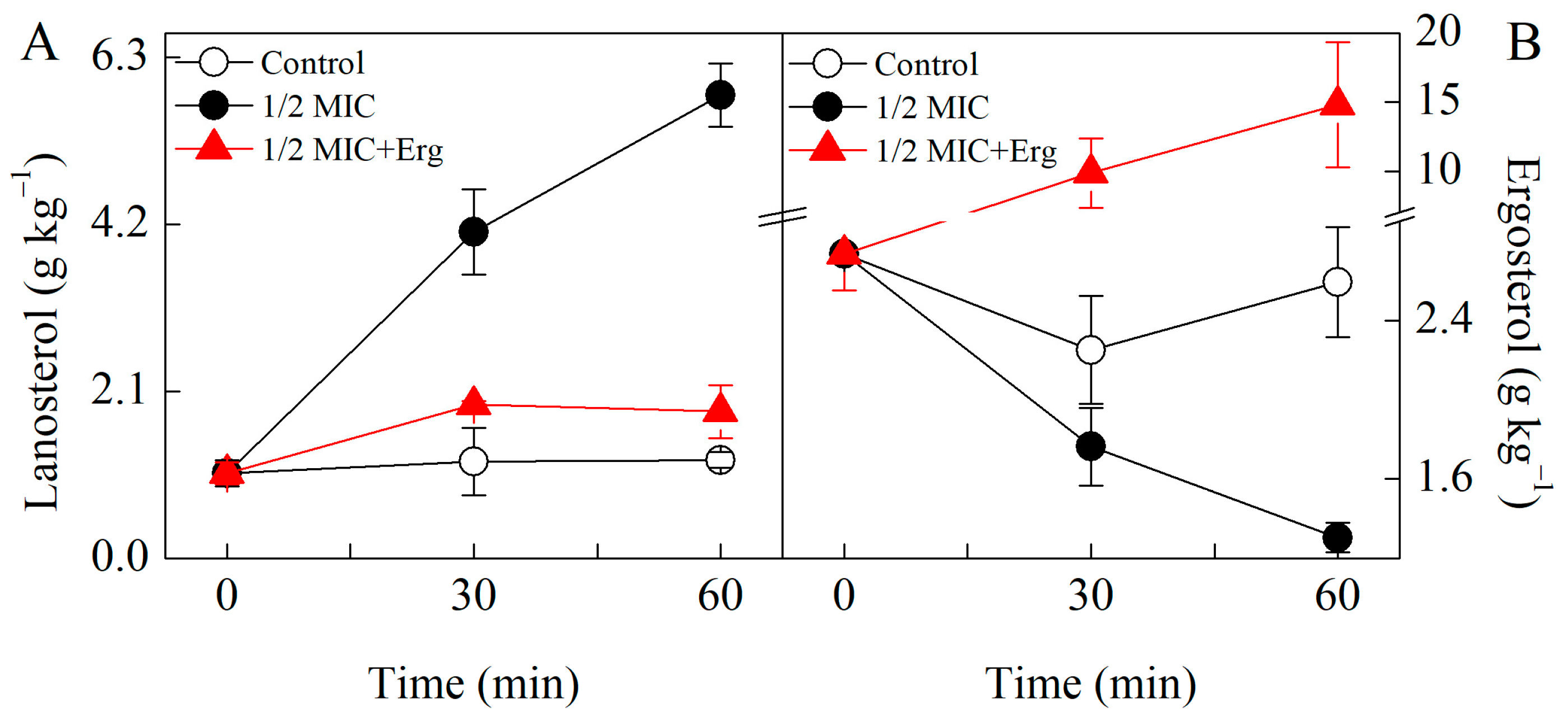
The lanosterol content in citronellal treated P. digitatum samples was markedly higher than those of the control groups (Figure 3). Following 30 min of treatment with citronellal, lanosterol content in the test group was 3.4 times higher than in control group. External addition of ergosterol resulted into much lower lanosterol values in the test group, being only 1.6 times higher than those of the corresponding control group (Figure 3A).
Citronellal treatment reduced the ergosterol content of P. digitatum but 1/2MIC+Erg treatment induced a higher ergosterol content compared to the control sample (Figure 3B). After 30 min of treatment with citronellal, the ergosterol content of the test group was 21.7% lower than that of the control group. However, treatment with exogenous ergosterol for the same duration of time, raised ergosterol content to a value 4.4 times higher than in control group (p < 0.05) (Figure 3B).

3.5. Analysis of Sterols Composition by GC-MS

GC-MS results showed that addition of both citronellal and ergosterol could significantly change the steroid components in P. digitatum (Table 3). Ergosterol, lanosterol and squalene were continuously detected in both test and control groups all through the experiment. Whereas both ergosta-5,7,22,24(28)-tetraenol and ergosta-7,22-dienol were detected in the control groups, with the former being detected at 30 min and the latter at 60 min, ergosta-7,22-dienol was also detected in the citronellal treated samples at both 30 and 60 min and in the citronellal + ergosterol group at 30 min of treatment. A new compound, eburicol, was detected only in the citronellal treated samples at 30 and 60 min but not in the control group or citronellal + ergosterol group.

3.6. Effect of Citronellal on Gene Expression Levels in Ergosterol Biosynthesis

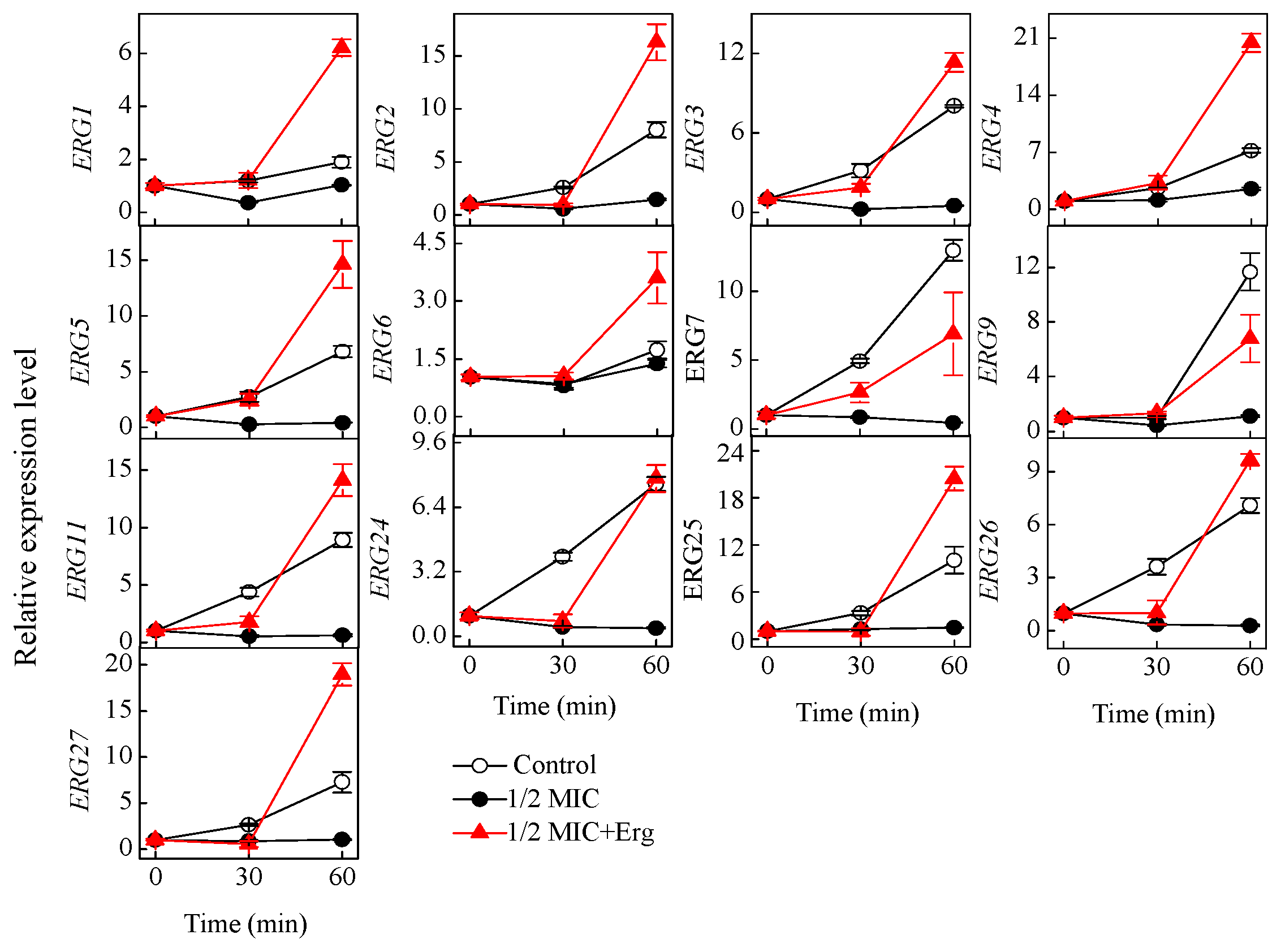
To explore the effect of citronellal on ergosterol biosynthesis of P. digitatum at the molecular level, the transcription levels of 13 genes involved in the biosynthesis of ergosterol was investigated. After citronellal treatment, the expression levels of all the 13 ERG genes in the test group (except ERG6 gene whose level did not change) were repressed (Figure 4).
P. digitatum showed different responses at the molecular level when the citronellal-treated cells were supplemented with ergosterol, with the ERG1, ERG3, ERG5, and ERG7 genes showing increased expression levels at 30 min, and by 60 min, the expression levels of nearly all genes (except ERG7, ERG9, and ERG24) in the test groups being significantly higher than in the control groups (p < 0.05) (Figure 4).

4. Discussion

Ergosterol is the most abundant and main sterol component of fungal membranes [32], involved in regulation of membrane permeability and fluidity; regulation, activity, and distribution of integral membrane proteins; and control of the cell cycle [33]. It is thus key to the structural integrity of the cell membranes and, as such, the main target of the majority of the available antifungals [32,33], which interfere with its function either through inhibition of various steps in ergosterol biosynthesis or by complexing directly with membrane ergosterol [24]. Numerous essential oils and their components such as Curcuma longa L. essential oil, citral, perillaldehyde, and thymol have an impact on the cell membrane integrity by targeting ergosterol metabolism [22,23,24,25,26].
Citronellal is a promising antifungal compound with good inhibitory effects on P. digitatum both in vitro and in vivo. Our results show that treatment of P. digitatum with citronellal led to a significant inhibition of ergosterol biosynthesis and damage to the cell membrane. This observation corresponds to that reported by Singh et al. [18] in which citronellal had adverse effects on cell membrane integrity and ergosterol levels of C. albicans. Addition of 150 mg/L ergosterol to the growth medium greatly enhanced the tolerance of P. digitatum to citronellal, restoring the cell membrane integrity and ergosterol content to normal levels. This again is consistent with reports that the presence of exogenous ergosterol in growth medium promotes increased survival of some fungi under test agents stress [24,28,34,35]. Abe and Hiraki [36] and Liu et al. [35] also reported about the restoration of ergosterol content and cell membrane integrity to normal levels following exogenous ergosterol treatment of Saccharomyces cerevisiae. These findings confirm a direct correlation between ergosterol levels and cell membrane integrity and allude to the possibility that the antifungal effect of citronellal on P. digitatum is attributed to its inhibition of ergosterol biosynthesis.
Ergosterol biosynthesis is tightly regulated by ERG genes [37]. Our previous study showed that citral, another terpene aldehyde, decreased the expression levels of genes ERG3, ERG5, ERG6, and ERG11 and acted on the ergosterol biosynthesis of P. digitatum by targeting the ERG11 gene [25], and Xu et al. [38] also noted that linalool (a terpene alcohol) downregulated the expression of genes in the ergosterol biosynthesis pathway. We have found similar results in P. digitatum in which the expression of most of the ERG genes in citronellal-treated samples was markedly lower (p < 0.05) than in the control samples during the entire experimental time. In this pathway, catalyzing C14-demethylation of lanosterol by ERG11 is critical for ergosterol biosynthesis. Obviously, citronellal induced the downregulated of ERG11 gene and then led the exclusive accumulation of lanosterol, indicating that the ergosterol synthesis was hindered. Meanwhile, the side chain of ergosterol biosynthesis was activated, inducing the production of eburicol in citronellal-treated samples. These results are consistent with those reported by Ottilie et al. [39] that posaconazole, a TcCyp51 inhibitor, can increase the accumulation of both lanosterol and eburicol. It should also be noted that the loss of the ERG3 led to the typical fungal sterol ergosterol and ergosta-5,7,22,24(28)-tetraenol depletion, activating another side chain of ergosterol biosynthesis hence inducing the generation of ergosta-7,22-dienol [40]. This implies that the action site of citronellal against P. digitatum might be between the ERG11 and ERG3.
Exogenous ergosterol is significant in improving the synthetic ability of ergosterol in S. cerevisiae and C. glabrata [34,35]. In this study, ergosterol content markedly increased after 30 min of treatment with 1/2 MIC + Erg. This might have resulted from the coordinated uptake and biosynthesis of the sterols [34]. We also discovered that the expression of genes involved in ergosterol biosynthesis in P. digitatum was altered after supplementation of exogenous ergosterol in the citronellal treatment samples. In the citronellal-treated cells supplemented with ergosterol, genes ERG1, ERG3, ERG5, and ERG7 responded quickly and in just 30 min had already been upregulated more than those in citronellal-treated samples. By 60 min of treatment, all the genes in the test samples supplemented with ergosterol had been upregulated more than those in only citronellal treatment. This corresponds to the result by Liu et al. [35] that ergosterol supplementation triggered the upregulation of genes encoding ergosterol biosynthesis, leading to increase in sterol levels. Furthermore, upregulation of ERG3 and ERG5 genes led to a decrease in lanosterol content to the normal value and a decrease in eburicol (a branch chain product) content to the detection limit after 30 min of treatment with exogenous ergosterol. This implies that ERG3 is the vital regulatory gene.
In summary, the present study has demonstrated that the suppression of ergosterol biosynthesis in P. digitatum cells by citronellal treatment occurs between ERG11 and ERG3 genes and that gene ERG3 in P. digitatum may be the key regulatory site gene in response to citronellal treatment. These findings provide new insights into the antifungal mechanism of citronellal.

Author Contributions

Conceptualization, N.T.; data curation, N.T.; formal analysis, N.T.; funding acquisition, Q.O. and N.T.; investigation, Q.O. and Y.L.; methodology, Q.O. and Y.L.; supervision, N.T.; writing—original draft, Q.O.; writing—review and editing, Q.O., O.R.O., M.Z., X.S., and N.T. All authors have read and agreed to the published version of the manuscript.

Funding

This study was supported by the National Natural Science Foundation of China (No. 31772364), Research Foundation of Education Bureau of Hunan Province (No. 19A476), Collaborative Innovation Center of New Chemical Technologies for Environmental Benignity and Efficient Resource Utilization, the Natural Science Foundation of Hunan Province (No. 2020JJ5536), and the Scientific Research Foundation for Doctors of Xiangtan University (No. 19QDZ12).

Institutional Review Board Statement

Not applicable.

Informed Consent Statement

Not applicable.

Data Availability Statement

The datasets generated during and/or analyzed during the current study are available from the corresponding author upon reasonable request.

Conflicts of Interest

The authors declare no conflict of interest.

References

  1. Kanetis, L.; Förster, H.; Adaskaveg, J.E. Comparative efficacy of the new postharvest fungicides azoxystrobin, fludioxonil, and pyrimethanil for managing citrus green mold. Plant Dis. 2007, 91, 1502–1511. [Google Scholar] [CrossRef] [PubMed] [Green Version]
  2. Sun, X.P.; Wang, J.Y.; Feng, D.; Ma, Z.H.; Li, H.Y. PdCYP51B, a new putative sterol 14a–demethylase gene of Penicillium digitatum involved in resistance to imazalil and other fungicides inhibiting ergosterol synthesis. Appl. Microbiol. Biotechnol. 2011, 91, 1107–1119. [Google Scholar] [CrossRef]
  3. Camele, I.; Elshafie, H.S.; Caputo, L.; De Feo, V. Anti-quorum sensing and antimicrobial effect of Mediterranean plant essential oils against phytopathogenic bacteria. Front. Microbiol. 2019, 10, 2619. [Google Scholar] [CrossRef] [PubMed]
  4. Elshafie, H.S.; Caputo, L.; De Martino, L.; Grul’ova, D.; Zheljazkov, V.Z.; De Feo, V.; Camele, I. Biological investigations of essential oils extracted from three Juniperus species and evaluation of their antimicrobial, antioxidant and cytotoxic. J. Appl. Microbiol. 2020, 129, 1261–1271. [Google Scholar] [CrossRef]
  5. Gru’ová, D.; Caputo, L.; Elshafie, H.S.; Baranová, B.; De Martino, L.; Sedlák, V.; Goga’ová, Z.; Poráčová, J.; Camele, I.; De Feo, V. Thymol chemotype Origanum vulgare L. essential oil as a potential selective bio-based herbicide on monocot plant species. Molecules 2020, 25, 595. [Google Scholar] [CrossRef] [Green Version]
  6. Kesdek, M.; Kordali, S.; Bozhuyuk, A.U.; Gudek, M. Larvicidal effect of Achillea biebersteinii Afan. (Asteraceae) essential oil against larvae of pine processionary moth, Thaumetopoea pityocampa (Denis & Schiffermüller, 1775) (Lepidoptera: Notodontidae). Turk. J. Agric. For. 2020, 44, 451–460. [Google Scholar] [CrossRef]
  7. Duan, X.F.; OuYang, Q.L.; Tao, N.G. Effect of applying cinnamaldehyde incorporated in wax on green mould decay in citrus fruits. J. Sci. Food Agric. 2017, 98, 527–533. [Google Scholar] [CrossRef] [PubMed]
  8. Hasheminejad, N.; Khodaiyan, F. The effect of clove essential oil loaded chitosan nanoparticles on the shelf life and quality of pomegranate arils. Food Chem. 2020, 309, 125520. [Google Scholar] [CrossRef] [PubMed]
  9. Yue, Q.; Shao, X.F.; Wei, Y.Y.; Jiang, S.; Xu, F.; Wang, H.F.; Gao, H.Y. Optimized preparation of tea tree oil complexation and their antifungal activity against Botrytis cinerea. Postharvest Biol. Technol. 2020, 162, 111114. [Google Scholar] [CrossRef]
  10. Kaur, H.; Bhardwaj, U.; Kaur, R. Cymbopogon nardus essential oil: A comprehensive review on its chemistry and bioactivity. J. Essent. Oil Res. 2021. [Google Scholar] [CrossRef]
  11. Rammanee, K.; Hongpattarakere, T. Effects of tropical citrus essential oils on growth, aflatoxin production, and ultrastructure alterations of Aspergillus flavus and Aspergillus parasiticus. Food Bioprocess Technol. 2011, 4, 1050–1059. [Google Scholar] [CrossRef]
  12. Scora, K.M.; Scora, R.W. Effect of volatiles on mycelium growth of Penicillium digitatum, P. italicum, and P. ulaiense. J. Basic Microbiol. 1998, 38, 405–413. [Google Scholar] [CrossRef]
  13. Morcia, C.; Tumino, G.; Ghizzoni, R.; Bara, A.; Salhi, N.; Terzi, A. In vitro evaluation of sub-lethal concentrations of plant-derived antifungal compounds on Fusaria growth and mycotoxin production. Molecules 2017, 22, 1271. [Google Scholar] [CrossRef] [Green Version]
  14. Wang, H.W.; Yang, Z.X.; Ying, G.Y.; Yang, M.H.; Nian, Y.J.; Wei, F.; Kong, W.J. Antifungal evaluation of plant essential oils and their major components against toxigenic fungi. Ind. Crop. Prod. 2018, 120, 180–186. [Google Scholar] [CrossRef]
  15. Wu, Y.L.; OuYang, Q.L.; Tao, N.G. Plasma membrane damage contributes to antifungal activity of citronellal against Penicillium digitatum. J. Food Sci. Technol. 2016, 53, 3853–3858. [Google Scholar] [CrossRef] [PubMed] [Green Version]
  16. OuYang, Q.L.; Okwong, R.O.; Chen, Y.P.; Tao, N.G. Synergistic activity of cinnamaldehyde and citronellal against green mold in citrus fruit. Postharvest Biol. Technol. 2020, 162, 111095. [Google Scholar] [CrossRef]
  17. Zore, G.B.; Thakre, A.D.; Jadhav, S.; Karuppayil, S.M. Terpenoids inhibit candida albicans growth by affecting membrane integrity and arrest of cell cycle. Phytomedicine 2011, 18, 1181–1190. [Google Scholar] [CrossRef] [PubMed]
  18. Singh, S.; Fatima, Z.; Hameed, S. Citronellal-induced disruption of membrane homeostasis in Candida albicans and attenuation of its virulence attributes. Rev. Soc. Bras. Med. Trop. 2016, 49, 465–472. [Google Scholar] [CrossRef] [Green Version]
  19. Liu, J.; Yuan, Y.Z.; Wu, Z.; Li, N.; Chen, Y.L.; Qin, T.T.; Geng, H.; Xiong, L.; Liu, D.L. A novel sterol regulatory element–binding protein gene (sreA) identified in Penicillium digitatum is required for prochloraz resistance, full virulence and erg11 (cyp51) regulation. PLoS ONE 2015, 10, e0117115. [Google Scholar] [CrossRef] [Green Version]
  20. Elshafie, H.S.; Mancini, E.; Camele, I.; De Martino, L.; De Feo, V. In vivo antifungal activity of two essential oils from Mediterranean plants against postharvest brown rot disease of peach fruit. Ind. Crop. Prod. 2015, 66, 11–15. [Google Scholar] [CrossRef]
  21. Elshafie, H.S.; Mancini, E.; Sakr, S.; De Martino, L.; Mattia, C.A.; De Feo, V.; Camele, I. Antifungal activity of some constituents of Origanum vulgare L. essential oil against postharvest disease of peach fruit. J. Med. Food 2015, 18, 929–934. [Google Scholar] [CrossRef] [PubMed]
  22. Ahmad, A.; Khan, A.; Akhtar, F.; Yousuf, S.; Xess, I.; Khan, L.A.; Manzoor, N. Fungicidal activity of thymol and carvacrol by disrupting ergosterol biosynthesis and membrane integrity against Candida. Eur. J. Clin. Microbiol. 2011, 30, 41–50. [Google Scholar] [CrossRef] [PubMed]
  23. Hua, H.J.; Xing, F.G.; Selvaraj, J.N.; Wang, Y.; Zhao, Y.J.; Zhou, L.; Liu, X.; Liu, Y. Inhibitory effect of essential oils on Aspergillus ochraceus growth and ochratoxin A production. PLoS ONE 2014, 9, e108285. [Google Scholar] [CrossRef] [Green Version]
  24. Khan, M.S.A.; Ahmad, I.; Cameotra, S.S. Phenyl aldehyde and propanoids exert multiple sites of action towards cell membrane and cell wall targeting ergosterol in Candida albicans. AMB Express 2013, 3, 54. [Google Scholar] [CrossRef] [Green Version]
  25. OuYang, Q.L.; Tao, N.G.; Jing, G.X. Transcriptional profiling analysis of Penicillium digitatum, the causal agent of citrus green mold, unravels an inhibited ergosterol biosynthesis pathway in response to citral. BMC Genom. 2016, 17, 599. [Google Scholar] [CrossRef] [Green Version]
  26. Tian, J.; Wang, Y.Z.; Zeng, H.; Zhang, P.; Tessema, A.; Peng, X. Efficacy and possible mechanisms of perillaldehyde in control of Aspergillus niger, causing grape decay. Int. J. Food Microbiol. 2015, 202, 27–34. [Google Scholar] [CrossRef] [PubMed]
  27. Tao, N.G.; Jia, L.; Zhou, H.E. Anti-fungal activity of Citrus reticulata Blanco essential oil against Penicillium italicum and Penicillium digitatum. Food Chem. 2014, 153, 265–271. [Google Scholar] [CrossRef] [PubMed]
  28. Nóbrega, R.O.; Teixeira, A.P.C.; Oliveira, W.A.; Lima, E.D.; Lima, I.O. Investigation of the antifungal activity of carvacrol against strains of Cryptococcus neoformans. Pharma. Biol. 2016, 54, 2591–2596. [Google Scholar] [CrossRef] [Green Version]
  29. OuYang, Q.L.; Tao, N.G.; Zhang, M.L. A damaged oxidative phosphorylation mechanism is involved in the antifungal activity of citral against Penicillium digitatum. Front. Microbiol. 2018, 9, 239. [Google Scholar] [CrossRef] [PubMed] [Green Version]
  30. Müller, C.; Staudacher, V.; Krauss, J.; Giera, M.; Bracher, F.A. Convenient cellular assay for the identification of the molecular target of ergosterol biosynthesis inhibitors and quantification of their effects on total ergosterol biosynthesis. Steroids 2013, 78, 483–493. [Google Scholar] [CrossRef]
  31. Livak, K.J.; Schmittgen, T.D. Analysis of relative gene expression data using Real–Time quantitative PCR and the 2−ΔΔCT method. Methods 2001, 25, 402–408. [Google Scholar] [CrossRef]
  32. Alcazar-Fuoli, L.; Mellado, E. Ergosterol biosynthesis in Aspergillus fumigatus: Its relevance as an antifungal target and role in antifungal drug resistance. Front. Microbiol. 2013, 3, 439. [Google Scholar] [CrossRef] [PubMed] [Green Version]
  33. Dhingra, S.; Cramer, R.A. Regulation of sterol biosynthesis in the human fungal pathogen Aspergillus fumigatus: Opportunities for therapeutic development. Front. Microbiol. 2017, 8, 92. [Google Scholar] [CrossRef] [PubMed] [Green Version]
  34. Li, Q.Q.; Tsai, H.F.; Mandal, A.; Walker, B.A.; Noble, J.A.; Fukuda, Y.; Bennett, J.E. Sterol uptake and sterol biosynthesis act coordinately to mediate antifungal resistance in Candida glabrata under azole and hypoxic stress. Mol. Med. Rep. 2018, 17, 6585–6597. [Google Scholar] [CrossRef] [PubMed]
  35. Liu, J.D.; Zhu, Y.B.; Du, G.C.; Zhou, J.W.; Chen, J. Exogenous ergosterol protects Saccharomyces cerevisiae from D-limonene stress. J. Appl. Microbiol. 2013, 114, 482–491. [Google Scholar] [CrossRef]
  36. Abe, F.; Hiraki, T. Mechanistic role of ergosterol in membrane rigidity and cycloheximide resistance in Saccharomyces cerevisiae. BBA Biomembr. 2009, 1788, 743–752. [Google Scholar] [CrossRef] [PubMed] [Green Version]
  37. Bhattacharya, S.; Esquivel, B.D.; White, T.C. Overexpression or deletion of ergosterol biosynthesis genes alters doubling time, response to stress agents, and drug susceptibility in Saccharomyces cerevisiae. mBio 2018, 9, e01291-18. [Google Scholar] [CrossRef] [Green Version]
  38. Xu, Y.Q.; Tong, Z.C.; Zhang, X.; Wang, Y.Y.; Fang, W.G.; Li, L.; Luo, Z.S. Unveiling the mechanisms for the plant volatile organic compound linalool to control gray mold on strawberry fruits. J. Agric. Food Chem. 2019, 67, 9265–9276. [Google Scholar] [CrossRef]
  39. Ottilie, S.; Goldgof, G.M.; Calvet, C.M.; Jennings, G.K.; Lamonte, G.; Schenken, J.; Vigil, E.; McCall, L.; Lopes, E.S.C.; Gunawan, F.; et al. Rapid chagas disease drug target discovery using directed evolution in drug-sensitive yeast. ACS Chem. Biol. 2017, 12, 422–434. [Google Scholar] [CrossRef] [Green Version]
  40. Vale-Silva, L.A.; Coste, A.T.; Ischer, J.E.; Parker, J.E.; Kelly, E.; Pinto, E.; Sanglard, D. Azole resistance by loss of function of the sterol Δ5,6- desaturase gene (ERG3) in Candida albicans does not necessarily decrease virulence. Antimicrob. Agents Chemother. 2012, 56, 1960–1968. [Google Scholar] [CrossRef] [Green Version]
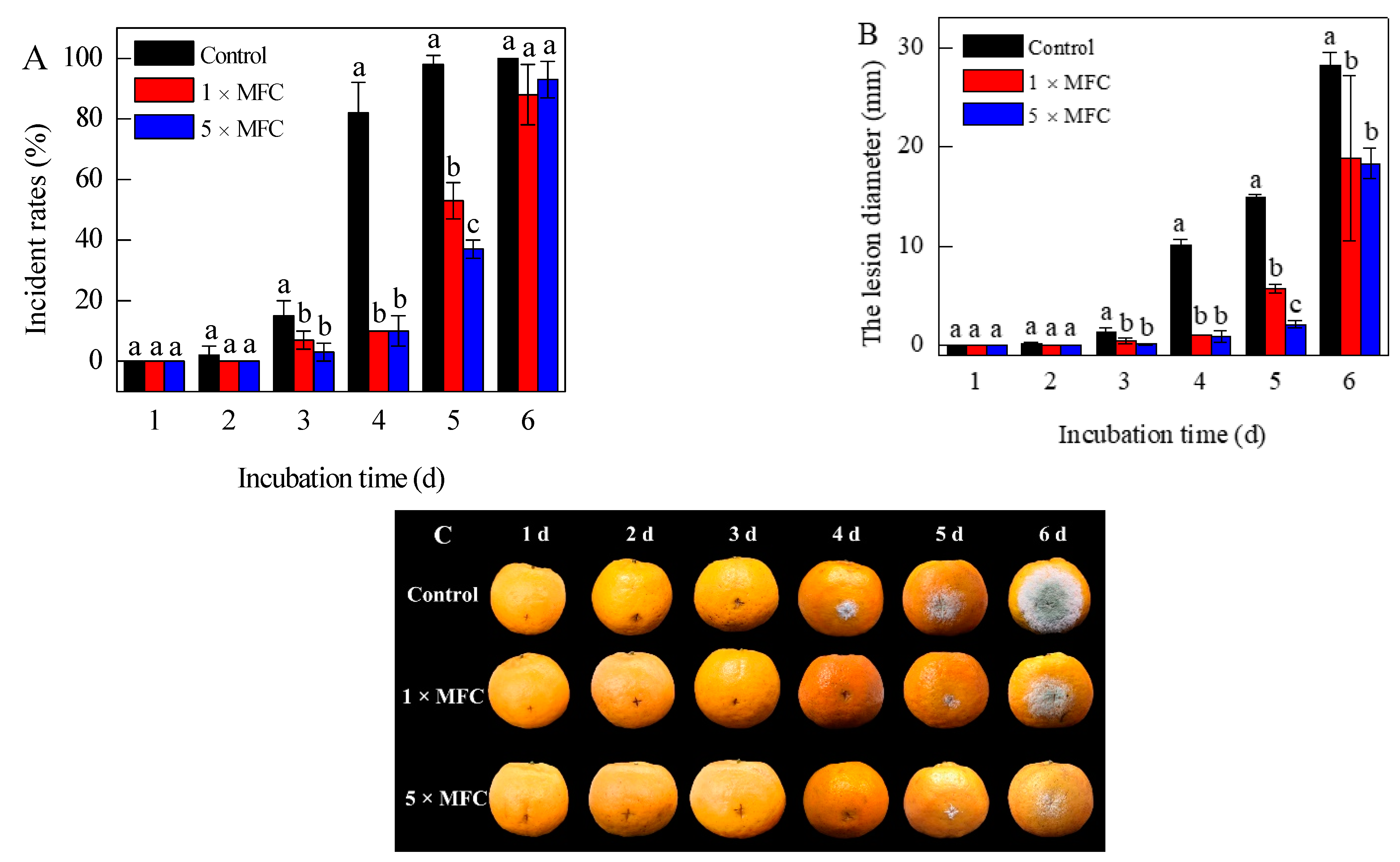
Figure 1. Disease incidence (A), the citrus lesion diameter (B) and the disease progression (C) in inoculated citrus fruits treated with citronellal (0 ×, 1 ×, and 5 ×  MFC) during storage at 25  ±  2  °C for 6 d and 85–90% RH. The data presented are the means of pooled data. Error bars indicate the SDs of the means (n = 3).
Jof 07 00432 g001
Figure 2. Effects of citronellal on the plasma membrane integrity of P. digitatum (A) PI staining images of P. digitatum mycelia treated with citronellal for 0, 30, and 60 min; (B) the fluorescence fold changes of the PI staining). The data presented are the means of pooled data. Error bars indicate the SDs of the means (n = 3).
Jof 07 00432 g002
Figure 3. HPLC analysis of lanosterol (A) and ergosterol (B) contents in control samples, 1/2 MIC citronellal treated samples, and 1/2 MIC citronellal + Erg treated sample. The data presented are the means of pooled data. Error bars indicate the SDs of the means (n = 3).
Jof 07 00432 g003
Figure 4. Changes in the expression of ergosterol biosynthesis genes of P. digitatum mycelia treated by control, 1/2 MIC citronellal treated samples, and 1/2 MIC citronellal + Erg treated samples for 0, 30, and 60 min. The data presented are the means of pooled data. Error bars indicate the SDs of the means (n = 3).
Jof 07 00432 g004
Table 1. Primer pair sequences designed for ERG genes in P. digitatum using RTFQ-PCR.
GenesPrimer (5′-3′)Length (bp)
ERG9TCTTTGTTGAGGCCGGGTTT
ACTTGGGGCGTTTCAACAGA
51
ERG1ATCCCCGATAACCTGCCTCT
CCCTTGACGCCTCCATTCTT
52
ERG7GCGCTGGCGATTGGTCGATG
CAGGCCCAGTTTCCGGGCTCC
219
ERG11GCGGAATCAAGAGGGACGAT
GCCCTAGCACACACTTCAGA
150
ERG24AGAGCTTCACAGTTCCAGCC
CGATGCCTCGCTGACAAATG
155
ERG25ATCGAAAGCTTCCTACGGGG
GCGCATCAATAGGCTGAGGA
80
ERG26ACCAGACCCCCTGCATCTAT
TTGGGATCCGTGCTCTAGGA
162
ERG27TTTCGATCTGCTGCCGTCTT
GCGCGCTTCGAGTTGTAAAT
91
ERG6CGCGTGATGCCGCCTTCAAC
TGAGCCTTGCGGGCCTCACG
184
ERG3CAGGCCATGGCCGCAATGCC
GGTGCAGGCCACGGTGGATCC
190
ERG5TCTCGCCATTGGCGGATGCG
GGCCAACAATGGCGCCCTTG
240
ERG4GCTGGAACCGCTACTTCCTT
AGACAAACAGGTAGGCGACG
51
ERG2ACATCTTCGACCCGGAACAC
TTGGGACCGACTTTCTGCTC
121
actin183TGCGCTGAACCGAACTGCCG
TCGGGAGCCTCGAAGCGCTC
183
Table 2. Effects of citronellal and exogenous ergosterol on the growth of P. digitatum.
TreatmentsInhibitory Rate (%)
1 d2 d3 d4 d
1/4 MIC40.48 ± 4.12 c19.42 ± 3.36 d16.67 ± 4.12 e14.57 ± 1.99 f
1/4 MIC+50 mg/LErg42.86 ± 7.14 c23.30 ± 3.67 d16.67 ± 2.38 f21.19 ± 1.15 e
1/4 MIC+150 mg/LErg40.48 ± 4.12 c5.82 ± 3.36 f3.17 ± 2.75 g5.30 ± 3.03 g
1/4 MIC+250 mg/LErg50.00 ± 7.14 c16.50 ± 3.36 e19.84 ± 5.99 e15.23 ± 5.00 f
1/2 MIC66.67 ± 8.25 b76.70 ± 2.91 c68.25 ± 5.99 c59.60 ± 9.18 c
1/2 MIC+50 mg/LErg66.67 ± 8.25 b80.58 ± 6.06 b65.08 ± 4.96 c57.62 ± 8.96 c
1/2 MIC+150 mg/LErg52.38 ± 4.12 bc76.70 ± 2.91 c50.79 ± 7.27 d42.38 ± 7.16 d
1/2 MIC+250 mg/LErg52.38 ± 8.25 b82.52 ± 5.04 b74.60 ± 7.65 c65.56 ± 11.30 c
MIC100.00 ± 0.00a100.00 ± 0.00a95.24 ± 0.00 b90.73 ± 1.15 b
MIC+50 mg/LErg100.00 ± 0.00 a100.00 ± 0.00 a95.24 ± 0.00 b85.43 ± 14.91 b
MIC+150 mg/LErg100.00 ± 0.00 a100.00 ± 0.00 a95.24 ± 0.00 b93.38 ± 1.15 b
MIC+250 mg/LErg100.00 ± 0.00 a100.00 ± 0.00 a95.24 ± 0.00 b93.38 ± 1.15 b
MFC100.00 ± 0.00 a100.00 ± 0.00 a100.00 ± 0.00 a100.00 ± 0.00 a
2 MIC+50 mg/LErg100.00 ± 0.00 a100.00 ± 0.00 a100.00 ± 0.00 a100.00 ± 0.00 a
2 MIC+150 mg/LErg100.00 ± 0.00 a100.00 ± 0.00 a100.00 ± 0.00 a100.00 ± 0.00 a
2 MIC+250 mg/LErg100.00 ± 0.00 a100.00 ± 0.00 a100.00 ± 0.00 a100.00 ± 0.00 a
Note: ‘Erg’ represents ergosterol. Data presented are the means ± standard error of pooled data (n = 3). Different lowercase letters indicate significant differences of columns at each time point according to Duncan’s multiple range test (p < 0.05).
Table 3. Effects of citronellal on the sterols composition of P. digitatum by GC-MS.
SubstancesControl1/2 MIC1/2 MIC + Erg
Time (min)
0306030603060
squalene+++++++
ergosterol+++++++
ergosta-5,7,22,24(28)-tetraenol+
ergosta-7,22-dienol++++
lanosterol+++++++
eburicol++-
Note: ‘+’: detected; ‘−’: not detected.
Publisher’s Note: MDPI stays neutral with regard to jurisdictional claims in published maps and institutional affiliations.