
Identifying Conserved Generic Aspergillus spp. Co-Expressed Gene Modules Associated with Germination Using Cross-Platform and Cross-Species Transcriptomics

 ,
,  , ,
, , 
Abstract
1. Introduction
2. Materials and Methods
2.1. Transcriptomic Data
2.2. Orthology Inference Using Reciprocal Best Hits (RBH) Method
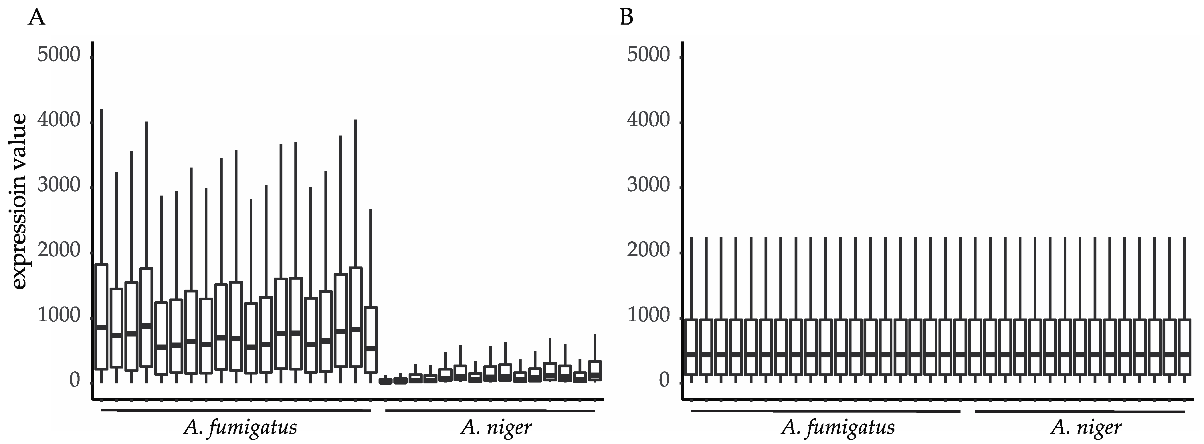
2.3. Data Integration and Exploratory Analysis
2.4. Consensus Weighted Gene Co-Expression Network Analysis (consensusWGCNA)
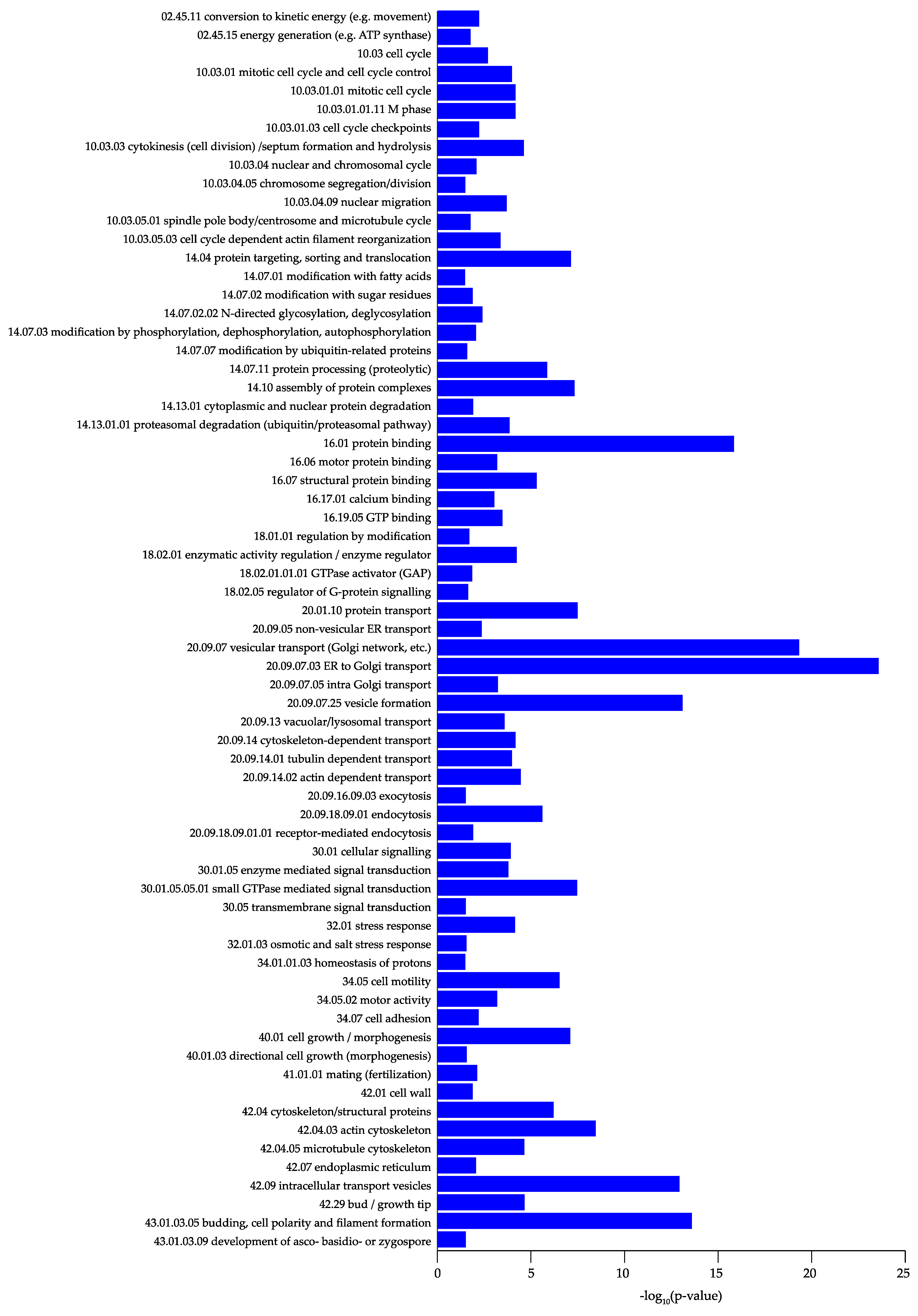
2.5. Functional Classification
3. Results
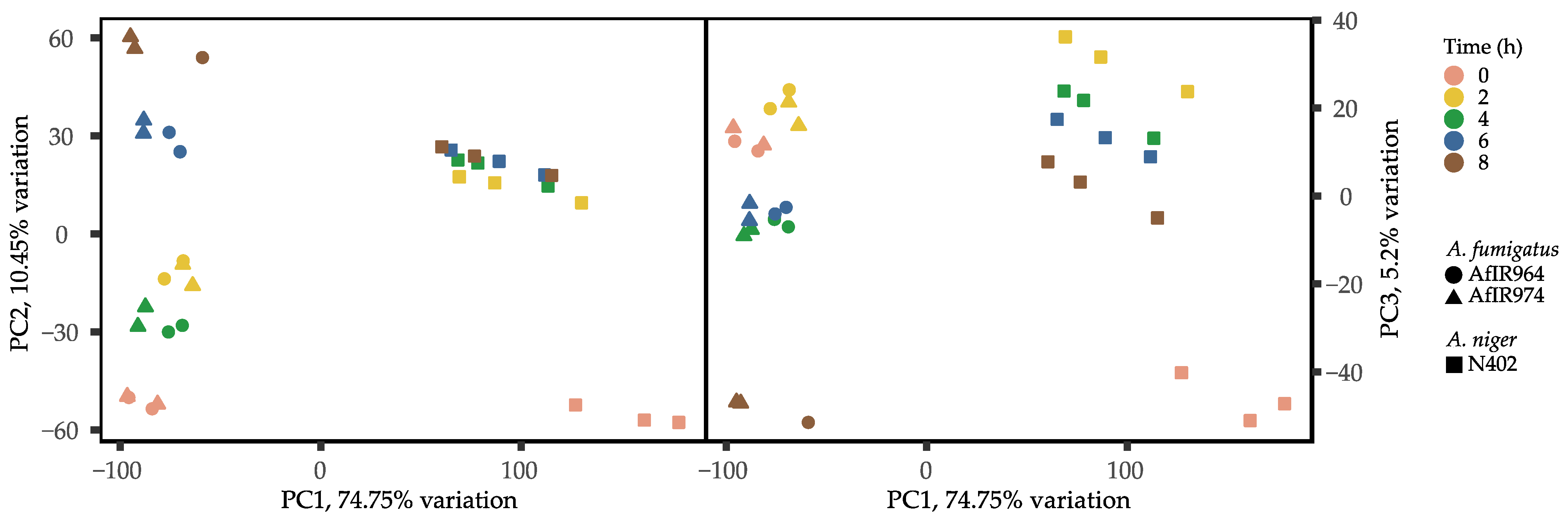
3.1. Orthology Inference, Data Integration, and Exploratory Analysis
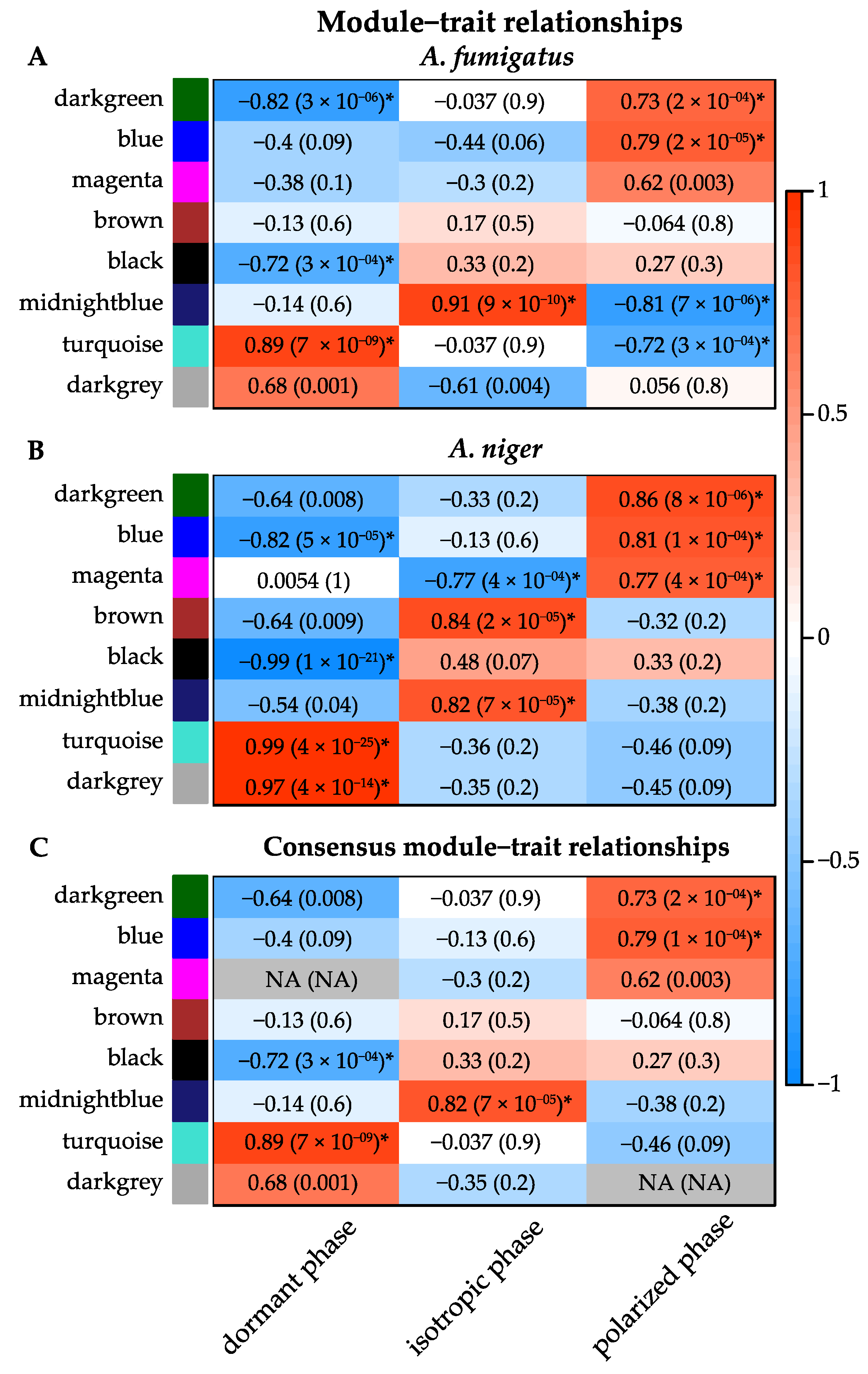
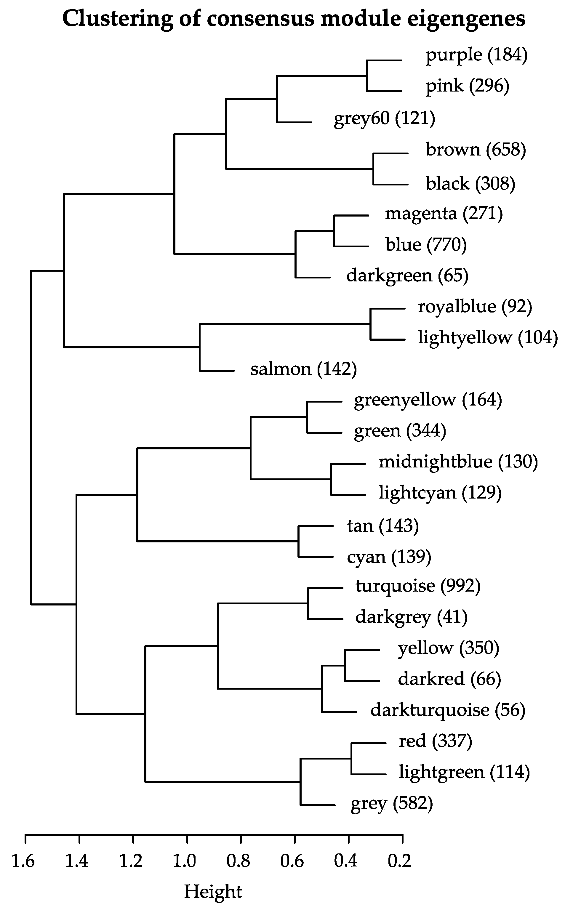
3.2. Consensus Co-Expression Network Analysis
3.3. Modules
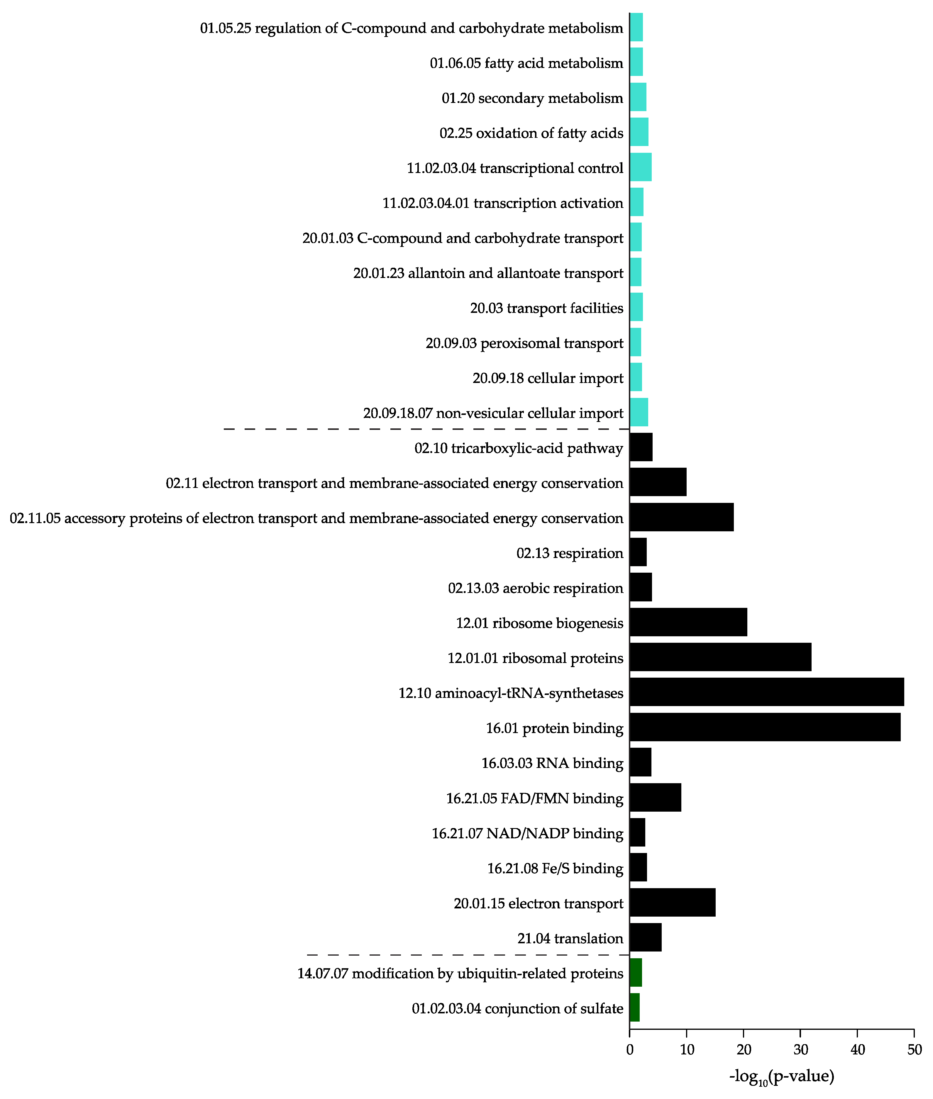
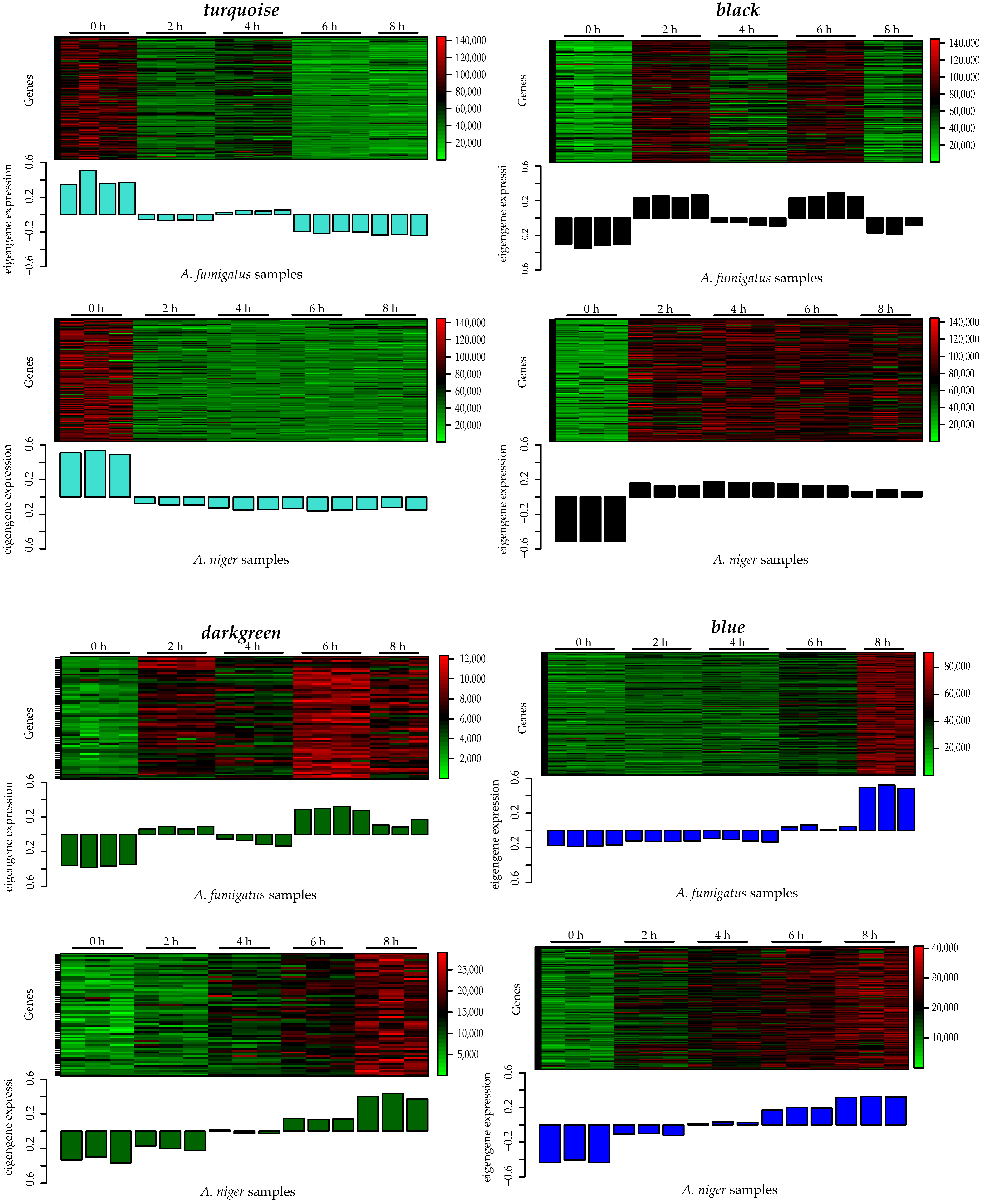
3.3.1. Turquoise
3.3.2. Black
3.3.3. Darkgreen
3.3.4. Blue
Mitosis and Septum Formation
Protein Processing
Onset of Polarization
Establishment of Polarized Growth, Hyphal Elongation
4. Discussion
5. Conclusions
Supplementary Materials
Author Contributions
Funding
Institutional Review Board Statement
Informed Consent Statement
Data Availability Statement
Acknowledgments
Conflicts of Interest
References
- Houbraken, J.; Kocsubé, S.; Visagie, C.; Yilmaz, N.; Wang, X.-C.; Meijer, M.; Kraak, B.; Hubka, V.; Bensch, K.; Samson, R.; et al. Classification of Aspergillus, Penicillium, Talaromyces and related genera (Eurotiales): An overview of families, genera, subgenera, sections, series and species. Stud. Mycol. 2020, 95, 5–169. [Google Scholar] [CrossRef] [PubMed]
- Shinn, E.A.; Smith, G.W.; Prospero, J.M.; Betzer, P.; Hayes, M.L.; Garrison, V.; Barber, R.T. African dust and the demise of Caribbean Coral Reefs. Geophys. Res. Lett. 2000, 27, 3029–3032. [Google Scholar] [CrossRef]
- Silva, J.J.; Bertoldo, R.; Fungaro, M.H.P.; Massi, F.P.; Taniwaki, M.H.; Sant’Ana, A.S.; Iamanaka, B.T. Black aspergilli in Brazilian onions: From field to market. Int. J. Food Microbiol. 2021, 337, 108958. [Google Scholar] [CrossRef] [PubMed]
- Golan, J.J.; Pringle, A. Long-Distance Dispersal of Fungi. In the Fungal Kingdom; ASM Press: Washington, DC, USA, 2017; pp. 309–333. [Google Scholar]
- Van De Veerdonk, F.L.; Gresnigt, M.S.; Romani, L.; Netea, M.G.; Latgé, J.-P. Aspergillus fumigatus morphology and dynamic host interactions. Nat. Rev. Genet. 2017, 15, 661–674. [Google Scholar] [CrossRef] [PubMed]
- Carrion, S.D.J.; Leal, S.M.; Ghannoum, M.A.; Aimanianda, V.; Latgé, J.-P.; Pearlman, E. The RodA Hydrophobin on Aspergillus fumigatus Spores Masks Dectin-1– and Dectin-2–Dependent Responses and Enhances Fungal Survival In Vivo. J. Immunol. 2013, 191, 2581–2588. [Google Scholar] [CrossRef] [PubMed]
- Aimanianda, V.; Bayry, J.; Bozza, S.; Kniemeyer, O.; Perruccio, K.; Elluru, S.R.; Clavaud, C.; Paris, S.; Brakhage, A.A.; Kaveri, S.V.; et al. Surface hydrophobin prevents immune recognition of airborne fungal spores. Nat. Cell Biol. 2009, 460, 1117–1121. [Google Scholar] [CrossRef]
- Brown, G.D.; Denning, D.W.; Gow, N.A.R.; Levitz, S.M.; Netea, M.G.; White, T.C. Hidden Killers: Human Fungal Infections. Sci. Transl. Med. 2012, 4, 165rv13. [Google Scholar] [CrossRef]
- Hayes, G.E.; Novak-Frazer, L. Chronic Pulmonary Aspergillosis—Where Are We? and Where Are We Going? J. Fungi 2016, 2, 18. [Google Scholar] [CrossRef]
- Verweij, P.E.; Rijnders, B.J.A.; Brüggemann, R.J.M.; Azoulay, E.; Bassetti, M.; Blot, S.; Calandra, T.; Clancy, C.J.; Cornely, O.A.; Chiller, T.; et al. Review of influenza-associated pulmonary aspergillosis in ICU patients and proposal for a case definition: An expert opinion. Intensiv. Care Med. 2020, 46, 1524–1535. [Google Scholar] [CrossRef]
- Koehler, P.; Bassetti, M.; Chakrabarti, A.; Chen, A.S.C.; Colombo, A.L.; Hoenigl, M.; Klimko, N.; Lass-Flörl, C.; Oladele, O.R.; Vinh, D.C.; et al. Defining and managing COVID-19-associated pulmonary aspergillosis: The 2020 ECMM/ISHAM consensus criteria for research and clinical guidance. Lancet Infect. Dis. 2020. [Google Scholar] [CrossRef]
- Bertuzzi, M.; Hayes, G.E.; Icheoku, U.J.; Van Rhijn, N.; Denning, D.W.; Osherov, N.; Bignell, E.M. Anti-Aspergillus Activities of the Respiratory Epithelium in Health and Disease. J. Fungi 2018, 4, 8. [Google Scholar] [CrossRef] [PubMed]
- Baltussen, T.J.H.; Zoll, J.; Verweij, P.E.; Melchers, W.J.G. Molecular Mechanisms of Conidial Germination in Aspergillus spp. Microbiol. Mol. Biol. Rev. 2019, 84, 84. [Google Scholar] [CrossRef] [PubMed]
- Krijgsheld, P.; Bleichrodt, R.; van Veluw, G.; Wang, F.; Müller, W.; Dijksterhuis, J.; Wösten, H. Development in Aspergillus. Stud. Mycol. 2013, 74, 1–29. [Google Scholar] [CrossRef]
- Hayer, K.; Stratford, M.; Archer, D.B. Structural Features of Sugars That Trigger or Support Conidial Germination in the Filamentous Fungus Aspergillus niger. Appl. Environ. Microbiol. 2013, 79, 6924–6931. [Google Scholar] [CrossRef] [PubMed]
- Danion, F.; Van Van Rhijn, N.; Dufour, A.C.; Legendre, R.; Sismeiro, O.; Varet, H.; Olivo-Marin, J.-C.; Mouyna, I.; Chamilos, G.; Bromley, M.; et al. Aspergillus fumigatus, One Uninucleate Species with Disparate Offspring. J. Fungi 2021, 7, 30. [Google Scholar] [CrossRef] [PubMed]
- Hayer, K.; Stratford, M.; Archer, D.B. Germination of Aspergillus niger Conidia Is Triggered by Nitrogen Compounds Related to l-Amino Acids. Appl. Environ. Microbiol. 2014, 80, 6046–6053. [Google Scholar] [CrossRef]
- Van Leeuwen, M.; Krijgsheld, P.; Bleichrodt, R.; Menke, H.; Stam, H.; Stark, J.; Wösten, H.; Dijksterhuis, J. Germination of conidia of Aspergillus niger is accompanied by major changes in RNA profiles. Stud. Mycol. 2013, 74, 59–70. [Google Scholar] [CrossRef]
- Novodvorska, M.; Hayer, K.; Pullan, S.T.; Wilson, R.; Blythe, M.J.; Stam, H.; Stratford, M.; Archer, D.B. Trancriptional landscape of Aspergillus niger at breaking of conidial dormancy revealed by RNA-sequencing. BMC Genom. 2013, 14, 246. [Google Scholar] [CrossRef]
- Van Leeuwen, M.; Krijgsheld, P.; Wyatt, T.; Golovina, E.; Menke, H.; Dekker, A.; Stark, J.; Stam, H.; Bleichrodt, R.; Wösten, H.; et al. The effect of natamycin on the transcriptome of conidia of Aspergillus niger. Stud. Mycol. 2013, 74, 71–85. [Google Scholar] [CrossRef]
- Van Leeuwen, M.R.; Van Doorn, T.M.; Golovina, E.A.; Stark, J.; Dijksterhuis, J. Water- and Air-Distributed Conidia Differ in Sterol Content and Cytoplasmic Microviscosity. Appl. Environ. Microbiol. 2009, 76, 366–369. [Google Scholar] [CrossRef]
- Van Leeuwen, M.; Smant, W.; De Boer, W.; Dijksterhuis, J. Filipin is a reliable in situ marker of ergosterol in the plasma membrane of germinating conidia (spores) of Penicillium discolor and stains intensively at the site of germ tube formation. J. Microbiol. Methods 2008, 74, 64–73. [Google Scholar] [CrossRef]
- Baltussen, T.J.; Coolen, J.P.; Zoll, J.; Verweij, P.E.; Melchers, W.J. Gene co-expression analysis identifies gene clusters associated with isotropic and polarized growth in Aspergillus fumigatus conidia. Fungal Genet. Biol. 2018, 116, 62–72. [Google Scholar] [CrossRef]
- Oda, K.; Bignell, E.; Kang, S.E.; Momany, M. Transcript levels of theAspergillus fumigatusCdc42 module, polarisome, and septin genes show little change from dormancy to polarity establishment. Med Mycol. 2016, 55, 445–452. [Google Scholar] [CrossRef][Green Version]
- Lamarre, C.; Sokol, S.; Debeaupuis, J.-P.; Henry, C.; Lacroix, C.; Glaser, P.; Coppée, J.-Y.; François, J.-M.; Latgé, J.-P. Transcriptomic analysis of the exit from dormancy of Aspergillus fumigatus conidia. BMC Genom. 2008, 9, 417. [Google Scholar] [CrossRef]
- Tiwari, S.; Thakur, R.; Goel, G.; Shankar, J. Nano-LC-Q-TOF Analysis of Proteome Revealed Germination of Aspergillus flavus Conidia is Accompanied by MAPK Signalling and Cell Wall Modulation. Mycopathologia 2016, 181, 769–786. [Google Scholar] [CrossRef]
- Momany, M.; Taylor, I. Landmarks in the early duplication cycles of Aspergillus fumigatus and Aspergillus nidulans: Polarity, germ tube emergence and septation. Microbiology 2000, 146, 3279–3284. [Google Scholar] [CrossRef]
- Cagas, S.E.; Jain, M.R.; Li, H.; Perlin, D.S. The Proteomic Signature of Aspergillus fumigatus During Early Development. Mol. Cell. Proteom. 2011, 10. [Google Scholar] [CrossRef] [PubMed]
- Suh, M.-J.; Fedorova, N.D.; Cagas, E.S.; Hastings, S.; Fleischmann, R.D.; Peterson, S.N.; Perlin, D.S.; Nierman, W.C.; Pieper, R.; Momany, M. Development stage-specific proteomic profiling uncovers small, lineage specific proteins most abundant in the Aspergillus Fumigatus conidial proteome. Proteome Sci. 2012, 10, 30. [Google Scholar] [CrossRef] [PubMed]
- Teutschbein, J.; Albrecht, D.; Pötsch, M.; Guthke, R.; Aimanianda, V.; Clavaud, C.; Latgé, J.-P.; Brakhage, A.A.; Kniemeyer, O. Proteome Profiling and Functional Classification of Intracellular Proteins from Conidia of the Human-Pathogenic MoldAspergillus fumigatus. J. Proteome Res. 2010, 9, 3427–3442. [Google Scholar] [CrossRef] [PubMed]
- Kubitschek-Barreira, P.H.; Curty, N.; Neves, G.W.; Gil, C.; Lopes-Bezerra, L.M. Differential proteomic analysis of Aspergillus fumigatus morphotypes reveals putative drug targets. J. Proteom. 2013, 78, 522–534. [Google Scholar] [CrossRef] [PubMed]
- Thakur, R.; Shankar, J. Proteome Profile of Aspergillus terreus Conidia at Germinating Stage: Identification of Probable Virulent Factors and Enzymes from Mycotoxin Pathways. Mycopathologia 2017, 182, 771–784. [Google Scholar] [CrossRef] [PubMed]
- Novodvorska, M.; Stratford, M.; Blythe, M.J.; Wilson, R.; Beniston, R.G.; Archer, D.B. Metabolic activity in dormant conidia of Aspergillus niger and developmental changes during conidial outgrowth. Fungal Genet. Biol. 2016, 94, 23–31. [Google Scholar] [CrossRef] [PubMed]
- Hagiwara, D.; Takahashi, H.; Kusuya, Y.; Kawamoto, S.; Kamei, K.; Gonoi, T. Comparative transcriptome analysis revealing dormant conidia and germination associated genes in Aspergillus species: An essential role for AtfA in conidial dormancy. BMC Genom. 2016, 17, 1–18. [Google Scholar] [CrossRef] [PubMed]
- Breakspear, A.; Momany, M. Aspergillus nidulans Conidiation Genes dewA, fluG, and stuA Are Differentially Regulated in Early Vegetative Growth. Eukaryot. Cell 2007, 6, 1697–1700. [Google Scholar] [CrossRef] [PubMed]
- Asif, A.R.; Oellerich, M.; Amstrong, V.W.; Riemenschneider, B.; Monod, M.; Reichard, U. Proteome of Conidial Surface Associated Proteins of Aspergillus fumigatus Reflecting Potential Vaccine Candidates and Allergens. J. Proteome Res. 2006, 5, 954–962. [Google Scholar] [CrossRef] [PubMed]
- Bos, C.J.; Debets, A.J.M.; Swart, K.; Huybers, A.; Kobus, G.; Slakhorst, S.M. Genetic analysis and the construction of master strains for assignment of genes to six linkage groups in Aspergillus niger. Curr. Genet. 1988, 14, 437–443. [Google Scholar] [CrossRef] [PubMed]
- Overbeek, R.; Fonstein, M.; D’Souza, M.; Pusch, G.D.; Maltsev, N. The use of gene clusters to infer functional coupling. Proc. Natl. Acad. Sci. USA 1999, 96, 2896–2901. [Google Scholar] [CrossRef]
- Nierman, W.C.; Pain, A.; Anderson, M.J.; Wortman, J.R.; Kim, H.S.; Arroyo, J.; Berriman, M.; Abe, K.; Archer, D.B.; Bermejo, C.; et al. Genomic sequence of the pathogenic and allergenic filamentous fungus Aspergillus fumigatus. Nat. Cell Biol. 2005, 438, 1151–1156. [Google Scholar] [CrossRef]
- Pel, H.J.; De Winde, J.H.; Archer, D.B.; Dyer, P.S.; Hofmann, G.; Schaap, P.J.; Turner, G.; De Vries, R.P.; Albang, R.; Albermann, K.; et al. Genome sequencing and analysis of the versatile cell factory Aspergillus niger CBS 513.88. Nat. Biotechnol. 2007, 25, 221–231. [Google Scholar] [CrossRef]
- Ward, N.; Moreno-Hagelsieb, G. Quickly Finding Orthologs as Reciprocal Best Hits with BLAT, LAST, and UBLAST: How Much Do We Miss? PLoS ONE 2014, 9, e101850. [Google Scholar] [CrossRef]
- R Core Computing Team. R: A Language and Environment for Statistical Computing; R Found. Stat. Comput: Vienna, Austria, 2019. [Google Scholar]
- Blighe, K. PCAtools: Everything Principal Components Analysis; R Packag. Version 1.2.0; R Team: Vienna, Austria, 2019. [Google Scholar]
- Ritchie, M.E.; Phipson, B.; Wu, D.; Hu, Y.; Law, C.W.; Shi, W.; Smyth, G.K. limma powers differential expression analyses for RNA-sequencing and microarray studies. Nucleic Acids Res. 2015, 43, e47. [Google Scholar] [CrossRef]
- Castillo, D.; Gálvez, J.M.; Herrera, L.J.; Román, B.S.; Rojas, F.; Rojas, I. Integration of RNA-Seq data with heterogeneous microarray data for breast cancer profiling. BMC Bioinform. 2017, 18, 506. [Google Scholar] [CrossRef]
- Wickham, H. Ggplot2: Elegant Graphics for Data Analysis; Springer: Verlag, NY, USA, 2016; ISBN 978-3-319-24277-4. [Google Scholar]
- Langfelder, P.; Horvath, S. WGCNA: An R package for weighted correlation network analysis. BMC Bioinform. 2008, 9, 1–13. [Google Scholar] [CrossRef]
- Langfelder, P.; Horvath, S. FastRFunctions for Robust Correlations and Hierarchical Clustering. J. Stat. Softw. 2012, 46, 1–17. [Google Scholar] [CrossRef]
- Zhang, B.; Horvath, S. A General Framework for Weighted Gene Co-Expression Network Analysis. Stat. Appl. Genet. Mol. Biol. 2005, 4, 17. [Google Scholar] [CrossRef] [PubMed]
- Priebe, S.; Kreisel, C.; Horn, F.; Guthke, R.; Linde, J. FungiFun2: A comprehensive online resource for systematic analysis of gene lists from fungal species. Bioinform. 2015, 31, 445–446. [Google Scholar] [CrossRef] [PubMed]
- Ruepp, A.; Zollner, A.; Maier, D.; Alberman, K.; Hani, J.; Mokrejs, M.; Tetko, I.; Güldener, U.; Mannhaupt, G.; Münsterkö, M.; et al. The Funcat, a functional annotation scheme for systematic classification of proteins from whole genomes. Nucleic Acids Res. 2004, 32, 5539–5545. [Google Scholar] [CrossRef] [PubMed]
- Love, M.; Huber, W.; Anders, S. Moderated estimation of fold change and dispersion for RNA-seq data with DESeq2. Genome Biol. 2014, 15, 550. [Google Scholar] [CrossRef] [PubMed]
- Langfelder, P.; Horvath, S. Eigengene networks for studying the relationships between co-expression modules. BMC Syst. Biol. 2007, 1, 54. [Google Scholar] [CrossRef] [PubMed]
- D’Enfert, C. Fungal Spore Germination: Insights from the Molecular Genetics of Aspergillus nidulansandNeurospora crassa. Fungal Genet. Biol. 1997, 21, 163–172. [Google Scholar] [CrossRef]
- Lindsey, R.; Cowden, S.; Hernández-Rodríguez, Y.; Momany, M. Septins AspA and AspC Are Important for Normal Development and Limit the Emergence of New Growth Foci in the Multicellular Fungus Aspergillus nidulans. Eukaryot. Cell 2009, 9, 155–163. [Google Scholar] [CrossRef]
- Bartnicki-Garcia, S.; Bartnicki, D.D.; Gierz, G. Determinants of fungal cell wall morphology: The vesicle supply center. Can. J. Bot. 1995, 73, 372–378. [Google Scholar] [CrossRef]
- Fischer-Parton, S.; Parton, R.M.; Hickey, P.C.; Dijksterhuis, J.; Atkinson, H.A.; Read, N.D. Confocal microscopy of FM4-64 as a tool for analysing endocytosis and vesicle trafficking in living fungal hyphae. J. Microsc. 2000, 198, 246–259. [Google Scholar] [CrossRef]
- Bracker, C.E. Diversity and dynamics of the Spitzenkörper in growing hyphal tips of higher fungi. Protoplasma 1996, 195, 90–111. [Google Scholar] [CrossRef]
- Köhli, M.; Galati, V.; Boudier, K.; Roberson, R.W.; Philippsen, P. Growth-speed-correlated localization of exocyst and polarisome components in growth zones of Ashbya gossypii hyphal tips. J. Cell Sci. 2008, 121, 3878–3889. [Google Scholar] [CrossRef]
- Harris, S.D.; Read, N.D.; Roberson, R.W.; Shaw, B.; Seiler, S.; Plamann, M.; Momany, M. Polarisome Meets Spitzenkörper: Microscopy, Genetics, and Genomics Converge. Eukaryot. Cell 2005, 4, 225–229. [Google Scholar] [CrossRef]
- Pinar, M.; Pantazopoulou, A.; Arst, H.N.; Peñalva, M.A. Acute inactivation of theAspergillus nidulans Golgi membrane fusion machinery: Correlation of apical extension arrest and tip swelling with cisternal disorganization. Mol. Microbiol. 2013, 89, 228–248. [Google Scholar] [CrossRef] [PubMed]
- Virag, A.; Lee, M.P.; Si, H.; Harris, S.D. Regulation of hyphal morphogenesis by cdc42 and rac1 homologues in Aspergillus nidulans. Mol. Microbiol. 2007, 66, 1579–1596. [Google Scholar] [CrossRef] [PubMed]
- Araujo-Palomares, C.L.; Richthammer, C.; Seiler, S.; Castro-Longoria, E. Functional Characterization and Cellular Dynamics of the CDC-42–RAC–CDC-24 Module in Neurospora crassa. PLoS ONE 2011, 6, e27148. [Google Scholar] [CrossRef] [PubMed]
- Si, H.; Rittenour, W.R.; Harris, S.D. Roles of Aspergillus nidulans Cdc42/Rho GTPase regulators in hyphal morphogenesis and development. Mycologia 2016, 108, 543–555. [Google Scholar] [CrossRef]
- Riquelme, M.; Aguirre, J.; Bartnicki-García, S.; Braus, G.H.; Feldbrügge, M.; Fleig, U.; Hansberg, W.; Herrera-Estrella, A.; Kämper, J.; Kück, U.; et al. Fungal Morphogenesis, from the Polarized Growth of Hyphae to Complex Reproduction and Infection Structures. Microbiol. Mol. Biol. Rev. 2018, 82. [Google Scholar] [CrossRef]
- Virag, A.; Harris, S.D. The Spitzenkörper: A molecular perspective. Mycol. Res. 2006, 110, 4–13. [Google Scholar] [CrossRef] [PubMed]
- Takeshita, N.; Fischer, R. On the role of microtubules, cell end markers, and septal microtubule organizing centres on site selection for polar growth in Aspergillus nidulans. Fungal Biol. 2011, 115, 506–517. [Google Scholar] [CrossRef] [PubMed]
- Higashitsuji, Y.; Herrero, S.; Takeshita, N.; Fischer, R. The Cell End Marker Protein TeaC Is Involved in Growth Directionality and Septation in Aspergillus nidulans. Eukaryot. Cell 2009, 8, 957–967. [Google Scholar] [CrossRef]
- Harris, S.D.; Momany, M. Polarity in filamentous fungi: Moving beyond the yeast paradigm. Fungal Genet. Biol. 2004, 41, 391–400. [Google Scholar] [CrossRef] [PubMed]
- Meyer, V.; Arentshorst, M.; Hondel, C.A.V.D.; Ram, A.F. The polarisome component SpaA localises to hyphal tips of Aspergillus niger and is important for polar growth. Fungal Genet. Biol. 2008, 45, 152–164. [Google Scholar] [CrossRef]
- Machesky, L.M.; Gould, K.L. The Arp2/3 complex: A multifunctional actin organizer. Curr. Opin. Cell Biol. 1999, 11, 117–121. [Google Scholar] [CrossRef]
- Lipschutz, J.H.; Mostov, E.K. Exocytosis: The Many Masters of the Exocyst. Curr. Biol. 2002, 12, R212–R214. [Google Scholar] [CrossRef]
- TerBush, D.R.; Maurice, T.; Roth, D.; Novick, P. The Exocyst is a multiprotein complex required for exocytosis in Saccharomyces cerevisiae. EMBO J. 1996, 15, 6483–6494. [Google Scholar] [CrossRef]
- Seiler, S.; Plamann, M. The Genetic Basis of Cellular Morphogenesis in the Filamentous FungusNeurospora crassa. Mol. Biol. Cell 2003, 14, 4352–4364. [Google Scholar] [CrossRef]
- Punt, P.J.; Seiboth, B.; Weenink, X.O.; Van Zeijl, C.; Lenders, M.; Konetschny, C.; Ram, A.F.J.; Montijn, R.; Kubicek, C.P.; Van Den Hondel, C.A.M.J.J. Identification and characterization of a family of secretion-related small GTPase-encoding genes from the filamentous fungus Aspergillus niger: A putative SEC4 homologue is not essential for growth. Mol. Microbiol. 2001, 41, 513–525. [Google Scholar] [CrossRef] [PubMed]
- Powers-Fletcher, M.V.; Feng, X.; Krishnan, K.; Askew, D.S. Deletion of the sec4 Homolog srgA from Aspergillus fumigatus Is Associated with an Impaired Stress Response, Attenuated Virulence and Phenotypic Heterogeneity. PLoS ONE 2013, 8, e66741. [Google Scholar] [CrossRef] [PubMed]
- Gupta, G.D.; Heath, I.B. Predicting the distribution, conservation, and functions of SNAREs and related proteins in fungi. Fungal Genet. Biol. 2002, 36, 1–21. [Google Scholar] [CrossRef]
- Atkinson, H.A.; Daniels, A.; Read, N.D. Live-cell imaging of endocytosis during conidial germination in the rice blast fungus, Magnaporthe grisea. Fungal Genet. Biol. 2002, 37, 233–244. [Google Scholar] [CrossRef]
- Van Leeuwen, M.; Golovina, E.; Dijksterhuis, J. The polyene antimycotics nystatin and filipin disrupt the plasma membrane, whereas natamycin inhibits endocytosis in germinating conidia of Penicillium discolor. J. Appl. Microbiol. 2009, 106, 1908–1918. [Google Scholar] [CrossRef] [PubMed]
- Steinberg, G. Endocytosis and early endosome motility in filamentous fungi. Curr. Opin. Microbiol. 2014, 20, 10–18. [Google Scholar] [CrossRef]
- De Vries, R.P.; Riley, R.; Wiebenga, A.; Aguilar-Osorio, G.; Amillis, S.; Uchima, C.A.; Anderluh, G.; Asadollahi, M.; Askin, M.; Barry, K.; et al. Comparative genomics reveals high biological diversity and specific adaptations in the industrially and medically important fungal genus Aspergillus. Genome Biol. 2017, 18, 1–45. [Google Scholar] [CrossRef]
- Merrick, B.A.; Phadke, D.P.; Auerbach, S.S.; Mav, D.; Stiegelmeyer, S.M.; Shah, R.R.; Tice, R.R. RNA-Seq Profiling Reveals Novel Hepatic Gene Expression Pattern in Aflatoxin B1 Treated Rats. PLoS ONE 2013, 8, e61768. [Google Scholar] [CrossRef]
- Zhao, S.; Fung-Leung, W.-P.; Bittner, A.; Ngo, K.; Liu, X. Comparison of RNA-Seq and Microarray in Transcriptome Profiling of Activated T Cells. PLoS ONE 2014, 9, e78644. [Google Scholar] [CrossRef]
- Steenwyk, J.L.; Shen, X.-X.; Lind, A.L.; Goldman, G.H.; Rokas, A. A Robust Phylogenomic Time Tree for Biotechnologically and Medically Important Fungi in the Genera Aspergillus and Penicillium. mBio 2019, 10, e00925-19. [Google Scholar] [CrossRef]
- Kang, S.E.; Momany, M. Sporulation environment drives phenotypic variation in the pathogen Aspergillus fumigatus. bioRxiv 2019, 797076. [Google Scholar] [CrossRef]
- Wan, Q.; Tang, J.; Han, Y.; Wang, D. Co-expression modules construction by WGCNA and identify potential prognostic markers of uveal melanoma. Exp. Eye Res. 2018, 166, 13–20. [Google Scholar] [CrossRef] [PubMed]
- Guo, Y.; Ma, J.; Xiao, L.; Fang, J.; Li, G.; Zhang, L.; Xu, L.; Lai, X.; Pan, G.; Chen, Z. Identification of key pathways and genes in different types of chronic kidney disease based on WGCNA. Mol. Med. Rep. 2019, 20, 2245–2257. [Google Scholar] [CrossRef] [PubMed]
- Di, Y.; Chen, D.; Yu, W.; Yan, L. Bladder cancer stage-associated hub genes revealed by WGCNA co-expression network analysis. Hereditas 2019, 156, 1–11. [Google Scholar] [CrossRef]
- Hu, V.W.; Bi, C. Phenotypic Subtyping and Re-analyses of Existing Transcriptomic Data from Autistic Probands in Simplex Families Reveal Differentially Expressed and ASD Trait-Associated Genes. Front. Neurol. 2020, 11, 578972. [Google Scholar] [CrossRef] [PubMed]
- Li, X.; He, Y.; Hao, C.; Li, X.; Li, X. Weighted gene correlation network analysis reveals novel regulatory modules associated with recurrent early pregnancy loss. Biosci. Rep. 2020, 40, 40. [Google Scholar] [CrossRef]
- Li, Y.-J.; Zhou, T.; Zhang, J.; Zhang, L.; Ke, H.; Zhang, C.; Li, P. Clinical trait-connected network analysis reveals transcriptional markers of active psoriasis treatment with Liangxue-Jiedu decoction. J. Ethnopharmacol. 2021, 268, 113551. [Google Scholar] [CrossRef]
- Fischer, R.; Zekert, N.; Takeshita, N. Polarized growth in fungi–interplay between the cytoskeleton, positional markers and membrane domains. Mol. Microbiol. 2008, 68, 813–826. [Google Scholar] [CrossRef]
- Riquelme, M.; Sánchez-León, E. The Spitzenkörper: A choreographer of fungal growth and morphogenesis. Curr. Opin. Microbiol. 2014, 20, 27–33. [Google Scholar] [CrossRef]
- Takeshita, N. Coordinated process of polarized growth in filamentous fungi. Biosci. Biotechnol. Biochem. 2016, 80, 1693–1699. [Google Scholar] [CrossRef]


| Biological Process | A. niger | A. fumigatus | Description |
|---|---|---|---|
| Secretion-related GTPases and interacting proteins | sarA/An01g04040 | Afu1g04940 | Small GTPase of the ARF family, likely a component of COPII coat of transport vesicles |
| An01g06060 | rab11/Afu1g02190 | Ras GTPase | |
| An08g03690 | Afu1g11730 | Ortholog(s) have GTPase activity and role in Golgi to plasma membrane transport | |
| srgA/An14g00010 | srgA/Afu4g04810 | Putative Rab GTPase | |
| An02g07780 | Afu3g12080 | Ortholog(s) have GTPase activity, enzyme activator activity, mRNA binding activity | |
| geaA/An18g02490 | Afu5g11900 | Putative guanine nucleotide exchange factor | |
| Exocyst | An08g05570 | Afu1g12790 | Ortholog(s) have a role in golgi to plasma membrane transport, endoplasmic reticulum inheritance, establishment or maintenance of cell polarity, exocyst assembly |
| An08g07370 | Afu6g11370 | Ortholog(s) have role in golgi to plasma membrane transport, exocyst assembly, exocyst localization | |
| An14g00010 | srgA/Afu4g04810 | Putative Rab GTPase | |
| cftA/An02g14200 | cdc42/Afu2g05740 | Rho GTPase | |
| rhoA/An18g05980 | rho1/Afu6g06900 | Ras GTPase | |
| SNAREs and SNARE interactions | An02g01580 | sec17/Afu2g12870 | Putative vesicular fusion protein |
| An04g07020 | Afu4g10040 | Ortholog(s) have golgi cisterna, endosome localization | |
| An07g02170 | Afu7g05735 | Putative v-SNARE | |
| An07g09960 | Afu1g07420 | Putative v-SNARE | |
| An15g01380 | Afu6g04150 | Ortholog(s) have SNAP receptor activity and a role in ER to golgi vesicle-mediated transport, retrograde vesicle-mediated transport, golgi to ER, vesicle fusion with golgi apparatus | |
| Cell wall biosynthesis | cfcD/An01g05360 | Afu1g02800 | Putative chitinase |
| An03g06220 | gel5/Afu8g02130 | Putative 1,3-beta-glucanosyltransferase | |
| An05g00130 | Afu2g07590 | Ortholog(s) have role in (1->6)-beta-D-glucan biosynthetic process | |
| crhB/An07g07530 | crh2/Afu2g03120 | Putative cell wall glucanase | |
| An08g07350 | gel2/Afu6g11390 | GPI-anchored 1,3-beta-glucanosyltransferase | |
| chsD/An09g02290 | chsF/Afu8g05630 | Chitin synthase | |
| agtA/An09g03100 | amyA/Afu3g00900 | Alpha-amylase | |
| An16g02850 | crh3/Afu3g09250 | Putative beta-glucanase | |
| An16g07040 | btgE/Afu8g05610 | Putative cell wall glucanase | |
| Cell end markers | An18g04780 | Afu1g06090 | Ortholog(s) have a role in apical protein localization, regulation of cell shape and cell septum, plasma membrane of cell tip, spitzenkorper localization |
| Cdc42 complex | cftA/An02g14200 | modA/Afu2g05740 | Rho GTPase |
| cdc24/An04g05150 | cdc24/Afu4g11450 | Ortholog(s) have Rho guanyl-nucleotide exchange factor activity | |
| racA/An11g10030 | racA/Afu3g06300 | Rho GTPase involved in regulation of cell polarity | |
| Polarisome | spaA/An07g08290 | Afu2g03710 | Ortholog(s) have role in establishment of cell polarity, establishment, or maintenance of cell polarity, hyphal growth and hyphal tip polarisome localization |
| Arp 2/3 complex | An08g06400 | Afu1g13330 | Ortholog(s) have actin filament binding activity, role in Arp2/3 complex-mediated actin nucleation, endocytosis, spore germination and Arp2/3 protein complex, actin cortical patch localization |
| An01g05510 | Afu1g02670 | Ortholog(s) have actin filament binding activity, role in Arp2/3 complex-mediated actin nucleation and Arp2/3 protein complex localization | |
| An12g08380 | Afu6g02370 | Ortholog(s) have actin filament binding, molecular adaptor activity, role in Arp2/3 complex-mediated actin nucleation, actin cortical patch organization, spore germination and Arp2/3 protein complex localization | |
| An16g01570 | Afu5g01860 | Ortholog(s) have actin filament binding activity |
Publisher’s Note: MDPI stays neutral with regard to jurisdictional claims in published maps and institutional affiliations. |
© 2021 by the authors. Licensee MDPI, Basel, Switzerland. This article is an open access article distributed under the terms and conditions of the Creative Commons Attribution (CC BY) license (https://creativecommons.org/licenses/by/4.0/).
Share and Cite
Baltussen, T.J.H.; Coolen, J.P.M.; Verweij, P.E.; Dijksterhuis, J.; Melchers, W.J.G. Identifying Conserved Generic Aspergillus spp. Co-Expressed Gene Modules Associated with Germination Using Cross-Platform and Cross-Species Transcriptomics. J. Fungi 2021, 7, 270. https://doi.org/10.3390/jof7040270
Baltussen TJH, Coolen JPM, Verweij PE, Dijksterhuis J, Melchers WJG. Identifying Conserved Generic Aspergillus spp. Co-Expressed Gene Modules Associated with Germination Using Cross-Platform and Cross-Species Transcriptomics. Journal of Fungi. 2021; 7(4):270. https://doi.org/10.3390/jof7040270
Chicago/Turabian StyleBaltussen, Tim J. H., Jordy P. M. Coolen, Paul E. Verweij, Jan Dijksterhuis, and Willem J. G. Melchers. 2021. "Identifying Conserved Generic Aspergillus spp. Co-Expressed Gene Modules Associated with Germination Using Cross-Platform and Cross-Species Transcriptomics" Journal of Fungi 7, no. 4: 270. https://doi.org/10.3390/jof7040270
APA StyleBaltussen, T. J. H., Coolen, J. P. M., Verweij, P. E., Dijksterhuis, J., & Melchers, W. J. G. (2021). Identifying Conserved Generic Aspergillus spp. Co-Expressed Gene Modules Associated with Germination Using Cross-Platform and Cross-Species Transcriptomics. Journal of Fungi, 7(4), 270. https://doi.org/10.3390/jof7040270