Article

A Pilot Study on Baseline Fungi and Moisture Indicator Fungi in Danish Homes

1
Division of Energy Efficiency, Indoor Climate and Sustainability of Buildings, Department of the Built Environment, Aalborg University, A.C. Meyers Vænge 15, DK-2450 Copenhagen SV, Denmark
2
Section for Synthetic Biology, Department of Biotechnology and Biomedicine, Technical University of Denmark, Søltofts Plads 221, DK-2800 Kongens Lyngby, Denmark
3
Department of Applied Ecology, Campus Box 7617, NC State University Campus, Raleigh, NC 27695-7617, USA
4
Wood and Biomaterials, Building and Construction, Danish Technological Institute, Gregersensvej 1, DK-2630 Taastrup, Denmark
*
Author to whom correspondence should be addressed.
J. Fungi 2021, 7(2), 71; https://doi.org/10.3390/jof7020071
Submission received: 22 December 2020 / Revised: 8 January 2021 / Accepted: 18 January 2021 / Published: 20 January 2021
(This article belongs to the Section Environmental and Ecological Interactions of Fungi)

Abstract

:
In many complaint cases regarding bad indoor environments, there is no evidence of visible fungal growth. To determine if the problems are fungi-related, dust sampling is the method of choice among building surveyors. However, there is a need to differentiate between species belonging to a normal, dry indoor environment and species belonging to a damp building envelope. The purposes of this pilot study were to examine which fungal species are present in problem-free Danish homes and to evaluate different detection and identification methods. Analyses showed that the fungal diversity outside was different from the diversity inside and that the composition of fungal species growing indoors was different compared to those found as spores, both indoors and outdoors. Common for most homes were Pseudopithomyces chartarum, Cladosporium allicinum and Alternaria sect. Infectoriae together with Botrytis spp., Penicillium digitatum and Pen. glabrum. The results show that ITS sequencing of dust samples is adequate if supported by thorough building inspections and that food products play as large a role in the composition of the baseline spora as the outdoor air and surrounding vegetation. This pilot study provides a list of baseline fungal species found in Danish homes with a good indoor environment.

1. Introduction

Fungal spores are ubiquitous, but not all fungal species can grow everywhere. In water-damaged buildings, Penicillium chrysogenum is almost omnipresent because it can grow on both wet and semi-dry materials [1], whereas Stachybotrys chartarum is restricted to very wet gypsum wallboard, wallpaper and cardboard [2]. Much is known about the mycobiota or fungal biota or fungal “flora” or funga (fungal species in active growth [1]) in damp or water-damaged buildings [3,4,5,6], but less is known about the spora or fungal diversity (fungal species present as dormant spores [7]) in the air and on surface dust in buildings that have no humidity problems and no dissatisfied occupants [8,9].
Sampling, detection and identification methods to determine the fungal spora in indoor environments at the species level have always been problematic and the subject for discussion and controversy: e.g., air sampling versus material sampling, culture methods versus molecular methods or morphology versus gene sequencing. Older studies using culture methods on air and dust samples reported fungal identity at the genus or species group level (e.g., Penicillium or Aspergillus/Penicillium group) [10,11,12]. Newer studies using next-generation sequencing on dust and swab samples reported fungal identity at the genus level [13], but mostly at the order or class level (e.g., Eurotiales or Eurotiomycetes) [14,15,16], or even at the phylum level [17].
Further complications in fungal identification have arisen in the last decade. Many name changes have occurred with the increased use of molecular tools and the adoption of the International Code of Nomenclature for algae, fungi and plants in 2017 [18]. Many genera have changed names, some genera have merged, and others have been split into several new ones [6]. However, current and correct fungal identification to the species level is necessary to distinguish between mycobiota and spora, to explore their origin, and to compare their properties in the scientific literature.
In a water-damaged home with visible fungal growth, action is usually straight forward (i.e., repair/renovation), but often occupants complain about their indoor environment without there being any visible signs of fungal growth. Fungal growth indoors can, however, be invisible to the naked eye: e.g., Chaetomium globosum on the reverse of a wet gypsum wall or Aspergillus versicolor under the floorboards. But an unsatisfactory indoor environment can also have other causes than fungal growth: e.g., off-gassing of chemicals from furniture and building materials. In either case, dust sampling becomes the proxy method of choice for building surveyors to determine the cause of the complaint. When the identity of the fungal species in a dust sample is known, the source of origin can often be inferred. Studies show that geographic location, climate zone and outdoor air [13,19] are determining factors for the spora in indoor air and dust. But what is the normal fungal spore diversity or baseline spora in indoor dust in Danish homes?
Denmark is an agricultural country located in the temperate oceanic climate zone with distinct seasons. Outside, there are 15–30 times as many viable fungal spores in the air in summer compared to the wintertime [20]. The indoor spora might reflect these outdoor patterns in the summer, but in wintertime, during which Danes rarely air their homes [21], the outdoor and indoor spora are expected to diverge. Therefore, this small pilot study was conducted in the winter to determine if it is possible to find what constitutes a normal Danish fungal spore diversity (the baseline spora) in indoor air and subsequently in dust and to pinpoint some of the factors that shape them. One hypothesis is that fungi brought into homes, e.g., on the food products we buy, play a role in the overall picture of the Danish spora indoors. The aims of the study were (1) to determine the fungal spore diversity in dust in and between private homes that were perceived to be dry and healthy by their owners, (2) to infer the origin of the dominant fungal genera/species found, and (3) to compare detection (culture-dependent and -independent) and identification (morphology and ITS sequencing) methods.

2. Materials and Methods

The study was inspired by a previous study by Dunn et al. [22]. Samples were taken both indoors and outside nine private homes that were perceived as dry, without visible fungal growth and with no indoor environment problems according to the owners. The samples were analyzed using both culture-dependent methods and culture-independent methods, because both methods are used by building inspectors in Denmark during a house inspection. Each home was also subjected to an inspection for visible fungal growth at the same time as the samples were taken and later revisited if data treatment showed unusual findings.

2.1. Sampling Locations

Samples were taken from the nine private homes between 21 January 2015 and 18 March 2015. In this period, the weather was fairly dry (ca. 56 mm in total) and mild (min./max. −9/14 °C, average ca. 2.4 °C) compared to the climate norm for 2006–2015 and with no snow cover. The nine homes were located in North Zealand and the Greater Copenhagen area (Table 1) with ca. 50 km between the most northerly and southerly homes in rural as well as suburban and urban settings. None of the homes had mechanical ventilation systems.

2.2. Sampling and Treatment Protocols

Samples for both DNA and culture were taken on the upper doorframe on the outside surface of the front door and on the upper doorframe on an interior door in the hallway of the residence. These sampling sites were selected because they are not likely to be cleaned frequently and therefore can serve as passive reservoirs for environmental dust and particles. Swab samples were taken for both DNA sequencing (dual-tipped sterile polyester swab (BD, Le Pont-de-Claix, France)) and culturing (single-tipped sterile cotton swab (Deltalab, Barcelona, Spain)) both outside and in accordance with the following protocol: the sampling area was first swabbed with the DNA swab (one wipe with both tips touching an area of ca. 1 cm × 10 cm), followed by the culture swab of approximately the same area (four wipes rolling the tip forwards and backwards on area of ca. 1.5 cm × 10 cm). Immediately after being returned to the lab, the culture swabs were streaked out onto Petri dishes and the DNA swabs were stored in the freezer until shipment to the Cooperative Institute for Research in Environmental Sciences, University of Colorado at Boulder, Boulder, CO, USA.

2.3. Additional Samples

None of homes had—according to the owner—any humidity problems, but three had a history of water damage (Table 1). The damage had been repaired well before samples were taken. However, each home was examined for any visible mold growth, which was found in two cases (Table 1), and additional swab samples were taken from the moldy areas and treated according to the protocol mentioned earlier. During the course of the sampling, a student flat in Kgs. Lyngby experienced water damage resulting in massive mold growth in the bathroom, which was constructed of gypsum wallboard. Two moldy areas were sampled and treated according to protocol. To match these samples, two samples of new gypsum wallboard with mold growth from an ongoing experiment by Andersen et al. [2] were also included and treated according to protocol.

2.4. Culture-Independent Method: DNA Sequencing

DNA swabs (25 in total) were sent to Cooperative Institute for Research in Environmental Sciences, for extraction, PCR, and sequencing. At the Cooperative Institute, swab tips were placed into 2 mL 96-well plates (Axygen Inc., Union City, CA, USA). Plates were processed using the Extract-N-Amp PCR kit (Sigma-Aldrich, Inc., St. Louis, MO, USA), following a modified version of the manufacturers’ instructions (as in Barberan et al. [23]). The first internal transcribed spacer (ITS1) region of the rRNA operon was sequenced, using the ITS1-F (CTTGGTCATTTAGAGGAAGTAA) and ITS2 (GCTGCGTTCTTCATCGATGC) primer pair. The primers included the appropriate Illumina adapters, with the reverse primers also having an error-correcting 12 bp barcode unique to each sample to permit multiplexing of samples. PCR products from all samples were quantified using the PicoGreen dsDNA assay and pooled together in equimolar concentrations for sequencing on an Illumina MiSeq instrument.

2.5. Data Treatment

A total of 1312 operational taxonomic units (OTUs), each trimmed to 251 base pairs, were returned from the Cooperative Institute for Research in Environmental Sciences with identities from the UNITE database. All sequences were first clustered using Clustal Omega (EMBL-EBI, http://www.ebi.ac.uk, standard settings) to facilitate initial work on excluding non-fungal and unidentified fungal sequences. Clusters of OTUs identified as virus and bacteria in GenBank and outliers of fungal OTUs with no identification were excluded, as were fungal OTUs where there was no agreement between UNITE and GenBank (206 OTUs in total). The identity of each OTU according to the UNITE database was manually cross-checked with GenBank (NCBI) using the “Percent identity” and “Distance Tree of Results” functions after three BLAST runs: (1) including “All sequences”, (2) excluding “Uncultured/environmental sample sequences”, and (3) limited to “Sequences from type of material” and only comparing the identity with sequences from cultures with strain numbers from well-known curated fungal collections (Supplementary Table S1) and type cultures, if possible. Synonyms and current fungal names were checked and adopted from Index Fungorum (http://www.indexfungorum.org), Mycobank (http://www.mycobank.org/) and the current taxonomic literature for the most frequently occurring genera and species and for all food and indoor fungi in the dataset. OTUs were pooled if UNITE, GenBank, FunCBS (Westerdijk Institute, Utrecht, The Netherlands) and Indoor.txt (Westerdijk Institute, Utrecht, The Netherlands) agreed on species identity and the sequences were located in the same cluster according to the Clustal Omega dendrogram. When two or more fungal species names of strains from curated collections were equally likely, identity was set at the section or clade or complex level. Selected OTU sequences were rechecked in November 2020 (Supplementary Table S1). Unscrambler (version X 10.4, CAMO) was used to perform principal component analysis (PCA) on a reduced and pooled dataset (18 indoor and outdoor samples × 41 common genera, species complexes, sections, series, or species indoors and out (29 marked with * in Table 2 and Table 3)). The PCAs were done with standardization (X/SDev) and with log-transformation (X = log(X + 1)) followed by standardization (X/SDev).
Table 2. Culture-independent identification. The 15 most abundant fungal genera outdoors and indoors at the nine homes (H) and the total reads for the different pooled operational taxonomic units (OTUs). High occurrence indoors compared to outdoors and vice versa are in bold.
GenusTotal DNA ReadsOutdoorsIndoors
OutInH-1H-2H-3H-4H-5H-6H-7H-8H-9H-1H-2H-3H-4H-5H-6H-7H-8H-9
Alternaria2785759609081892255045224877062444618651931550241816193
Aspergillus3646784163620126917014027674053862772101295571
Aureobasidium *3724189314912594990225107902162423924550911005552110
Blumeria *4275183714755959014632156932459035355244000010530
Botrytis *14885652071101470171005943702831018329427480
Cladosporium519310,645671202327155852579614567381110194212972023173571313266664
Devriesia *3740410130965124301189679220025089654178089480
Exophiala *71981290124510773224439543817717003947000430
Itersonilia *324216937111440534482511688143978224002142070
Knufia *261823400247320843886701062086500000980
Penicillium466510305242101391629611065258480179208109084389
Pseudopithomyces *2647069698207909700975451988162600172810850
Scoliciosporum *5317251081399111604085139092201000020
Taphrina *34203530050282123553710172929016500095640
Wallemia *174610000000001070054809100
Total DNA reads 104,85885,701585315,95010,57110,49117,56614,32813,33776116,001892811,36617,04610,0828604597712,24511,273180
* Included in the PCA analysis in Figure 1. Total reads for all OTUs outdoors, indoors and for each sample site.
Table 3. Culture-independent identification. The most abundant indoor fungal species at the nine homes (H) and the total reads for the different OTUs out and in. Samples from the interior doorframe and on three samples from painted brick wall with visible fungal growth (G). High occurrences on the building materials compared to the corresponding dust sample are in bold.
SpeciesTotal DNA ReadsIndoorsFungal Growth
OutInH-1H-2H-3H-4H-5H-6H-7H-8H-9H-3-GH-4-GaH-4-Gb
Acremonium charticola55900000590000348350
Akanthomyces lecanii00000000000001289
Alternaria sect. Alternaria *154232751191614942581015476941020
Alternaria sect. Infectoriae *120140511252973601656008267861000
Alternaria sect. Ulocladium14590300550001000
Aspergillus canadensis64361027100008901118560
Aspergillus domesticus *2212829079270255040100330000
Aspergillus flavus15170001700000000
Aspergillus fumigatus40370037000000000
Aspergillus glaucus *1211101980008314700000
Aspergillus hiratsukae12001000010100
Aspergillus niger0150014000010000
Aspergillus penicillioides clade *09100710002000000
Aspergillus salinarum11800800000000000
Aspergillus series Versicolores *04280127119013903850016204108
Aspergillus vitricola481302301363690601170001
Aspergillus westerdijkiae70000000000000
Botryotrichum murorum *11269023300003600000
Chaetomium globosum19690609000000000
Cladosporium allicinum *270069095881636655982115568931984011000
Cladosporium cladosporioides complex *1110322252218258983558135361864000
Cladosporium dominicanum *26414001001380001004762
Cladosporium halotolerans *805720240000480033140
Cladosporium sphaerospermum *29185003813900080081725
Debaryomyces hansenii2037041251162703950271409413861536
Monocillium tenue12320000003200008540
Penicillium adametzioides011200011200000000
Penicillium bialowiezense10210070140000001
Penicillium brevicompactum *10311162330480744081100007
Penicillium chrysogenum *44909162306745409013840011
Penicillium citreonigrum23138000130036089000
Penicillium citrinum0690000690000000
Penicillium corylophilum *149000000000000115
Penicillium digitatum *482114763281902750037740000
Penicillium glabrum *235115301137840235190000
Penicillium lanosum0100000100000000
Penicillium olsonii *0620055010060000
Penicillium oxalicum70000000000000
Penicillium roqueforti *7410000004100000
Penicillium roseopurpureum00000000000002156
Saccharomyces cerevisiae *111266004151462520423300000
Stachybotrys chartarum48380000000380001
Verrucocladosporium dirinae243970026071000003520
Wallemia ichthyophaga0240000002400000
Wallemia muriae172201070054806700000
* Included in the PCA analysis in Figure 1. Cladosporium species as complexes (Cla. herbarum complex: Cla. allicinum; Cla. sphaerospermum complex: Cla. dominicanum, Cla. halotolerans and Cla. sphaerospermum).

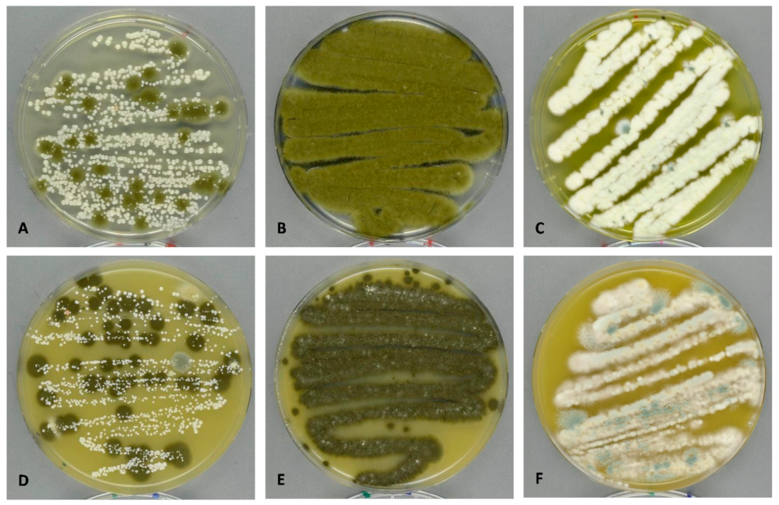
2.6. Culture-Dependent Method: Fungal Growth, Identification and Enumeration

Each growth swab was streaked out onto two Petri dishes with V8 (Campbell’s V8 juice agar [6] with chloramphenicol (0.05 g/L) and chlortetracycline (0.05 g/L)) and two Petri dishes with DG18 (Dichloran 18% Glycerol agar [6]). The Petri dishes were incubated at 20 °C and read after 7 and 14 days. After 7 days of growth on DG18 and V8 media, the plates were photographed and fungal and yeast colonies counted. Colonies were identified to genus level using a dissection microscope and representative fungal colonies were isolated and inoculated for species identification. Fungal colonies were identified to species level using a compound microscope: Alternaria were identified according to Simmons [24], Aspergillus and Penicillium according to Samson et al. [6], Valdez et al. [25], Chen et al. [26], Chaetomium according to Wang et al. [27], Cladosporium according to Bensch et al. [28] and other fungi according to Samson et al. [29]. Pictures were taken of each Petri dish and non-sporulating colonies were marked and checked again for sporulation after 14 days of growth. After species identification, each colony forming unit (CFU) in the pictures were annotated with species ID and counted across all samples. Non-sporulating colonies were transferred to malt extract agar (MEA, [6]) and labelled “Mycelia sterilia” if no spores were formed after 14 days.

3. Results

3.1. Culture-Independent Methods (DNA Sequencing)

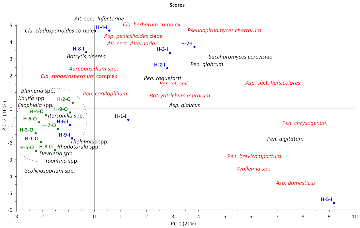
3.1.1. OTU Diversity and Abundance

A total of 1106 fungal operational taxonomic units (OTUs) were found across all 21 samples (nine indoor samples, nine outdoor samples and three samples with fungal growth). The fungal diversity and abundance were greater outdoors than indoors. Across the nine outdoor samples, 732 fungal OTUs (104,858 reads) were found, compared to 591 fungal OTUs (85,701 reads) for the nine indoor samples. Outdoors, the diversity and abundance of ascomycetes (468 OTUs/73,873 reads) were both greater than those of basidiomycetes (208 OTUs/18,793 reads), lichens (46 OTUs/11,507 reads), and mucoromycetes (10 OTUs/685 reads). Indoors, the diversity in ascomycetes and basidiomycetes was almost the same (284 and 283 OTUs, respectively) despite the greater number of reads of ascomycetes than of basidiomycetes (60,722 and 24,130 reads, respectively). Lichens (16 OTUs/327 reads) and mucoromycetes (8 OTUs/168 reads) were neither diverse nor particularly abundant. Samples from areas with fungal growth were quite different from both outdoor and indoor samples. Across the three samples of a painted brick wall with fungal growth, ascomycetes were represented by many reads, but a low diversity of OTUs (35 OTUs/35,568 reads) was observed, and the occurrence of basidiomycetes was negligible. Across the four gypsum wallboard samples with fungal growth, the diversity and read numbers of ascomycetes was higher than for the growth on brick (66 OTUs/67,522 reads), but lower than for dust samples. Generally, dust samples were characterized by high diversity (hundreds of OTUs), whereas areas of fungal growth were characterized by lower diversity (tens of OTUs).

3.1.2. Genus Diversity and Abundance

Exophiala spp. (7198 reads) was the most abundant genus outside (Table 2), followed by Scoliciosporum sp. (5317 reads (as S. umbrinum)) and Cladosporium spp. (5193 reads). Indoors, Cladosporium spp. (10,645 reads) was most abundant, with Alternaria spp. (7596 reads) and Pseudopithomyces sp. (7069 reads as Pse. chartarum) second and third. As can be seen from Table 2, Exophiala and Scoliciosporum spp. were rarely found indoors (129 and 25 reads, respectively), while Pse. chartarum was rare outdoors (264 reads). Aspergillus, Penicillium and Wallemia spp. were much more abundant indoors (6784, 5103 and 746 reads, respectively) than outdoors (364, 466 and 1 reads, respectively). For the yeasts and yeast-like fungi, Saccharomyces cerevisiae and Cryptococcus spp. were the most abundant species indoors (1266 and 1019 reads, respectively) compared to outdoors (11 and 425 reads, respectively).

3.1.3. Species Diversity and Abundance Indoors

Some fungal species were found in all homes, whereas other species were located in only one home (Table 3). Cla. cladosporioides complex (3222 reads) was the most common Cladosporium group and was found in all homes, followed by Cla. allicinum (6909 reads) and Alt. sect. Alternaria (3275 reads), found in eight of the nine homes. Alt. sect. Infectoriae (4051 reads), Pen. digitatum (2114 reads), and Pen. chrysogenum (909 reads) were detected in seven of the nine homes. Pse. chartarum, the most abundant species indoors, was only found in six out of the nine homes, but with high read numbers when present. Other species, such as Asp. domesticus and Asp. glaucus, were found in high numbers indoors (2250 and 1980 reads, respectively), but only in one home each (H-5 and H-2, respectively).
Table 3 also shows the results of three samples with visible fungal growth due to local condensation. An inner wall (painted brick wall) in the bathroom at H-3 showed moderate growth with Cla. allicinum, Cla. halotolerans, and Debaryomyces hansenii, while two samples from outer walls (painted brick walls) in a bedroom at H-4 showed massive growth that included Acremonium charticola, Asp. canadensis, Asp. series Versicolores, Cla. sphaerospermum, Deb. hansenii, Akanthomyces lecanii, Monocillium tenue, and Pen. roseopurpureum. However, none of these fungal species were found in higher amounts in the corresponding dust samples compared to dust samples from the other seven homes (Table 3), except for Cla. sphaerospermum in H-4.
The moisture indicator fungus, Stachybotrys chartarum, was only found in low numbers and only in one home (38 reads on H-8-I, Table 3). Other moisture indicator fungi, such as Fusarium, Didymella, and Trichoderma spp., were only detected sporadically indoors and in low numbers (<10 reads) in this study.
Table 4 shows the result of the three brick wall samples with fungal growth from Table 3 compared with four gypsum wallboard samples with fungal growth. The gypsum samples are unrelated to the home study but have been included to enable comparisons between material types. As can be seen, Sta. chartarum was found on the gypsum wallboard samples (Ub and Pa), but not on the brick wall samples. Similarly, Alt. sect. Ulocladium, Asp. hiratsukae, Candida parapsilosis, and Pen. chrysogenum were abundant on the gypsum samples, but almost absent on the brick wall samples. On the other hand, Asp. canadensis, D. hansenii, Acr. lecanii, Mon. tenue, and Pen. roseopurpureum were found on the brick wall samples, but not on the gypsum samples.
Principal component analysis (PCA) based on the most abundant genera overall and the most abundant indoor species (marked with * in Table 2 and Table 3, Cladosporium pooled into complexes) showed that the outdoor samples were more similar to each other than they were to any of the indoor samples (Figure 1). All outdoor sampling sites grouped together due to high reads of genera such as Blumeria, Knufia, Devriesia, and Taphrina and low reads of Aspergillus, Penicillium, and Pseudopithomyces (Table 2). Two indoor sampling sites (H-6-I and H-9-I) also grouped with the outdoor sample mostly due to the absence of indoor fungi compared to the other indoor samples. The seven remaining homes were scattered throughout the plot, reflecting apparently idiosyncratic differences from one house to the next. All homes with a history of water damage (H-2, H-7 and H-8) or found to have fungal growth (H-3 and H-4) were located together in the upper part of the plot. H-5-I, located in the bottom right corner, is an outlier due to high reads of Asp. domesticus, Pen. brevicompactum and Wal. muriae compared to other indoor samples (Table 3).

3.2. Culture-Dependent Methods (Growth on DG18 and V8)

3.2.1. Colony Forming Unit (CFU) and Genus Diversity and Abundance

A total of 467 fungal colony forming units (CFUs) were identified across the 18 samples (nine indoor samples and nine outdoor samples) using DG18 and V8 as culture media. The three swab samples of fungal growth (H-3-G, H-4-Ga and H-4-Gb) were not countable due to high numbers of CFUs and coalescing colonies (Figure 2). The total CFUs for outdoors was not particularly different from the total CFUs found indoors (262 and 205, respectively) and, as expected, both the diversity and the abundance in CFUs were much lower compared to the culture-independent samples. No basidiomycetes or lichens could be identified on DG18 and V8, but there were 45 CFUs with no sporulation (Mycelia sterilia) outside and 25 CFUs inside, which may be basidiomycetes; however, no clamp connections were found either. Cladosporium spp. were the most abundant fungi detected outside (87 CFUs in total), while the total CFU for Cladosporium spp. inside was much lower (seven CFUs in total) (Table 5). Similar low distributions indoors were seen for Didymella and different yeast spp. (zero and two CFUs, respectively). Penicillium was the most prevalent genus inside (132 CFUs in total), while the total Penicillium CFUs outside was five-fold lower. Similar differences in indoor to outdoor CFU numbers were seen for Aspergillus spp. (30 CFUs indoors vs. one CFU outdoors).

3.2.2. Species Diversity and Abundance Indoors

No fungal species was found to be common in all nine homes using the culture methods. Pseudopithomyces chartarum, the most prevalent fungus indoors according to the culture-independent method, was not found in any sample using the culture-dependent method. Likewise, Cla. allicinum was not found in any culture-dependent sample indoors, while Cla. cladosporioides (four CFUs) and Cla. sphaerospermum (three CFUs) were found inside three and two homes, respectively, and in very low CFU numbers (Table 5). The most common Penicillium species, Pen. brevicompactum (16 CFUs) and Pen. glabrum (15 CFUs), were found in five of the nine homes, whereas Pen. digitatum (69 CFUs) was found in three homes. Pen. chrysogenum, which was detected very frequently using the culture-independent method, was only detected in three homes with five CFUs. Asp. glaucus and Asp. domesticus were not found either, but contrary to the culture-independent method, 20 CFUs of Asp. niger were found in home H-3 and 17 CFUs of Didymella sp. were found outside home H-8.

4. Discussion

The results of this Danish pilot study show that a closed building envelope shields well against airborne fungal spores from the outside, but at the same time traps spores from fungi that grow or have grown somewhere inside the building. Contrary to studies conducted in Mediterranean or subtropical climates [8,9,13,14], a clear difference between the compositions of fungal species indoors and outdoors was seen. This was also seen in other studies conducted in a Scandinavian or temperate climate [5,30] where households keep windows and doors closed as much as possible during the winter. However, both nature and culture play a role on the fungal composition indoors. In North Zealand, Denmark, where all nine homes are located, there are large expanses with deciduous forests, agricultural fields, and pastures that change with the seasons, resulting in variations in outdoor fungi [20]. Since our results were obtained in the colder winter months, when airing and/or ventilation was sparse, the influx and influence of fungal spores from the outside environment was limited, giving a clearer picture of the fungal species that have indoor sources. In the summertime, the differences would probably be much smaller because it is warm enough to have doors and windows open most of the day. The results show that previous water damages may interfere when culture--independent methods are used, because fungal spores from old damage may linger on for months or perhaps years in undisturbed dust. The results also show that occupants may perceive their home to be problem-free, even though there might be minor undiscovered dampness somewhere in the home, especially in basements or bathrooms.
For some fungal genera, such as Pseudopithomyces and Wallemia, the source is inside, while for Blumeria and Itersonilia, an outdoor source is a determining factor. For example, one home (H-1) had slightly higher reads indoors of Scoliciosporum spp., which are strictly outdoor lichenized fungi. A second inspection of the home showed that the exterior window ledge outside the bedroom was covered with lichens, showing that the proximity of an outdoor fungal source plays a role in the overall fungal composition indoors. The opposite was the case in another home (H-4) where the bedroom of a teenager had massive growth of, amongst others, Aspergillus spp. These fungi were, however, not detected in the dust sample, probably due to the remoteness of the bedroom compared to the interior sampling point. These results show that the proximity of the fungal source, inside or out, plays a role in the overall composition of spores in indoor dust samples.
For genera such as Aspergillus and Penicillium, which both seem to have indoor sources, species identification is necessary to pinpoint if their source indoors is either food- or building-associated. This was seen in H-1 and H-5, where moldy tangerines were found. High reads and CFUs of Pen. digitatum were later detected at both sites. Pen. digitatum, which is associated with moldy citrus fruit [6], was detected using the culture-independent method in most homes, but only in three homes using the culture-dependent method. Pen. glabrum was also detected frequently indoors due to its association with the brown dried scales found on most onions [6]. Asp. glaucus was detected in several homes because of its association with white bread, dried herbs and spices [6], and a very moldy baguette was subsequently found at H-2 after a second inspection. These results show that food-borne fungi play a key role in Danish homes compared to, e.g., American homes [8,13], probably due to differences in food and cooking culture. Many Danes still prepare lunch packs and cook meals from scratch on a daily basis and therefore always have a certain amount of (moldy) bread, fruit and vegetables in their kitchens.
A presence of spores from moisture indicator fungi was seen in the three homes with a history of water damage (H-2, H-7, and H-8). Even though renovation had been completed at least 6 months prior to sampling, low reads of Botryotrichum murorum, Chaetomium globosum, and Stachybotrys chartarum, all indicator fungi for severe water damage [1,27], were still found in one or more homes using the culture-independent method, whereas only one colony of Cha. globosum was detected in H-2 using the culture-dependent method. This suggests that culture-independent methods, such as DNA sequencing, can detect fungal spores after old, repaired water damage, due to the longevity of the DNA compared to the viability of the spores, and shows that knowledge of the history of the home is important, in order not to draw the wrong conclusions, i.e., ongoing problems with water damage. On the other hand, at home 5, where no visible fungal growth was discovered and no history of water damage, high reads of Asp. domesticus, Pen. brevicompactum and Wallemia muriae were found with the culture-independent method and some CFUs of Pen. brevicompactum using the culture-dependent method. These fungal species are associated with moderate humid indoor environments, especially moist plaster [6], and this fungal combination suggested that this home might have humidity problems, even though the owners considered their home dry and problem-free. A second inspection revealed that a remote part of the basement had elevated moisture levels and a moldy odor, but no visible fungal growth. These findings show that a thorough inspection or even re-examination is important when high reads of several species of moisture indicating fungi are detected.

4.1. Detection Methods

Absolute quantitative measurements of fungal diversity and abundance between species are not possible with either culture-dependent (CFUs) or culture-independent (reads) methods. One CFU on a growth medium may be the result of several fresh spores clustering together and thereby giving a false lower count than what is really present. On the other hand, the number of reads may vary between species depending on the number of cells in the spores, giving a false higher count, especially for fungal species such as Alternaria and Epicoccum [31]. However, comparison between sampling sites (i.e., indoors and out) and relative quantitative evaluation between individual species is possible.
Spore mortality and the ability to grow on the chosen media are probably the two major reasons for the big difference between the results of the culture-independent method (reads) and the culture-dependent method (CFUs). The ITS primer will detect both viable and dead fungal spores, while culture methods select only for living spores. The viability of fungal spores may also vary between species, suggesting that melanized spores (e.g., Chaetomium and Cladosporium) survive better than hyaline spores [32,33,34]. Furthermore, not all viable spores will germinate and grow on the chosen media; in this case, DG18 and V8. Culture-dependent methods therefore exclude most basidiomycetes (e.g., Itersonilia), lichen (e.g., Scoliciosporum), and plant pathogenic ascomycetes (e.g., Taphrina and Blumeria). However, most ascomycetous fungi related to indoor environments (e.g., food products and building materials) are able to grow on DG18 and/or V8 agars [6], which makes the method useful when read by skilled mycologists.
Another reason for the skewed result may be in the sampling method itself, where the swab for ITS sequencing was taken first followed by the swab for cultivation. The swab for sequencing may contain more viable spores than the swab for cultivation. Secondly, not all viable spores may have been transferred from the swab to the media by simple streaking. An alternative strategy for future studies would be to take the swabs in reverse order and place the cultivation swab in a spore suspension, treat the suspension in ultrasound and spread it onto the media. Another option could be to divide the spore suspension into equal parts and culture one part and sequence the other. A third reason for the difference between culture-independent and -dependent methods is that spore deposits in surface dust may be unevenly distributed across the sample area, especially when there are a low number of reads or CFU. This might be the case with Asp. niger in H-3, where the CFU count was larger than the reads.
Differences in fungal species composition were also seen between dust samples and areas with fungal growth, regardless of detection and identification methods. Some fungal spores are not readily airborne or the source of fungal growth may be too remote from the sampling point. These results show that just because a fungus is not detected, it does not mean that it is not there and again shows that a thorough inspection is particularly important.

4.2. Fungal Identification

The paradigm or basic assumption that only 1% of microbes are culturable [35], and therefore identifiable, seems not to apply to the fungal kingdom. All of the OTUs with reads higher than 250 and the majority of OTUs with low reads could be assigned at least a genus identity, suggesting that most fungi found in indoor environments are culturable since they have been named and described morphologically on agar media [6]. Identification to species level was, however, only possible when each sequence was aligned/blasted manually using a combination of curated (UNITE and FunCBS) and un-curated (GenBank) databases. Automated identification of fungi using sequences is only as good as the repository that is used and each of the abovementioned repositories have their strong points. UNITE is good for basidiomycetes, whereas FunCBS is good for food and indoor ascomycetes. GenBank is good for rare and obscure fungi, which are not present in UNITE or FunCBS, but comprises up to 20% misidentifications [36]. Correct naming is also hampered by the adoption of the Amsterdam Declaration [37,38], which has divided the scientific community regarding which names to use and by the discovery of many new species due to genetics and sequence-based phylogeny (e.g., Houbraken et al. [39]). Even though databases such as MycoBank and Index Fungorum are good sources for synonyms and name changes and are up to date with many of the most common genera, correct fungal names are sometimes omitted in the literature (e.g., Reboux et al. [40]) on purpose. Repositories such as GenBank also contain many new entries derived from metagenomics studies with either no fungal identity or uncertain identity of the uncultured fungus. Completely automated assignment of identity to the species level is therefore not possible for a growing number of OTUs, because none of the databases/repositories are fully updated when it comes to the new species, synonyms, and correct names according to the Amsterdam Declaration, which makes laborious manual checking unavoidable. In this pilot study, 117 (9%) out of 1312 OTUs remain uncertain or unidentifiable.

4.3. Baseline Spora in a Danish Setting

In order to determine the cause(s) of an unsatisfactory indoor environment, there is a need to know which fungal species are normally present in a home with a satisfactory, problem-free indoor environment. The baseline spora is here defined as the collection of fungal species, which spores are found indoors in dust samples, but not found naturally growing on humid or wet building materials, fabrics, furniture or other materials normally associated with the interior of the building. In other words, the baseline spora is the total spora minus spores from the building mycobiota (building funga).
Identification to the species level is here essential to distinguish between fungal spores originating from the outside air, dirt, and soil, or food products (the baseline spora of the home) and fungal spores originating from growth on humid or wet building materials inside the building (the mycobiota of the building), particularly species within the genera of Aspergillus and Penicillium that can be soil- and food-borne as well as building-related [6]. Misidentifying or lumping these species together, combined with inadequate inspection and a lack of home history, may result in the wrong action being taken: health-damaging dampness and fungal growth may be overlooked and the problems may persist or worsen. Alternatively, hygiene or environmental problems may be misconstrued and end in unnecessary and expensive remediation.
Table 6 gives a conservative bid on a list of fungal species that should be considered the baseline in a Danish setting. Fungal species typically associated with common Danish food products, such as Asp. chevalieri, Pen. commume, and Pen. expansum [6], were not detected in this pilot study, but may also be a part of the baseline spora. Furthermore, species in the Cla. cladosporioides complex may also belong to the baseline spora, since they are rarely isolated from moldy building materials [28]. A larger, ongoing study will test these hypotheses.
Moreover, the baseline spora may be different from country to country, depending on climate (e.g., cold winters, heavy rainfall or high winds), culture (e.g., type and amount of raw food products, number of potted plants or storage of firewood indoors) and nature (e.g., type and proximity to farming, forests or compost heaps outside). Therefore, other countries’ data and tools, such as the Environmental Relative Moldiness Index (ERMI), which has been developed for dust samples in USA [41], may not work as well in Scandinavia [42]. Baseline fungi must be determined locally and not globally.

5. Conclusions

This pilot study presented here suggests that the fungal diversities in Danish homes, which are perceived to have a good and dry indoor environment, have fungal species in common, which could constitute a general Danish baseline spora. A larger study is in progress in order to verify and extend the species list in Table 6. Dust samples may be a good proxy method to examine the indoor air quality of a home and culture-independent methods would give correct fungal identity as well as semi-quantitative amounts. However, two or more sampling areas are necessary to represent larger or multi-story homes together with a questionnaire and two inspections, one during sampling and one after sequencing and data treatment, to fully compare the fungal results with the conditions of the home.
In quality assessment of indoor environments per se, all fungal spores—dead or alive and from outdoors as well as food and building—are important in relation to the health and wellbeing of the occupants. Allergens, glucans, and mycotoxins can be present on all fungal spores and may have health implications if spores are present in high numbers [43]. Daily airing of the home and weekly vacuum cleaning and wiping of all surfaces are important measures that can reduce spore load indoors [44]. Furthermore, if the household comprises asthmatics or allergy sufferers, the number of fungal spores in the home can be reduced even further by having a HEPA filter [45,46] fitted in the vacuum cleaner.

Supplementary Materials

The following are available online at https://www.mdpi.com/2309-608X/7/2/71/s1, Table S1: GenBank data from BLAST on selected OUTs.

Author Contributions

Conceptualization, B.A. and R.R.D.; data curation, B.A., J.C.F. and U.T.; formal analysis, B.A., J.C.F. and U.T.; funding acquisition, B.A.; investigation, B.A.; methodology, B.A. and R.R.D.; project administration, B.A.; writing—original draft, B.A., J.C.F., R.R.D. and U.T.; writing—review and editing, B.A., J.C.F., R.R.D. and U.T. All authors have read and agreed to the published version of the manuscript.

Funding

This research was funded by the VILLUM fonden, Grundejernes Investeringsfond and Landsbyggefonden.

Institutional Review Board Statement

Not applicable.

Informed Consent Statement

Not applicable.

Data Availability Statement

Data are contained within the article or supplementary material. The data presented in this study are available in [Supplementary Table S1].

Acknowledgments

The authors would like to thank family, friends, and colleagues for allowing us into their homes and Lisette Knoth-Nielsen for technical assistance.

Conflicts of Interest

The authors declare no conflict of interest. The funders had no role in the design of the study; in the collection, analyses, or interpretation of data; in the writing of the manuscript, or in the decision to publish the results.

References

  1. Andersen, B.; Frisvad, J.C.; Søndergaard, I.; Rasmussen, I.S.; Larsen, L.S. Associations between fungal species and water-damaged building materials. Appl. Environ. Microbiol. 2011, 77, 4180–4188. [Google Scholar] [CrossRef] [PubMed] [Green Version]
  2. Andersen, B.; Dosen, I.; Lewinska, A.M.; Nielsen, K.F. Pre-contamination of new gypsum wallboard with potentially harmful fungal species. Indoor Air 2017, 27, 6–12. [Google Scholar] [CrossRef] [PubMed] [Green Version]
  3. Prezant, B.; Weekes, D.M.; Miller, J.D. (Eds.) Recognition, Evaluation, and Control of Indoor Mold; American Industrial Hygiene Association: Fairfax, VA, USA, 2008. [Google Scholar]
  4. Adan, O.; Samson, R. Fundamentals of Mold Growth in Indoor Environments and Strategies for Healthy Living; Wageningen Academic Publishers: Wageningen, The Netherlands, 2011. [Google Scholar]
  5. Nevalainen, A.; Täubel, M.; Hyvärinen, A. Indoor fungi: Companions and contaminants. Indoor Air 2015, 25, 125–156. [Google Scholar] [CrossRef] [PubMed]
  6. Samson, R.A.; Houbraken, J.; Thrane, U.; Frisvad, J.C.; Andersen, B. Food and Indoor Fungi, 2nd ed.; Westerdijk Laboratory Manual Series 2; Westerdijk Fungal Biodiversity Institute: Utrecht, The Netherlands, 2019. [Google Scholar]
  7. Hunter, C.A.; Grant, C.; Flannigan, B.; Bravery, A.F. Mould in buildings: The air spora of domestic dwellings. Int. Biodeterior. 1988, 24, 81–101. [Google Scholar] [CrossRef]
  8. Horner, W.E.; Worthan, A.G.; Morey, P.R. Air- and dust-borne mycoflora in houses free of water damage and fungal growth. Appl. Environ. Microbiol. 2004, 70, 6394–6400. [Google Scholar] [CrossRef] [Green Version]
  9. Ara, K.; Aihara, M.; Ojima, M.; Toshima, Y.; Yabune, C.; Tokuda, H.; Kawai, S.; Ueda, N.; Tanaka, T.; Akiyama, K.; et al. Survey of fungal contamination in ordinary houses in Japan. Allergol. Int. 2004, 53, 369–377. [Google Scholar] [CrossRef] [Green Version]
  10. Ren, P.; Jankun, T.M.; Belanger, K.; Bracken, M.B.; Leaderer, B.P. The relation between fungal propagules in indoor air and home characteristics. Allergy 2001, 56, 419–424. [Google Scholar] [CrossRef] [Green Version]
  11. Gent, J.F.; Ren, P.; Belanger, K.; Triche, E.; Bracken, M.B.; Holford, T.R.; Leaderer, B.P. Levels of household mold associated with respiratory symptoms in the first year of life in a cohort at risk for asthma. Environ. Health Perspect. 2002, 110, A781–A786. [Google Scholar] [CrossRef] [Green Version]
  12. Chew, G.L.; Rogers, C.; Burge, H.A.; Muilenberg, M.L.; Gold, D.R. Dustborne and airborne fungal propagules represent a different spectrum of fungi with differing relations to home characteristics. Allergy 2003, 58, 13–20. [Google Scholar] [CrossRef]
  13. Adams, R.I.; Miletto, M.; Taylor, J.W.; Bruns, T.D. Dispersal in microbes: Fungi in indoor air are dominated by outdoor air and show dispersal limitation at short distances. ISME J. 2013, 7, 1262–1273. [Google Scholar] [CrossRef] [Green Version]
  14. Adams, R.I.; Miletto, M.; Taylor, J.W.; Bruns, T.D. The diversity and distribution of fungi on residential surfaces. PLoS ONE 2013, 8, e78866. [Google Scholar] [CrossRef] [PubMed]
  15. Fouquier, J.; Schwartz, T.; Kelley, S.T. Rapid assemblage of diverse environmental fungal communities on public restroom floors. Indoor Air 2016, 26, 869–879. [Google Scholar] [CrossRef] [PubMed]
  16. Balolong, M.P.; Dalmacio, L.M.M.; Magabo, M.L.V.; Sy, D.N.L.; Hallare, A.V. Next-generation sequencing revealed dominant fungal populations in collected dust from selected public-school classrooms in Metro Manila. Aerobiologia 2017, 33, 127–135. [Google Scholar] [CrossRef]
  17. Gangneux, J.P.; Sassi, M.; Lemire, P.; Le Cann, P. Metagenomic characterization of indoor dust bacterial and fungal microbiota in homes of asthma and non asthma patients using next generation sequencing. Front. Microbiol. 2020, 11, 1671. [Google Scholar] [CrossRef] [PubMed]
  18. Turland, N.J.; Wiersema, J.H.; Barrie, F.R.; Greuter, W.; Hawksworth, D.L.; Herendeen, P.S.; Knapp, S.; Kusber, W.-H.; Li, D.-Z.; Marhold, K.; et al. (Eds.) International Code of Nomenclature for algae, fungi, and plants (Shenzhen Code) adopted by the Nineteenth International Botanical Congress Shenzhen, China, July 2017. In Regnum Vegetabile 159; Koeltz Botanical Books: Glashütten, Germany, 2018. [Google Scholar] [CrossRef]
  19. Amend, A.S.; Seifert, K.A.; Samson, R.A.; Bruns, T.D. Indoor fungal composition is geographically patterned and more diverse in temperate zones than in the tropics. Proc. Natl. Acad. Sci. USA 2010, 107, 13748–13753. [Google Scholar] [CrossRef] [Green Version]
  20. Larsen, L.; Gravesen, S. Seasonal variation of outdoor airborne viable microfungi in Copenhagen, Denmark. Grana 1991, 30, 467–471. [Google Scholar] [CrossRef]
  21. Frankel, M.; Bekö, G.; Timm, M.; Gustavsen, S.; Hansen, E.W.; Madsen, A.M. Seasonal variations of indoor microbial exposures and their relation to temperature, relative humidity, and air exchange rate. Appl. Environ. Microbiol. 2012, 78, 8289–8297. [Google Scholar] [CrossRef] [Green Version]
  22. Dunn, R.R.; Fierer, N.; Henley, J.B.; Leff, J.W.; Menninger, H.L. Home life: Factors structuring the bacterial diversity found within and between homes. PLoS ONE 2013, 8, e64133. [Google Scholar] [CrossRef] [Green Version]
  23. Barberán, A.; Dunn, R.R.; Reich, B.J.; Pacifici, K.; Laber, E.B.; Menninger, H.L.; Morton, J.M.; Henley, J.B.; Leff, J.W.; Miller, S.L.; et al. The ecology of microscopic life in household dust. Proc. R. Soc. B Biol. Sci. 2015, 282, 20151139. [Google Scholar] [CrossRef]
  24. Simmons, E.G. Alternaria: An Identification Manual; CBS Fungal Biodiversity Centre: Utrecht, The Netherlands, 2007. [Google Scholar]
  25. Valdez, J.G.; Makuch, M.A.; Ordovini, A.F.; Frisvad, J.C.; Overy, D.P.; Masuelli, R.W.; Piccolo, R.J. Identification, pathogenicity and distribution of Penicillium spp. isolated from garlic in two regions in Argentina. Plant Pathol. 2009, 58, 352–361. [Google Scholar] [CrossRef]
  26. Chen, A.J.; Hubka, V.; Frisvad, J.C.; Visagie, C.M.; Houbraken, J.; Meijer, M.; Varga, J.; Demirel, R.; Jurjević, Ž.; Kubátová, A.; et al. Polyphasic taxonomy of Aspergillus section Aspergillus (formerly Eurotium), and its occurrence in indoor environments and food. Stud. Mycol. 2017, 88, 37–135. [Google Scholar] [CrossRef] [PubMed] [Green Version]
  27. Wang, X.W.; Houbraken, J.; Groenewald, J.Z.; Meijer, M.; Andersen, B.; Nielsen, K.F.; Crous, P.W.; Samson, R.A. Diversity and taxonomy of Chaetomium and chaetomium-like fungi from indoor environments. Stud. Mycol. 2016, 84, 145–224. [Google Scholar] [CrossRef] [PubMed] [Green Version]
  28. Bensch, K.; Groenewald, J.Z.; Meijer, M.; Dijksterhuis, J.; Jurjević, Ž.; Andersen, B.; Houbraken, J.; Crous, P.W.; Samson, R.A. Cladosporium species in indoor environments. Stud. Mycol. 2018, 89, 177–301. [Google Scholar] [CrossRef] [PubMed]
  29. Samson, R.A.; Visagie, C.M.; Houbraken, J.; Hong, S.B.; Hubka, V.; Klaassen, C.H.; Perrone, G.; Seifert, K.A.; Susca, A.; Tanney, J.B.; et al. Phylogeny, identification and nomenclature of the genus Aspergillus. Stud. Mycol. 2014, 78, 141–173. [Google Scholar] [CrossRef] [PubMed] [Green Version]
  30. Hyvärinen, A.; Reponen THusman, T.; Ruuskanen, J.; Nevalainen, A. Characterizing mold problem buildings–concentrations and flora of viable fungi. Indoor Air 1993, 3, 337–343. [Google Scholar] [CrossRef]
  31. Tonge, D.P.; Pashley, C.H.; Gant, T.W. Amplicon–based metagenomic analysis of mixed fungal samples using proton release amplicon sequencing. PLoS ONE 2014, 9, e93849. [Google Scholar] [CrossRef] [Green Version]
  32. Page, W.M. Viability of spores of some coprophilous species of Sordaria and Chaetomium. Trans. Br. Mycol Soc. 1951, 34, 539. [Google Scholar] [CrossRef]
  33. Valero, A.; Begum, M.; Leong, S.L.; Hocking, A.D.; Ramos, A.J.; Sanchis, V.; Marin, S. Effect of germicidal UVC light on fungi isolated from grapes and raisins. Lett. Appl. Microbiol. 2007, 45, 238–243. [Google Scholar] [CrossRef]
  34. Latorre, B.A.; Rojas, S.; Díaz, G.A.; Chuaqui, H. Germicidal effect of UV light on epiphytic fungi isolated from blueberry. Int. J. Agric. Nat. Resour. 2012, 39, 473–480. [Google Scholar]
  35. Martiny, A.C. High proportions of bacteria are culturable across major biomes. ISME J. 2019, 13, 2125–2128. [Google Scholar] [CrossRef]
  36. Nilsson, R.H.; Ryberg, M.; Kristiansson, E.; Abarenkov, K.; Larsson, K.H.; Kõljalg, U. Taxonomic reliability of DNA sequences in public sequence databases: A fungal perspective. PLoS ONE 2006, 1, e59. [Google Scholar] [CrossRef] [PubMed]
  37. Hawksworth, D.L.; Crous, P.W.; Redhead, S.A.; Reynolds, D.R.; Samson, R.A.; Seifert, K.A.; Taylor, J.W.; Wingfield, M.J.; Abaci, Ö.; Aime, C.; et al. The Amsterdam declaration on fungal nomenclature. IMA Fungus. 2011, 2, 105–111. [Google Scholar] [CrossRef] [PubMed] [Green Version]
  38. Pitt, J.I.; Taylor, J.W. Aspergillus, its sexual states and the new International Code of Nomenclature. Mycologia 2014, 106, 1051–1062. [Google Scholar] [CrossRef] [PubMed] [Green Version]
  39. Houbraken, J.; Frisvad, J.C.; Seifert, K.A.; Overy, D.P.; Tuthill, D.M.; Valdez, J.G.; Samson, R.A. New penicillin-producing Penicillium species and an overview of section Chrysogena. Persoonia 2012, 29, 78. [Google Scholar] [CrossRef] [Green Version]
  40. Reboux, G.; Rocchi, S.; Vacheyrou, M.; Millon, L. Identifying indoor air Penicillium species: A challenge for allergic patients. J. Med. Microbiol. 2019, 68, 812–821. [Google Scholar] [CrossRef]
  41. Vesper, S.; McKinstry, C.; Haugland, R.; Wymer, L.; Bradham, K.; Ashley, P.; Cox, D.; Dewalt, G.; Friedman, W. Development of an environmental relative moldiness index for US homes. J. Occup. Environ. Med. 2007, 49, 829–833. [Google Scholar] [CrossRef]
  42. Pitkäranta, M.; Meklin, T.; Hyvärinen, A. Molecular profiling of fungal communities in moisture damaged buildings before and after remediation-a comparison of culture-dependent and culture-independent methods. BMC Microbiol. 2011, 11, 235. [Google Scholar] [CrossRef] [Green Version]
  43. Miller, J.D.; McMullin, D.R. Fungal secondary metabolites as harmful indoor air contaminants: 10 years on. Appl. Microbiol. Biotechnol. 2014, 98, 9953–9966. [Google Scholar] [CrossRef]
  44. Prahl, P. Reduction of indoor airborne mould spores. Allergy 1992, 47, 362–365. [Google Scholar] [CrossRef]
  45. Cheong, C.D.; Neumeister-Kemp, H.G.; Dingle, P.W.; Hardy, G.S.J. Intervention study of airborne fungal spora in homes with portable HEPA filtration units. J. Environ. Monit. 2004, 6, 866–873. [Google Scholar] [CrossRef]
  46. Wong, W.C.; Zalesak, S.; Yoets, A.; Capriotti, J.; Smith, M.J.; Castro, V.A.; Pierson, D.L. Engineering case report: Effectiveness of HEPA filter vacuum in removing transient microbial contaminants on cargo bags destined for the International Space Station. J. Occup. Environ. Hyg. 2013, 10, D71–D75. [Google Scholar] [CrossRef] [PubMed]
Figure 1. Score plot from a PCA (18 × 41) based on the 9 homes (H) (indoors (I) in blue and outdoors (O) in green) and 41 of the most common fungal genera and species. Selected fungal genera and species have been transferred from the biplot. Fungal species in red are humidity indicator fungi. The plot axes PC-1 and PC-2 are principal components with arbitrary score values. Alt.: Alternaria; Asp.: Aspergillus; Pen.: Penicillium. Cladosporium species as complexes (Cla. herbarum complex: Cla. allicinum and Cla. limoniforme; Cla. sphaerospermum complex: Cla. dominicanum, Cla. halotolerans and Cla. sphaerospermum).
Jof 07 00071 g001
Figure 2. Culture-dependent swab samples taken after DNA swabs on H-3-G (A,D), H-4-Ga (B,E) and H-4-Gb (C,F) with visible growth. (AC): DG18 after 7 days at 20 °C and (DF): V8 after 7 days at 20 °C.
Jof 07 00071 g002
Table 1. Location (zip code and town) and description of the nine private Danish homes (H) sampled from January to March 2015.
HomeLocationType of
Residence
Previous
Water Damage
Fungal Growth
Detected in 2015
H-13460 BirkerødFlat, mezzanineNoneNone
H-22800 Kgs. Lyngby2-storey houseIn basement, 2011None
H-32840 HolteTerraced houseNoneInner wall in bathroom
H-43520 FarumBungalowNoneOuter wall in bedroom
H-52860 Søborg2-storey houseNoneNone—dampness in basement
H-63200 HelsingeBungalowNoneNone
H-72200 KøbenhavnFlat, fifth floorIn attic, 2013None
H-82500 Valby3-storey houseIn attic, 2014None
H-9 2500 ValbyFlat, third floor NoneNone
Table 4. Culture-independent identification. The most abundant fungal species growing on humid gypsum compared to humid brick wall and the total reads for the different OTUs out and in. High occurrences on building materials compared to total OTUs indoors and outdoors in bold.
Species *Total DNA ReadsGypsum Wallboard (GW)Painted Brick Wall §
OutInUa Ub Pa Pb H-3-GH-4-GaH-4-Gb
Acr. charticola000085600348350
Aka. lecanii000000001289
Alt. sect. Ulocladium0590020770000
Asp. canadensis643610000118560
Asp. hiratsukae128123882400100
Asp. penicillioides clade02000098000
Asp. sect. Versicolores04280033480016204108
Asp. vitricola4813000688001
Can. parapsilosis1110016,045100
Cha. globosum1969023500000
Cla. allicinum270069090007711000
Cla. dominicanum26414000610104762
Cla. halotolerans805720012431733140
Cla. sphaerospermum29185004447828081725
Deb. hansenii20370400009413861536
Exo. lecanii-corni00000162000
Gib. nigrescens1000051050000
Mey. guilliermondii0221000359000
Mon. tenue12320000008540
Mor. alpina2140000129000
Pen. chrysogenum4490987180410011
Pen. corylophilum1490000000115
Pen. roseopurpureum000000002156
Sta. chartarum48380465544770001
Tau. pullulans29487000108000
Wal. muriae1000090000
* Acr.: Acremonium; Aka.: Akanthomyces; Alt.: Alternaria; Asp.: Aspergillus; Can.: Candida; Cha.: Chaetomium; Cla.: Cladosporium; Deb.: Debaryomyces; Exo.: Exophiala; Gib.: Gibellulopsis; Mey.: Meyerozyma; Mon.: Monocillium; Mor.: Mortierella; Pen.: Penicillium; Sta.: Stachybotrys; Tau.: Tausonia; Wal.: Wallemia. Untreated gypsum wallboard samples from an experiment on fungal growth in new wallboard conducted at DTU, Kgs. Lyngby, spring 2015 (Andersen et al. [2]). Painted gypsum wallboard samples from a water damaged bathroom in a student accommodation next to DTU Campus, Kgs. Lyngby. Samples were taken the day after sampling at H-2 in Kgs. Lyngby. § No wallpaper on the brick walls. Samples from Table 3.
Table 5. Culture-dependent identification (DG18 and V8). Fungal species detected outdoors and indoors at the nine homes (H). Colony forming units (CFUs) counted and pooled from two DG18 and two V8 plates.
Species *Total CFUsOutdoorsIndoors
OutInH-1H-2H-3H-4H-5H-6H-7H-8H-9H-1H-2H-3H-4H-5H-6H-7H-8H-9
Alt. arborescens32000100002000000020
Alt. infectoria40001000003000000000
Arthrinium spp.02000000000000100010
Asp. fumigatus02000000000100000001
Asp. glaucus03000000000030000000
Asp. niger0200000000000020000000
Asp. sydowii11001000000000010000
Asp. versicolor04000000000003000100
Aureobasidium spp.20000100001000000000
Botrytis spp.30102000000000000000
Cha. globosum22000110000010100000
Cla. allicinum2801004000023000000000
Cla. cladosporioides234119016005000010120
Cla. sphaerospermum36302613001212000002010
Didymella spp.2000200001170000000000
Epi. nigrum50001000004000000000
Fusarium sp.10000000001000000000
Pen. allii0130000000000003100000
Pen. atramentosum30000000300000000000
Pen. brevicompactum13161010010001202160050
Pen. chrysogenum05000000000100000310
Pen. citreonigrum03000000000002000100
Pen. crustosum40000004000000000000
Pen. decumbens10000000010000000000
Pen. digitatum26900100001090290310000
Pen. echinulatum01000000000000100000
Pen. glabrum0150000000000012100101
Pen. olsonii07000000000100000600
Pen. phoeniceum20000000002000000000
Pen. polonicum02000000000200000000
Pen. spatulatum01000000000000000010
Scopulariopsis sp.01000000000001000000
Sta. chartarum01000000000100000000
Trichoderma sp.01000000000000000010
Mycelia sterilia452502541212641004020144
Yeast6425011312121614000000110
Total CFUs26220597382715238637227462961215196
* Alt.: Alternaria; Asp.: Aspergillus; Cha.: Chaetomium; Cla.: Cladosporium; Epi.: Epicoccum; Pen.: Penicillium; Sta.: Stachybotrys.
Table 6. Baseline fungi (spora) and moisture indicator fungi (mycobiota/funga) in a Danish setting. Current and previous species names, the detection method, and the most common origin are based on this study and studies by Andersen et al. [1] and Samson et al. [6].
Previous NameCurrent Name(s)Detection
Method
Substratum/Origin
Baseline fungi
Alternaria infectoriaAlt. infectoriaV8/ITSCereal plants, grasses/outside
Aspergillus fumigatusAsp. fumigatusDG18/ITSWood chip, compost/outside
Botrytis cinereaBot. cinereaV8/ITSOnions, cabbage, soft fruit
Eurotium herbariorumAsp. glaucusDG18/ITSDried foods, herbs and spices
Epicoccum nigrumEpi. nigrumV8/ITSSoil, dead plant debris/outside
Penicillium digitatumPen. digitatumDG18/ITSLemons, oranges
Pen. glabrumPen. glabrumDG18/ITSOnions
Pen. roquefortiPen. roquefortiDG18/ITSRye bread, blue cheese
Moisture indicator fungi
Asp. versicolorAsp. creber,
Asp. jensenii,
Asp. versicolor
V8, DG18/ITSMost building materials
Asp. penicillioidesAsp. penicillioides,
Asp. tardicrescens,
Asp. vitricola
DG18/ITSTextiles, leather, paintings, dried grain
Chaetomium globosumCha. globosumV8/ITSGypsum wallboard, plywood
Cha. murorumBotryotrichum murorumV8/ITSCeiling tile
Cladosporium bruhneiCla. allicinumV8, DG18/ITSWoodwork, plaster, grain
Cla. sphaerospermumCla. dominicanum,
Cla. halotolerans,
Cla. sphaerospermum
V8, DG18/ITSWoodwork, plaster, paint, plywood, textiles
Pen. chrysogenumPen. chrysogenum,
Pen. rubens
DG18/ITSMost building materials
Stachybotrys chartarumSta. chartarumV8/ITSGypsum wallboard, cardboard
Ulocladium alternariaeAlt. alternariaeV8/ITSWallpaper, textiles, plant debris
Wallemia sebiWal. ichthyophaga,
Wal. muriae, Wal. sebi
DG18/ITSPlaster, brickwork, hay, dried food
Publisher’s Note: MDPI stays neutral with regard to jurisdictional claims in published maps and institutional affiliations.

