Article

Deletion of the Stress Response Gene DDR48 from Histoplasma capsulatum Increases Sensitivity to Oxidative Stress, Increases Susceptibility to Antifungals, and Decreases Fitness in Macrophages

by
Logan T. Blancett
1,2,*,
Kauri A. Runge
1,3,
Gabriella M. Reyes
1,
Lauren A. Kennedy
1,4,
Sydney C. Jackson
1,
Sarah E. Scheuermann
1,5,6,
Mallory B. Harmon
1,5,
Jamease C. Williams
1,5 and
Glenmore Shearer, Jr.
1
1
Center for Molecular and Cellular Biology, The University of Southern Mississippi, Hattiesburg, MS 39406, USA
2
Division of Infectious Diseases, Department of Internal Medicine, University of Cincinnati College of Medicine, Cincinnati, OH 45267, USA
3
ThruPore Technologies, Inc., New Castle, DE 19720, USA
4
Department of Cell and Molecular Biology, University of Mississippi Medical Center, Jackson, MS 39216, USA
5
Mississippi INBRE Research Scholars Program, Mississippi INBRE, The University of Southern Mississippi, Hattiesburg, MS 39406, USA
6
High Containment Research Performance Core, Tulane National Primate Research Center, Covington, LA 70433, USA
*
Author to whom correspondence should be addressed.
J. Fungi 2021, 7(11), 981; https://doi.org/10.3390/jof7110981
Submission received: 11 October 2021 / Revised: 15 November 2021 / Accepted: 16 November 2021 / Published: 18 November 2021

Abstract

:
The stress response gene DDR48 has been characterized in Saccharomyces cerevisiae and Candida albicans to be involved in combating various cellular stressors, from oxidative agents to antifungal compounds. Surprisingly, the biological function of DDR48 has yet to be identified, though it is likely an important part of the stress response. To gain insight into its function, we characterized DDR48 in the dimorphic fungal pathogen Histoplasma capsulatum. Transcriptional analyses showed preferential expression of DDR48 in the mycelial phase. Induction of DDR48 in Histoplasma yeasts developed after treatment with various cellular stress compounds. We generated a ddr48∆ deletion mutant to further characterize DDR48 function. Loss of DDR48 alters the transcriptional profile of the oxidative stress response and membrane synthesis pathways. Treatment with ROS or antifungal compounds reduced survival of ddr48∆ yeasts compared to controls, consistent with an aberrant cellular stress response. In addition, we infected RAW 264.7 macrophages with DDR48-expressing and ddr48∆ yeasts and observed a 50% decrease in recovery of ddr48∆ yeasts compared to wild-type yeasts. Loss of DDR48 function results in numerous negative effects in Histoplasma yeasts, highlighting its role as a key player in the global sensing and response to cellular stress by fungi.

1. Introduction

The stress response gene DDR48 is required to combat oxidative stress, antifungal drugs, and numerous other stressors encountered by fungi. An identifying feature of DDR48 is that it contains multiple repeats of S-Y-G, which seem to be conserved between all fungal species [1,2]. The SYG motif has been characterized as a prion-like domain with low complexity sequence that exhibits RNA binding properties [3,4]. SYG motif-containing proteins have been identified as components of stress granules, which are unique protein/RNA aggregates involved in RNA quality control, such as maintaining the balance between translating RNAs and mRNA degradation [3,5,6]. DDR48 was identified in stress granules in the pathogenic fungus C. albicans [7], and overexpression of DDR48 in S. cerevisiae resulted in a ~60% decrease in global RNA accumulation [8]. These findings demonstrate that DDR48 functions in RNA metabolism in fungi.
DDR48 has been extensively characterized in C. albicans; however, its function remains unknown. DDR48 is among a unique subset of genes that are induced by the presence of ketoconazole, amphotericin-B, and 5-fluorocytosine [9]. Interestingly, DDR48 is highly expressed during in vivo infections as well and is required for detoxification of the potent reactive oxygen species (ROS) hydrogen peroxide [1,2,10,11,12]. A DDR48 mutant was more susceptible to the UV-like damage exerted by 4-nitroquinoline-1-oxide (4NQO) [12]. Hromatka et al. performed a genomic DNA microarray on C. albicans after exposure to reactive nitrogen species (RNS) and found DDR48 levels to be upregulated by ~2-fold [13]. DDR48 expression increases in response to amino acid starvation as well, which is dependent upon the general amino acid biosynthesis transcriptional activator GCN4 [14]. A DDR48 mutant strain is more susceptible to killing by the antifungal compounds itraconazole, fluconazole, and ketoconazole when compared to a wild-type strain [15,16]. Another study performed in 2014 expounded on the implications of DDR48′s role in antifungal drug resistance, in which clinical isolates of C. albicans that were fluconazole-resistant exhibited significantly increased quantities of DDR48 mRNAs than fluconazole-sensitive isolates [11]. Interestingly, DDR48 was identified as one of the 50 most highly abundant proteins in C. albicans during stationary growth [17]. To gain insight into the function of DDR48, we sought to characterize it in the dimorphic fungal pathogen Histoplasma capsulatum.
Histoplasma capsulatum is the etiological agent of histoplasmosis, one of the leading endemic mycoses in the world [18,19]. In the United States, Histoplasma is found primarily in the Mississippi and Ohio River Valley regions, with roughly 80% of the population having been exposed to the fungus, and over 500,000 new cases diagnosed annually [20,21]. Histoplasma is a thermally dimorphic fungus, existing in two distinct, temperature-dependent, forms. At environmental temperatures (25 °C), the fungus grows as a multicellular, spore-producing mycelia [22]. When soil containing Histoplasma spores is disturbed, the fungal elements are aerosolized and inhaled into mammalian lungs. The increase in temperature (37 °C) within the host triggers a transcriptional growth program in Histoplasma that promotes a shift to unicellular, pathogenic yeasts [23,24,25]. A unique feature of Histoplasma is the ability to become an intracellular pathogen of phagocytes, thus shielding it from the host immune system and providing a permissive niche for replication and dissemination [26,27,28,29]. Histoplasma yeasts produce several key virulence factors to promote survival and establish residence within phagocytic cells. As such, investigating these virulence factors and their pathways of regulation is important in understanding Histoplasma pathogenesis.
In this study, we characterized DDR48 in the clinically relevant pathogen Histoplasma capsulatum. Transcriptional analyses showed preferential expression of DDR48 in the mycelial phase; DDR48 was induced in Histoplasma yeasts that were treated with various cellular stress compounds. We generated a ddr48∆ deletion mutant to characterize DDR48 function. Loss of DDR48 alters the transcriptional profile of the oxidative stress response and membrane synthesis pathways. Treatment of mutant ddr48∆ yeasts with ROS or antifungal compounds reduced survival. In addition, mutant yeasts did not thrive as well in macrophages as compared to wild-type yeasts. Thus, the absence of DDR48 perturbs the response by H. capsulatum to stressors.

2. Materials and Methods

2.1. Strains and Culture Conditions

All H. capsulatum strains used in this study were derived from G186AS, a kind gift from William Goldman at University of North Carolina, Chapel Hill, which are listed in Table 1. G186AS is an attenuated variant [30] of G186AR (ATCC 26029), which was used in all experiments in this study due to biosafety concerns, as the majority of experiments involved undergraduate research students. H. capsulatum was cultured in Histoplasma macrophage medium (HMM), a basal cell culture medium that supports H. capsulatum growth [31]. All cultures were supplemented with ampicillin (50 µg/mL) and streptomycin (100 µg/mL) to decrease the likelihood bacterial contamination over long periods of growth and/or multiple samplings. H. capsulatum strains auxotrophic for uracil were supplemented with uracil (100 µg/mL). H. capsulatum yeasts were grown at 37 °C with shaking at 200 rpm until mid/late log phase before performing experiments. Growth of H. capsulatum yeasts was determined by culture turbidity at 600 nm or colony forming-united (CFUs). For measurement of growth kinetics, H. capsulatum yeasts were grown to mid/late log phase (OD600 = 1.8–2.2) then diluted with pre-warmed, pre-aerated HMM to a final OD600 = 0.1 and incubated at 37 °C with shaking at 200 rpm for a duration of 7 days. Turbidity measurements were taken every 24 h in triplicate to assess growth. For experiments involving Histoplasma mycelia, fresh yeast cultures were passed at a 1:10 dilution into room temperature, pre-aerated HMM, and incubated for 5–7 days at 25 °C to promote yeast-to-mycelia dimorphism. The cultures were then passed once more at a 1:10 dilution and used for experiments on day 3.

2.2. Generation of the ddr48∆ Mutant and DDR48-Complemented Strains

The ddr48∆ deletion strain was previously created in our laboratory using allelic replacement technology described previously [32]. All primers used are listed in Table S1. Briefly, two fragments of DDR48 genomic DNA (gDNA) were PCR amplified. A 5′ region fragment (5′) yielded a 660 bp product containing 603 bp upstream of the DDR48 open reading frame (ORF) and 57 bp of DDR48 ORF using primers ddr48-5′-F and ddr48-5′-R. A 3′ region fragment (3′) yielded a 766 bp product containing 680 bp downstream of the DDR48 ORF and 86 bp of DDR48 ORF using primers ddr48-3′-F and ddr48-3′-R. ddr48-5′-R and ddr48-3′-F both contain sequence of the hygromycin phosphotransferase gene (hph) to enable fusion cloning. Fragment 1 and fragment 2 combined would delete ~85% of the DDR48 ORF. hph was amplified from a plasmid containing the hph ORF, pAN7-1, to yield a 3100 bp product (hph) using primers hph1-F and hph1-R.
Fusion PCR was conducted to fuse 5′, hph, and 3′ PCR products together to yield the DDR48 knockout construct. A volume of 1 µL of each PCR product was used as template for the fusion PCR reactions and added to a mixture containing all PCR reagents except primers. Fusion PCR #1 was run as follows: (1) 95 °C for 1 min followed by (2) 95 °C for 20 s, 55 °C for 2 min, and 68 °C for 5 min repeated for a total of 14 cycles. Next, the tube was incubated at 68 °C for 10 min and placed on ice. A volume of 1 µL of product from Fusion PCR #1 was used as template for Fusion PCR #2. Fusion PCR #2 was primed by nested primers nest1-F and nest1-R to yield a ~4400 bp product containing the DDR48 knockout construct with flanking EcoRV restriction endonuclease sites on each end. The product of Fusion PCR #2 was cloned into pCR2.1 TOPO Vector (Invitrogen, Waltham, MA, USA) following manufacturer’s recommendations to yield pDDR48-KO. pDDR48-KO was digested with EcoRV (New England Biolabs, Ipswich, MA, USA) and the DDR48 knockout fragment and ligated into the MscI site of the telomeric expression vector pRPU1 [33], generating pDC01. This process is schematically represented in Figure S1a.
pDC01 was digested with PmeI to expose the telomeric repeats. Linearized pDC01 was then electroporated as previously described [33] into H. capsulatum strain WU27. Genomic replacement of DDR48 was conducted using a modified positive and negative selection system previously described [34,35]. Briefly, H. capsulatum yeasts containing pDC01 were grown on solid HMM without uracil for uracil selection. Colonies were streaked onto a fresh HMM plate without uracil and incubated at 37 °C until colonies appeared. Individual transformants were inoculated into 1 mL of liquid HMM without uracil in 15 mL tubes. A 20 gauge needle was used to make a small hole on the screw cap of each tube to allow oxygenation. Samples were incubated at 37 °C on a roller drum for 7 days to allow for the double crossover event to occur. 200 µL from each tube was streaked onto selection plates containing HMM supplemented with 50 µg/mL uracil, 1 mg/mL 5-fluoroorotic acid (5-FOA), and 125 µg/mL hygromycin B. The plates were incubated at 37 °C for 14 days to select for hygromycin resistance (hygromycin B) and loss of pDC01 (5-FOA + uracil). gDNA was extracted from individual colonies and screened for deletion of DDR48. The recombination process is schematically represented in Figure S1b.
For complementation of DDR48, the DDR48 gene and 1000 bp upstream of the start codon (to encompass the DDR48 promoter region) were PCR amplified from parent strain WU27 genomic DNA using primers DDR48Comp-F and DDR48Comp-R. The PCR product was then cloned into the MscI site of pRPU1, creating pLE05. The DDR48-expressing telomeric vector (pLB05) was then transformed into the ddr48∆ strain by electroporation as described previously [33]. URA+ transformants were screened for DDR48 gene expression and positive transformants were chosen and confirmed rescue of DDR48 expression by qRT-PCR and Northern blot.

2.3. Nucleic Acid Extractions and Blotting

DNA and RNA were extracted from mid/late log phase Histoplasma yeasts as previously described [36]. Briefly, cells were harvested by centrifugation and re-suspended in RNA extraction buffer (0.1 M NaCH3COO, pH 5.0, 0.2 M NaCl, 0.2% w/v sodium dodecyl sulfate) or DNA extraction buffer (0.1 M TRIS–pH 8.0, 0.1 M EDTA, 0.25 M NaCl) along with 0.5 mm acid washed glass beads and phenol:chloroform (5:1). The nucleic acids were then liberated by ballistic disruption with a MP Fast Prep-24 vortexer (MP Biomedicals, Irvine, CA, USA) until roughly 80% of cells were lysed as determined by microscopy. The nucleic acids were precipitated from the aqueous fraction with 100% ethanol. The pellets were air dried and resuspended in TE buffer (10 mM TRIS-HCl–pH 8.0, 0.5 M EDTA) (DNA) or RNase-free water (RNA) and stored at −80 °C until use. Northern blotting was performed as previously described [32]. Probes for northern blotting were generated by PCR amplification of WU27 genomic DNA using primers DDR48probe-F and DDR48probe-R for DDR48 and HHT1probe-F and HHT1probe-R for HHT1 (Table S1).

2.4. Quantitative Real-Time PCR

Primer pairs for each gene target used in this study can be found in Table S1. All primers were pre-tested to ensure high PCR efficiency and the lack of hairpins and/or dimer formations. RNA was extracted per the protocol listed above. Extracted RNA was subjected to DNAse treatment prior to cDNA synthesis using TURBO DNA-free kit (Invitrogen, Waltham, MA, USA) per the manufacturer’s instructions. A total of 500 ng of RNA was then reverse-transcribed using the Maxima First Strand cDNA Synthesis Kit for qRT-PCR, with dsDNase (Thermo Fisher Scientific, Waltham, MA, USA) per the manufacture’s protocol. The cDNA was quantified via A260/A280 using a NanoDrop (Thermo Fisher Scientific, Waltham, MA, USA) spectrophotometer and diluted to a final concentration of 500 ng/μL with nuclease-free water. The diluted cDNA stocks were stored at 4 °C until use.
Quantitative, real-time PCR (qRT-PCR) was performed on triplicate samples containing 500 ng of reverse-transcribed RNA using the Maxima SYBR Green/ROX qPCR Master Mix (2X) (Thermo Fisher Scientific, Waltham, MA, USA). Each reaction contained 0.2 μM forward primer, 0.2 μM reverse primer, 12.5 μL 2X SYBR Green/ROX Master Mix, 500 ng of cDNA, and nuclease-free water to a total volume of 25 μL. To reduce pipetting errors, a master mix was assembled for each gene-specific primer set containing all reagents except cDNA and dispensed into each tube. A volume of 1 μL of cDNA was then individually added to each tube. The qRT-PCR reactions were performed on a CFX96 Touch™ Real-Time PCR Detection System (Bio-Rad Laboratories, Hercules, CA, USA) using the following conditions: 95 °C for 10 min followed by 40 cycles of 95 °C for 15 s, 60 °C for 30 s, 72 °C for 30 s. Integrity of each run was measured by melt-curve analysis. Relative expression was determined using the Livak method (2−∆∆Ct) [37] after normalizing to levels of the constitutively expressed house-keeping gene Histone H3 (HHT1) transcript.

2.5. Cellular Stress Challenge

For cellular stress challenges, 500 mL of H. capsulatum yeasts were grown to mid-log phase (OD600 = 1.8–2.2) in HMM at 37 °C (yeast) and mycelia were grown for 5 days at 25 °C (mycelia) and then split into 50 mL aliquots. The various cellular stressors were then added to their respective 50 mL aliquot at optimized concentrations. Optimized concentrations were defined as the amount of each compound in which no growth inhibition of the ddr48∆ strain was observed in liquid HMM. The stressors used include 0.1 µM paraquat-superoxide anions; 2.5 mM hydrogen peroxide–peroxides; 0.1 µg/mL amphotericin-B–membrane disruption; 0.25 µg/mL ketoconazole–sterol synthesis inhibition; 50 µg/mL 5-fluorocytosine–DNA/RNA biosynthesis inhibition 1 mM methyl methanesulfonate–DNA damage, methylating agent; 0.25 µM 4-nitroquinoline-1-oxide–UV-like damage; and heat-shock at 42 °C. The cultures were then incubated at 37 °C or 25 °C, respectively, with shaking, and 10 mL samples were taken at 15, 30, and 60 min after treatment for gene expression analysis. Samples were then subjected to RNA extraction, DNase treatment, cDNA synthesis, and gene expression measurement, as described above. Data was normalized to samples taken before the addition of each respective stress reagent.

2.6. Susceptibility to Superoxides and Peroxides

Yeast phase Histoplasma cultures were grown to mid-log phase (OD600 = 1.8–2.2). The cultures were then diluted with pre-warmed, pre-aerated, HMM to an OD600 = 0.1. To generate the superoxide anions, a 20 mM stock of paraquat dichloride was used to bring 50 mL aliquots of the diluted H. capsulatum cultures, in triplicate, to a final concentration of 0.5, 0.75, and 1.0 μM, respectively. Immediately before performing the experiment, a 33% v/v hydrogen peroxide solution (Sigma-Aldrich, St. Louis, MO, USA) was diluted with 1X PBS to make a fresh 50 mM hydrogen peroxide stock solution. The 50 mM stock was then used to bring 50 mL aliquots of diluted H. capsulatum cultures, in triplicate, to a final concentration of 2.5 mM, 5.0 mM, and 7.5 mM, hydrogen peroxide, respectively. Controls were also prepared by taking triplicate 50 mL aliquots of diluted H. capsulatum cultures with no additions. The cultures were then incubated at 37 °C in a humidified chamber with shaking at 200 rpm. After 72 h of incubation, aliquots from each culture were diluted and 100 µL spread onto HMM plates. Colony forming units (CFUs) were calculated from the plates once visible colonies were present.

2.7. ROS Measurements

ROS were detected using the fluorogenic dye 2′,7′-dichlorofluorescin diacetate (DCFDA), which is deacetylated by cellular esterases to a non-fluorescent compound within the cell and later oxidized by ROS into 2′,7′-dichlorofluorescin (DCF). DCF is highly fluorescent and can be detected by flow cytometry [38]. To measure ROS levels, a fresh 5 mM working stock of DCFDA (Invitrogen, Waltham, MA, USA) for each experiment per the manufacturer’s instructions. A total of 1 × 10⁷ H. capsulatum yeasts per ml were prepared in 10 mL volumes of HMM in 15 mL conical tubes, in triplicate. Each tube was supplemented with paraquat at a final concentration of 0.1 µM or an equivalent volume of PBS. DCFDA was then added to all tubes at a final concentration of 10 µM and incubated in the dark at 37 °C for 60 min. The tubes were then washed three times with 1X PBS and re-suspended in FACS buffer (1X PBS + 1 mM EDTA, 0.05% NaN3, and 2% FBS) containing 100 µg/mL propidium iodide (PI) (Abcam, Cambridge, UK) and incubated for 10 min in the dark at 4 °C. The samples were then analyzed by flow cytometry on an Aurora Spectral Flow Cytometer (Cytek Biosciences, Fremont, CA, USA) and subsequently gated on singlets, live cells (PI), and DCF. Analysis of flow cytometry data was analyzed in FlowJo software (BD, Franklin Lakes, NJ, USA). All flow cytometric data were acquired using equipment maintained by the Research Flow Cytometry Core in the Division of Rheumatology at Cincinnati Children’s Hospital Medical Center, Cincinnati, Ohio.

2.8. Protein Extraction

Histoplasma capsulatum yeast cultures were grown in liquid HMM at 37 °C with rotary shaking until they reached mid exponential/late exponential growth phase (OD600 = 1.8–2.2) before harvest. Five milliliter aliquots of each culture were placed into 15 mL conical tubes (Corning Inc., Corning, NY, USA) and supplemented with 5 μL (0.25 mg) of 50 mg/mL cycloheximide (Sigma). The tubes were then incubated at room temperature for 10 min and centrifuged at 500× g for 5 min. Next, the supernatant was removed, and the cells were washed twice with an equal volume of ice-cold water and re-suspended in 2 mL of ice-cold water. The cells were then equally divided between two 2 mL screw cap tubes and allowed to equilibrate to 4 °C on ice for 5 min before being centrifuged at 500× g for 5 min. Once the supernatant was removed 200 μL of glass beads, 600 μL of protein extraction buffer (60 mM Na2HPO4, 40 mM NaH2PO4, 10 mM KCl, and 1 mM MgSO4–pH 7.5), and 6 μL of fungal protease inhibitor cocktail (Thermo Fisher Scientific, Waltham, MA, USA) was added to each screw-cap tube. The tubes were allowed to equilibrate to 4 °C on ice for 5 min. Total protein was then liberated by ballistic disruption on a MP Fast Prep–24 sample preparation system (MP Biomedicals, Irvine, CA, USA) at 4.0 m/s in 20 s intervals followed by 1-min intervals of resting on ice for a total of 4 cycles. Each tube was supplemented with 15 μL of 20% sodium dodecyl sulfate (SDS) and allowed to incubate on ice for 5 min. The lysate was centrifuged at 10,000× g for 5 min at 4 °C to pellet any cellular debris. The supernatant containing soluble protein was then recovered and placed into a clean, pre-chilled 1.5 mL microcentrifuge tube and stored at −80 °C until use.

2.9. Catalase and Superoxide Dismutase (SOD) Assays

The superoxide dismutase (SOD) assay was performed using the Superoxide Dismutase Assay Kit (Cayman Chemical) per the manufacturer’s protocol. The hydrogen peroxide destruction assay was performed using a modified protocol previously described [39,40,41] to extrapolate relative catalase activity. In brief, 33% hydrogen peroxide (HPO) (Sigma-Aldrich, St. Louis, MO, USA) was diluted to a final concentration of 10 mM using phosphate buffer (PB) (137 mM NaCl, 10 mM Na2HPO4, 2.7 mM KCl, and 1.8 mM KH2HPO4–pH 7.4), added to a clean 100 mL glass bottle, and placed on ice, immediately before performing the experiment. Eight micrograms of total protein lysate from each H. capsulatum strain was diluted to a final volume of 200 μL using PB and allowed to equilibrate to 4 °C on ice. The reactions were assembled immediately before use by adding 800 μL of the 10 mM HPO solution to 200 μL H. capsulatum protein extract, mixing briefly so not to create bubbles, and placed into a 1 mL quartz cuvette (Sigma-Alrich, St. Louis, MO, USA). The quartz cuvette was then immediately placed into the cuvette holder of a microplate reader (Molecular Devices, San Jose, CA, USA) and absorbance at 240 nm and 600 nm was recorded every 5 s for a total of 5 min. The above methods were repeated for each protein extract examined right before reading the absorbance to ensure the linear phase of catalase activity occurred in the measured time frame of the assay. One unit (U) of catalase activity was defined as the amount required to catalyze the decomposition of 1 mmol of HPO per minute in a 0.05 M HPO solution at 25 °C. The equation ∆OD 240 nm (1 min) × 1000/43.6 × mg protein was used to calculate the specific catalase activity of each reaction in unit per mg of protein per min. Degradation rate was calculated by transforming the ∆OD 240 nm data points into molarity using a standard curve of HPO concentration versus ∆OD 240 nm to calculate the change in molarity per minute.

2.10. Susceptibility to Antifungal Agents

Susceptibility of H. capsulatum strains to the antifungal drugs amphotericin-B and ketoconazole was determined using a modified microplate-based growth assay optimized for H. capsulatum developed by Goughenour et al. 2015 [42]. Briefly, Histoplasma yeasts were grown to mid/late log (OD600 = 1.8–2.2) and diluted to 4.0 × 10⁶ yeasts/mL in a total volume of 10 mL pre-warmed 2X HMM and 50 μL aliquots were added to each well of a 96-well, untreated flat-bottomed microplate. Next, 50 μL of water or water supplemented with amphotericin-B or ketoconazole, respectively, at twice the desired concentration (two-fold dilutions from 32 μg/mL to 0.03 μg/mL final concentrations) was added to each well for a total volume equaling 100 μL. Each well was mixed gently using a multichannel micropipette by gently pipetting up and down for 10 s, to avoid creating bubbles. The lid was placed on the microplate and sealed with Blenderm™ (3M, Saint Paul, MN, USA) breathable tape. The plates were incubated for 72 h at 37 °C in a humidified chamber with twice daily aeration by incubating plates on a bench-top rocker for 30-min intervals every 12 h. The absorbance of each well at 600 nm was measured using a Synergy microplate reader (BioTek, Winooski, VT, USA) on the fourth day of the experiment. The measurements were then entered in to GraphPad Prism software and 50% inhibition values (IC50) were calculated by nonlinear regression.

2.11. In Vitro Macrophage Infections

RAW 264.7 macrophages (ATCC TIB-71) from Mus musculus were thawed from a frozen DMSO stock and added to a T-75 cell culture flask containing complete DMEM (C-DMEM; DMEM supplemented with 10% v/v FBS, 50 µg/mL ampicillin, and 100 µg/mL streptomycin) and incubated at 37 °C in a humidified incubator with 5% CO2 and 95% room air atmosphere for 24 h. Once the culture reached ~80% confluence, the macrophages were dissociated with 0.25% trypsin-EDTA (Gibco, Waltham, MA, USA), quantified by microscopy with a hemacytometer, and 5 × 10⁴ macrophages were seeded into each well of a 24-well tissue culture plate in 1 mL volumes. For assays that required macrophage stimulation, the macrophages were allowed to adhere to the culture plate for 20 min before murine, recombinant interferon-gamma (IFNγ) (Invitrogen, Waltham, MA, USA) was added to each well at 10 ng/mL and incubated at 37 °C for 24 h. For infection, Histoplasma yeasts were quantified via a hemacytometer and diluted in HMM-M (HMM + 10% FBS, 584 mg/L L-glutamine, and 3.7 g/L NaHCO3) to a multiplicity of infection (MOI) = 5:1. The plates were then incubated at 37 °C for 2 h to facilitate phagocytosis. Each well was then washed with 1X Dulbecco’s phosphate-buffered saline (DPBS) (2.7 mM KCl, 1.5 mM KH2PO4, 136.9 mM NaCl, 8.9 mM Na2HPO4•7H2O–pH 7.4) three times to remove any extracellular H. capsulatum yeasts. The wells were re-suspended in an equal volume of HMM-M and incubated at 37 °C for 24 h. The culture medium was then removed from each well, washed with DPBS three times, and an equal volume of water added to lyse macrophages. Each well also underwent mechanical lysing by scraping the bottom of the well with a sterile micropipette tip. Dilutions of each lysate were plated on to triplicate HMM plates and incubated at 37 °C until visible colonies appeared. Yeast survival was determined by recovery of viable colony forming units (CFUs).

2.12. Phagocytosis Assay

Phagocytosis assays were performed using a modified protocol from Cordero et al. [43]. A total of 1.5 × 10⁵ RAW 264.7 macrophages were seeded into wells of a 6-well tissue culture plate, each containing an 18 mm diameter poly-L-lysine coated glass coverslip. The macrophages were incubated for 24 h at 37 °C in a humidified incubator with 5% CO2 and 95% room air atmosphere to allowed adherence to the coverslip. Histoplasma yeast cultures were grown in HMM at 37 °C to mid-log growth phase before being labelled with 40 μg/mL of NHS Rhodamine (Thermo Fisher Scientific, Waltham, MA, USA) for 30 min at 25 °C. The cells were then washed with an equal volume of DPBS three times before re-suspending in an equal volume of pre-warmed HMM and quantified by a hemacytometer. For infection, the media was removed from each well of macrophages, washed with DPBS three times, and H. capsulatum cells were added to each well in pre-warmed HMM-M at an MOI = 5:1. The plates were incubated for two hours at 37 °C to facilitate phagocytosis of the Rhodamine-labelled H. capsulatum yeasts. The plates were then washed with DPBS three times, fixed for 15 min at 25 °C in 4% formaldehyde in DPBS, and washed three times. Each coverslip was then removed, placed onto a clean glass microscope slide containing Vecta-Shield hard set mounting medium (Vector Laboratories, Inc., Burlingame, CA, USA), and allowed to set for 10 min before being sealed with clear nail polish. The slides were allowed to dry horizontally in the dark overnight at 25 °C before being stored vertically long term in a slide box at 4 °C until microscopic analysis. Images were captured using a Zeiss 510 Meta confocal microscope (Carl Zeiss AG, Jena, Germany) using differential interfering contrast (DIC) and the 650 nm laser at 60X oil immersion magnification. At least 8 different images were obtained, each of a different field of view. The number of macrophages and the number of H. capsulatum yeasts within each were recorded. At least 200 macrophages were counted for each H. capsulatum strain being analyzed. Percent phagocytosis was determined by calculating the ratio of macrophages containing H. capsulatum yeasts divided by the total number of macrophages quantified. The phagocytic index was calculated as the average number of H. capsulatum yeasts inside each macrophage.

2.13. Statistical Analysis

All data are represented as individual data points and the mean ± standard deviation (SD). Data were analyzed by Students t test, one-way analysis of variance (ANOVA), or two-way ANOVA followed by Tukey’s multiple comparisons test or Sidak’s multiple comparisons test using GraphPad Prism v9 (GraphPad Software, San Diego, CA, USA). A p-value ≤ 0.05 was considered significant.

3. Results

3.1. Identification of DDR48 in H. capsulatum

The H. capsulatum DDR48 gene (HCBG_02772) was originally isolated in our lab from a subtractive cDNA hybridization library enriched to identify transcripts whose expression was up-regulated in Histoplasma mycelia compared to Histoplasma yeasts [44]. Transcripts identified in this library were named as “mold-specific” plus the number corresponding to when it was identified chronologically (e.g., mold-specific 8; MS8). Using this naming system, DDR48 was originally referred to as mold-specific 95 (MS95). Reciprocal BLAST of MS95 using the Saccharomyces Genome Database (SGD) found it to be an orthologue of the S. cerevisiae DDR48 gene, sharing 48.6% identity. For continuity between fungal species in the literature, we updated the name from MS95 to DDR48. Using the NCBI Conserved Protein Domain Family tool, we found Histoplasma DDR48 to contain a conserved PTZ00110 (CDD) helicase domain belonging to the CL36512 domain superfamily. The PTZ00110 domain family consists of DEAD-box ATP-dependent RNA helicases involved in various aspects of RNA biosynthesis, maturation, and degradation [45,46]. Specifically, these RNA binding proteins contain a disordered amino acid region consisting of SYG repeats which are present in various numbers in fungal DDR48 proteins. The putative Histoplasma DDR48 amino acid sequence contains a total of 15 SYG repeats, compared to 36 SYG repeats found in S. cerevisiae DDR48 (Figure 1a). To validate the results of the cDNA library that DDR48 preferentially expressed in Hc mycelia vs. yeasts, we compared DDR48 transcript levels between the two morphotypes quantitatively via quantitative real-time PCR and qualitatively via Northern blot. As expected, DDR48 was expressed at 6–7-fold higher levels in mycelia versus yeasts (Figure 1b), suggesting a possible mycelia-specific function.
To enable functional analysis of DDR48 in H. capsulatum, we created a mutant strain lacking the DDR48 gene. We replaced the DDR48 gene with a constitutively expressed hygromycin resistance gene, hph. Roughly one kilobases of DNA sequence directly up-stream and down-stream of the DDR48 gene flanked the hph gene to provide homologous sequences for recombination. The deletion strain ddr48∆ was created by a homologous recombination event and the rare recombinants were screened by sequential positive and negative selections, schematically presented in (Figure S1). We next created a DDR48-complemented strain (ddr48∆/DDR48), which served to validate that any functional deficits observed in ddr48∆ yeasts were due to the lack of DDR48 and not off-target effects. A telomeric plasmid containing a functional copy of DDR48, its native promoter, and the URA5 gene was inserted into the ddr48∆ strain. The parental strain (WU27) lacks a functional URA5 gene, which enables positive selection using uracil prototrophy [47]. We confirmed the absence of DDR48 transcripts in the ddr48∆ strain and successful complementation in the ddr48∆/DDR48 strain by measuring DDR48 and HHT1 expression levels by qRT-PCR and northern blot. There were no DDR48 transcripts detected in the ddr48∆ strain and the complemented strain (ddr48∆/DDR48) restored DDR48 expression to wildtype levels (Figure S2a). These results confirm successful generation of both a strain devoid of DDR48 expression and a functional complemented strain restoring DDR48 expression. To determine if there were any growth deficits between the DDR48(+), ddr48∆, and ddr48∆/DDR48 strains, we cultured yeasts in liquid HMM and observed their growth (Figure S2b). There were no detectable changes in growth rate of yeasts between the wild-type and mutant strains when cultured in HMM. To ensure that the chosen housekeeping gene, HHT1, was not fluctuating between wildtype and ddr48∆ strains, we confirmed transcript levels of HHT1 normalized to other common Histoplasma housekeeping genes: ACT1, PFK1, TEF1, and GPD3 (Figure S2c). We found no significant differences in HHT1 expression between wildtype, ddr48∆, and ddr48∆/DDR48 yeasts.

3.2. DDR48 Is Constitutively Expressed in Histoplasma Mycelia and Inducible in Histoplasma Yeasts under Stress

To enable functional analysis of DDR48 in H. capsulatum, we created a mutant strain lacking the DDR48 gene. In other fungi, DDR48 is a known stress response gene that is upregulated in response to numerous cellular stressors [1,11,12,15,48,49,50]. We examined DDR48 mRNA expression upon treatment of mycelia or yeasts with numerous stress-inducing compounds. Cells were grown in liquid culture to mid-late log phase, supplemented with each respective compound, and incubated at 37 °C (yeasts) or 25 °C (mycelia), respectively. In mycelial samples, DDR48 was expressed constitutively and not altered by stress (Figure 2). In Histoplasma yeasts, however, DDR48 expression was induced following exposure to each compound; expression peaked at 30 min post-treatment and returned to basal levels (Figure 2). Peak DDR48 expression in yeasts mirrored basal DDR48 expression in mycelia. Since mycelia grow at a much slower rate than yeasts, the lack of changes in DDR48 transcript could simply be attributed to altered kinetics. To address this, we measured DDR48 gene expression in mycelia at 3, 6, and 12 h post-treatment. DDR48 expression remained at basal levels observed in the no treatment group (Figure S3). Thus, DDR48 is constitutively expressed in mycelia, but inducible in Histoplasma yeasts under stress.

3.3. Loss of DDR48 Alters Intracellular Oxidative Stress Response and Increases Sensitivity to Oxidative Stress

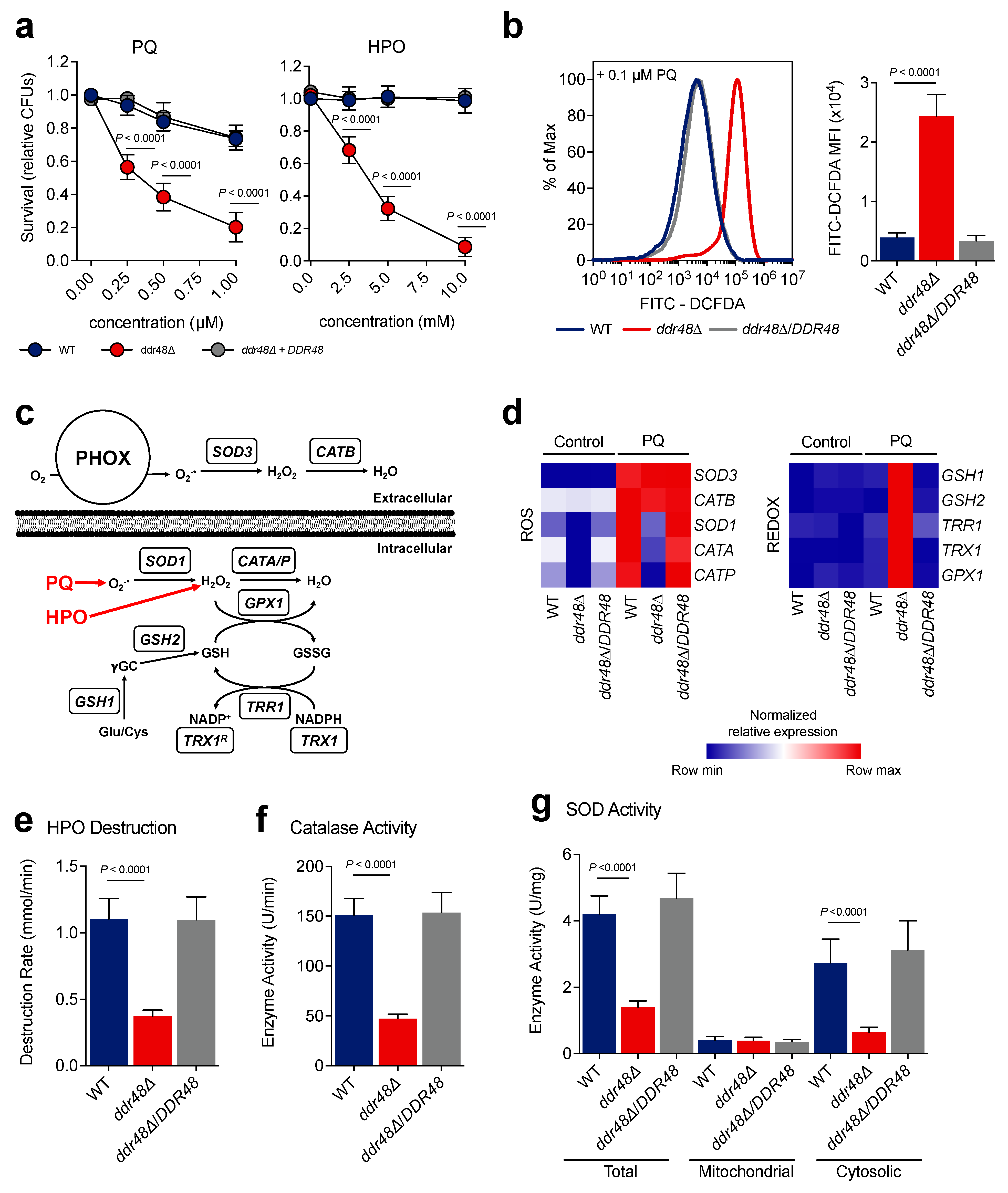
Since expression of DDR48 increases when treated with oxidative stress agents, we next assessed if DDR48 plays a role in combatting oxidative stress in yeasts. To test if DDR48 contributes to protection from reactive oxygen species (ROS), wildtype, ddr48∆, and ddr48∆/DDR48 yeasts were challenged in vitro with various concentrations of paraquat (0.25 µM, 0.5 µM, and 0.75 µM) or hydrogen peroxide (2.5 mM, 5 mM, and 7.5 mM) to determine survival of each strain (colony forming units (CFUs)). Paraquat was used to generate superoxide [51]. Because superoxides are broken down to liberate peroxides, hydrogen peroxide was included as a ROS source. Wild-type, DDR48-expressing yeasts challenged with paraquat, or hydrogen peroxide exhibited greater than 80% survival (Figure 3a). However, only 20% of ddr48∆ yeasts survive the highest concentration of paraquat and less than 5% survived the highest concentration of hydrogen peroxide (Figure 3a). Complementation of the ddr48∆ deletion strain (ddr48∆/DDR48) restored survivability comparable to that observed in wild-type yeasts. To determine if the increased killing of ddr48∆ yeasts with oxidative stress is due to less clearance of ROS, we treated wild-type, ddr48∆, and ddr48∆/DDR48 yeasts with 0.1 µM paraquat and quantified ROS levels using the DCFDA assay via flow cytometry [38]. ddr48∆ cells contained 3x more ROS than wild-type cells 1-h after treatment with paraquat (Figure 3b). Complementation of the ddr48∆ mutant restored the ability to clear ROS generated by paraquat. These results demonstrate that DDR48 contributes to protection of yeasts from both superoxide and peroxide species.
Histoplasma yeasts possess enzymes for detoxification of intracellular and extracellular reactive oxygen species (Figure 3c) [39,40,52,53]. To determine if the increased killing of ddr48∆ yeasts by ROS is due to changes in gene expression of oxidative stress genes, we examined the transcriptional profile of the oxidative stress and redox pathways before and after treatment with 0.1 µM paraquat. Expression of the intracellular catalases (CATA and CATP) and cytosolic superoxide dismutase (SOD1) were decreased ~10-fold in ddr48∆ yeasts before paraquat treatment, suggesting the loss of DDR48 alters basal transcription of these genes (Figure 3d). In wild-type, DDR48-expressing yeasts, transcripts of CATA, CATP, and SOD1 are upregulated after exposure to paraquat (Figure 3d). In paraquat-exposed ddr48∆ yeasts, expression of CATA, CATP, and SOD1 increased, albeit by the same fold-change but the magnitude differed. Complementation of the ddr48∆ mutant restored gene expression to wild-type values. Expression of the extracellular catalase CATB and the extracellular superoxide dismutase SOD3 was not affected by DDR48, as transcript levels were comparable between wild-type and ddr48∆ yeasts before and after treatment (Figure 3d). These results suggest that DDR48 is involved in combatting intracellular oxidative stress and not extracellular oxidative stress.
We next determined the transcriptional profile of the glutathione-dependent redox system before and after paraquat treatment, as it operates synergistically with ROS detoxification enzymes to maintain cellular redox homeostasis (Figure 3c) [54,55,56,57]. Gene expression of cytosolic thioredoxin reductase TRR1, cytosolic thioredoxin TRX1, glutathione peroxidase GPX1, glutamate-cysteine ligase GSH1, and glutathione synthetase GSH2 before and after paraquat treatment was performed in wild-type, ddr48∆, and ddr48∆/DDR48 yeasts. There were no significant changes in expression levels of wild-type yeasts after paraquat treatment; however, TRR1, TRX1, GPX1, GSH1, and GSH2 were significantly upregulated in ddr48Δ yeasts after paraquat exposure (Figure 3d). Expression of ddr48Δ/DDR48-complemented yeasts remained unchanged, as in wild-type cells. To ensure that the changes in gene expression observed were not due to fluctuating HHT1 mRNA levels, we confirmed transcript levels of HHT1 normalized to ACT1 before and after treatment with PQ or HPO. We found no significant differences in HHT1 expression between wild-type and ddr48∆ yeasts before or after treatment with PQ (Figure S4a) or HPO (Figure S4b). These results show that the glutathione-dependent oxidative stress system could be attempting to compensate for the loss of catalase and superoxide dismutase expression see in ddr48Δ yeasts.
To confirm that the changes in catalase and superoxide dismutase gene expression observed in the ddr48∆ mutant corresponded with actual enzyme activity, we determined catalase and superoxide dismutase (SOD) enzymatic activity in wildtype, ddr48Δ, and ddr48Δ/DDR48 yeasts. We measured catalase activity by absorption of light at 240 nm [41]. We optimized the assay for the concentration of Histoplasma protein and the time in which linear measurements could be taken (Figure S5). The rate of hydrogen peroxide decomposition decreased by 57% in ddr48∆ yeasts compared to wild-type, DDR48-expressing yeasts (Figure 3e). Complementation of the ddr48∆ strain returned the hydrogen peroxide decomposition rate to that resembling wildtype. The corresponding catalase activity of ddr48∆ yeasts was 64% lower than wild-type and ddr48∆/DDR48 strains (Figure 3f). We next subjected wildtype, ddr48∆, and ddr48∆/DDR48 yeasts to 0.1 µM paraquat and measured superoxide dismutase activity levels from the corresponding cytoplasmic extracts. ddr48∆ yeasts exhibited an 80% decrease in cytosolic and 64% decrease in total SOD enzyme activity compared to wild-type samples (Figure 3g). Interestingly, mitochondrial SOD activity was unaffected by the loss of DDR48. Cytosolic and total SOD activity levels were restored to wild-type levels in complemented ddr48∆/DDR48 yeasts. These data demonstrate that the loss of DDR48 results in decreased gene expression and corresponding decreases in enzymatic activity of intracellular superoxide dismutases and catalases, decreasing Histoplasma yeast’s response and survival against oxidative stress.

3.4. ddr48∆ Yeasts Are More Susceptible to Killing by Amphotericin-B and Ketoconazole

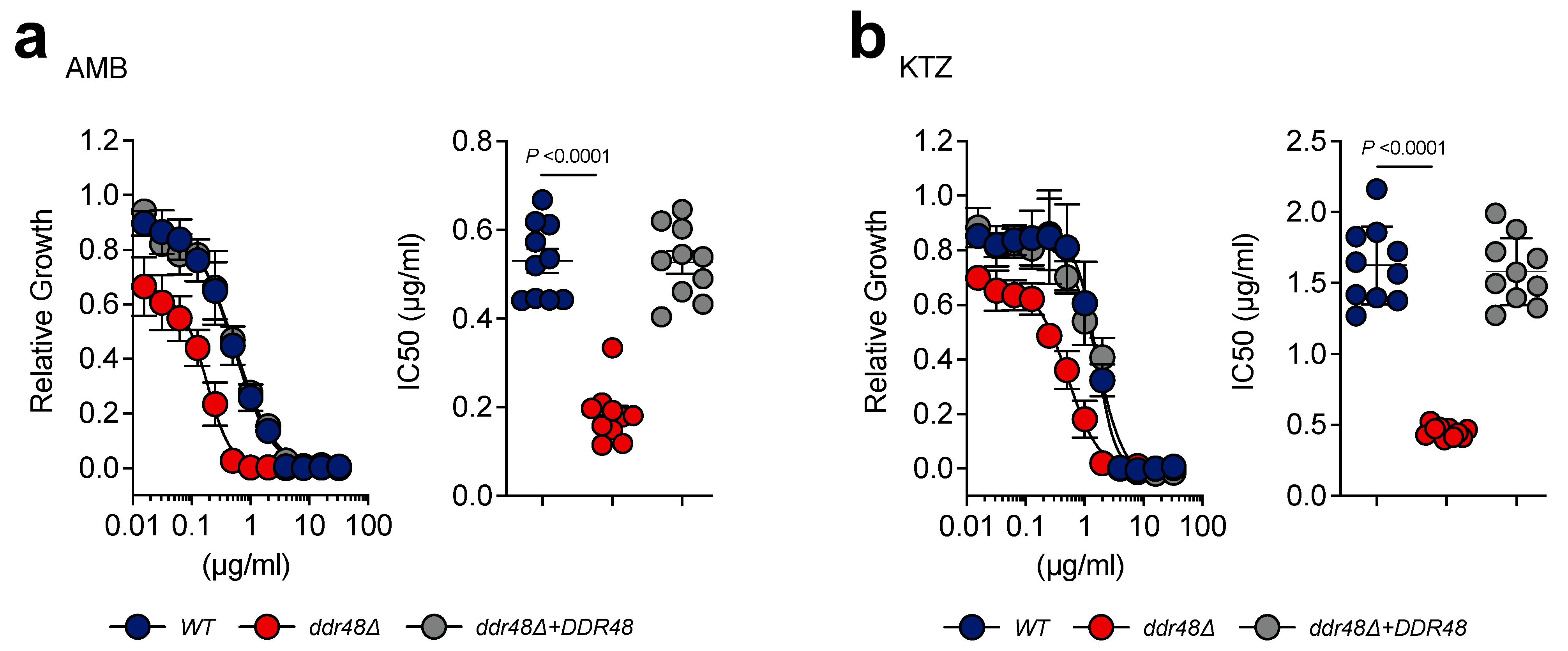
Increased DDR48 expression has been linked to antifungal drug resistance in clinical isolates of C. albicans [11]. We performed dose–response tests on wild-type, ddr48∆, and ddr48∆/DDR48 strains to determine the concentrations for 50% inhibition (IC50) against amphotericin-B (polyene) and ketoconazole (azole) [58]. Wild-type yeasts treated with amphotericin-B showed an IC50 of 0.5296 ± 0.0858 µg/mL (mean ± standard deviation (SD)), whereas ddr48∆ yeasts were 55% more susceptible to this antifungal with an IC50 of 0.1831 ± 0.0617 µg/mL (mean ± SD) (Figure 4a). ddr48∆ yeasts (IC50 = 1.624 ± 0.2749 µg/mL (mean ± SD)) were 66% more susceptible to ketoconazole than wild-type yeasts (IC50 = 0.4514 ± 0.0397 µg/mL (mean ± SD)) (Figure 4b). No significant difference in the susceptibility to amphotericin-B or ketoconazole were found between wild-type and the complemented ddr48∆/DDR48 strains. These data indicate that DDR48 contributes to resistance to ketoconazole and amphotericin-B.

3.5. ddr48∆ Yeasts Exhibit an Aberrant Ergosterol Biosynthesis Transcriptional Program before and after Antifungal Drug Treatment

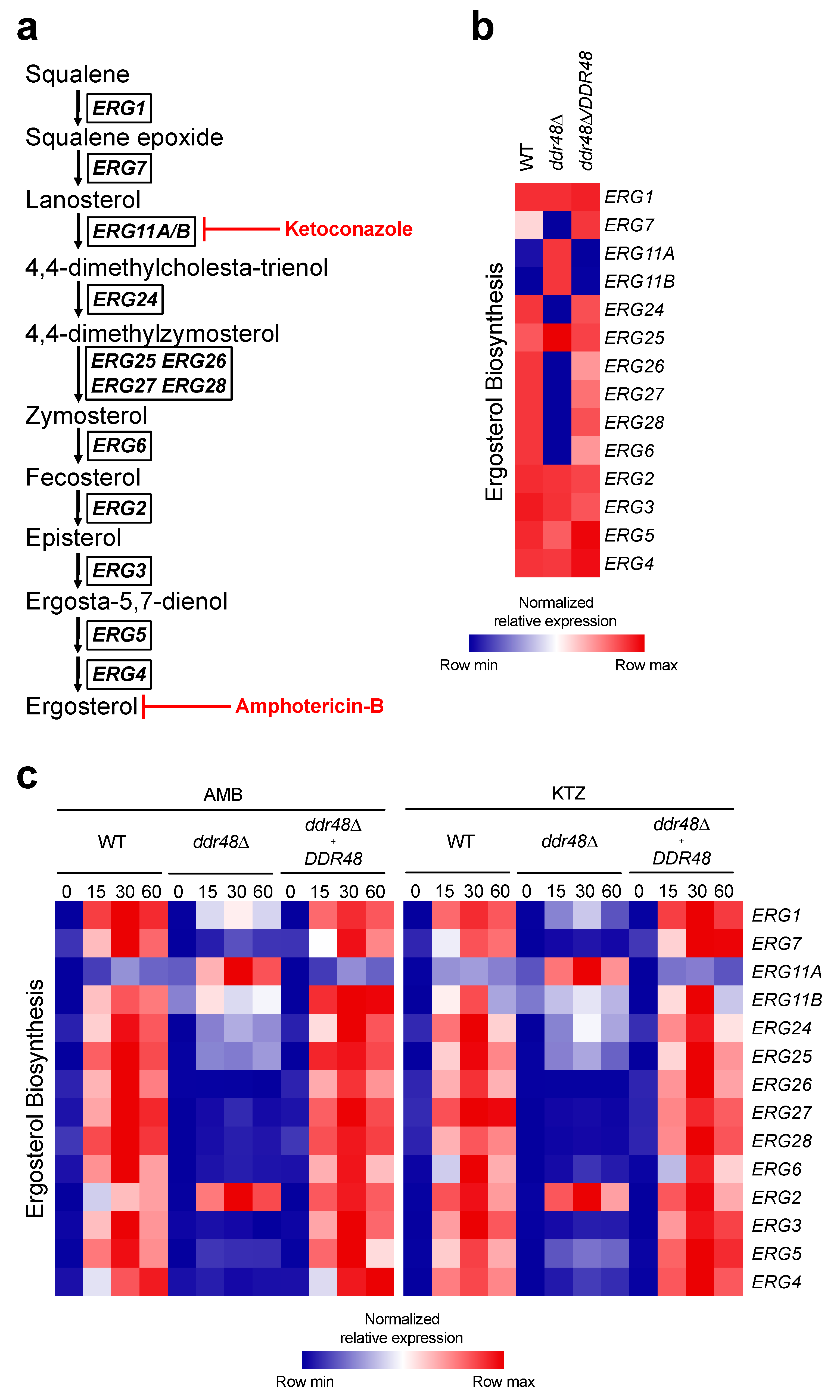
Amphotericin-B and ketoconazole exert their antifungal effects by targeting components of the ergosterol biosynthesis pathway as shown in (Figure 5a) [59,60,61]. Since deletion of DDR48 affected susceptibility to amphotericin-B and ketoconazole, we examined the transcriptional profile of the ergosterol biosynthesis pathway before and 15, 30, and 60 min after treatment with these agents. Some ergosterol biosynthesis genes exhibited changes in basal expression between wild-type and ddr48∆ yeasts before antifungal treatment (Figure 5b). ERG11A and ERG11B were upregulated by 5-fold and 10-fold, respectively, in the ddr48∆ mutant strain. In contrast, ERG7 (-3-fold), ERG24 (-10-fold), ERG26 (-3-fold), ERG27 (-7-fold), ERG28 (-16-fold), and ERG6 (-12-fold) were downregulated in ddr48∆ yeasts (Figure 5b). These results suggest that DDR48 affects basal transcription rates of 8 out of 14 (57%) of ergosterol biosynthesis genes. As expected, transcript levels of all the ergosterol biosynthesis genes tested increased upon treatment with amphotericin-B or ketoconazole in wildtype yeasts (Figure 5c).
Among ddr48Δ yeasts there were no significant changes in transcript levels of ERG3, ERG4, ERG5, ERG11B, and ERG26 after treatment with amphotericin-B or ketoconazole (Figure 5c). Transcript levels of ERG1, ERG6, ERG7, ERG24, ERG25, ERG27, and ERG28 increased in ddr48Δ yeasts, but did not reach magnitude of wildtype yeasts. ERG11A expression increased beyond wildtype after treatment, and no significant changes in ERG2 expression were observed after treatment (Figure 5c). The complemented strain behaved similar to wildtype yeasts.
One possible explanation for these changes is that the housekeeping gene HHT1 used to normalize expression is affected by the loss of DDR48. To ensure that the changes in gene expression were not caused by altered HHT1 mRNA levels, we compared transcript levels of HHT1 with ACT1. No significant differences in HHT1 expression between wild-type and ddr48∆ yeasts were observed after AMB (Figure S4c) or KTZ (Figure S4d) treatments. These data demonstrate that the loss of DDR48 disrupts basal gene expressional levels of over half of ergosterol biosynthesis genes. The loss of DDR48 disrupts the transcriptional program of the ergosterol biosynthesis pathway in responding to amphotericin-B and ketoconazole, leading to less resistance of yeasts to antifungal drugs.

3.6. ddr48∆ Yeasts Exhibit Decreased Survival in Macrophages

Macrophages respond to H. capsulatum infection by initiating a respiratory burst that generates ROS [62]. Since ddr48∆ yeasts are more sensitive to killing by ROS in vitro, we asked if deletion of DDR48 affects intracellular survival within resting and stimulated RAW 264.7 macrophages. We first sought to determine if DDR48 gene expression is upregulated once Histoplasma yeasts are phagocytosed by macrophages. At 4 h post-infection (hpi) DDR48 expression was elevated by 10-fold and remained increased at 24 hpi (Figure 6a). We confirmed there were no changes in HHT1 mRNA levels at 4 and 24 hpi (Figure S6a) Next, we infected resting and IFNγ-stimulated (10 ng/mL) RAW 264.7 macrophages with wild-type, ddr48∆, or ddr48∆/DDR48 yeasts and examined Histoplasma CFUs at 24 hpi. ddr48Δ yeasts manifested a 50% decrease in CFUs in resting and IFNγ-stimulated macrophages compared to wild-type (Figure 6b). Recovery of the complemented ddr48∆/DDR48 strain was similar to wildtype in resting and stimulated samples. IFNγ activation did increase macrophage killing of mutant and wildtype yeasts.
The changes in CFUs could be caused by differences in phagocytosis. Hence, we calculated the phagocytic index of wildtype, ddr48∆, and ddr48∆/DDR48 yeasts and found no significant changes in the number of yeasts being phagocytized by each macrophage (Figure S6b). We found an average of 4 yeasts per macrophage. Next, we determined the percent of macrophages that contained Histoplasma yeasts of each strain and found no significant differences in phagocytosis between DDR48-expressing and ddr48∆ yeasts (Figure S6c). These results affirm that ddr48∆ yeasts are less fit (~50%) within macrophages compared to wild-type Histoplasma yeasts.

4. Discussion

In this study, we aimed to increase our understanding of the H. capsulatum DDR48 gene, as little is known about DDR48 in general, and there are no prior studies in H. capsulatum. DDR48 was identified as a 315 amino acid protein containing fifteen repeats of the characteristic SYG amino acid sequence that is present in all identified DDR48 amino acid sequences. The repetitive SYG motif is a putative RNA binding sequence believed to be involved in RNA regulation [3]. Given this information, we postulate that DDR48 possesses RNA binding activity, providing a potential route in which it exerts its effects on the fungal stress response. Jin et al. identified DDR48 as a component of processing bodies (p-bodies) and glycolytic bodies (G-bodies) in C. albicans [7], both of which are stress granules that function to maintain the balance between translating mRNAs and mRNA degradation under stress [63,64,65]. Each of these mRNA granules relies upon RNA binding proteins for proper structure and function [5,66], thus providing a link between DDR48 and RNA metabolism. The H. capsulatum DDR48 amino acid sequence contains a conserved domain belonging to a group of ATP-dependent RNA helicases (PTZ00110), which are known to be involved in RNA regulatory activities and localize to stress granules under stress [45,67]. PTZ00110 RNA helicases use ATP molecules to achieve their RNA un-winding activities, hence they also exhibit ATP hydrolysis [68]. A study in S. cerevisiae concluded that DDR48 possesses ATP and GTP hydrolysis abilities [1], though no other studies have confirmed these results. These studies provide a foundation in which more investigations can begin to determine if DDR48 has RNA binding properties and elucidate a possible role in RNA metabolism.
Although DDR48 has been characterized as a mycelial phase-specific gene, our results demonstrated that DDR48 is expressed in Histoplasma yeasts under cellular stress to levels observed in mycelia. Thirty minutes after treatment with hydrogen peroxide, DDR48 expression was statistically similar (p > 0.05) to mycelial phase mRNA levels. A potential reason that DDR48 is constitutively expressed in the mycelial phase is that the environmental conditions encountered by mycelia are variable, and depend on geographic location, nutrient availability, and weather conditions [69,70,71,72]. Constitutive expression in mycelia may enhance their ability to confront fluctuating environmental conditions.
Our studies are the first investigating the mode of action of DDR48 in H. capsulatum. To gain more insight into DDR48, we subjected yeasts to a battery of cellular stressors and showed that DDR48 was induced by all stressors used in this study, demonstrating the far-reaching effects of DDR48 regarding fungal stress response. An interesting pattern emerged in which DDR48 expression peaked 30 min post-treatment and began to decline thereafter. The expression pattern is similar to fungal heat shock proteins [73]. As an example, Histoplasma heat shock protein HSP82 is induced transiently when the temperature is shifted to 34 °C [74]. Like HSP82, DDR48 responded to heat shock, but at 42 °C. It is conceivable that DDR48 acts like a heat shock protein since this family contributes to stress response and RNA metabolism [75,76,77]. On the other hand, RNA binding proteins that attach to and stabilize pre-existing mRNAs during the cellular stress response exhibit the same pattern of expression, peaking early on after stress treatment and quickly waning to basal levels [78,79,80,81].
We found that deletion of DDR48 compromised Histoplasma yeast’s survival when treated with either superoxide or hydrogen peroxide. At the highest concentrations tested, ddr48∆ yeasts were ~12-fold more susceptible to hydrogen peroxide and ~4-fold more susceptible to superoxides generated by paraquat. These results are consistent with experiments performed in S. cerevisiae and C. albicans [82,83]. In this work, we tracked gene expression of the intracellular and extracellular superoxide dismutases and catalases in wild-type and ddr48∆ mutant strains. Youseff et al. previously showed that H. capsulatum contains an extracellular catalase and superoxide dismutase, CATB and SOD3, respectively, which are specialized to detoxify extracellular ROS produced by phagocytes [40,52,53]. When treated with oxidative stress, CATB and SOD3 expression was unaffected by deletion of DDR48. This suggests that DDR48 is required for Histoplasma survival of intracellular, but not exogenous oxidative stress. Our data did show that DDR48 controls basal expression of the intracellular oxidative stress genes in addition to their response to oxidative stress. These findings suggest that DDR48 has a function in RNA turnover. Overexpression of DDR48 in S. cerevisiae led to a ~60% decrease in global RNA accumulation [8], which conflicts with our findings. However, these two fungi are genetically very different. Histoplasma yeasts compensate for the loss of DDR48 by activating alternative ROS detoxification pathways like the glutathione/thioredoxin system to aid in survival. This is consistent with studies in S. cerevisiae in which yeasts lacking the cytosolic thioredoxin system were more susceptible to killing by oxidative stress [54,84]. A potential reason for the glutathione/thioredoxin system to be upregulated in the ddr48∆ strain is that paraquat induces ROS and glutathione/thioredoxin. However, the glutathione/thioredoxin system was unaltered in wild-type yeasts incubated with paraquat. This finding explains why the ddr48∆ mutant is still able to proliferate, though at lower levels than wild-type, DDR48-expressing strains.
The antifungals fluconazole and itraconazole, in combination with amphotericin-B, are mainline treatments for infection with Histoplasma capsulatum [85]. One of the leading causes of treatment failure in HIV-positive patients with H. capsulatum infections is resistance to azole antifungals, thus new therapeutic approaches are needed to decrease relapse prevalence [86]. ddr48Δ yeasts are more susceptible to killing by ketoconazole and amphotericin-B. These results are consistent with studies in C. albicans; fluconazole-resistant isolates of C. albicans from patients exhibited higher expression of DDR48. Moreover, a strong correlation exists between overexpression of DDR48 and azole resistance genes [11]. DDR48 may exert its protective effects by modulating the ergosterol biosynthesis pathway. In this regard, the loss of DDR48 impacted several genes of the ergosterol biosynthesis pathway.
ddr48∆ yeasts were less fit within resting and activated macrophages, exhibiting a 50% reduction in survivability compared to wild-type yeasts. This was not caused by the exogenous ROS produced by phagocytes, as CATB and SOD3, the predominant enzymes that detoxify host-generated ROS were unaffected by deletion of DDR48. A probable explanation for the decreased survival of ddr48∆ yeasts within macrophages is the intracellular ROS generated from cellular metabolism is accruing in cells, thus the ddr48∆ yeasts are unable to cope with the harsh intramacrophage environment as efficiently as wild-type yeasts.
In summary, we have demonstrated that H. capsulatum relies on DDR48 to adapt and recover to cellular stress. We have shown that it is needed for optimal survival in macrophages, response to oxidative stress, and response to antifungal drugs. More research into the function of DDR48 will no doubt uncover more pathways in which DDR48 is involved. Given the broad range of processes that are dependent upon DDR48, thought should be given to determining if it could be a successful therapeutic target for those predisposed or suffering from endemic fungal pathogens, as a pathogen that is not poised to adapt when exposed to various cellular stressors could tip the balance in favor of the host’s immune system and eventual elimination of the fungal pathogen.

Supplementary Materials

The following are available online at https://www.mdpi.com/article/10.3390/jof7110981/s1. Figure S1. Deletion, complementation, and confirmation of HcDDR48. Figure S2. Confirmation of ddr48Δ, growth characteristics, and qRT-PCR controls. Figure S3. DDR48 transcript levels are constitutive in Histoplasma mycelia subjected to cellular stressors. Figure S4. HHT1 gene expression remains constant in WT, ddr48Δ, and ddr48Δ/DDR48 yeasts after exposure to cellular stress. Figure S5. Optimization of HPO decomposition assay. Figure S6. HHT1 expression levels remain unchanged within macrophages. Table S1: Primers used in this study.

Author Contributions

Conceptualization, L.T.B. and G.S.J.; methodology, L.T.B., K.A.R., G.M.R., L.A.K., and G.S.J.; validation, L.T.B. and G.S.J.; formal analysis, L.T.B. and G.S.J.; investigation, L.T.B., K.A.R., L.A.K., S.C.J., S.E.S., M.B.H. and J.C.W.; resources, G.S.J.; data curation, L.T.B., K.A.R., and G.M.R.; writing—original draft preparation, L.T.B.; writing—review and editing, L.T.B. and G.S.J.; supervision, L.T.B. and G.S.J.; project administration, G.S.J.; funding acquisition, G.S.J. All authors have read and agreed to the published version of the manuscript.

Funding

This research was supported by the Mississippi INBRE Institutional Development Award (IDeA) grant P20GM103476 to G.S.J. from the from the National Institute of General Medical Sciences of the National Institutes of Health.

Institutional Review Board Statement

Not applicable.

Informed Consent Statement

Not applicable.

Data Availability Statement

The data that support the findings of this study are available from the corresponding author, L.T.B., upon reasonable request.

Acknowledgments

We thank Davida Crossley for construction of the ddr48Δ Histoplasma strain previously in the Shearer laboratory. We are grateful to Jonathan Lindner and the MS INBRE Imaging Facility for equipment use and helpful discussions regarding data collection. We thank Erin Smith and Paige Brady for assistance in screening DDR48 complement strains and running qRT-PCR plates. We thank George Deepe for helpful edits and critiques. We would like to acknowledge the assistance of the Research Flow Cytometry Core in the Division of Rheumatology at Cincinnati Children’s Hospital Medical Center.

Conflicts of Interest

The authors declare no conflict of interest. The sponsors had no role in the design, execution, interpretation, or writing of the study.

References

  1. Treger, J.M.; McEntee, K. Structure of the DNA Damage-Inducible Gene DDR48 and Evidence for Its Role in Mutagenesis in Saccharomyces cerevisiae. Mol. Cell Biol. 1990, 10, 3174–3184. [Google Scholar]
  2. Sheng, S.; Schuster, S.M. Purification and characterization of Saccharomyces cerevisiae DNA damage-responsive protein 48 (DDRP 48). J. Biol. Chem. 1993, 268, 4752–4758. [Google Scholar] [CrossRef]
  3. Mitsuhashi, K.; Ito, D.; Mashima, K.; Oyama, M.; Takahashi, S.; Suzuki, N. De novo design of RNA-binding proteins with a prion-like domain related to ALS/FTD proteinopathies. Sci. Rep. 2017, 7, 16871. [Google Scholar] [CrossRef] [Green Version]
  4. Hennig, S.; Kong, G.; Mannen, T.; Sadowska, A.; Kobelke, S.; Blythe, A.; Knott, G.J.; Iyer, K.S.; Ho, D.; Newcombe, E.A.; et al. Prion-like domains in RNA binding proteins are essential for building subnuclear paraspeckles. J. Cell Biol. 2015, 210, 529–539. [Google Scholar] [CrossRef]
  5. Anderson, P.; Kedersha, N. RNA granules: Post-transcriptional and epigenetic modulators of gene expression. Nat. Rev. Mol. Cell Biol. 2009, 10, 430–436. [Google Scholar] [PubMed]
  6. Anderson, P.; Kedersha, N. RNA granules. J. Cell Biol. 2006, 172, 803–808. [Google Scholar] [CrossRef] [PubMed]
  7. Jin, M.; Fuller, G.G.; Han, T.; Yao, Y.; Alessi, A.F.; Freeberg, M.A.; Roach, N.P.; Moresco, J.J.; Karnovsky, A.; Baba, M.; et al. Glycolytic Enzymes Coalesce in G Bodies under Hypoxic Stress. Cell Rep. 2017, 20, 895–908. [Google Scholar] [CrossRef] [PubMed] [Green Version]
  8. Li, Z.; Barajas, D.; Panavas, T.; Herbst, D.A.; Nagy, P.D. Cdc34p Ubiquitin-Conjugating Enzyme Is a Component of the Tombusvirus Replicase Complex and Ubiquitinates p33 Replication Protein. J. Virol. 2008, 82, 6911–6926. [Google Scholar] [CrossRef] [Green Version]
  9. Banerjee, D.; Martin, N.; Nandi, S.; Shukla, S.; Dominguez, A.; Mukhopadhyay, G.; Prasad, R. A genome-wide steroid response study of the major human fungal pathogen Candida albicans. Mycopathologia 2007, 164, 1–17. [Google Scholar] [CrossRef]
  10. Enjalbert, B.; Smith, D.A.; Cornell, M.J.; Alam, I.; Nicholls, S.; Brown, A.J.P.; Quinn, J. Role of the Hog1 Stress-Activated Protein Kinase in the Global Transcriptional Response to Stress in the Fungal Pathogen Candida albicans. Mol. Biol. Cell 2006, 17, 1018–1032. [Google Scholar] [CrossRef] [Green Version]
  11. Mostafa, M.S.; Awad, A.R. Association of ADH1 and DDR48 expression with azole resistance in Candida albicans. Int. Arab. J. Antimicrob. Agents 2014, 4, 1–10. [Google Scholar] [CrossRef] [Green Version]
  12. Cleary, I.A.; MacGregor, N.B.; Saville, S.P.; Thomas, D.P. Investigating the Function of Ddr48p in Candida albicans. Eukaryot. Cell 2012, 11, 718–724. [Google Scholar] [CrossRef] [PubMed] [Green Version]
  13. Hromatka, B.S.; Noble, S.M.; Johnson, A.D. Transcriptional response of Candida albicans to nitric oxide and the role of the YHB1 gene in nitrosative stress and virulence. Mol. Biol. Cell 2005, 16, 13. [Google Scholar] [CrossRef] [PubMed] [Green Version]
  14. Tournu, H.; Tripathi, G.; Bertram, G.; Macaskill, S.; Mavor, A.; Walker, L.; Odds, F.C.; Gow, N.A.R.; Brown, A.J.P. Global role of the protein kinase Gcn2 in the human pathogen Candida albicans. Eukaryot. Cell 2005, 4, 1687–1696. [Google Scholar] [CrossRef] [PubMed] [Green Version]
  15. Dib, L.; Hayek, P.; Sadek, H.; Beyrouthy, B.; Khalaf, R.A. The Candida albicans DDR48 Protein Is Essential for Filamentation, Stress Response, and Confers Partial Antifungal Drug Resistance. Med. Sci. Monit. 2008, 14, 113–121. [Google Scholar]
  16. Singh, V.; Sinha, I.; Sadhale, P.P. Global Analysis of Altered Gene Expression during Morphogenesis of Candida Albicans In Vitro. Biochem. Biophys. Res. Commun. 2005, 334, 1149–1158. [Google Scholar] [CrossRef]
  17. Kusch, H.; Engelmann, S.; Bode, R.; Albrecht, D.; Morschhäuser, J.; Hecker, M. A Proteomic View of Candida Albicans Yeast Cell Metabolism in Exponential and Stationary Growth Phases. Int. J. Med. Microbiol. 2008, 298, 291–318. [Google Scholar] [CrossRef]
  18. Walsh, T.J.; Dixon, D.M. Spectrum of Mycoses. In Medical Microbiology; University of Texas Medical Branch at Galveston: Galveston, TX, USA, 1996; pp. 919–925. [Google Scholar]
  19. Kauffman, C.A. Histoplasmosis: A Clinical and Laboratory Update. Clin. Microbiol. Rev. 2007, 20, 115–132. [Google Scholar] [CrossRef] [PubMed] [Green Version]
  20. Ajello, L. The Medical Mycological Iceberg. HSMHA Health Rep. 1971, 86, 437–448. [Google Scholar] [CrossRef]
  21. Maiga, A.W.; Deppen, S.; Scaffidi, B.K.; Baddley, J.; Aldrich, M.C.; Dittus, R.S.; Grogan, E.L. Mapping Histoplasma Capsulatum Exposure, United States. Emerg. Infect. Dis. 2018, 24, 1835–1839. [Google Scholar] [CrossRef]
  22. Rippon, J.W. Dimorphism in Pathogenic Fungi. CRC Crit. Rev. Microbiol. 1980, 8, 49–97. [Google Scholar] [CrossRef] [PubMed]
  23. Webster, R.H.; Sil, A. Conserved Factors Ryp2 and Ryp3 Control Cell Morphology and Infectious Spore Formation in the Fungal Pathogen Histoplasma Capsulatum. Proc. Natl. Acad. Sci. USA 2008, 105, 14573–14578. [Google Scholar] [CrossRef] [Green Version]
  24. Nguyen, V.Q.; Sil, A. Temperature-Induced Switch to the Pathogenic Yeast Form of Histoplasma Capsulatum Requires Ryp1, a Conserved Transcriptional Regulator. Proc. Natl. Acad. Sci. USA 2008, 105, 4880–4885. [Google Scholar] [CrossRef] [Green Version]
  25. Nemecek, J.C. Global Control of Dimorphism and Virulence in Fungi. Science 2006, 312, 583–588. [Google Scholar] [CrossRef]
  26. Deepe, G.S.; Gibbons, R.S.; Smulian, A.G. Histoplasma Capsulatum Manifests Preferential Invasion of Phagocytic Subpopulations in Murine Lungs. J. Leukoc. Biol. 2008, 84, 669–678. [Google Scholar] [CrossRef] [Green Version]
  27. Ray, S.C.; Rappleye, C.A. Flying under the Radar: Histoplasma Capsulatum Avoidance of Innate Immune Recognition. Semin Cell Dev. Biol. 2019, 89, 91–98. [Google Scholar] [CrossRef] [PubMed]
  28. Howard, D.H. Intracellular growth of Histoplasma capsulatum. J. Bacteriol. 1965, 89, 518–523. [Google Scholar] [CrossRef] [PubMed] [Green Version]
  29. Newman, S.L.; Bucher, C.; Rhodes, J.; Bullock, W.E. Phagocytosis of Histoplasma Capsulatum Yeasts and Microconidia by Human Cultured Macrophages and Alveolar Macrophages. Cellular Cytoskeleton Requirement for Attachment and Ingestion. J. Clin. Investig. 1990, 85, 223–230. [Google Scholar] [CrossRef] [Green Version]
  30. Klimpel, K.R.; Goldman, W.E. Isolation and Characterization of Spontaneous Avirulent Variants of Histoplasma capsulatum. Infect. Immun. 1987, 55, 528–533. [Google Scholar] [CrossRef] [Green Version]
  31. Worsham, P.L.; Goldman, W.E. Quantitative Plating of Histoplasma Capsulatum without Addition of Conditioned Medium or Siderophores. J. Med. Vet. Mycol. 1988, 26, 137–143. [Google Scholar] [CrossRef]
  32. Tian, X.; Shearer, G. The Mold-Specific MS8 Gene Is Required for Normal Hypha Formation in the Dimorphic Pathogenic Fungus Histoplasma capsulatum. Eukaryot. Cell 2002, 1, 249–256. [Google Scholar] [CrossRef] [Green Version]
  33. Crossley, D.; Naraharisetty, V.; Shearer, G. The Mould-Specific M46 Gene Is Not Essential for Yeast-Mould Dimorphism in the Pathogenic Fungus Histoplasma capsulatum. Med. Mycol. 2016, 54, 876–884. [Google Scholar] [CrossRef] [PubMed] [Green Version]
  34. Sebghati, T.S.; Engle, J.T.; Goldman, W.E. Intracellular Parasitism by Histoplasma Capsulatum: Fungal Virulence and Calcium Dependence. Science 2000, 290, 1368–1372. [Google Scholar] [CrossRef] [PubMed]
  35. Woods, J.P.; Retallack, D.M.; Heinecke, E.L.; Goldman, W.E. Rare Homologous Gene Targeting in Histoplasma Capsulatum: Disruption of the URA5Hc Gene by Allelic Replacement. J. Bacteriol. 1998, 180, 9. [Google Scholar] [CrossRef] [PubMed] [Green Version]
  36. Carr, J.; Shearer, G. Genome Size, Complexity, and Ploidy of the Pathogenic Fungus Histoplasma Capsulatum. J. Bacteriol. 1998, 180, 7. [Google Scholar] [CrossRef] [Green Version]
  37. Livak, K.J.; Schmittgen, T.D. Analysis of Relative Gene Expression Data Using Real-Time Quantitative PCR and the 2−ΔΔCT Method. Methods 2001, 25, 402–408. [Google Scholar] [CrossRef]
  38. Eruslanov, E.; Kusmartsev, S. Identification of ROS Using Oxidized DCFDA and Flow-Cytometry. In Advanced Protocols in Oxidative Stress II; Armstrong, D., Ed.; Methods in Molecular Biology; Humana Press: Totowa, NJ, USA, 2010; Volume 594, pp. 57–72. ISBN 978-1-60761-410-4. [Google Scholar]
  39. Guimarães, A.J.; Hamilton, A.J.; de M. Guedes, H.L.; Nosanchuk, J.D.; Zancopé-Oliveira, R.M. Biological Function and Molecular Mapping of M Antigen in Yeast Phase of Histoplasma Capsulatum. PLoS ONE 2008, 3, e3449. [Google Scholar] [CrossRef] [Green Version]
  40. Holbrook, E.D.; Smolnycki, K.A.; Youseff, B.H.; Rappleye, C.A. Redundant Catalases Detoxify Phagocyte Reactive Oxygen and Facilitate Histoplasma Capsulatum Pathogenesis. Infect. Immun. 2013, 81, 2334–2346. [Google Scholar] [CrossRef] [PubMed] [Green Version]
  41. Li, Y.; Schellhorn, H.E. Rapid Kinetic Microassay for Catalase Activity. J. Biomol. Tech. 2007, 18, 3. [Google Scholar]
  42. Goughenour, K.D.; Balada-Llasat, J.-M.; Rappleye, C.A. Quantitative Microplate-Based Growth Assay for Determination of Antifungal Susceptibility of Histoplasma Capsulatum Yeasts. J. Clin. Microbiol. 2015, 53, 3286–3295. [Google Scholar] [CrossRef] [Green Version]
  43. Cordero, R.J.B.; Liedke, S.C.; de S. Araújo, G.R.; Martinez, L.R.; Nimrichter, L.; Frases, S.; Peralta, J.M.; Casadevall, A.; Rodrigues, M.L.; Nosanchuk, J.D.; et al. Enhanced Virulence of Histoplasma Capsulatum through Transfer and Surface Incorporation of Glycans from Cryptococcus Neoformans during Co-Infection. Sci. Rep. 2016, 6, 21765. [Google Scholar]
  44. Tian, X.; Shearer, G. Cloning and Analysis of Mold-Specific Genes in the Dimorphic Fungus Histoplasma Capsulatum. Gene 2001, 275, 107–114. [Google Scholar] [CrossRef]
  45. Cordin, O.; Banroques, J.; Tanner, N.K.; Linder, P. The DEAD-Box Protein Family of RNA Helicases. Gene 2006, 367, 17–37. [Google Scholar] [CrossRef] [PubMed]
  46. de la Cruz, J.; Kressler, D.; Linder, P. Unwinding RNA in Saccharomyces Cerevisiae: DEAD-Box Proteins and Related Families. Trends Biochem. Sci. 1999, 24, 192–198. [Google Scholar] [CrossRef]
  47. Worsham, P.L.; Goldman, W.E. Development of a Genetic Transformation System for Histoplasma Capsulatum: Complementation of Uracil Auxotrophy. Mol. Gen. Genet. 1990, 221, 358–362. [Google Scholar] [CrossRef]
  48. Barker, K.S. Genome-Wide Expression Profiling Reveals Genes Associated with Amphotericin B and Fluconazole Resistance in Experimentally Induced Antifungal Resistant Isolates of Candida Albicans. J. Antimicrob. Chemother. 2004, 54, 376–385. [Google Scholar] [CrossRef]
  49. Ramírez-Quijas, M.D.; López-Romero, E.; Cuéllar-Cruz, M. Proteomic Analysis of Cell Wall in Four Pathogenic Species of Candida Exposed to Oxidative Stress. Microb. Pathog. 2015, 87, 1–12. [Google Scholar] [CrossRef]
  50. Salmon, T.B. Biological Consequences of Oxidative Stress-Induced DNA Damage in Saccharomyces cerevisiae. Nucleic Acids Res. 2004, 32, 3712–3723. [Google Scholar] [CrossRef] [PubMed] [Green Version]
  51. Bus, J.S.; Gibson, J.E. Paraquat: Model for Oxidant-Initiated Toxicity. Environ. Health Perspect. 1984, 55, 37–46. [Google Scholar] [CrossRef]
  52. McEwen, J.E.; York, J.L.; Kruft, V.; Johnson, C.H.; Klotz, M.G. Redundancy, Phylogeny and Differential Expression of Histoplasma Capsulatum Catalases. Microbiology 2002, 148, 1129–1142. [Google Scholar]
  53. Youseff, B.H.; Holbrook, E.D.; Smolnycki, K.A.; Rappleye, C.A. Extracellular Superoxide Dismutase Protects Histoplasma Yeast Cells from Host-Derived Oxidative Stress. PLoS Pathog. 2012, 8, e1002713. [Google Scholar] [CrossRef] [Green Version]
  54. Ross, S.J.; Findlay, V.J.; Malakasi, P.; Morgan, B.A. Thioredoxin Peroxidase Is Required for the Transcriptional Response to Oxidative Stress in Budding Yeast. Mol. Biol. Cell 2000, 11, 2631–2642. [Google Scholar] [CrossRef] [PubMed] [Green Version]
  55. Leith, K.M.; Hazen, K.C. Paraquat Induced Thiol Modulation of Histoplasma capsulatum Morphogenesis. Mycopathologia 1988, 103, 21–27. [Google Scholar] [CrossRef]
  56. Briones-Martin-del-Campo, M.; Orta-Zavalza, E.; Cañas-Villamar, I.; Gutiérrez-Escobedo, G.; Juárez-Cepeda, J.; Robledo-Márquez, K.; Arroyo-Helguera, O.; Castaño, I.; De Las Peñas, A. The Superoxide Dismutases of Candida Glabrata Protect against Oxidative Damage and Are Required for Lysine Biosynthesis, DNA Integrity and Chronological Life Survival. Microbiology 2015, 161, 300–310. [Google Scholar] [CrossRef]
  57. Gutiérrez-Escobedo, G.; Orta-Zavalza, E.; Castaño, I.; De Las Peñas, A. Role of Glutathione in the Oxidative Stress Response in the Fungal Pathogen Candida Glabrata. Curr. Genet. 2013, 59, 91–106. [Google Scholar] [CrossRef]
  58. Li, R.-K.; Ciblak, M.A.; Nordoff, N.; Pasarell, L.; Warnock, D.W.; McGinnis, M.R. In Vitro Activities of Voriconazole, Itraconazole, and Amphotericin B against Blastomyces Dermatitidis, Coccidioides Immitis, and Histoplasma Capsulatum. Antimicrob. Agents Chemother. 2000, 44, 1734–1736. [Google Scholar] [CrossRef] [Green Version]
  59. Delhom, R.; Nelson, A.; Laux, V.; Haertlein, M.; Knecht, W.; Fragneto, G.; Wacklin-Knecht, H.P. The Antifungal Mechanism of Amphotericin B Elucidated in Ergosterol and Cholesterol-Containing Membranes Using Neutron Reflectometry. Nanomaterials 2020, 10, 2439. [Google Scholar] [CrossRef]
  60. Borgers, M. The Mechanism of Action of the New Antimycotic Ketoconazole. Am. J. Med. 1983, 74, 2–8. [Google Scholar] [CrossRef]
  61. Gray, K.C.; Palacios, D.S.; Dailey, I.; Endo, M.M.; Uno, B.E.; Wilcock, B.C.; Burke, M.D. Amphotericin Primarily Kills Yeast by Simply Binding Ergosterol. Proc. Natl. Acad. Sci. USA 2012, 109, 2234–2239. [Google Scholar] [CrossRef] [PubMed] [Green Version]
  62. Lam, G.Y.; Huang, J.; Brumell, J.H. The Many Roles of NOX2 NADPH Oxidase-Derived ROS in Immunity. Semin. Immunopathol. 2010, 32, 415–430. [Google Scholar] [CrossRef] [PubMed]
  63. Decker, C.J.; Parker, R. P-Bodies and Stress Granules: Possible Roles in the Control of Translation and mRNA Degradation. Cold Spring Harb. Perspect. Biol. 2012, 4, a012286. [Google Scholar] [CrossRef] [PubMed] [Green Version]
  64. Buchan, J.R.; Muhlrad, D.; Parker, R. P Bodies Promote Stress Granule Assembly in Saccharomyces cerevisiae. J. Cell Biol. 2008, 183, 441–455. [Google Scholar] [CrossRef]
  65. Balagopal, V.; Parker, R. Polysomes, P Bodies and Stress Granules: States and Fates of Eukaryotic MRNAs. Curr. Opin. Cell Biol. 2009, 21, 403–408. [Google Scholar] [CrossRef] [PubMed] [Green Version]
  66. Sun, Z.; Diaz, Z.; Fang, X.; Hart, M.P.; Chesi, A.; Shorter, J.; Gitler, A.D. Molecular Determinants and Genetic Modifiers of Aggregation and Toxicity for the ALS Disease Protein FUS/TLS. PLoS Biol. 2011, 9, e1000614. [Google Scholar] [CrossRef] [PubMed]
  67. Hilliker, A. Analysis of RNA Helicases in P-Bodies and Stress Granules. In Methods in Enzymology; Elsevier: Amsterdam, The Netherlands, 2012; Volume 511, pp. 323–346. ISBN 978-0-12-396546-2. [Google Scholar]
  68. Jankowsky, E.; Fairman, M.; Yang, Q. RNA Helicases: Versatile ATP-Driven Nanomotors. J. Nanosci. Nanotechnol. 2005, 5, 1983–1989. [Google Scholar] [CrossRef]
  69. Wang, J.-J.; Lee, C.-L.; Pan, T.-M. Improvement of Monacolin K, Gamma-Aminobutyric Acid and Citrinin Production Ratio as a Function of Environmental Conditions of Monascus Purpureus NTU 601. J. Ind. Microbiol. Biotechnol. 2003, 30, 669–676. [Google Scholar] [CrossRef]
  70. Pitt, R.E. A Descriptive Model of Mold Growth and Aflatoxin Formation as Affected by Environmental Conditions. J. Food Prot. 1993, 56, 139–146. [Google Scholar] [CrossRef]
  71. Bohnert, H.J. Adaptations to Environmental Stresses. Plant Cell 1995, 7, 1099–1111. [Google Scholar] [CrossRef]
  72. Gasch, A.P.; Werner-Washburne, M. The Genomics of Yeast Responses to Environmental Stress and Starvation. Funct. Integr. Genom. 2002, 2, 181–192. [Google Scholar] [CrossRef]
  73. Tiwari, S.; Thakur, R.; Shankar, J. Role of Heat-Shock Proteins in Cellular Function and in the Biology of Fungi. Biotechnol. Res. Int. 2015, 2015, 1–11. [Google Scholar] [CrossRef] [Green Version]
  74. Minchiotti, G.; Gargano, S.; Maresca, B. Molecular Cloning and Expression of Hsp82 Gene of the Dimorphic Pathogenic Fungus Histoplasma capsulatum. Biochim. Biophys. Acta (BBA) Gene Struct. Expr. 1992, 1131, 103–107. [Google Scholar] [CrossRef]
  75. Haslbeck, M.; Vierling, E. A First Line of Stress Defense: Small Heat Shock Proteins and Their Function in Protein Homeostasis. J. Mol. Biol. 2015, 427, 1537–1548. [Google Scholar] [CrossRef] [PubMed] [Green Version]
  76. Vogel, J.L.; Parsellt, D.A.; Lindquist, S. Heat-Shock Proteins Hspl04 and Hsp70 Reactivate mRNA Splicing after Heat Inactivation. Curr. Biol. 1995, 5, 306–317. [Google Scholar] [CrossRef] [Green Version]
  77. Mühlhofer, M.; Berchtold, E.; Stratil, C.G.; Csaba, G.; Kunold, E.; Bach, N.C.; Sieber, S.A.; Haslbeck, M.; Zimmer, R.; Buchner, J. The Heat Shock Response in Yeast Maintains Protein Homeostasis by Chaperoning and Replenishing Proteins. Cell Rep. 2019, 29, 4593–4607. [Google Scholar] [CrossRef] [Green Version]
  78. Bose, S.; Dutko, J.A.; Zitomer, R.S. Genetic Factors That Regulate the Attenuation of the General Stress Response of Yeast. Genetics 2005, 169, 1215–1226. [Google Scholar] [CrossRef] [Green Version]
  79. Canadell, D.; García-Martínez, J.; Alepuz, P.; Pérez-Ortín, J.E.; Ariño, J. Impact of High PH Stress on Yeast Gene Expression: A Comprehensive Analysis of MRNA Turnover during Stress Responses. Biochim. Biophys. Acta (BBA) Gene Regul. Mech. 2015, 1849, 653–664. [Google Scholar] [CrossRef]
  80. Gutin, J.; Joseph-Strauss, D.; Sadeh, A.; Shalom, E.; Friedman, N. Genetic Screen of the Yeast Environmental Stress Response Dynamics Uncovers Distinct Regulatory Phases. Mol. Syst. Biol. 2019, 15, e8939. [Google Scholar] [CrossRef]
  81. Weng, M.; Yang, Y.; Feng, H.; Pan, Z.; Shen, W.-H.; Zhu, Y.; Dong, A. Histone Chaperone ASF1 Is Involved in Gene Transcription Activation in Response to Heat Stress in A Rabidopsis Thaliana: AtASF1 in Heat Stress Response. Plant Cell Environ. 2014, 37, 2128–2138. [Google Scholar] [CrossRef]
  82. Boorsma, A.; de Nobel, H.; ter Riet, B.; Bargmann, B.; Brul, S.; Hellingwerf, K.J.; Klis, F.M. Characterization of the Transcriptional Response to Cell Wall Stress in Saccharomyces cerevisiae. Yeast 2004, 21, 413–427. [Google Scholar] [CrossRef] [PubMed]
  83. Roetzer, A.; Gregori, C.; Jennings, A.M.; Quintin, J.; Ferrandon, D.; Butler, G.; Kuchler, K.; Ammerer, G.; Schüller, C. Candida glabrata Environmental Stress Response Involves Saccharomyces cerevisiae Msn2/4 Orthologous Transcription Factors. Mol. Microbiol. 2008, 69, 603–620. [Google Scholar] [CrossRef] [Green Version]
  84. Zaki, N.Z. Candida albicans TRR1 Heterozygotes Show Increased Sensitivity to Oxidative Stress and Decreased Pathogenicity. Afr. J. Microbiol. Res. 2012, 6, 1796–1805. [Google Scholar]
  85. Wheat, J. Histoplasmosis: Recognition and Treatment. Clinical Infectious Diseases Supplemental S19–S27. Available online: https://www.jstor.org/stable/pdf/4458030.pdf (accessed on 12 February 2021).
  86. Wheat, J.; Marichal, P.; Vanden Bossche, H.; Le Monte, A.; Connolly, P. Hypothesis on the Mechanism of Resistance to Fluconazole in Histoplasma capsulatum. Antimicrob. Agents Chemother. 1997, 41, 410–414. [Google Scholar] [CrossRef] [PubMed] [Green Version]
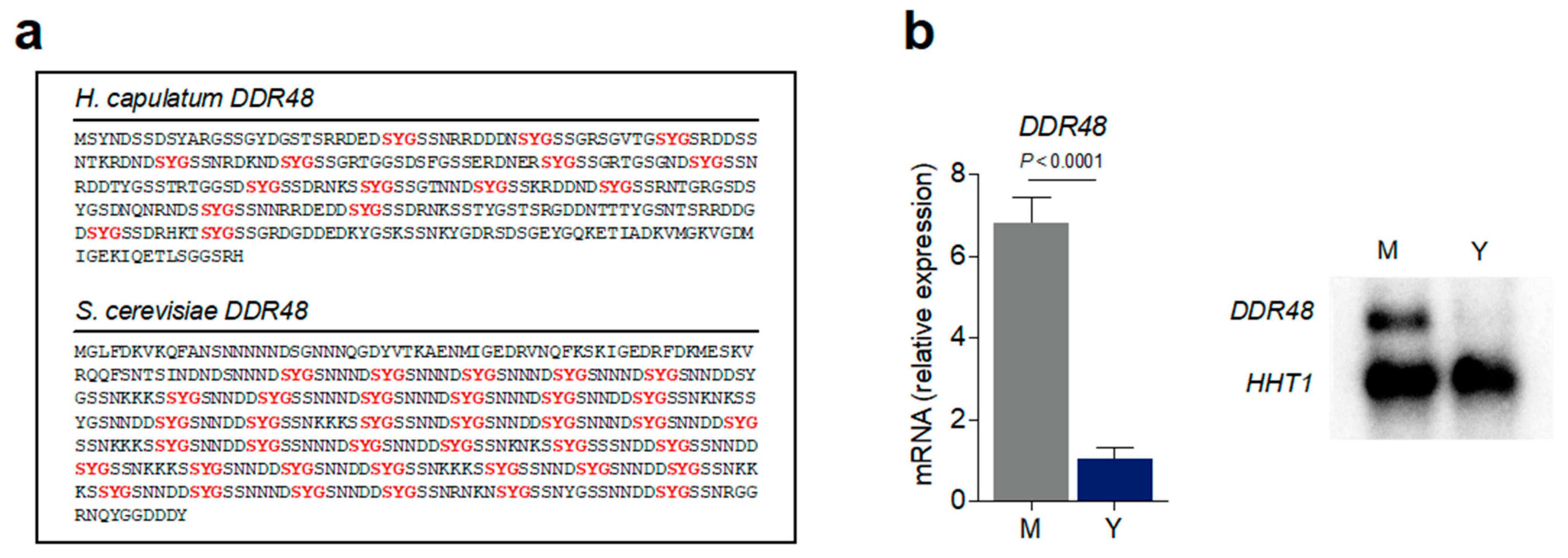
Figure 1. DDR48 gene expression levels are enriched in Histoplasma mycelia. (a) amino acid sequence of H. capsulatum DDR48 (top) and S. cerevisiae DDR48 (bottom) showing SYG repeats (red). (b) Expression of DDR48 in yeasts (Y) (blue bar) and mycelia (M) (grey bar) growth phases in liquid HMM measured by qRT-PCR (left) and representative northern blot (right) (n = 12) calculated relative to HHT1 transcript levels. All data generated were performed on three technical replicates and at least two biological replicates. Data is represented as the mean ± standard deviation (SD). Statistical analyses were performed using Student’s t test.
Jof 07 00981 g001
Figure 2. DDR48 transcript levels are increased in Histoplasma yeasts subjected to cellular stressors. Expression of DDR48 in yeast (blue bar) and mycelia (grey bar) growth phases measured by qRT-PCR (n = 9) at 0, 15, 30, and 60 min after the addition of various cellular stressors. The stressors include 0.1 µM paraquat (PQ)-superoxide anions, 2.5 mM hydrogen peroxide (HPO)-peroxides, 0.1 µg/mL amphotericin-B (AMB)-membrane disruption, 0.25 µg/mL ketoconazole (KTZ)-sterol synthesis inhibition, 50 µg/mL 5-fluorocytosine (5FC)-DNA/RNA biosynthesis inhibition, 1 mM methyl methanesulfonate (MMS)-DNA damage;methylating agent, 0.25 µM 4-nitroquinoline-1-oxide (4NQO)-UV-like damage, and heat-shock at 42 °C (HS). All data generated were performed on three technical replicates and at least two biological replicates. Data is represented as the mean ± standard deviation (SD). Statistical analyses were performed using one-way ANOVA with Tukey’s multiple comparisons test.
Jof 07 00981 g002
Figure 3. ddr48∆ yeasts possess decreased antioxidant activity, resulting in increased ROS levels and killing. (a) Survival (relative CFUs) of wildtype (DDR48(+)), ddr48∆, and ddr48∆/DDR48 strains measured (n = 8) after 4 days of growth in basal cell culture medium (HMM) supplemented with various concentrations of paraquat (PQ) or hydrogen peroxide (HPO). Survival was normalized to relative CFUs recovered from wildtype (DDR48(+)) yeasts with no treatment. (b) ROS levels measured (n = 8) in wildtype (DDR48(+)), ddr48∆, and ddr48∆/DDR48 yeasts by DCFDA fluorescence and flow cytometry. Data is shown as a representative histogram (left) and median fluorescence intensity (MFI) measurements (right). (c) ROS/Antioxidant pathway utilized by H. capsulatum yeasts with representative genes and their function. The type of oxidant generated by paraquat (PQ) and hydrogen peroxide (HPO) are shown in bold red. (d) Transcriptional profile of wildtype (WT), ddr48∆, and ddr48∆/DDR48 yeasts left untreated (Control) or treated with 0.1 µM PQ for 30 min (n = 9) measured by qRT-PCR relative to HHT1 levels. Expression of genes is presented as normalized row intensity (blue and red tiles) grouped by ROS or REDOX functions. (e) HPO destruction and (f) catalase activity of wildtype (WT), ddr48∆, and ddr48∆/DDR48 protein extracts measured (n = 9) 10 s after the addition of 5 mM HPO. (g) Superoxide dismutase (SOD) enzymatic activity (n = 6) of mid-log phase wildtype (WT), ddr48∆, and ddr48∆/DDR48 yeasts in HMM. Data is represented as the mean ± standard deviation (SD). Statistical analyses were performed using one-way ANOVA with Tukey’s multiple comparisons test.
Jof 07 00981 g003
Figure 4. Loss of DDR48 increases sensitivity to the antifungal drugs ketoconazole and amphotericin-B. Dose response curves (n = 10) of wildtype (WT), ddr48∆, and ddr48∆/DDR48 yeasts for (a) amphotericin-B (AMB) and (b) ketoconazole (KTZ). IC50 values were determined by non-linear regression. Data is represented as the mean ± standard deviation (SD). Statistical analyses were performed using one-way ANOVA with Tukey’s multiple comparisons test.
Jof 07 00981 g004
Figure 5. DDR48-Depleted Histoplasma Yeasts Contain an Aberrant Ergosterol Biosynthesis Pathway That is Exacerbated by Antifungal Treatment. (a) Ergosterol biosynthesis pathway utilized by H. capsulatum yeasts with representative genes and their function. The step at which each class of antifungals interferes with the pathway are shown in bold red. (b) Transcriptional profile of wildtype (WT), ddr48∆, and ddr48∆/DDR48 yeasts grown in basal cell culture medium (HMM) and (c) left untreated (0) or treated with 0.1 µg/mL amphotericin-B (AMB; polyene) or 0.25 µg/mL ketoconazole (KTZ; azole) for 15, 30, and 60 min (n = 9) measured by qRT-PCR relative to HHT1 levels. Expression of genes is presented as normalized row intensity (blue and red tiles) grouped by chronological step in the ergosterol biosynthesis pathway. All data generated were performed on three technical replicates and at least two biological replicates.
Jof 07 00981 g005
Figure 6. Deletion of DDR48 decreases fitness of Histoplasma yeasts in macrophages. (a) RAW 264.7 macrophages were infected with wildtype yeasts (MOI 5:1) and DDR48 mRNA levels were measured (n = 9) from uninfected (Ctrl) yeasts or from yeasts 4 h and 24 h post-infection by qRT-PCR and calculated relative to HHT1 levels. (b) Resting (left) or stimulated (right; 10 ng/mL IFNγ) RAW 264.7 macrophages were infected with wildtype (WT), ddr48∆, or ddr48∆/DDR48 yeasts (MOI 5:1) and in vitro macrophage growth of each strain was determined by lysis of macrophages and comparison of levels of viable yeasts (CFUs) in the lysate 24 h post-infection. All data generated were performed on three technical replicates and at least two biological replicates. Data is represented as the mean ± standard deviation (SD). Statistical analyses were performed using one-way ANOVA with Sidak’s multiple comparisons test.
Jof 07 00981 g006
Table 1. Histoplasma strains used in this study.
Strain 1Genotype 2Other
Designation
WU27ura5∆WT, DDR48(+)
USM10ura5∆ ddr48-3∆::hphddr48∆
USM13ura5∆ ddr48-3∆::hph/pLE04 (URA5, DDR48)ddr48∆/DDR48
1 Strains were constructed in the Histoplasma G186AS background, a “smooth” colony variant [30] of G186A (ATCC 26029). 2 Gene designations: hph: hygromycin B phosphotransferase (hygromycin resistance). URA5: Podospora anserina orotate phosphoribosyltransferase. DDR48: stress response gene DDR48.
Publisher’s Note: MDPI stays neutral with regard to jurisdictional claims in published maps and institutional affiliations.