Article

Entomopathogenic Fungi as Endophytes for Biological Control of Subterranean Termite Pests Attacking Cocoa Seedlings

by
Chaba F. Ambele
1,2,
Sunday Ekesi
1,
Hervé D. B. Bisseleua
3,
Olubukola O. Babalola
2,
Fathiya M. Khamis
1,
Christian T. L. Djuideu
4 and
Komivi S. Akutse
1,*
1
International Centre of Insect Physiology and Ecology (icipe), P.O. Box 30772-00100 Nairobi, Kenya
2
Food Security and Safety, Faculty of Agriculture, Science and Technology, North-West University, South Africa, Private Bag X2046, Mmabatho 2745, South Africa
3
World Cocoa Foundation, P.O. Box 217 Accra, Ghana
4
Zoology Laboratory, Faculty of Science, University of Yaoundé I, P.O. Box 812 Yaoundé, Cameroon
*
Author to whom correspondence should be addressed.
J. Fungi 2020, 6(3), 126; https://doi.org/10.3390/jof6030126
Submission received: 23 June 2020 / Revised: 21 July 2020 / Accepted: 3 August 2020 / Published: 5 August 2020

Abstract

:
This study was conducted in the scope of developing a sustainable effective approach against subterranean termite pests using entomopathogenic and endophytic fungus-based biopesticides. Termites, Odontotermes spp. workers, were tested for their susceptibility to 15 entomopathogenic fungal isolates through the direct spraying of conidia suspensions at 1 × 10⁸ conidia/mL. In general, all the isolates screened were pathogenic, with 100% mortality 4–7 days post-inoculation. However, the most virulent isolates were Metarhizium brunneum Cb15-III; the M. anisopliae isolates ICIPE 30 and ICIPE 60; Hypocrea lixii F3ST1; and the Beauveria bassiana isolates ICIPE 279, ICIPE 706 and ICIPE 662. These isolates were further tested for their endophytic colonization of cocoa seedlings using seed soaking, soil drench and foliar spray at 1 × 10⁸ conidia/mL. The colonization of the plant tissues by the fungi was determined using a culture-based technique. Only the B. bassiana isolates ICIPE 706 and ICIPE 279, and H. lixii F3ST1 colonized the cocoa seedlings, with varied colonization rates among isolates and inoculation methods. Three naturally occurring endophytes—Trichoderma asperellum, Fusarium solani and F. redolens—were also isolated from the cocoa seedling tissues. These findings suggest that cocoa seedlings are conducive to endophytic fungal growth either occurring naturally or from artificial inoculation Our findings could possibly lead to an innovative approach to the management of herbivory and subterranean termite pests in cocoa agroforests.

1. Introduction

Termites are becoming major pests in cocoa agroforests, causing significant losses [1,2,3,4]. Their control in the past relied on persistent organochlorine insecticides [5], which are now under restrictive use due to increasing concerns over negative effects on human and environmental health. Farmers in many parts of Africa use various traditional termite control methods, often with limited success [2]. The majority of termite control practices are ineffective and ecologically unsustainable and, above all, do not address the root attacks of termite infestation [2]. There is, therefore, a need for the identification of sustainable eco-friendly and locally available alternative methods for termite control in cocoa agroforests. Biological control using naturally occurring microorganisms such as entomopathogenic fungi (EPF) as part of integrated pest management could be the most sustainable long-term solution for termite control in cocoa agroforests. Entomopathogenic fungi (EPF) have long been considered as a promising approach for sustainable termite control [6]. This is because termites are assumed to live in environments conducive to entomopathogens [7,8]. The use of entomopathogens for termite control started as early as 1965 [9,10]. Most of these studies focused on the use of the EPF Beauveria bassiana (Balsamo) Vuillemin and Metarhizium anisopliae (Metschnikoff) Sorokin [6,8,11]. These EPF have generally been proved to be effective against termites in laboratory studies but have had little success in field trials when applied as conidial suspensions or granules [12]. This is due to repellency or host avoidance and because termites can deploy a diversity of defense mechanisms against the pathogens [13,14]. The efficacy of EPF against termite pests is also limited by abiotic factors (e.g., UV radiation, temperature and low humidity) that reduce the viability of fungal conidia [15]. Because of these reasons, the biological control of termites using EPF has generally been regarded as unfeasible [11,12,16].
However, apart from their direct mode of action against insect pests, several fungal entomopathogens have been reported to be able to grow internally within plant tissues, paving the way for possible use as endophytes in plant protection measures [17]. Mburu et al. [14] showed a co-evolutionary phenomenon in which termites’ behavioral response to either M. anisopliae or B. bassiana isolates is directly related to the potential infections that these fungi can inflict on them, and thus, termites can detect a virulent isolate from a distance and avoid direct contact. The foragers of the termite species Coptotermes formosanus Shiraki were significantly repelled by tree-based organic mulches coated with M. anisopliae, and the mulch consumption was highly reduced by up to 71% [18]. The repellent action of conidia against termites has also been utilized to protect maize crops from termites, which significantly reduced maize plant logging and increased grain yield in Kenya [19]. Therefore, another possible approach against subterranean termite pests of cocoa could be the inoculation of cocoa plants with highly virulent fungal entomopathogen as endophytes [20]. Several species of fungal entomopathogens are now known to have complex relationships with host plants as endophytes [15]. Beauveria bassiana for example, has been reported as an endophyte of maize [20,21], bananas [22], tomatoes [23], Theobroma gileri [24], coffee [25], and cocoa seedlings [26]. One advantage of the application of endophytes over the conventional direct application of fungal entomopathogens is the ability to colonize plants systemically, thereby offering continuous protection against insect pests. There is a plethora of recent studies on the insect-repellent, antifeedant and toxicity attributes of endophytic EPF [27,28,29]. However, whether virulent EPF against termites could colonize host tissues in cocoa seedlings and protect the seedlings against subterranean termites’ damage has not been evaluated.
In the scope of investigating the potential use of EPF to control subterranean termite pests in cocoa agroforests, this study was designed to (1) screen and select virulent entomopathogenic fungal strains on key subterranean termite species attacking cocoa and (2) determine if the most virulent entomopathogenic fungal isolates could be established as endophytes in cocoa seedlings, thereby establishing an important first step in the possible use of virulent entomopathogenic fungal endophytes as biocontrol agents against termites in cocoa agroforests.

2. Materials and Methods

2.1. Termite Collection and Maintenance

Termites (Odontotermes spp.) were collected from infested wood around icipe Duduville campus, Nairobi, Kenya (1.221 S, 36.896 E; 1616 m above sea level) and placed on sterilized soil in plastic containers. They were then transferred into an incubator (26 ± 2 °C and 75 ± 5% RH in the dark) and kept for 1 h for acclimatization before being used in the bioassays. The relative humidity in the incubator was controlled using a saturated solution of K2SO4 (Supelco, Sigma-Aldrich, UK). Active and non-injured worker castes with a uniform size were selected for the various treatments.

2.2. Fungal Culture and Suspension Preparation

The identity, origin and source of the fungal isolates used in this study are summarized in Table 1. Six Metarhizium anisopliae fungal isolates, seven Beauveria bassiana isolates and one Hypocrea lixii isolate were obtained from the icipe’s Arthropod Pathology Unit Germplasm, while M. brunneum Cb15-III was obtained from Bielefeld University, Germany. The Metarhizium isolates were cultured on Sabouraud Dextrose Agar (SDA), while the Beauveria and Hypocrea isolates were cultured on Potatoes Dextrose Agar (PDA) medium in 90 mm Petri dishes in complete darkness at 25 ± 1 °C. Prior to bioassays, conidia were harvested from 2- to 3-week-old fungal cultures by scraping the plate surfaces with a sterile spatula. After harvesting, the conidia were suspended in 10 mL of sterile distilled water containing 0.05% Triton X-100 (Fluka, Sigma-Aldrich, UK) in 30 mL universal bottles. Five glass beads (ɸ = 3 mm) were added to each bottle to facilitate the breakdown of the hyphae. The bottles were stoppered and then vortexed in a Vortex (Genie 2 Scientific Industries, Bohemia, NY, USA) for 5 min at about 700 rpm to ensure a homogeneous conidial suspension. We then filtered the suspensions using cheesecloth to separate the conidia from the hyphae and to remove conidial clumps and mycelia debris. Conidial concentrations were quantified using an Improved Neubauer haemocytometer (Weder Scientific International Ltd., Teddington, UK) under a light microscope (Leica Microsystems, Cambridge, UK). The conidial suspensions were adjusted to 1 × 10⁸ conidia/mL through dilution with distilled water prior to the bioassays.
For the viability test, a concentration of 3 × 10⁶ conidia/mL was prepared, and 0.1 mL of the suspension was evenly spread on SDA or PDA plates. Four sterile microscope cover slips were randomly placed on the surface of each inoculated plate. The inoculated plates were then sealed with Parafilm and incubated at 25 ± 2 °C in complete darkness for 18 h. To stop the germination and assess the viability of the conidia, lactophenol cotton blue was added to the plates to stain the spores for ease of counting, following the method described by Goettel and Inglis [30]. One hundred (100) conidia were randomly counted under the cover slips at 400× magnification using a light microscope (Leica Microsystems, Cambridge, UK). Conidia were considered to have germinated when the length of the germ tube was at least twice the diameter of the conidium [30,31]. Four replicate plates were used per isolate, and viability of each isolate was determined, where more than 95% germination was obtained for all the isolates (Table 1).

2.3. Screening of Fungal Isolates for Time–Mortality Responses

Fifteen fungal isolates (7 Metarhizium, 7 Beauveria and 1 Hypocrea) were screened against the worker termites of Odontotermes spp. The termites used were larger workers involved in foraging, hence the most vulnerable to pathogens, and mostly act as carriers of pathogens [32]. Twenty (20) termites per treatment were placed in round plastic containers of 12 cm diameter × 6 cm. The control samples were sprayed with 10 mL of sterile distilled water containing 0.05% Triton X-100 (Fluka, Sigma Aldrich, UK) (without any fungal conidia) prior to fungal treatment inoculation using a Potter spray tower (Burkard Manufacturing Co Ltd., Rickmansworth, UK). For the fungal treatment groups, each plastic bowl containing the test termites was sprayed with 10 mL of a suspension of a concentration of 1 × 10⁸ conidia/mL. The spray tower was cleaned with 70% alcohol and sterile distilled water after each treatment. To maintain social cohesion within the group, two soldier termites were also added into each plastic bowl after conidial inoculation [33]. A piece of wet cotton wool was used to maintain high humidity in each plastic bowl throughout the experiment. The lids of the plastic bowls had aeration holes to ensure the free flow of air. Pieces of wood that the termites were found feeding on were collected, sterilized and added as food. Sterilized soil (autoclaved at 121 °C for 1 h), was provided as shelter after applications of conidia on the termites in each plastic bowl. The various treatment bowls were placed on moist cotton wool spread on a metal stand. The metal stand was immersed in a saturated solution of K2SO4 to maintain moisture in bigger plastic containers (34 × 24 × 20 cm). The plastic containers were then placed in an incubator maintained at 23 ± 2° and 75 ± 5% RH. Mortality was recorded daily for seven days, and the LT50 values (time needed to cause 50% mortality in the exposed termite population) for each of the fungal isolates and controls were calculated.
To confirm that mortality was due to fungal infection, the cadavers were removed from treatments then surface-sterilized in 1% sodium hypochlorite solution and then in 70% alcohol for 3 s in each solution and rinsed for 3 min in sterile distilled water. They were then placed into 90 mm Petri dishes lined with Whatman filter paper moistened with sterile distilled water. The Petri dishes were covered with their lids, the edges sealed with Parafilm, and placed in an incubator. Mycosis was confirmed by daily microscopic examinations of hyphae and spores at a magnification of 400×. The cadavers were monitored for fungal growth for at least two weeks.

2.4. Cocoa Seeds and Seedlings

Cocoa seeds from ripe cocoa pods were obtained from the Institute of Agricultural Research for Development (IRAD), Yaoundé (Cameroon), and used for the inoculation experiments. All experiments were conducted at the International Centre of Insect Physiology and Ecology (icipe), Nairobi, Kenya. The cocoa seeds were surface sterilized by submersion in 70% ethanol for 2 min, followed by submersion in 1.5% sodium hypochlorite for 3 min, and rinsed in sterile distilled water thrice. The seeds were air-dried on sterile paper towels under a safe hood guard prior to inoculation or planting. Seedlings were grown on sterilized planting soil substrate mixed with livestock manure in a 5:1 ratio and at the density of one plant per pot.

2.5. Inoculation Methods

For seed inoculation, the sterilized cocoa seeds were soaked for 12 h in a conidial suspension of 1 × 10⁸ conidia/mL prepared from the most virulent isolates (M. brunneum Cb15-III; Hypocrea lixii F3ST1; M. anisopliae ICIPE 30 and ICIPE 60; and B. bassiana ICIPE 279 and ICIPE 706), obtained from the above pathogenicity bioassay. The control seeds were soaked in a sterile distilled water solution of 0.05% Triton X-100. The planting soil substrate was mixed with livestock manure in a 5:1 ratio, sterilized in an autoclave for 2 h at 121 °C, and allowed to cool down to ambient temperature for about 72 h before use. After soaking, each seedling was transferred individually into pots containing the sterile mixture of soil and manure in a screenhouse. The pots were watered thrice a week. The experiment with each isolate was replicated 4 times, and all the treatments were randomized in a complete block design.
For seedling inoculation, surface-sterilized seeds, as described above, were raised in plastic buckets with sterile planting substrate and maintained in a screenhouse at room temperature (25 ± 2 °C and 60% RH) for two months. Two inoculation methods (foliar spray and soil drench) were applied. For the soil drench method, each cocoa seedling root area was drenched with 25 mL of the fungal isolates’ inoculum mentioned above of each conidia suspension at the concentration of 1 × 10⁸ conidia/mL, applied to the soil surface around the base of the plants. Control seedlings were inoculated with 25 mL of sterile distilled water containing 0.1% Triton X-100. Each treatment was replicated four times, and the plants were maintained under conditions similar to those for seed inoculation. For foliar spray, a spore stock suspension of each isolate was prepared, and the spore concentration was adjusted to 1 × 10⁸ conidia/mL. The foliar application was performed with a hand sprayer using an average of 25 mL per plant. The spray was directed mainly to the leaves. The top of each pot was covered with aluminum foil while spraying to avoid conidial runoff to the soil. The experiment was arranged in a completely randomized design where the treatments and controls were replicated 4 times. After inoculation, the plastic containers containing the inoculated cocoa seedlings were kept in the screenhouse and watered thrice a week.

2.6. Assessment of Colonization

To determine the colonization rates for each fungus-inoculated plant, evaluation was conducted at two months after inoculation, where four separate plants per isolate were used. The pots containing cocoa seedlings were transferred from the screenhouse to the laboratory, where the plants were uprooted and individually washed with running tap water. After washing, the plants were placed on paper towels on a clean bench to dry. Leaves were randomly selected and cut into small pieces (approximately 5 × 5 mm) using a sterile scalpel and forceps. Similarly, the stems and roots were also cut into small pieces (approximately 5 mm long). Five parts of each plant tissue were randomly selected, surface-sterilized in 70% ethanol for 2 min and 1.5% sodium hypochlorite suspension for 3 min, and rinsed three times in sterile distilled water. The surface-sterilized plant parts were placed on sterile filter paper on a clean working surface in a cabinet until the residual water had evaporated and then placed in a uniform distribution on SDA and PDA plates amended with antibiotics (tetracycline and streptomycin sulfate salt at 0.05%). The final rinse water was plated on SDA and PDA plates amended with antibiotics, and a tissue imprint was conducted to assess the effectiveness of the surface sterilization procedure [17]. The plates were evaluated for seven days after incubation. Plates with plant tissues were incubated at 25 °C, and the assessment of fungal endophyte occurrence was performed daily after the first day of incubation until 14 days after incubation. Positive colonization was scored by counting the number of pieces of the different plant parts with the growth of the inoculated endophytes. The inoculated fungal isolate, together with other fungal endophytes found, was isolated. The isolated fungi were sub-cultured five times to obtain pure cultures. To confirm whether the growing endophytes were the ones initially inoculated, slides were prepared from the mother and re-isolated sub-cultured plates used for morphological comparison [34,35]. The isolated fungal endophytes that occurred naturally from the plant tissues were identified using molecular tools.

2.7. Morphological Identification of the Inoculated Endophytes

The fungal endophytes that were isolated from the cocoa seedling tissues were sub-cultured five times to obtain pure cultures. Morphological identification was performed following procedures described by Akutse et al. [36], Goettel and Inglis [30], Humber [34,35], and Burgess et al. [37]. These procedures were used to assess the major fungal colony features (appearance, texture and pigmentation) by examining the top and reverse of the plates. In addition, slides were also prepared from the pure cultures and used for observing key fungal characteristics such as mycelium type, microconidia and macroconidia, conidium size and shape, and chlamydospore and microconidia chains.

2.8. Molecular Identification of Naturally Occurring Endophytes Isolated from Cocoa Seedlings

2.8.1. DNA Extraction and PCR Amplification

For the molecular identification of the three isolated fungal endophytes from cocoa seedling tissues, the fungal colonies were harvested from two-week-old cultures by scrapping the conidia into 1.5 mL Eppendorf tubes containing three sterile beads. We extracted the Genomic DNA using a Plant DNA extraction kit (Bioline, London, UK) following the manufacturer’s instructions. The extracted DNA was quantified using a Nanodrop 2000/2000c spectrophotometer (Thermo Fischer Scientific, Wilmington, USA). The samples were then stored at −20 °C until further processing.
The universal fungal ITS primers—forward primer ITS5, (5′-TCCTCCGCTTATTGATATGC-3′), and reverse primer ITS4, (5′-GGAAGTAAAAGTCGTAACAAGG-3′) [38]—were used to carry out the PCR amplification of the fungal isolates. A final volume of 20 µL containing 5× MyTaq reaction buffer (Bioline, London, UK) (5 mM dNTPs, 15 mM MgCl2, stabilizers and enhancers), 0.5 pmol µL−1 of each primer, 0.25 mM MgCl2, 0.0625 U µL−1 of MyTaq DNA polymerase (Bioline, London, UK), and 15 ng µl-1 of DNA template was used to carry out all the PCR reactions [36]. A Mastercycler Nexus thermal cycler (Eppendorf, Hamburg, Germany) was used to set up the PCR reactions, with the cycling conditions of (i) an initial denaturation step at 95 °C for 10 min, (ii) 35 cycles of a denaturation step at 94 °C for 1 min, and (iii) an annealing step at 54 °C for 1 min, followed by (iv) an extension step at 72 °C for 1 min and (v) an extension at 72 °C for 10 min. The expected product size ranged between 450 and 600 bp [36].
A 1.2% agarose gel was used to resolve the amplified PCR products at 70 V for 80 min. Using a KETA GL imaging system trans-illuminator (Wealtec Corp, Meadowvale Way Sparks, Nevada, USA), the obtained DNA bands from the gel were analyzed and documented. Following the manufacturer’s instructions, an Isolate II PCR and Gel Kit (Bioline, London, UK) was used to excise and purify the successfully amplified products.

2.8.2. Sequencing and Analysis

The purified samples were shipped to Macrogen Inc Europe Laboratory, the Netherlands, for bi-directional sequencing. The sequences obtained were assembled and edited using the BioEdit Sequence Alignment Editor Version 7.2.5 software [39], and multiple alignment was performed in Clustal X (version 2.1) [40]. For conclusive identification, the sequences were queried via BLAST at the GenBank database hosted by NCBI [41,42,43].

2.9. Data Analysis

Mortality data were corrected for natural mortality in controls using Abbott’s formula [44]. Time-mortality data were analyzed with the Generalized Linear Model (GLM), using the function “dose.p” from the MASS library, to estimate the lethal time to 50% mortality (LT50) for each isolate that caused more than 50% mortality. The corrected percent mortalities of the worker termites 3 days after inoculation were calculated using a binomial generalized linear model with logit link [45]. Mycosis data were also analyzed using one-way analysis of variance (ANOVA). Prior to performing ANOVA, the assumption of homogeneity of variance was tested and satisfied using Bartlett’s test.
The success rate (%) of the fungal colonization of the host plants’ parts was calculated following the Fisher and Petrini [46] formula:
Colonization   % = Number   of   pieces   exhbiting   fungal   growth Total   number   of   pieces   plated   out   × 100
The percentage values of plant colonization were square-root transformed to stabilize the variance, and the transformed data were then subjected to ANOVA. When there were significant differences between treatments, the means were separated using Student–Newman–Keuls (SNK) post hoc tests. All analyses were performed using R version 3.6.1 [47].

3. Results

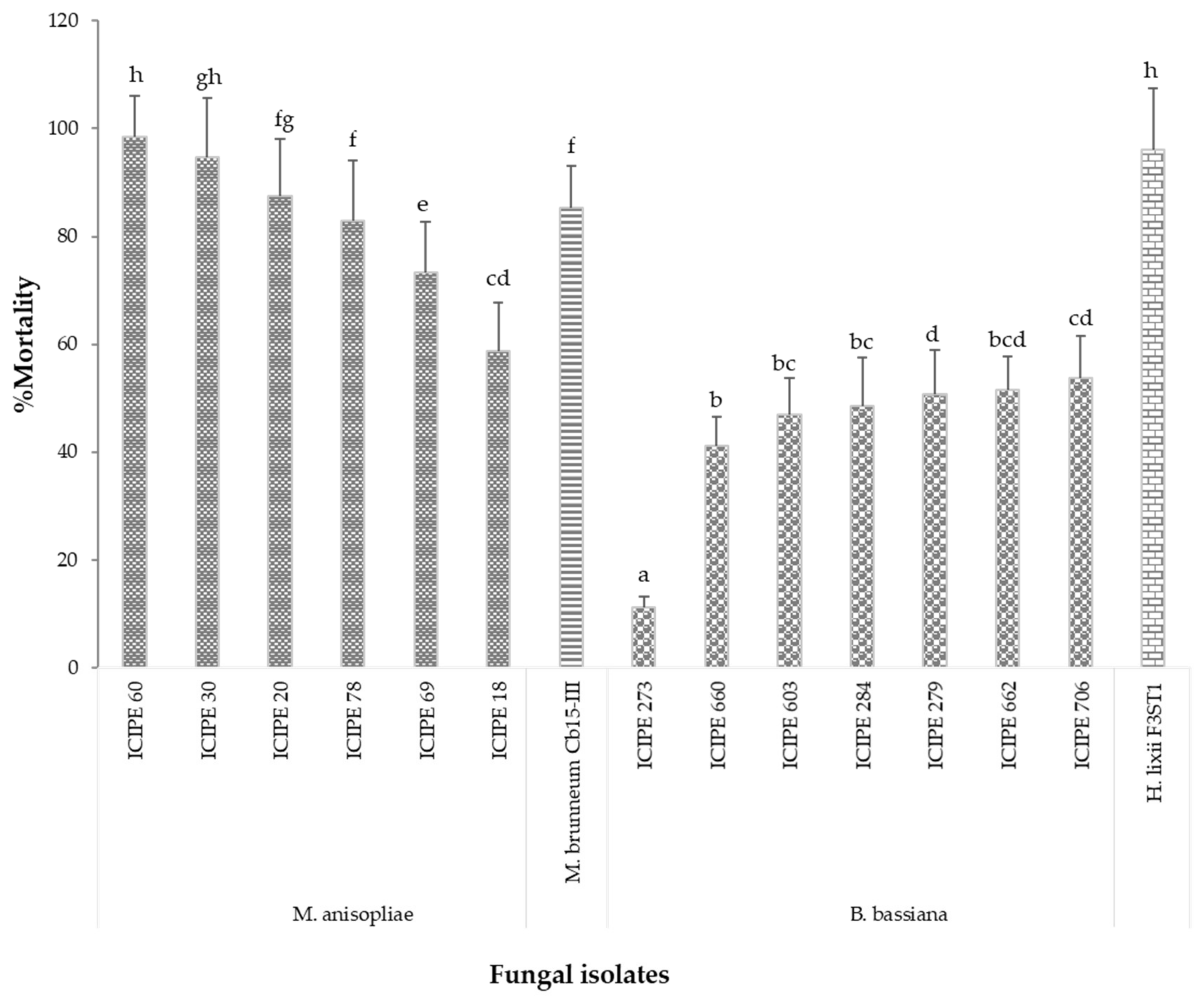
3.1. Pathogenicity of EPF Isolates against Termites

The screening for pathogenicity of the 15 isolates at the concentration of 1 × 10⁸ conidia/mL indicated that all the isolates were pathogenic to the termites, causing a significantly higher mortality level (100%) between 4 and 7 days. The calculated LT50 values for the isolates (Table 2) and the corrected percent mortality of the worker termites three days after inoculation (Figure 1) showed that, in general, the Metarhizium isolates elicited quicker mortality with lower LT50 (1.50–2.84 days) values than the Beauveria isolates (2.23–4.41 days).
The most virulent isolates with LT50 less than two days were M. brunneum Cb15-III, M. anisopliae isolates (ICIPE 60 and ICIPE 30) and Hypocrea lixii F3ST1, followed by isolates with LT50 between 2 and 3 days, and these included M. anisopliae isolates (ICIPE 20, ICIPE 18, ICIPE 78 and ICIPE 69) and B. bassiana isolates (ICIPE 279, ICIPE 284, ICIPE 706, ICIPE 662 and ICIPE 660), while B. bassiana isolates (ICIPE 603 and ICIPE 273) were the least virulent with LT50 of more than 3 days (Table 2). There were significant differences (F = 18.38, df = 14, P = 0.001) in the mycoses of cadavers among the isolates (Table 2).
Generally, the sign of penetration of conidia observed on the termite cuticle treated with Metarhizium isolates appeared faster than on those treated with Beauveria and Hypocrea isolates. After the penetration, the conidia emerged on the surfaces of the cadavers. Termites started to change in colour after few days of infection (became darker) and became more fragile when touched.

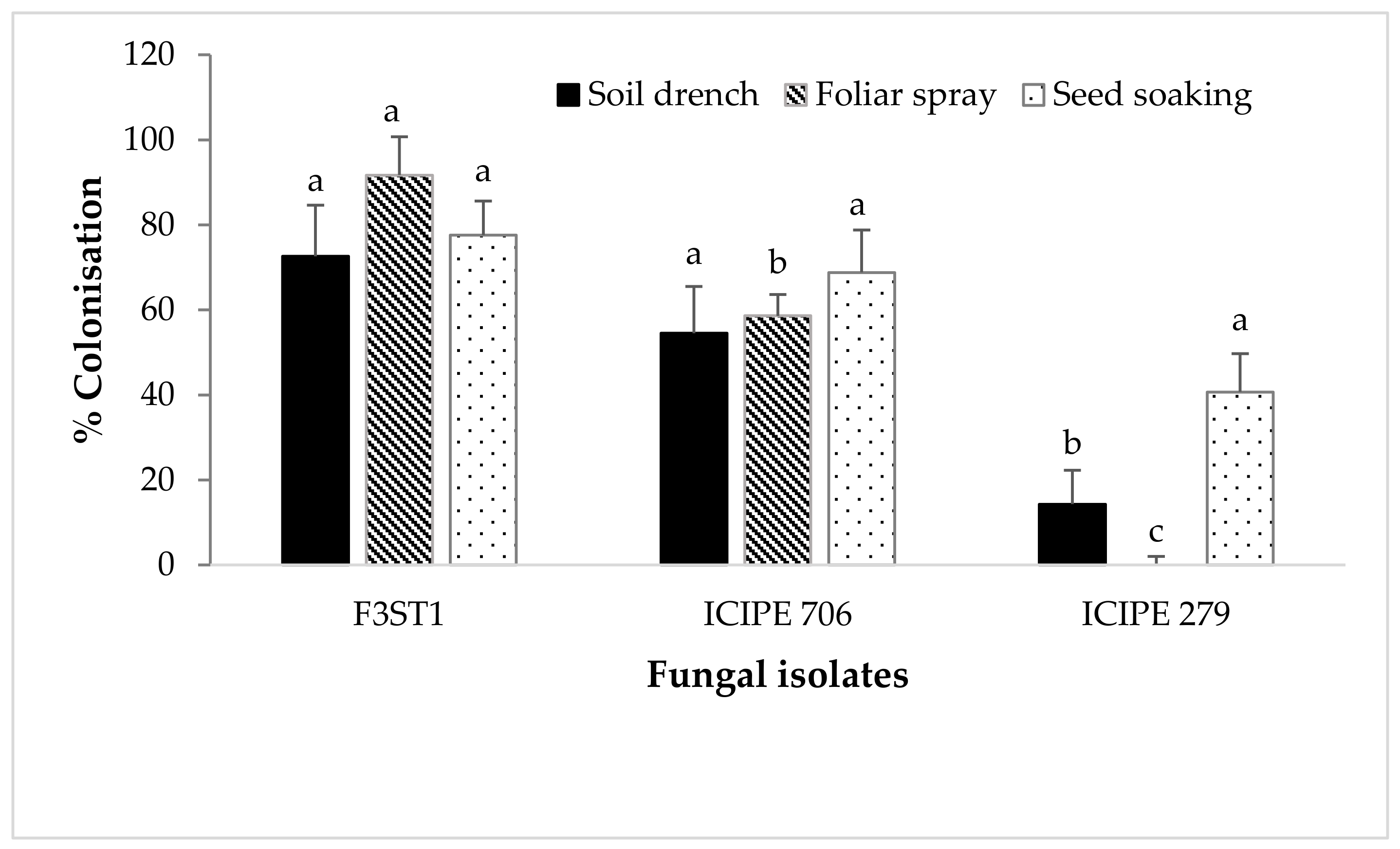
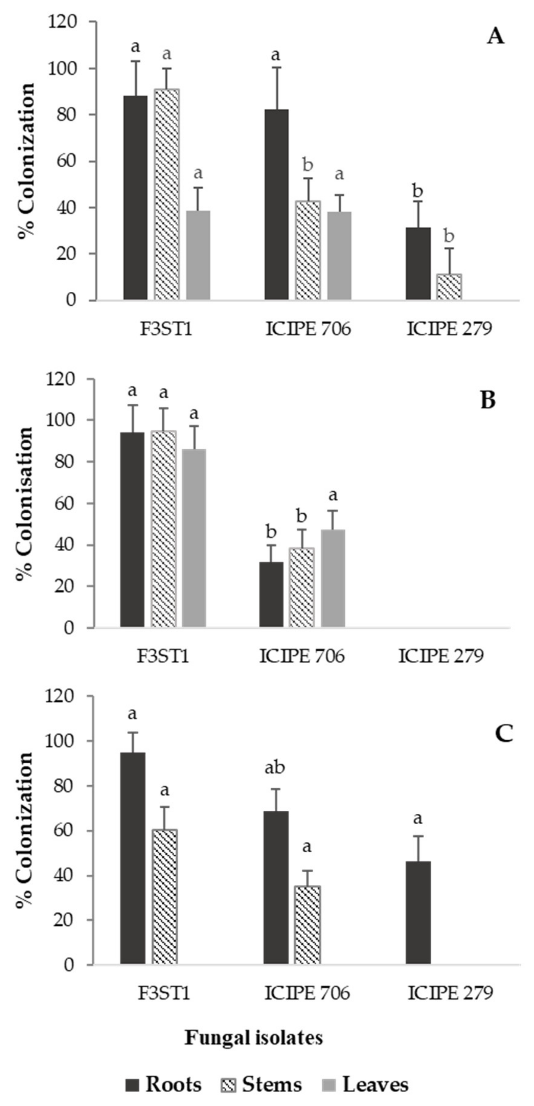
3.2. Endophytic Colonization of Cocoa Seedlings by Artificially Inoculated Fungal Isolates

For the endophytic colonization experiment, all the Metarhizium isolates tested did not colonize any of the cocoa seedling parts. However, only the B. bassiana isolates (ICIPE 706 and ICIPE 279) and H. lixii F3ST1 were able to successfully colonize the cocoa seedlings. The last rinse water used to rinse the tissues after sterilization, the imprinting plants and the sterilized soil yielded no microorganisms, thus confirming that the external surface of the plant parts was properly surface sterilized and that any ensuing fungal growth from surface-sterilized tissues was inferred to have originated from internal plant tissues (i.e., as endophytes). Significant variations were observed in the percentage colonization between the different fungal isolates with regard to the inoculation method used (Figure 2). For the seed soaking method, no significant differences (F2,17 = 3.58, P = 0.05) were observed in the colonization of the different plant parts by the various fungal isolates (Figure 2). However, significant differences were observed in the colonization of the different isolates when the cocoa seedlings were sprayed with the fungal inoculum (F2,19 = 4.9, P < 0.001), as well as with the soil drench method (F2,33 = 10.76, P = 0.0002) (Figure 2).
The endophytic colonization of the cocoa seedlings also varied significantly between the fungal isolates with regard to the plant parts and the inoculation methods. The interaction between the percent colonization of the different plant parts and the method of inoculation of conidia was significant (F4,97 = 4.382, P = 0.003). For instance, for the soil drench method (Figure 3A), H. lixii F3ST1 showed the highest percent colonization for roots (88.2%) and stems (90.8%) but lower colonization for leaves (38.7%), while ICIPE 706 showed the highest colonization for roots (82.5%) but lower percent colonization for the leaf (38.2%) and stem (42.8%). ICIPE 279 showed no colonization for leaf parts but the lowest colonization for the stem (11.2%) and roots (31.6%). Significant differences (F2,9 = 6.18, P = 0.02) were observed for the root colonization of the different isolates in the soil drench method, where more root parts were colonized by H. lixii F3ST1 and ICIPE 706 than ICIPE 279. More stem parts (F2,9 =11.1, P = 0.003) were also colonized by H. lixii F3ST1 than ICIPE 706 and 279. However, no significant differences (F2,9 = 2.17, P = 0.17) were observed for the colonization of the leaf parts. For the foliar spray method (Figure 3B), H. lixii F3ST1 showed high colonization of the roots (94.3%), stems (94.7%) and leaves (82.2%). ICIPE 706 exhibited higher colonization of the leaves (47.4%) than stems (38.2%) and roots (31.6%), while no colonization was observed for any of the plant tissues for ICIPE 279. For this method, more root parts (F2,9 = 4.87, P = 0.0005) were colonized by H. lixii F3ST1, as well as the stem parts (F2,9 = 2.54, P < 0.001) and leaves (F2,9 = 12.67, P = 0.002), than ICIPE 706, whereas no plant part was colonized by ICIPE 279. For the seed soaking method (Figure 3C), H. lixii F3ST1 showed high colonization for roots (94.7%) and stems (60.5%), and ICIPE 706 colonized the roots (68.7%) and stems (35.2%), while ICIPE 279 colonized only the roots (46.2%). No isolate colonized the leaves in the seed soaking method of inoculation (Figure 3C). Hypocrea lixii F3ST1 significantly colonized the roots (F2,19 = 4.5, P = 0.004) and stem (F2,9 = 5.2, P = 0.03) compared to ICIPE 706 and ICIPE 279.

3.3. Naturally Occurring Fungal Endophytes Isolated from the Cocoa Seedlings

In addition to the artificially inoculated isolates that successfully colonized the cocoa seedlings (B. bassiana isolates ICIPE 279 and 706 and H. lixii F3ST1), three additional fungal endophytes that naturally occurred and were identified using molecular techniques as Trichoderma asperellum, Fusarium redolens and F. solani (Table 3), were isolated from the leaves, stems and roots.

4. Discussion

All the fungal isolates screened were pathogenic to the termites, causing 100% mortality within 4–7 days post-inoculation. However, M. brunneum Cb15-III, M. anisopliae ICIPE 60 and ICIPE 30, and Hypocrea lixii F3ST1 were able to kill 50% of the exposed population in less than two days, showing high virulence to Odontotermes spp. The results obtained in this study corroborate those of other studies, which showed that many fungal isolates are virulent to termites [8]. For instance, Zoberi and Grace [48] showed that 100% mortality of Reticulitermes flavipes Kollar (Isoptera: Rhinotermitidae) workers was attained at 1–3 days after exposure to a strain of B. bassiana isolated from R. flavipes workers. Similarly, under laboratory conditions, a strain of M. anisopliae has been reported to cause 100% mortality to Odontotermes formosanus Shiraki (Isoptera: Termitidae) three days post-inoculation at the concentration of 3 × 10⁸ conidia/mL [49]. Hoe et al. [50] reported that isolates of M. anisopliae were pathogenic against a subterranean termite, Coptotermes curvignathus, causing 100% mortality at 1 × 10⁷ conidia/mL within three days post-inoculation. Kramm and West [7] also reported that 100% mortality occurred within one day after the exposure of termites to M. anisopliae, while similar mortality rates were obtained within five days in the case of B. bassiana. Following this high susceptibility of termites to EPF, researchers suspect that the rapid killing by EPF such as M. anisopliae of their hosts could be caused not only through the direct physical invasion of the hyphae but also possibly due to some enzymatic mechanisms or toxic metabolites produced by the EPF [33]. In addition, the soft nature of the termite cuticle could also contribute a lot to the rapid killing by EPF.
Although all the isolates screened were pathogenic, there was significant variation in their virulence levels. Our results showed that Metarhizium and Hypocrea isolates elicited high mortality with quicker killing speed and lower LT50 values than Beauveria isolates. The higher virulence effect of Metarhizium isolates than that of Beauveria isolates confirms earlier reports on similar patterns of activity with these fungal pathogen species [7,13,51,52,53,54,55]. Metarhizium brunneum Cb15-III, M. anisopliae ICIPE 30 and ICIPE 60, and H. lixii F3ST1 were the most virulent against the tested termite species. A previous study has reported the virulence of M. brunneum Cb15-III encapsulated with CO2 for the control of subterranean termite pests [32]. Other studies have also reported that the Metarhizium genus, especially M. anisopliae and M. brunneum, is one of the EPF genera that has high potential for controlling subterranean termites [56,57]. Research conducted by Milner et al. [58] also revealed M. anisopliae to be the most effective fungal pathogen for termite control. However, highly virulent isolates may not be always ideal candidates for biological control programs, especially in the case of social insects such as termites, because other reports have shown that the conidia of highly virulent strains of EPF are very repellent to termites [14,59,60]. Therefore, it is very important to assess the systemic use of these virulent EPF through the artificial inoculation of the host plant to confer resistance against the subterranean termites.
Our results show that only B. bassiana isolates ICIPE 279 and 706, and H. lixii F3ST1 could successfully colonize and establish in cocoa seedlings as endophytes without causing any visual/visible symptoms or damage to the seedlings like in the control treatments. Beauveria bassiana has also been artificially introduced into many other plants such as the common bean [61,62], maize [21], cocoa [26], date palms [63], coffee [64], bananas [22], radiata pine [65], faba beans [66], opium poppies [67], cotton [68,69], tomatoes [69] and grapevine leaves [70]. Similarly, H. lixii F3ST1 has been shown to endophytically colonize the common bean [62,71], onion [72] and faba bean [71,73]. This suggests that these entomopathogens have the potential to also endophytically colonize several host plant species without causing any visual detrimental effects on the host plants. This observation corroborates with the findings of Posada and Vega [26], who also did not observe any visual symptoms on cocoa seedlings colonized by B. bassiana and, therefore, concluded that B. bassiana can colonize cocoa seedlings without causing any detriment to the hosts. Additionally, it is also reported that entomopathogenic fungi can colonize different plant tissues as symptomless endophytes [27]. None of the Metarhizium isolates colonized the cocoa seedlings in this study. Akutse et al. [71] and Mutune et al. [62] also reported a lack of colonization and establishment of M. anisopliae isolates in V. faba or P. vulgaris cultivars through seed inoculation. Metarhizium species are less well known as endophytes [74], even though some strains have been successfully introduced into tomatoes [75], fava beans [66], oilseed rape [76] and haricot beans [77,78]. Two unidentified Metarhizium species and M. anisopliae have also been found to act as endophytes by naturally colonizing the roots of wall barley (Hordeum murinum L.) [79].
Our soil drench inoculation method resulted mostly in the colonization of roots and stems compared to the leaves, while the foliar spray inoculation method resulted mostly in the colonization of the leaves and stems as compared to the roots. Some studies have shown that colonization by the applied EPF is more likely in the plant part that was in direct contact with the inoculum and less likely in plant parts distant from the application site [80,81]. The variation in the colonization rates among the tested fungal strains, as shown in this study, may be due to the differential growth rate and endophytic abilities of each fungal isolate as associated with different host plant species or cultivars [71,82]. For example, M. brunneum Cb15-III, which did not colonize the cocoa seedling in this study, had been shown to be an efficient endophyte in wheat (Triticum aestivum L.) [70] and faba beans [83].
Previous studies have reported the endophytic colonization of plants with B. bassiana for as long as eight months in coffee [25] and nine months in radiata pine [65]. It therefore remains to be tested whether the endophytic colonization of cocoa seedlings by B. bassiana isolates ICIPE 279 and 706, and H. lixii F3ST1 can persist for more than one month and confer protection of cocoa seedlings against termite damage in the field following transplantation. Plant colonization by B. bassiana has been reported to reduce the damage caused by many crop pests such as the lepidopteran cob and the stemborers Ostrinia nubilalis and Sesamia calamistis Hampson (Lepidoptera: Noctuidae) in maize [21,52]; the banana weevil, Cosmopolites sordidus Germar (Coleoptera: Curculionidae) in bananas [22]; the poppy stem gall wasp, Iraella luteipes Thompson (Hymenoptera: Cynipidae) in opium poppy [84]; and the stem weevil Apion corchori Marshall (Coleoptera: Curculionidae) in white jute [82]. Adverse effects against insect pests have also been reported as a result of plant colonization with other fungal endophytes, such as the black bean aphid, Aphis fabae Scopoli (Hemiptera: Aphididae), and the pea leafminer, Liriomyza huidobrensis Blanchard (Diptera: Agromyzidae), with H. lixii in broad beans [71,80]; the cotton aphid, Aphis gossypii Glover (Hemiptera: Aphididae), with B. bassiana or Lecanicillium lecanii in cotton [68]; onion thrips, Thrips tabaci Lindeman (Thysanoptera: Thripidae), with Clonostachys rosea, H. lixii, Trichoderma harzianum, T. asperellum, T. atroviride and Fusarium sp. in onions [85]; and the bean stem maggot, Ophiomyia phaseoli Tryon (Diptera: Agromyzidae), with H. lixii, T. asperellum and T. atroviride, in beans [62]. Future studies are therefore warranted to assess the fate of the B. bassiana isolates ICIPE 279 and 706 and H. lixii F3ST1, which successfully colonized the cocoa seedlings, and the ability of these endophytes to confer systemic resistance against termite pests from the laboratory to in the field.
Trichoderma asperellum, Fusarium redolens and F. solani were also isolated from the cocoa seedling parts as endophytes. Trichoderma species are typically considered soilborne organisms associated with many plant parts [86]. Other researchers have also isolated Trichoderma and Fusarium spp. from live sapwood immediately below the bark of trunks of wild and cultivated Theobroma cacao and other Theobroma species [24]. These fungi therefore appear to be cocoa-associated endophytes possibly transmitted through the seeds. The pathogenicity of these fungal endophytes isolated from cocoa seedlings for termites and other cocoa pests also remains important for investigation.

5. Conclusions

This study suggests that M. brunneum Cb15-III and M. anisopliae isolates ICIPE 60 and ICIPE 30 have the potential to be developed as microbial termiticides for controlling subterranean termite pests in cocoa agroforests. The recovery of B. bassiana isolates ICIPE 706 and 279 and H. lixii F3ST1 from cocoa tissues after artificial inoculation through seed soaking, soil drench and foliar spray indicates that these fungi could establish as cocoa endophytes when inoculated. In addition, the lack of any visual symptoms (retarded growth, discoloration, lesions, etc.) on the seedlings also indicate that fungal isolates can colonize cocoa plants without causing any detrimental effects on the plant. Future studies are warranted to determine the growth parameters of the endophytically colonized seedlings, the fate of these isolates in cocoa seedlings transplanted in the field and their ability to protect the plant against subterranean termite pest damage in cocoa plantations.

Author Contributions

S.E., H.D.B.B. and K.S.A. conceptualized the research idea. C.F.A. conducted experiments, analyzed the data and drafted the manuscript. K.S.A., F.M.K. and S.E. provided the fungal isolates, reagent materials, analytical tools and facilities. C.F.A., H.D.B.B., O.O.B., F.M.K, C.T.L.D. and K.S.A. wrote, reviewed and edited the manuscript. S.E., O.O.B., H.D.B.B. and K.S.A. supervised. C.F.A. and H.D.B.B., K.S.A. and S.E. secured funding. All authors have read and agreed to the published version of the manuscript.

Funding

This research is part of the fellowship project (VW-60420894) of B.D.H.B. funded by the Volkswagen Foundation, under the Funding Initiative Knowledge for Tomorrow—Cooperative Research Projects in Sub-Saharan Africa on Resources, their Dynamics, and Sustainability-Capacity Development in Comparative and Integrated Approaches.

Acknowledgments

The first author received a scholarship from the German Academic Exchange Service (DAAD) through the African Regional Postgraduate Programme in Insect Science (ARPPIS) of icipe. The authors gratefully acknowledge the icipe core funding provided by European Union (EU), the UK’s Department for International Development (DFID); the Swedish International Development Cooperation Agency (Sida); the Swiss Agency for Development and Cooperation (SDC); the Federal Democratic Republic of Ethiopia; and the Kenyan Government. The authors are also grateful to Fidelis L. Ombura and S. Wafula for the technical assistance. The views expressed herein do not necessarily reflect the official opinion of the donors.

Conflicts of Interest

The authors declare that there is no conflict of interest. The funders had no role in the design of the study; in the collection, analyses or interpretation of data; in the writing of the manuscript; or in the decision to publish the results.

References

  1. Tra Bi, C.S.; Soro, S.; Yéboué-Nguessan, L.; Tano, Y.; Konaté, S. Termites impact on different age of cocoa (Theobroma cacao L.) plantations with different fertilizer treatments in semi-deciduous forest zone (Oume, Ivory Coast). HJAFSR 2015, 4, 021–027. [Google Scholar]
  2. Ambele, F.C.; Bisseleua, D.H.B.; Babalola, O.O.; Ekesi, S. Soil-dwelling insect pests of tree crops in Sub-Saharan Africa, problems and management strategies—A review. J. Appl. Entomol. 2018, 142, 539–552. [Google Scholar] [CrossRef]
  3. Ambele, F.C.; Hervé, B.D.; Ekesi, S.; Akutse, K.S.; Djuideu, C.T.; Meupia, M.J.; Babalola, O.O. Consequences of shade management on the taxonomic patterns and functional diversity of termites (Blattodea: Termitidae) in cocoa agroforestry systems. Evol. Ecol. 2018, 8, 11582–11595. [Google Scholar]
  4. Djuideu, T.C.L.; Bisseleua, D.H.B.; Kekeunou, S.; Meupia, M.J.; Difouo, F.G.; Ambele, C.F. Plant community composition and functional characteristics define invasion and infestation of termites in cocoa agroforestry systems. Agrofor. Syst. 2020, 94, 185–201. [Google Scholar] [CrossRef]
  5. Su, N.Y.; Scheffrahn, R.H. A review of subterranean termite control practices and prospects for integrated pest management programmes. Integr. Pest Manag. Rev. 1998, 3, 1–13. [Google Scholar] [CrossRef]
  6. Grace, J.K. Biological control strategies for suppression of termites. J. Agric. Entomol. 1997, 14, 281–289. [Google Scholar]
  7. Kramm, K.R.; West, D.F. Termite pathogens: Effects of ingested Metarhizium, Beauveria, and Gliocladium conidia on worker termites (Reticulitermes sp.). J. Invertebr. Pathol. 1982, 40, 7–11. [Google Scholar] [CrossRef]
  8. Rath, A.C. The use of entomopathogenic fungi for control of termites. Biocontrol Sci. Technol. 2000, 10, 563–581. [Google Scholar] [CrossRef]
  9. Smythe, R.V.; Coppel, H.C. The susceptibility of Reticulitermes flavipes (Kollar) and other termite species to an experimental preparation of Bacillus thuringiensis Berliner. J. Invertebr. Pathol. 1965, 7, 423–426. [Google Scholar] [CrossRef]
  10. Yendol, W.G.; Paschke, J.D. Pathology of an Entomophthora infection in the eastern subterranean termite Reticulitermes flavipes (Kollar). J. Invertebr. Pathol. 1965, 7, 414–422. [Google Scholar] [CrossRef]
  11. Culliney, T.W.; Grace, J.K. Prospects for the biological control of subterranean termites (Isoptera: Rhinotermitidae), with special reference to Coptotermes formosanus. Bull. Entomol. Res. 2000, 90, 9–21. [Google Scholar] [CrossRef] [PubMed]
  12. Chouvenc, T.; Su, N.Y.; Grace, J.K. Fifty years of attempted biological control of termites-Analysis of a failure. Biol. Control 2011, 59, 69–82. [Google Scholar] [CrossRef]
  13. Yanagawa, A.; Yokohari, F.; Shimizu, S. Defense mechanism of the termite, Coptotermes formosanus Shiraki, to entomopathogenic fungi. J. Invertebr. Pathol. 2008, 97, 165–170. [Google Scholar] [CrossRef] [PubMed]
  14. Mburu, D.M.; Ochola, L.; Maniania, N.K.; Njagi, P.G.N.; Gitonga, L.M.; Ndung’u, M.W.; Hassanali, A. Relationship between virulence and repellency of entomopathogenic isolates of Metarhizium anisopliae and Beauveria bassiana to the termite Macrotermes michaelseni. J. Insect Physiol. 2009, 55, 774–780. [Google Scholar] [CrossRef] [PubMed]
  15. Vega, F.E. Insect pathology and fungal endophytes. J. Invertebr. Pathol. 2008, 98, 277–279. [Google Scholar] [CrossRef]
  16. Grace, J.K. Approaches to biological control of termites. Sociobiology 2003, 41, 115–121. [Google Scholar]
  17. Schulz, B.; Boyle, C. The endophytic continuum. Mycol. Res. 2005, 109, 661–686. [Google Scholar] [CrossRef] [Green Version]
  18. Sun, J.Z.; Fuxa, J.R.; Richter, A.; Ring, D. Interactions of Metarhizium anisoplae and tree-based mulches in repellence and mycoses against Coptotermes formosanus (Isoptera: Rhinotermitidae). Environ. Entomol. 2014, 37, 755–763. [Google Scholar] [CrossRef]
  19. Maniania, N.K.; Ekesi, S.; Songa, J.M. Managing termites in maize with the entomopathogenic fungus Metarhizium anisopliae. Int. J. Trop. Insect Sci. 2002, 22, 41–46. [Google Scholar] [CrossRef] [Green Version]
  20. Arnold, A.E.; Lewis, L.C. Ecology and Evolution of Fungal Endophytes, and Their Roles Against Insects. Insect-Fungal Associations: Ecology and Evolution; Oxford University Press: New York, NY, USA, 2005; pp. 74–96. [Google Scholar]
  21. Bing, L.A.; Lewis, L.C. Suppression of Ostrinia nubilalis (Hübner) (Lepidoptera: Pyralidae) by endophytic Beauveria bassiana (Balsamo) Vuillemin. Environ Entomol. 1991, 20, 1207–1211. [Google Scholar] [CrossRef]
  22. Akello, J.; Dubois, T.; Coyne, D.; Kyamanywa, S. Endophytic Beauveria bassiana in banana (Musa spp.) reduces banana weevil (Cosmopolites sordidus) fitness and damage. Crop Prot. 2008, 27, 1437–1441. [Google Scholar] [CrossRef]
  23. Leckie, B.M. Effects of Beauveria bassiana Mycelia and Metabolites Incorporated into Synthetic Diet and Fed to Larval Helicoverpa zea; and Detection of Endophytic Beauveria bassiana in Tomato Plants Using PCR and ITS Primers. Master’s Thesis, Department of Entomology, The University of Tennessee, Knoxville, TN, USA, 2002. [Google Scholar]
  24. Evans, H.C.; Holmes, K.A.; Thomas, S.E. Endophytes and mycoparasites associated with an indigenous forest tree, Theobroma gileri, in Ecuador and a preliminary assessment of their potential as biocontrol agents of cocoa diseases. Mycol. Prog. 2003, 2, 149–160. [Google Scholar] [CrossRef]
  25. Posada, F.; Aime, M.C.; Peterson, S.W.; Rehner, S.A.; Vega, F.E. Inoculation of coffee plants with the fungal entomopathogen Beauveria bassiana (Ascomycota: Hypocreales). Mycol. Res. 2007, 111, 748–757. [Google Scholar] [CrossRef] [PubMed] [Green Version]
  26. Posada, F.; Vega, F.E. Establishment of the fungal entomopathogen Beauveria bassiana (Ascomycota: Hypocreales) as an endophyte in cocoa seedlings (Theobroma cacao). Mycologia 2005, 97, 1195–1200. [Google Scholar] [CrossRef] [PubMed]
  27. Vidal, S.; Jaber, L.R. Entomopathogenic fungi as endophytes: Plant–endophyte–herbivore interactions and prospects for use in biological control. Curr. Sci. 2015, 109, 46–54. [Google Scholar]
  28. Vega, F.E.; Goettel, M.S.; Blackwell, M.; Chandler, D.; Jackson, M.A.; Keller, S.; Pell, J.K. Fungal entomopathogens: New insights on their ecology. Fungal Ecol. 2009, 2, 149–159. [Google Scholar] [CrossRef] [Green Version]
  29. Wemheuer, F.; Berkelmann, D.; Wemheuer, B.; Daniel, R.; Vidal, S.; Bisseleua, D.H.B. Agroforestry Management Systems Drive the Composition, Diversity, and Function of Fungal and Bacterial Endophyte Communities in Theobroma cacao Leaves. Microorganisms 2020, 8, 405. [Google Scholar] [CrossRef] [Green Version]
  30. Goettel, M.S.; Inglis, G.D. Fungi: Hyphomycetes. In Manual of Techniques in Insect Pathology; Elsevier: Amsterdam, The Netherlands, 1997; pp. 213–249. [Google Scholar]
  31. Inglis, G.D.; Enkerli, J.; Goettel, M.S. Laboratory techniques used for entomopathogenic fungi: Hypocreales. In Manual of Techniques in Invertebrate Pathology, 2nd ed.; Springer: Berlin, Germany, 2012; pp. 189–253. [Google Scholar]
  32. Ambele, C.F.; Bisseleua, H.D.; Akutse, K.S.; Babalola, O.O.; Humbert, P.; Patel, A.; Ekesi, S. Testing a co-formulation of CO2-releasing material with an entomopathogenic fungus for the management of subterranean termite pests. Mycol. Prog. 2019, 18, 1201–1211. [Google Scholar] [CrossRef]
  33. Sun, J.; Fuxa, J.R.; Henderson, G. Effects of virulence, sporulation, and temperature on Metarhizium anisopliae and Beauveria bassiana laboratory transmission in Coptotermes formosanus. J. Invertebr. Pathol. 2003, 84, 38–46. [Google Scholar] [CrossRef]
  34. Humber, R.A. Fungi: Identification. In Manual of Techniques in Insect Pathology; Lacey, L., Ed.; Academic Press: Washington, DC, USA, 1997; pp. 153–185. [Google Scholar]
  35. Humber, R.A. Identification of entomopathogenic fungi. In Manual of Techniques in Invertebrate Pathology, 2nd ed.; Lacey, L.A., Ed.; Academic Press: San Diego, CA, USA, 2012. [Google Scholar]
  36. Akutse, K.S.; Van den Berg, J.; Maniania, N.K.; Ekesi, S.; Fiaboe, K.K.M. Morphological and molecular characterization of Vicia faba and Phaseolus vulgaris seed-born fungal endophytes. Res. J. Seed Sci. 2017, 10, 1–16. [Google Scholar] [CrossRef] [Green Version]
  37. Burgess, L.W.; Liddell, C.M.; Summerell, B.A. Laboratory Manual for Fusarium Research: Incorporating a Key and Descriptions of Common Species Found in Australasia; FAO: Rome, Italy, 1988. [Google Scholar]
  38. White, T.J.; Bruns, T.; Lee, S.; Taylor, J.W. Amplification and Direct Sequencing of Fungal Ribosomal RNA Genes for Phylogenetics. PCR Protocols: A Guide to Methods and Applications; Academic Press, Inc.: New York, NY, USA, 1990; pp. 315–322. [Google Scholar]
  39. Hall, T.A. BioEdit: A user-friendly biological sequence alignment editor and analysis program for Windows 95/98/NT. Nucleic Acids Symp. Ser. 1999, 41, 95–98. [Google Scholar]
  40. Thompson, J.D.; Gibson, T.J.; Plewniak, F.; Jeanmougin, F.; Higgins, D.G. The CLUSTAL_X windows interface: Flexible strategies for multiple sequence alignment aided by quality analysis tools. Nucleic Acids Res. 1997, 25, 4876–4882. [Google Scholar] [CrossRef] [PubMed] [Green Version]
  41. Liu, D.; Coloe, S.; Baird, R.; Pedersen, J. Application of PCR to the identification of dermatophyte fungi. J. Med. Microbiol. 2000, 49, 493–497. [Google Scholar] [CrossRef] [PubMed] [Green Version]
  42. Landeweert, R.; Leeflang, P.; Kuyper, T.W.; Hoffland, E.; Rosling, A.; Wernars, K.; Smit, E. Molecular identification of ectomycorrhizal mycelium in soil horizons. Appl. Environ. Microbiol. 2003, 69, 327–333. [Google Scholar] [CrossRef] [PubMed] [Green Version]
  43. Javadi, M.A.; Ghanbary, M.A.T.; Tazick, Z. Isolation and molecular identification of soil inhabitant Penicillia. Ann. Biol. Res. 2012, 3, 5758–5761. [Google Scholar]
  44. Abbott, W.S. A method of computing the effectiveness of an insecticide. J. Econ. Entomol. 1925, 18, 265–267. [Google Scholar] [CrossRef]
  45. McCullagh, P.; Nelder, J. Generalized Linear Models, 2nd ed.; Chapman and Hall: London, UK, 1989. [Google Scholar] [CrossRef]
  46. Fisher, P.J.; Petrini, O. Location of fungal endophytes in tissues of Suaeda fruticosa: A preliminary study. Trans. Br. Mycol. Soc. 1987, 89, 246–249. [Google Scholar] [CrossRef]
  47. R Core Team R: A Language and Environment for Statistical Computing. R Foundation for Statistical Computing, Vienna, Austria. 2019. Available online: https://www.R-project.org/ (accessed on 23 June 2019).
  48. Zoberi, M.H.; Grace, J.K. Fungi associated with the subterranean termite Reticulitermes flavipes in Ontario. Mycologia 1990, 82, 289–294. [Google Scholar] [CrossRef]
  49. Dong, C.; Zhang, J.; Chen, W.; Huang, H.; Hu, Y. Characterization of a newly discovered China variety of Metarhizium anisopliae (M. anisopliae var. dcjhyium) for virulence to termites, isoenzyme, and phylogenic analysis. Microbiol. Res. 2007, 162, 53–61. [Google Scholar] [CrossRef]
  50. Hoe, P.; Bong, C.F.J.; Kadir, J.; Amartalingam, R. Evaluation of Metarhizium anisopliae var. anisopliae (Deuteromycotina: Hyphomycete) isolates and their effects on subterranean termite Coptotermes curvignathus (Isoptera: Rhinotermitidae). Am. J. Agric. Biol. Sci. 2009, 4, 289–297. [Google Scholar] [CrossRef] [Green Version]
  51. Wang, C.; Powell, J.E. Isolation and evaluation of Beauveria bassiana for control of Coptotermes formosanus and Reticulitermes flavipes (Isoptera: Rhinotermitidae). Sociobiology 2003, 41, 369–382. [Google Scholar]
  52. Cherry, A.J.; Abalo, P.; Hell, K. A laboratory assessment of the potential of different strains of the entomopathogenic fungi Beauveria bassiana (Balsamo) Vuillemin and Metarhizium anisopliae (Metschnikoff) to control Callosobruchus maculatus (F.) (Coleoptera: Bruchidae) in stored cowpea. J. Stored Prod. Res. 2005, 41, 295–309. [Google Scholar] [CrossRef]
  53. Singha, D.; Singha, B.; Dutta, B.K. Virulence of isolates of Metarhizium anisopliae (Metschnikoff) Sorokin and Beauveria bassiana (Ballsamo) Vuillemin on tea termite Microcerotermes beesoni Snyder (Isoptera: Termitidae) of Barak Valley of Assam. Indian J. Entomol. 2006, 68, 66–70. [Google Scholar]
  54. Singha, D.; Singha, B.; Dutta, B.K. Potential of Metarhizium anisopliae and Beauveria bassiana in the control of tea termite Microtermes obesi Holmgren in vitro and under field conditions. J. Pest Sci. 2011, 84, 69–75. [Google Scholar] [CrossRef]
  55. Sileshi, A.; Sori, W.; Dawd, M. Laboratory evaluation of entomopathogenic fungi Metarhizium anisopliae and Beauveria bassiana against Termite, Macrotermes (Isoptera: Termitidae). Asian J. Plant Sci. 2013, 12, 1–10. [Google Scholar]
  56. Denier, D.; Bulmer, M.S. Variation in subterranean termite susceptibility to fatal infections by local Metarhizium soil isolates. Insectes Soc. 2015, 62, 219–226. [Google Scholar] [CrossRef]
  57. Kuswanto, E.; Ahmad, I.; Dungani, R. Threat of subterranean termite attack in the Asian countries and their control: A Review. Asian J. Appl. Sci. 2015, 8, 227–239. [Google Scholar]
  58. Milner, R.J.; Staples, J.A.; Lenz, M. Options for termite management using the insect pathogenic fungus Metarhizium anisopliae. In Proceedings of the 27th Annual Meeting of the International Research Group on Wood Preservation, Guadaloupe, France, 19–24 May 1996. [Google Scholar]
  59. Staples, J.A.; Milner, R.J. A laboratory evaluation of the repellency of Metarhizium anisopliae conidia to Coptotermes lacteus (Isoptera: Rhinotermitidae). Sociobiology 2000, 36, 133–148. [Google Scholar]
  60. Myles, T.G. Alarm, aggregation, and defense by Reticulitermes flavipes in response to a naturally occurring isolate of Metarhizium anisopliae. Sociobiology 2002, 40, 243–256. [Google Scholar]
  61. Gathage, J.W.; Lagat, Z.O.; Fiaboe, K.K.M.; Akutse, K.S.; Ekesi, S.; Maniania, N.K. Prospects of fungal endophytes in the control of Liriomyza leafminer flies in common bean Phaseolus vulgaris under field conditions. Biocontrol 2016, 61, 741–753. [Google Scholar] [CrossRef]
  62. Mutune, B.; Ekesi, S.; Niassy, S.; Matiru, V.; Bii, C.; Maniania, N.K. Fungal endophytes as promising tools for the management of bean stem maggot Ophiomyia phaseoli on beans Phaseolus vulgaris. J. Pest Sci. 2016, 89, 993–1001. [Google Scholar] [CrossRef]
  63. Gómez-Vidal, S.; Lopez-Llorca, L.V.; Jansson, H.B.; Salinas, J. Endophytic colonization of date palm (Phoenix dactylifera L.) leaves by entomopathogenic fungi. Micron 2006, 37, 624–632. [Google Scholar] [CrossRef] [PubMed]
  64. Posada, F.; Vega, F.E. Inoculation and colonization of coffee seedlings (Coffea arabica L.) with the fungal entomopathogen Beauveria bassiana (Ascomycota: Hypocreales). Mycoscience 2006, 47, 284–289. [Google Scholar] [CrossRef]
  65. Brownbridge, M.; Reay, S.D.; Nelson, T.L.; Glare, T.R. Persistence of Beauveria bassiana (Ascomycota: Hypocreales) as an endophyte following inoculation of radiata pine seed and seedlings. Biol. Control 2012, 61, 194–200. [Google Scholar] [CrossRef]
  66. Akello, J.; Sikora, R. Systemic acropedal influence of endophyte seed treatment on Acyrthosiphon pisum and Aphis fabae offspring development and reproductive fitness. Biol. Control 2012, 61, 215–221. [Google Scholar] [CrossRef]
  67. Quesada-Moraga, E.; López-Díaz, C.; Landa, B.B. The hidden habit of the entomopathogenic fungus Beauveria bassiana: First demonstration of vertical plant transmission. PLoS ONE 2014, 9, e89278. [Google Scholar] [CrossRef] [Green Version]
  68. Gurulingappa, P.; Sword, G.A.; Murdoch, G.; McGee, P.A. Colonization of crop plants by fungal entomopathogens and their effects on two insect pests when in planta. Biol. Control 2010, 55, 34–41. [Google Scholar] [CrossRef]
  69. Ownley, B.H.; Griffin, M.R.; Klingeman, W.E.; Gwinn, K.D.; Moulton, J.K.; Pereira, R.M. Beauveria bassiana: Endophytic colonization and plant disease control. J. Invertebr. Pathol. 2008, 98, 267–270. [Google Scholar] [CrossRef]
  70. Jaber, L.R. Grapevine leaf tissue colonization by the fungal entomopathogen Beauveria bassiana s. l. and its effect against downy mildew. Biocontrol 2015, 60, 103–112. [Google Scholar] [CrossRef]
  71. Akutse, K.S.; Maniania, N.K.; Fiaboe, K.K.M.; Van den Berg, J.; Ekesi, S. Endophytic colonization of Vicia faba and Phaseolus vulgaris (Fabaceae) by fungal pathogens and their effects on the life-history parameters of Liriomyza huidobrensis (Diptera: Agromyzidae). Fungal Ecol. 2013, 6, 293–301. [Google Scholar] [CrossRef]
  72. Muvea, A.M.; Meyhöfer, R.; Maniania, N.K.; Poehling, H.M.; Ekesi, S.; Subramanian, S. Behavioral responses of Thrips tabaci Lindeman to endophyte-inoculated onion plants. J. Pest Sci. 2015, 88, 555–562. [Google Scholar] [CrossRef]
  73. Akutse, K.S.; Fiaboe, K.K.; Van den Berg, J.; Ekesi, S.; Maniania, N.K. Effects of endophyte colonization of Vicia faba (Fabaceae) plants on the life–history of leafminer parasitoids Phaedrotoma scabriventris (Hymenoptera: Braconidae) and Diglyphus isaea (Hymenoptera: Eulophidae). PLoS ONE 2014, 9, e109965. [Google Scholar] [CrossRef] [PubMed]
  74. Greenfield, M.; Gómez-Jiménez, M.I.; Ortiz, V.; Vega, F.E.; Kramer, M.; Parsa, S. Beauveria bassiana and Metarhizium anisopliae endophytically colonize cassava roots following soil drench inoculation. Biol. Control 2016, 95, 40–48. [Google Scholar] [CrossRef] [PubMed] [Green Version]
  75. Elena, G.J.; Beatriz, P.J.; Alejandro, P.; Lecuona, R.E. Metarhizium anisopliae (Metschnikoff) Sorokin promotes growth and has endophytic activity in tomato plants. Adv. Biol. Res. 2011, 5, 22–27. [Google Scholar]
  76. Batta, Y.A. Efficacy of endophytic and applied Metarhizium anisopliae (Metch.) Sorokin (Ascomycota: Hypocreales) against larvae of Plutella xylostella L. (Yponomeutidae: Lepidoptera) infesting Brassica napus plants. Crop Prot. 2013, 44, 128–134. [Google Scholar] [CrossRef]
  77. Behie, S.W.; Jones, S.J.; Bidochka, M.J. Plant tissue localization of the endophytic insect pathogenic fungi Metarhizium and Beauveria. Fungal Ecol. 2015, 13, 112–119. [Google Scholar] [CrossRef]
  78. Sasan, R.K.; Bidochka, M.J. The insect-pathogenic fungus Metarhizium robertsii (Clavicipitaceae) is also an endophyte that stimulates plant root development. Am. J. Bot. 2012, 99, 101–107. [Google Scholar] [CrossRef] [PubMed]
  79. Murphy, B.R.; Martin Nieto, L.; Doohan, F.M.; Hodkinson, T.R. Profundae diversitas: The uncharted genetic diversity in a newly studied group of fungal root endophytes. Mycology 2015, 6, 139–150. [Google Scholar] [CrossRef] [Green Version]
  80. Akello, J.; Dubois, T.; Gold, C.S.; Coyne, D.; Nakavuma, J.; Paparu, P. Beauveria bassiana (Balsamo) Vuillemin as an endophyte in tissue culture banana (Musa spp.). J. Invertebr. Pathol. 2007, 96, 34–42. [Google Scholar] [CrossRef]
  81. Tefera, T.; Vidal, S. Effect of inoculation method and plant growth medium on endophytic colonization of sorghum by the entomopathogenic fungus Beauveria bassiana. BioControl 2009, 54, 663–669. [Google Scholar] [CrossRef] [Green Version]
  82. Biswas, C.; Dey, P.; Satpathy, S.; Satya, P.; Mahapatra, B.S. Endophytic colonization of white jute (Corchorus capsularis) plants by different Beauveria bassiana strains for managing stem weevil (Apion corchori). Phytoparasitica 2013, 41, 17–21. [Google Scholar] [CrossRef]
  83. Jaber, L.R.; Enkerli, J. Effect of seed treatment duration on growth and colonization of Vicia faba by endophytic Beauveria bassiana and Metarhizium brunneum. Biol. Control 2016, 103, 187–195. [Google Scholar] [CrossRef]
  84. Quesada-Moraga, E.; Munoz-Ledesma, F.J.; Santiago-Alvarez, C. Systemic protection of Papaver somniferum L. against Iraella luteipes (Hymenoptera: Cynipidae) by an endophytic strain of Beauveria bassiana (Ascomycota: Hypocreales). Environ. Entomol. 2009, 38, 723–730. [Google Scholar] [CrossRef] [Green Version]
  85. Muvea, A.M.; Meyhöfer, R.; Subramanian, S.; Poehling, H.M.; Ekesi, S.; Maniania, N.K. Colonization of onions by endophytic fungi and their impacts on the biology of Thrips tabaci. PLoS ONE 2014, 9, e108242. [Google Scholar] [CrossRef] [Green Version]
  86. Harman, G.E.; Howell, C.R.; Viterbo, A.; Chet, I.; Lorito, M. Trichoderma species-opportunistic, avirulent plant symbionts. Nat. Rev. Microbiol. 2004, 2, 43–56. [Google Scholar] [CrossRef]
Figure 1. Mean percent mortality after 3 days post-inoculation of worker caste of Odontotermes spp. with fungal isolates of Metarhizium anisopliae, M. brunneum, Beauveria bassiana and Hypocrea lixii. Bars with different letters are significantly different (Tukey’s HSD, P ˂ 0.05).
Jof 06 00126 g001
Figure 2. Mean (±SE) percentage of colonization of cocoa seedlings by different fungal pathogens (Beauveria bassiana ICIPE 279 and 706 and Hypocrea lixii F3ST1), 2 months post-inoculation using different inoculation methods (soil drench, seed soaking and foliar spray). Bars with different letters differ significantly at P = 0.05 (SNK test after ANOVA).
Jof 06 00126 g002
Figure 3. Mean (±SE) percentage of colonization of different cocoa seedling parts (root, stem and leaves) by fungal pathogens, Beauveria bassiana ICIPE 279 and 706 and Hypocrea lixii F3ST1, 2 months post-inoculation using different inoculation methods; (A) = soil drench, (B) = foliar spray and (C) = seed soaking. Bars with different letters differ significantly at P = 0.05 (SNK test after ANOVA).
Jof 06 00126 g003
Table 1. Identity and % germination of fungal isolates screened against the termite Odontotermes spp. for virulence under laboratory conditions.
Fungal SpeciesIsolatesSourceLocality/CountryYear of Isolation% Germination
Metarhizium anisopliaeICIPE 20SoilMigori (Kenya)198998.25 ± 0.47 bcd
ICIPE 30Busseola fuscaKendubay (Kenya)198998.06 ± 0.47 cde
ICIPE 60SoilKakelo-Seme (Kenya)199099.10 ± 0.29 abc
ICIPE 18SoilMbita (Kenya)198999.77 ± 0.01 a
ICIPE 69SoilMatete (DRC)199099.34 ± 0.28 ab
ICIPE 78Temnoschoita quadripustulataUngoe (Kenya)199099.09 ± 0.03 abc
Metarhizium brunneumCb15-III-Germany-95.25 ± 0.48 f
Beauveria bassiana
ICIPE 273SoilMbita (Kenya)200696.96 ± 0.27 e
ICIPE 279Coleopteran larvaKericho (Kenya)200598.25 ± 0.85 bcd
ICIPE 284Coleopteran larvaMauritus200599.22 ± 0.38 ab
ICIPE 603HymenopteraTaita (Kenya)200797.26 ± 0.48 de
ICIPE 706MonocotsKenya200899.51 ± 0.01 a
ICIPE 660SoilChemokock (Kenya)200898.70 ± 0.06 abc
ICIPE 662SoilMariakani (Kenya)200899.33 ± 0.14 ab
Hypocrea lixiiF3ST1MaizeKenya200995.25 ± 0.45 f
Means within a column followed by the same letter are not significantly different according to Student–Newman–Keuls (SKN) tests (P ˂ 0.05).
Table 2. Median lethal time (LT50) 7 days after treatment and % mycosis of worker termites of Odontotermes spp. inoculated with different isolates of entomopathogenic fungi.
Fungal SpeciesIsolatesLT50 (Days) (95% FL)% Mycosis of Termite Cadavers
Beauveria bassianaICIPE 2792.23 (2.19–2.27)66.25 ± 5.91 a
ICIPE 6033.09 (3.04–3.14)47.50 ± 5.20 e
ICIPE 2843.00 (2.96–3.04)61.25 ± 9.44 ab
ICIPE 7062.85 (2.82–2.88)61.25 ± 4.27 ab
ICIPE 6622.85 (2.82–2.88)70.00 ± 4.56 a
ICIPE 6603.00 (2.96–3.04)62.50 ± 3.23 ab
ICIPE 2734.41 (4.37–4.45)46.25 ± 5.54 bcd
Metarhizium anisopliaeICIPE 202.23 (2.19–2.27)48.75 ± 9.66 de
ICIPE 601.51 (1.49–1.53)52.50 ± 7.77 abcd
ICIPE 301.72 (1.69–1.75)41.25 ± 3.15 cde
ICIPE 182.84 (2.80–2.88)53.75 ± 2.39 abcd
ICIPE 782.38 (2.35–2.41)47.50 ± 4.33 bcd
ICIPE 692.41 (2.38–2.44)67.50 ± 6.61 abc
Metarhizium brunneumCb15-III1.50 (1.44–1.56)57.50 ± 10.9 bcd
Hypocrea lixiiF3ST11.80 (1.51–2.09)62.54 ±7.11 ab
Means within a column followed by the same letters are not significantly different according to Student–Newman–Keuls (SNK) tests (P < 0.05). LT50 (in days); FL represents fiducially limit at 95%.
Table 3. Identified fungal endophytes species using ITS 5 and 4.
Sample CodeIdentified Fungal Samples from GenBankQuery Coverage %E-valueIdentities (%)GenBank IDSample Accession Numbers
CUTrichoderma asperellum isolate NBAIR TATP small subunit ribosomal RNA gene, partial sequence; internal transcribed spacer 1, 5.8S ribosomal RNA gene, and internal transcribed spacer 2, complete sequence; and large subunit ribosomal RNA gene, partial sequence100099.82MN727373.1MT767125
CLFusarium solani isolate 7184.01 internal transcribed spacer 1, partial sequence; 5.8S ribosomal RNA gene and internal transcribed spacer 2, complete sequence; and large subunit ribosomal RNA gene, partial sequence990100MN729431.1MT767126
CR1Fusarium redolens strain T1ST190421511 small subunit ribosomal RNA gene, partial sequence; internal transcribed spacer 1, 5.8S ribosomal RNA gene, and internal transcribed spacer 2, complete sequence; and large subunit ribosomal RNA gene, partial sequence100099.64MN486568.1MT767127

