Article

AoChk1 Is Required for Sporulation, Trap Formation, and Metabolic Process in Arthrobotrys oligospora

State Key Laboratory for Conservation and Utilization of Bio-Resources, Key Laboratory for Microbial Resources of the Ministry of Education, School of Life Sciences, Yunnan University, Kunming 650091, China
*
Author to whom correspondence should be addressed.
J. Fungi 2025, 11(8), 602; https://doi.org/10.3390/jof11080602
Submission received: 10 July 2025 / Revised: 11 August 2025 / Accepted: 18 August 2025 / Published: 19 August 2025

Abstract

Chk1, a highly conserved serine/threonine protein kinase, functions as a critical regulator of fungal cell cycle progression, mitotic fidelity, and DNA damage response. In this study, we characterized an orthologous Chk1 (AoChk1) in a ubiquitous nematode-trapping fungus, Arthrobotrys oligospora, through targeted gene knockout coupled with integrated phenotypic, metabolomic, and transcriptomic analyses. This study aims to elucidate the function and potential regulatory networks of AoChk1 in A. oligospora. Deletion of Aochk1 leads to significant reductions in nucleus number, hyphal cell length, conidial production, and trap formation, but an increase in the accumulation of lipid droplets and autophagy. In addition, transcriptomics data indicate that AoChk1 plays an important role in cell cycle and division, nuclear architecture and organelle dynamics, protein homeostasis maintenance, and membrane systems. In addition, the inactivation of the Aochk1 exhibited remarkably reduced metabolite abundance relative to the WT strain. In conclusion, our results identify AoChk1 as an important regulator of asexual development, pathogenicity, and metabolic processes in A. oligospora.

1. Introduction

Cell cycle checkpoint kinase 1 (Chk1), an evolutionarily conserved serine/threonine protein kinase initially characterized in Schizosaccharomyces pombe, has been subsequently identified through comparative genomic analyses as an ubiquitously expressed regulatory protein across multiple eukaryotic taxa, including mammalia (murine and human models), diptera (Drosophila melanogaster), anura (Xenopus laevis), and other phylogenetically diverse organisms [1,2,3,4,5,6]. Biochemical and genetic studies have further established its critical role in maintaining genomic integrity through coordinated regulation of cell cycle progression and DNA damage response pathways [7,8]. Chk1 exhibits a canonical tripartite architecture comprising the following: (1) an N-terminal kinase domain demonstrating significant evolutionary conservation, featuring a catalytically competent active site with a conserved ATP-binding pocket and phosphotransferase activity essential for substrate phosphorylation-mediated signal transduction; (2) a central scaffold region; and (3) a C-terminal regulatory module containing conserved SQ/TQ cluster domains that serve as phosphorylation-dependent regulatory motifs [9,10,11]. Chk1 demonstrates evolutionarily conserved functionality across diverse eukaryotic species [12], and it functions as a DNA damage checkpoint kinase that senses genomic insults and replication stress, initiating cell cycle arrest at the G2/M transition to ensure proper DNA repair execution [13,14,15]. Chk1 regulates cell cycle progression by interacting with key molecules, including Cdc25 and Wee1, during DNA damage, modulating cyclin-dependent kinase activity to maintain cell cycle integrity [16,17,18]. Chk1 ensures mitotic fidelity to prevent chromosome missegregation and genomic instability [9,19,20,21,22].
The function of Chk1 orthologs has been characterized in several yeasts and filamentous fungi. In Schizosaccharomyces pombe, phosphorylation of the mediator protein Crb2 induces direct complex formation with Chk1, enabling its targeted recruitment to DNA double-strand break sites. This phosphorylation-dependent interaction constitutes an essential molecular checkpoint for DNA damage surveillance and repair fidelity [23]. Furthermore, emerging studies demonstrate that Chk1 orchestrates critical regulatory functions in fungal developmental processes, conidiation, stress adaptation, and virulence. In Candida albicans, Chk1 mediates osmotic stress adaptation by phosphorylating downstream response regulators to activate the Hog1 MAPK signaling pathway [24]. It transduces oxidative stress signals by phosphorylating Ypd1-Ssk1, thereby activating the cAMP-PKA pathway [25], and coordinates with its downstream response regulator Skn7 to regulate the expression of catalase and superoxide dismutase, thus mitigating ROS-mediated damage [26]. It is also essential for fungal cell wall integrity, regulating the expression of glucan synthase and mannosyltransferase through the downstream response regulator protein Ssk1 and the Hog1 MAPK signaling pathway [27]. In S. pombe, wat1Δchk1Δ double mutants exhibit severe viability defects and elevated spontaneous recombination frequencies relative to single mutants. Cells lacking checkpoint kinase Chk1 display marked growth impairment, culminating in synthetic lethality [28]. In Cochliobolus heterostrophus, the causal agent of leaf blight in southern corn, chk1 ablation induces aerial hyphae hypoplasia, autolysis, and conidiation deficiency [29]. In Neurospora crassa, coordinated epigenetic modifications of chk1 and chk2 maintain rhythmic frq transcription and safeguard circadian homeostasis under genotoxic stress [30]. In Cryptococcus neoformans, Chk1 regulates dNTP supply by activating ribonucleotide reductase (RNR), which is essential for DNA damage repair and DNA synthesis under replication stress [31]. This mechanism coordinates the DNA damage checkpoint to maintain genome stability and genetically interacts with RRM1 (the large subunit of RNR) to mediate stress adaptation responses [32].
Nematode-trapping fungi are microorganisms that utilize nematodes as nutrient sources through parasitic and trapping mechanisms under nutrient-limited conditions, serving as biological regulators of nematode population dynamics [33]. Nematode-trapping fungi exhibit significant adaptive plasticity, dynamically modulating their trophic strategies (saprotrophic to predatory transitions) in response to environmental resource availability [34]. Under nutrient-replete conditions, nematode-trapping fungi exist in a saprophytic phase, whereas under nutrient deprivation or in the presence of nematodes, their hyphae differentiate into specialized trapping apparatuses (traps)—including three-dimensional networks, adhesive branches, adhesive knobs, and constricting rings—to capture and digest nematodes [35,36,37]. Nematode-trapping fungi exhibit ubiquitous distribution, thus holding significant research relevance and biotechnological potential. A. oligospora, a representative species of nematode-trapping fungi, develops adhesive three-dimensional mycelial networks (traps) upon nematode or chemical induction to ensnare nematodes [38]. It is a commonly utilized model fungal organism employed to study nematode–fungal interactions [39]. In recent studies, several signaling pathways, including the cAMP-PKA signaling pathway [40,41], the MAPK signaling cascade [42,43,44], and G protein signaling [45], have been proven to modulate mycelial growth, trap formation, and pathogenicity. In this study, a homologous Chk1 (AoChk1) was retrieved from the fungus A. oligospora, and its functions were characterized through multi-phenotypic comparisons, metabolomic profiling, and transcriptomic assays. Our results demonstrate that AoChk1 plays pleiotropic roles in the conidiation, trap formation, and pathogenicity of A. oligospora.

2. Materials and Methods

2.1. Strains and Culture Conditions

The fungus Arthrobotrys oligospora (ATCC 24927) served as the wild type (WT), and the ΔAochk1 mutant strain was generated via homologous recombination [46]. Escherichia coli DH5α competent cells (Takara) were utilized as the host for plasmids pRS426 (cloning vector) and pCSN44 (harboring the hygromycin resistance gene hph) to maintain and propagate plasmid constructs. Recombinant vectors were selected using Saccharomyces cerevisiae FY834 on SC-Ura medium [47].
YPD (Yeast Extract Peptone Dextrose) medium was utilized for the cultivation of the yeast strain S. cerevisiae FY834 [47]. CMY (Corn dextrose with Yeast Extract) medium was used for spore production, and hyphal growth rates were assessed on PDA (Potato Dextrose Agar), TG (Tryptone–Glucose), and TYGA (Tryptone–Yeast Extract–Glucose Agar) media as previously described [48]. The nematode (Caenorhabditis elegans, N2 strain) was maintained on sterile oat medium (30 g/L oats, 60 mL ddH2O) at 26 °C.

2.2. Phylogenetic Tree Construction and Sequence Analysis

Phylogenetic analysis of AoChk1 (AOL_s00080g44) was performed by identifying its orthologs through BlastP searches against the A. oligospora genome using the Chk1 sequence (XKU26349.1) from the model fungus S. cerevisiae. The molecular weight (MW) and isoelectric point (pI) of AoChk1 were calculated via the ExPASy Compute pI/MW tool (http://web.expasy.org, accessed on 10 March 2025). Based on the amino acid sequences of Chk1 homologs in different fungi, the Neighbor-Joining (NJ) method was applied in MEGA 7.0 software to construct the phylogenetic trees [49,50]. At the same time, the Batch CD-Search tool was used on the NCBI website as well as TBtools software v2.148 to analyze the structural domains of the Chk1 proteins [51].

2.3. Deletion of Aochk1

The target gene Aochk1 in A. oligospora was disrupted through a homologous recombination-mediated method [52,53]. Firstly, the genetic sequence of Aochk1 was obtained from the NCBI database, and primers for the 5F and 3R ends of the target gene were designed using Primer 5 software. The target gene fragment was amplified via PCR using the designed primers (Table S1). The hph fragment was amplified using the pSCN44 plasmid as the template. The pRS426 vector, digested with EcoRI and XhoI, was transformed into the S. cerevisiae FY834 strain via PEG/CaCl2-mediated transformation. Recombinant strains were selected on SC-Ura medium, and the recombinant plasmid was harvested. Then, the replacement fragment was amplified using primers. Subsequently, the replacement fragment was transformed into the protoplasts of A. oligospora, and plated on PDAS medium supplemented with 200 μg/mL hygromycin B. Transformants were validated by PCR and real-time quantitative PCR (RT-qPCR) [14,54,55]. The primers used in this experiment are listed in Supplementary Table S1.

2.4. Assays of Mycelial Growth and Spore Yield

The WT strain and the mutant strains were inoculated onto PDA medium for 5 days of activation each. The uniformly sized clumps were obtained by punching holes from the outer rim of the mycelium using a sterile puncher. They were inoculated onto PDA, TG, and TYGA media at 28 °C in the dark, and the diameter of the colony was measured every 24 h until the 5th day [56].
Fungal mycelial blocks were inoculated onto CMY medium and incubated at 28 °C in the dark for 14 days. Mycelia were washed with 20 mL ddH2O, filtered, and resuspended to obtain a spore suspension. A 1 μL aliquot of the suspension was quantified under a microscope, with three biological replicates per group. Additionally, to compare spore germination rates, approximately 20,000 spores were spread onto WA plates and incubated at 28 °C in the dark. Spore germination rates were determined at 4, 8, and 12 h post-inoculation [57,58].

2.5. Comparison of Trap Formation and Pathogenicity

A spore suspension containing 20,000 spores of the WT and the mutant strain was spread evenly on WA medium and incubated at 28 °C for 3 days. Trap formation was induced by the addition of 200 nematodes, after which the number of traps and nematode lethality were observed and counted under a light microscope (Olympus, Tokyo, Japan) at 12, 24, 36, and 48 h [42,59,60].

2.6. Staining and Observation of Mycelial Structures

To observe the mycelial septa, the activated strains were incubated on PDA medium for 5 days, and the mycelia were stained using 20 μg/mL of calcium fluorescent white (CFW) (Sigma-Aldrich, St. Louis, MO, USA) and visualized under the DAPI (blue light) channel. The fungal mycelia were stained with 20 μg/mL of CFW and 20 μg/mL of 4′,6-diamidino-2-phenylindole (DAPI) (Sigma-Aldrich, D9542) to observe the nuclei as previously described [61]. Similarly, the lipid droplets (LDs) were visualized by staining with 10 μg/mL of boron dipyrrole methylene (BODIPY) (Thermo Fisher Scientific, Waltham, MA, USA). In addition, the autophagosomes were stained with 100 μg/mL of Monodansylcadaverine (MDC). The treated mycelia were observed using a fluorescence microscope. In addition, the LDs and autophagosomes were observed by transmission electron microscopy (TEM, Hitachi, Tokyo, Japan).

2.7. RT-qPCR Analysis

In order to detect the changes in the transcription levels of corresponding target genes, RT-qPCR was utilized. The activated mycelia were incubated on CMY medium lined with cellophane at 28 °C in the dark, and the mycelia were collected at 3, 5, and 7 days, respectively, and frozen with liquid nitrogen. The total RNA of the mycelia was extracted by using RNA Extraction Kit (Axygen Scientific, Union City, CA, USA); then, the RNA was reverse-transcribed into cDNA by using PrimeScriptTM RT reagent (Takara, Otsu, Shiga, Japan). The expression of genes related to sporulation was measured by RT-qPCR, with the β-tubulin gene (AOL_s00076g640) as an internal reference (Table S2) [14,54,62].

2.8. Analysis of Metabolomics

The activated mycelial block was inoculated into liquid PDB medium at 28 °C and 180 rpm for 7 days. The fermentation broth was collected, and 250 mL of ethyl acetate was added and mixed thoroughly. Then, the mixture was sonicated for 20 min, and this was repeated three times. The supernatant was extracted with a vacuum pump, and the extracts were solubilized with 1.5 mL methanol (chromatographic-grade ≥ 99%) and preserved in brown light-proof glass bottles. The sample was subjected to liquid chromatography–mass spectrometry (Thermo Fisher Scientific) and analyzed using Compound Discoverer 3.0 software [63,64,65,66,67].

2.9. Transcriptomics Analysis

To probe the regulatory influence of Aochk1 on mycelial growth and trap formation in A. oligospora, comparative transcriptomic profiling was conducted between the WT and ΔAochk1 mutant strains. The fungal strains were inoculated onto a PDA plate at 28 °C for 5 days, and mycelia were harvested and induced with 800 nematodes for 0 and 24 h, respectively, in triplicate for each sample. All samples were frozen with liquid nitrogen and submitted for RNA sequencing by Genedenovo Biotechnology Co., Ltd. (Guangzhou, China). Bioinformatics analysis was performed using the OmicShare cloud platform (https://www.omicsmart.com/ accessed on 6 June 2025).

2.10. Data Analysis

Three biological replicates were performed for all data statistics, and all experimental data are expressed as the mean ± standard deviation (SD) of the three biological replicates. Data were analyzed using Prism 8.0 (Graph, San Diego, CA, USA) software using multiple t-tests, and statistically significant differences were indicated when p < 0.05.

3. Results

3.1. Bioinformatics Analysis and Knockout of the Aochk1 Gene

The Aochk1 encodes a 595-amino acid polypeptide with a predicted MW of 66.45 kDa and a pI of 4.88. Phylogenetic analysis revealed that AoChk1 shares the highest sequence identity (90.91%) with its ortholog in nematode-trapping fungi Arthrobotrys flagrans within the same clade, high sequence similarities with other nematode-trapping fungi Dactylellina haptotyla (69.57%) and Dactylellina cionopaga (68.33%), and moderate similarities with other filamentous ascomycetes (e.g., Aspergillus nidulans 43.19%, N. crassa 40.32% and S. cerevisiae 31.73%). Structural domain annotation confirmed the presence of a PKC-like superfamily domain, demonstrating a high conservation across various fungi (Figure S1A).
Using the homologous recombination method, transformants were picked from the hygromycin resistance plate and verified using PCR and RT-qPCR. We finally obtained three mutants (ΔAochk1-3, ΔAochk1-9, and ΔAochk1-49), which we used for subsequent experimental analysis (Figure S1B,C).

3.2. Aochk1 Gene Affects the Length of Mycelial Cells and the Number of Nuclei

The activated WT and ΔAochk1 strains were cultured on PDA, TG, and TYGA media at 28 °C for 5 days. Colony diameter was measured daily to assess growth rate. The result revealed that Aochk1 deletion did not alter mycelial growth rate (Figure 1A,B). Concurrently, CFW staining revealed a significant reduction in mean hyphal compartment length concomitant with increased septation frequency in the mutant strain relative to WT (Figure 1C,D). Furthermore, DAPI staining revealed a statistically significant reduction in mean nuclear count per mycelial cell in the mutant strain relative to WT, with WT exhibiting an approximately six-fold higher nuclear density (Figure 1E,F).

3.3. Deletion of Aochk1 Impairs Sporulation and Germination Rate

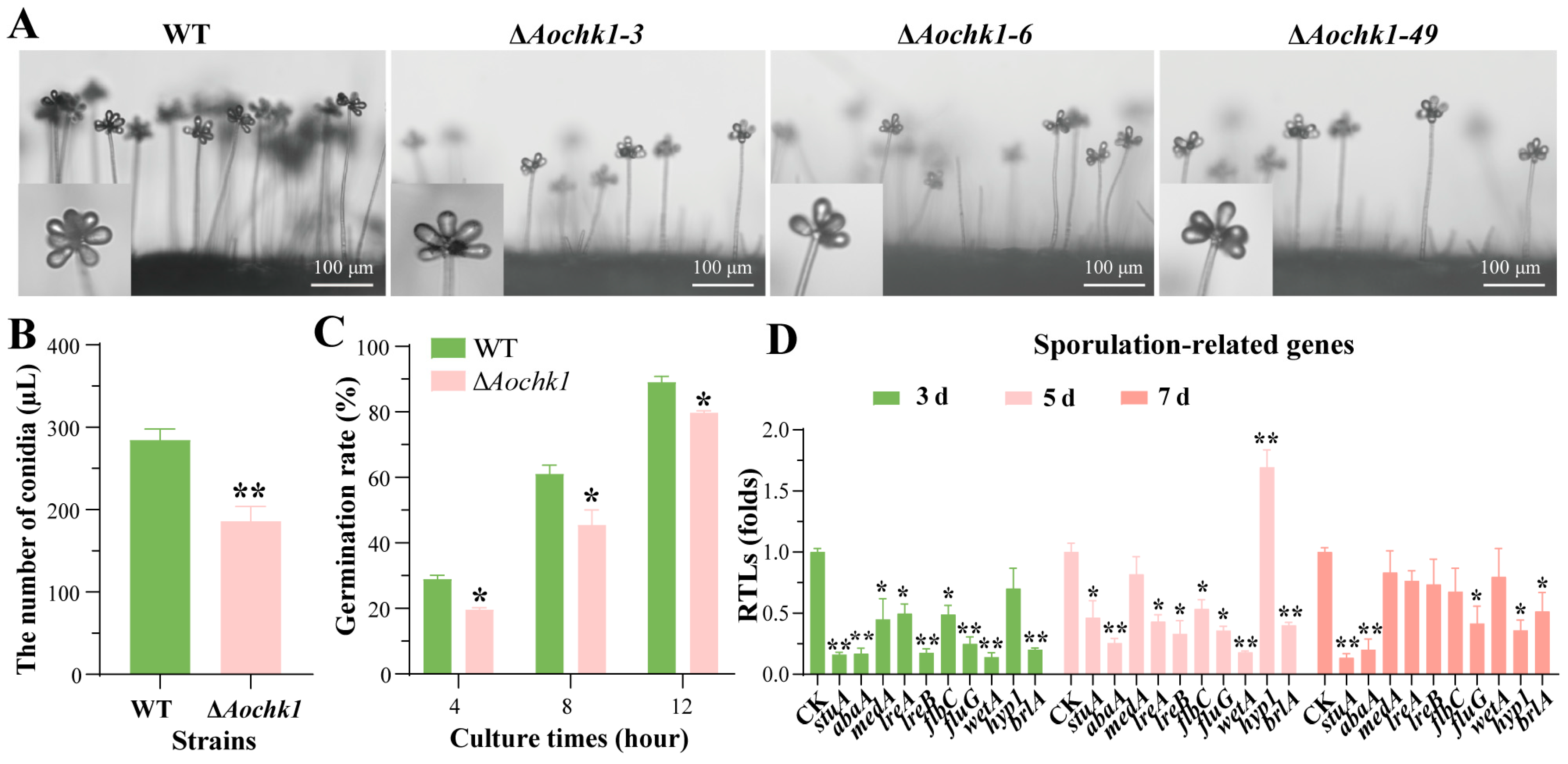
To assess the role of Aochk1 in conidiation, activated WT and ΔAochk1 strains were inoculated onto CMY medium at 28 °C for 14 days. The inactivation of Aochk1 resulted in a remarkable reduction in the number of conidiophores (Figure 2A). Microscopic quantification (1 μL aliquot) revealed that the ΔAochk1 mutant exhibited reduced conidial yield, with nearly 100 fewer spores per microliter than the WT strain (Figure 2B). In addition, the spore germination rate of the ΔAochk1 mutant was decreased relative to the WT strain (Figure 2C). Meanwhile, the transcription levels of most spore-producing-related genes, including the central regulatory genes abaA, wetA, and brlA, were remarkably downregulated in ΔAochk1 mutant on the third and fifth days (Figure 2D). Therefore, Aochk1 ablation significantly impairs sporulation capacity and compromises spore viability.

3.4. Aochk1 Is Involved in Regulating Trap Formation and Pathogenicity

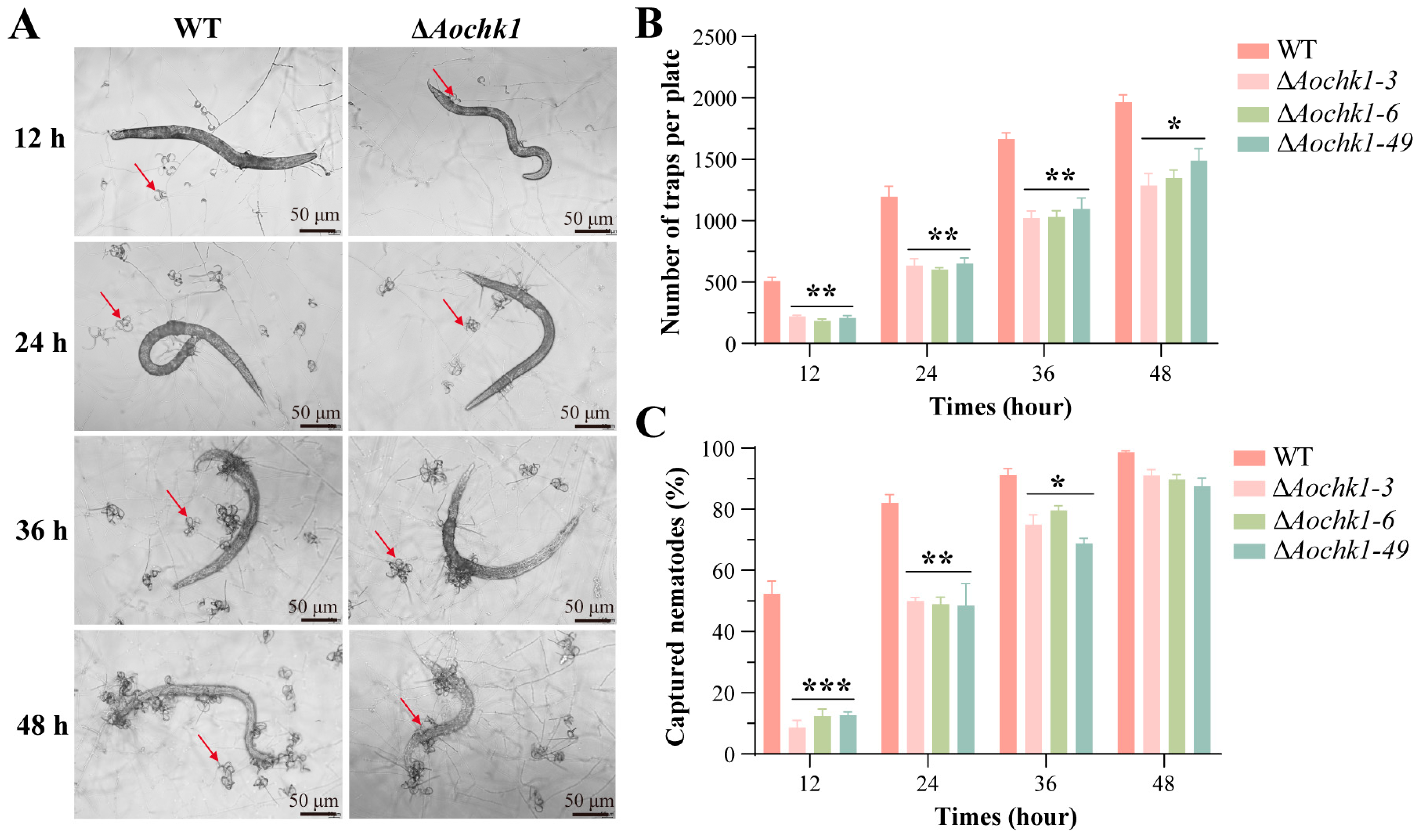
Trap morphogenesis is a critical step for the nematode predation and lifestyle transition of A. oligospora. To further investigate the role of Aochk1 in trap formation and pathogenicity, the trap formation was observed and quantified after the WT and ΔAochk1 mutant strains were induced in C. elegans. The results showed that the mutant strain exhibited a significant reduction in trap production and nematode predation efficiency compared to the WT (Figure 3A). Traps were formed in both WT and mutant strains at 12 h after induction, but the number of traps in the mutant strain was 50% lower than in the WT strain. This corresponded with markedly divergent nematocidal capacity; WT achieved 50% mortality versus 10% for the mutant. Although trap morphology showed no significant differences thereafter, the mutant consistently demonstrated fewer traps and substantially attenuated virulence at 24, 36, and 48 h post-induction (Figure 3B,C).

3.5. Transcriptomic Analysis of the Aochk1 Gene

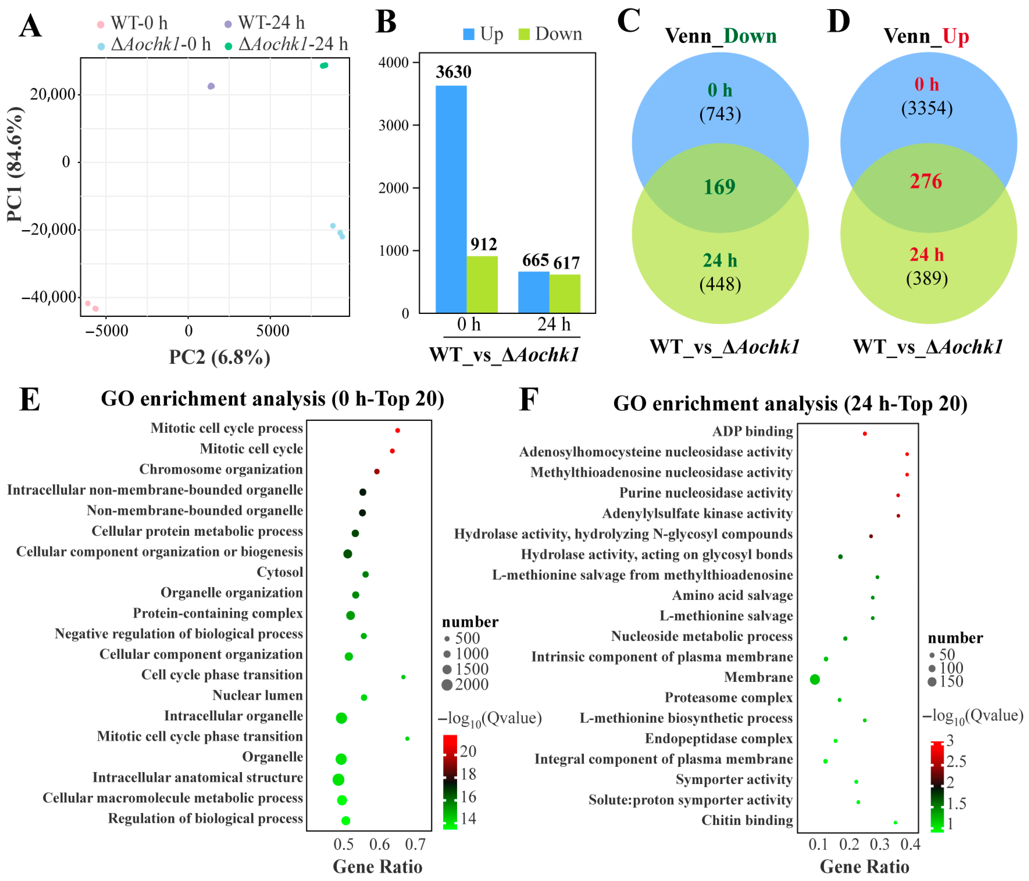
To elucidate the potential mechanisms of Aochk1, genome-wide transcriptome profiling was conducted on ΔAochk1 mutants. Biological triplicates per sample yielded 37.27–44.72 million high-quality reads per library. Quality metrics demonstrated Q20 > 99.08%, Q30 > 96.89%, and GC content > 47.88% (Table S3). We further validated the transcriptome data through RT-qPCR analysis. First, we conducted cluster analysis on 21 related genes in the cell cycle, glycolysis/gluconeogenesis, and MAPK signaling pathways. The results showed that six related genes in the cell cycle pathway had high expression levels in ΔAochk1 mutants, while these genes showed low expression levels in the other two pathways (Figure S2). Additionally, principal component analysis revealed clear segregation of WT and mutant clusters across temporal phases, with high intra-group correlation confirming robust biological reproducibility for downstream analyses (Figure 4A). Transcriptomic profiling revealed substantial differential gene expression in ΔAochk1 versus WT. Under the vegetative growth stage (0 h), 3630 genes were significantly upregulated and 912 downregulated. Following a 24 h nematode challenge, these shifts attenuated to 665 upregulated and 617 downregulated differentially expressed genes (DEGs) (Figure 4B). Venn analysis robustly identified 169 persistently downregulated and 276 consistently upregulated DEGs common to both 0 h and 24 h timepoints (Figure 4C,D). To delineate Aochk1-mediated regulatory networks in A. oligospora, GO enrichment analysis of DEGs at 0 h and 24 h was performed. At the 0 h timepoint, significantly enriched terms were categorized into three functional modules: (1) cell cycle and division, involving mitotic cell cycle process, cell cycle phase transition, and mitotic cell cycle phase transition; (2) nuclear architecture and organelle dynamics, involving chromosome organization, nuclear lumen, intracellular non-membrane-bounded organelle, and organelle organization; and (3) protein homeostasis and regulatory systems, involving cellular protein metabolic process, protein-containing complex assembly, negative regulation of biological process, and regulation of biological process (Figure 4E). The experimental data shows that, at the 24 h timepoint, the main enriched GO terms can be categorized into the following groups according to their functional description: (1) hub for purine/thionucleoside metabolism, involving ADP binding, adenylylsulfate kinase activity, methylthioadenosine nucleosidase activity, and nucleoside metabolic process; (2) protein turnover system, involving hydrolase activity, endopeptidase complex, and amino acid salvage (Figure 4F).
We further analyzed KEGG enrichment of DEGs that were upregulated at 0 h. The enriched pathways were mainly classified by function as follows: (1) protein homeostasis maintenance, involving ribosome, proteasome, aminoacyl-tRNA biosynthesis, and RNA degradation; (2) DNA integrity assurance system, involving mismatch repair, DNA replication, homologous recombination, and base excision repair; (3) cell cycle and genetic information transfer, involving cell cycle, meiosis, and nucleocytoplasmic transport; and (4) metabolic reprogramming, involving pyrimidine metabolism and glycosphingolipid biosynthesis (Figure S3A). In the downregulated pathway at 0 h, we found an enrichment of a variety of amino acid metabolic pathways, such as purine metabolism, carbon metabolism, aspartate and glutamate metabolism, phenylalanine metabolism, aspartate and glutamate metabolism, arginine and proline metabolism. At the same time, the MAPK signaling pathway was also downregulated (Figure S3B). Further KEGG enrichment analysis of upregulated DEGs at 24 h demonstrated enrichment of the nitrogen metabolism, proteasome, fatty acid degradation, amino metabolism, ubiquitin-mediated proteolysis, glycerolipid metabolism, DNA replication, protein processing in endoplasmic reticulum, glycolysis/gluconeogenesis, and other upregulated pathways (Figure S3C). In the 24 h downregulated pathway, biosynthesis of secondary metabolites, nitrogen metabolism, fatty acid elongation, glycerophospholipid metabolism, sphingolipid metabolism, and various types of N-glycan biosynthesis were mainly enriched (Figure S3D).
Transcriptomic profiling of A. oligospora during the initial stages of nematode interaction identified 525 genes constitutively enriched at both 0 h and 24 h post-induction. Furthermore, comparative analysis revealed 757 DEGs were specifically enriched following nematode exposure, implicating their potential involvement in trap morphogenesis (Figure S4A). Gene Ontology (GO) enrichment analysis of these DEGs highlighted significant overrepresentation of terms associated with membrane biology. The GO terms that were enriched can mainly be categorized as follows: (1) Terminology for membrane systems includes intrinsic component of plasma membrane, intrinsic component of external side of plasma membrane, integral component of membrane, rough endoplasmic reticulum membrane, and side of membrane. These genes focus on transmembrane proteins and may be involved in related pathways such as cell membrane signaling or protein secretion. (2) Macromolecular complex terminology includes viral capsid, proteasome core complex, beta-subunit complex, DASH complex, outer kinetochore, mannan polymerase complex, and magnesium chelatase complex. (3) Terms related to enzyme activity include alpha-1,2-galactosyltransferase activity, aspartic-type, endopeptidase/peptidase activity, serine-type endopeptidase inhibitor activity, and glutamate dehydrogenase activity. These genes may be involved in glycosylation modifications and regulation of protein hydrolysis and key enzymes of amino acid metabolism (Figure S4B). Additionally, KEGG pathway analysis further demonstrated significant enrichment of DEGs in diverse metabolic pathways; these included amino acid metabolism (tyrosine metabolism, tryptophan metabolism, arginine biosynthesis, alanine, aspartate and glutamate metabolism), glycerolipid metabolism, nitrogen metabolism, various types of N-glycan biosynthesis, and biosynthesis of secondary metabolites (Figure S4C).

3.6. Aochk1 Is Involved in Regulating Lipid Metabolism and Autophagy Levels

Intracellular LD distribution and morphology were assessed using BODIPY fluorometric staining. The WT hyphae exhibited uniformly dispersed LDs, whereas ΔAochk1 mutants displayed pronounced LD accumulation, as well as an increased number of LDs, and partial LDs had gathered together (Figure 5A). Ultrastructural observation via TEM also found LD accumulation, revealing a significant increase in both LD size and density within mutant hyphae relative to WT (Figure 5B). Clustering analysis of 20 lipid metabolism-associated genes showed that the transcripts of 17 genes were upregulated in the ΔAochk1 mutant, including the following: AOL_s00054g761 (serine palmitoyltransferase), AOL_s00004g426 (dihydroxyacetone kinase), AOL_s00006g298 (acyl-CoA-dependent ceramide synthase), AOL_s00043g488 (O-acyltransferase), AOL_s00043g619 (lysophospholipid acyltransferase, LPLAT), and AOL_s00004g254 (aldehyde dehydrogenase) (Figure 5C). Similarly, MDC staining revealed that increased autophagic vesicles were observed in ΔAochk1 hyphae compared to WT (Figure 5D). TEM observation also found autophagosomes in mutant cells (Figure 5E). Clustering analysis of 20 autophagy-associated genes showed that the transcript of 18 genes was upregulated, including the following: AOL_s00004g301 (autophagy-related protein 11), AOL_s00078g306 (serine/threonine-protein kinase), AOL_s00004g466 (vacuolar protein sorting Vps16), and AOL_s00078g405 (cAMP-dependent protein kinase A, Pka) (Figure 5F). Collectively, these findings demonstrate a central regulatory role for Aochk1 in modulating lipid homeostasis and autophagic flux in A. oligospora.

3.7. Aochk1 Is Involved in the Regulation of Secondary Metabolism

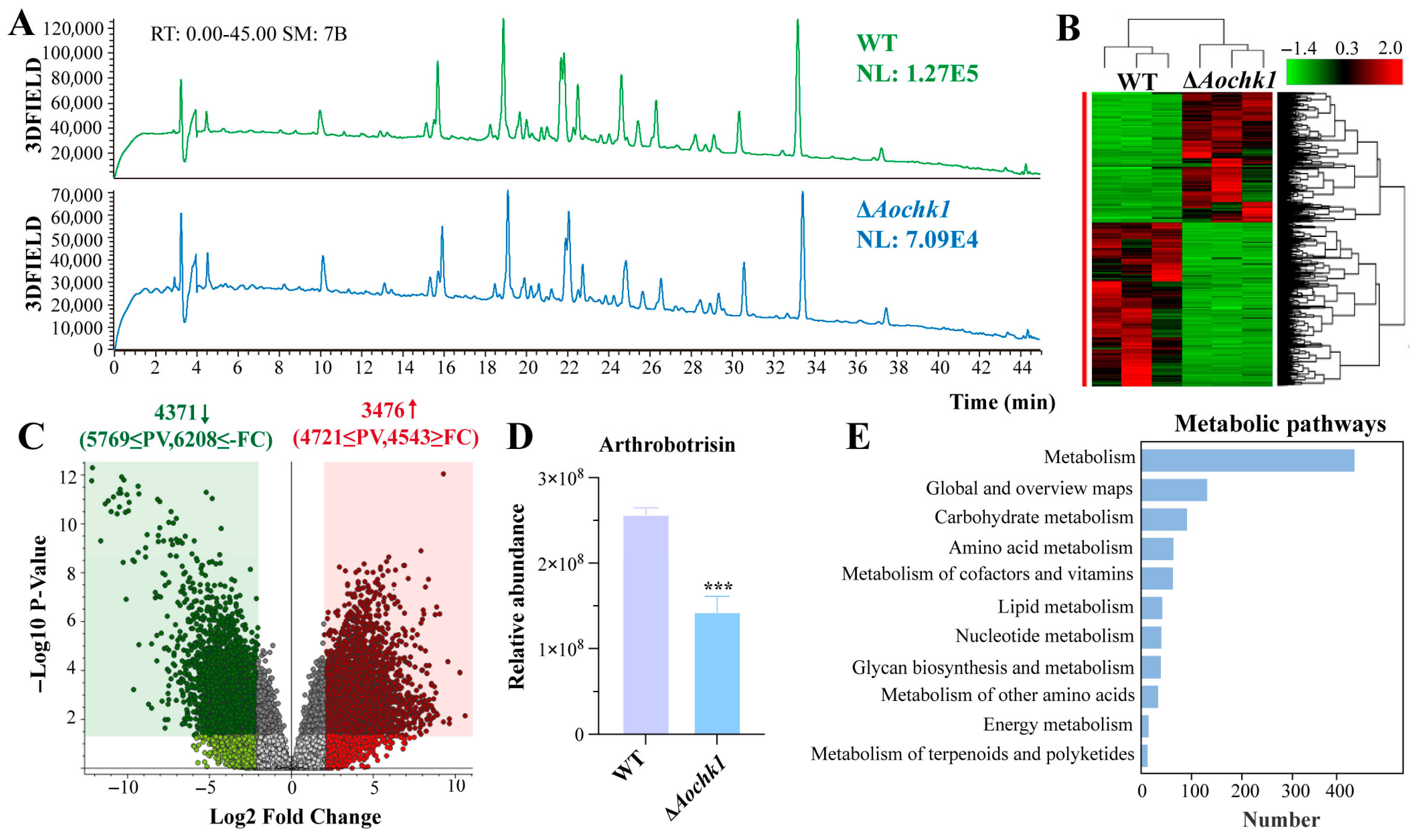
Metabolome profiling reveals Aochk1-dependent regulation in A. oligospora. LC-MS analysis of extracts of culture filtrates revealed distinct secondary metabolite profiles between the WT and ΔAochk1 mutant strains. Although overall chromatographic peaks remained consistent, the ΔAochk1 mutant exhibited remarkably reduced metabolite abundance relative to the WT strain (Figure 6A). Differential features were predominantly localized to the 14–34 min retention time. Clustering of metabolite intensities confirmed widespread downregulation in the mutant strain (Figure 6B). Volcano plot analysis identified 4371 significantly downregulated and 3476 upregulated compounds in ΔAochk1 versus WT (Figure 6C). Notably, arthrobotrisins-specific metabolites were detected in both WT and mutant strains, with significantly reduced accumulation observed in the mutant compared to WT (Figure 6D). The differential metabolites were enriched in several KEGG pathways, such as carbohydrate metabolism, amino acid metabolism, metabolism of cofactors and vitamins, lipid metabolism, nucleotide metabolism, and glycan biosynthesis and metabolism (Figure 6E).

4. Discussion

Chk1 is an evolutionarily conserved serine/threonine protein kinase that plays a pleiotropic role in the growth and development of fungal cells by regulating the cell cycle, responding to DNA damage and replication stresses, and ensuring mitotic fidelity, and this function remains highly consistent from yeast to mammals [1,9,68,69,70]. In this study, we characterized the role of AoChk1 in the sporulation, trap formation, pathogenicity, LD accumulation, autophagy and secondary metabolism of A. oligospora. Our results suggested Aochk1 is involved in various cellular processes and plays a crucial role in the proper development of nuclei, sporulation, lipid metabolism, and the nematode predation process.
It was shown that Chk1 orthologs are involved in mycelial growth and development and spore formation. In the corn leaf pathogen C. heterostrophus, the causal agent of southern corn leaf blight, chk1 ablation induces aerial hyphae hypoplasia, autolysis, and conidiation deficiency, while the variety of phenotypic changes suggests that Chk1 is involved in multiple developmental pathways, each of which responds to different signals. When involved in multiple pathways, different components or even pathways may be required to assist in specific responses to specific signals [29]. In the ink cap mushroom Coprinopsis cinerea, deletion of chk1 impairs fruiting body development; furthermore, they speculated that developmental defects were the result of cell cycle damage [71]. In the pathogenic yeast Candida albicans, Chk1 governs the transition from yeast to hyphal growth, a process critical for host invasion [72]. In our study, although deletion of Aochk1 did not influence the mycelial growth, the ∆Aochk1 mutant strains exhibited significant reductions in spore production, spore germination rate, and mycelial length compared to WT. Thus, we speculate that Chk1 participates in multiple developmental pathways, and the developmental defects of spores and changes in mycelial length are also the result of Chk1 causing cell cycle damage. Chk1 orthologs share multiple roles in vegetative growth and spore production in A. oligospora and other fungi.
In addition, Chk1 orthologs are involved in the pathogenic process of various fungi. In Candida albicans, chk1 modulates virulence and cell wall biosynthesis, with its deletion compromising virulence factor expression and restricting hyphal development to a non-pathogenic form [73,74]. During the virulence program induction process of the plant pathogenic fungus Ustilago maydis, cell cycle arrest at the G2 phase is essential for the implementation of the virulence infection process, which promotes the development of infective hyphae. The activation of Chk1 is also triggered during this process, leading researchers to conclude that the Chk1 gene is crucial for pathogenicity and plays a key regulatory role in host colonization and disease development [75]. In C. neoformans, knockout of Rad53 and Chk1 promotes macrophage phagosome maturation, reduced melanin production, and increased sensitivity to oxidative stress, thereby reducing their pathogenicity [13]. In C. heterostrophus, chk1 deletion results in severely reduced pigmentation, virulence, and conidiation [29]. In this study, deletion of Aochk1 resulted in decreased trap production and depressed nematode predation ability. Additionally, based on transcriptomic data, the GO term obtained from the GO enrichment analysis of 757 differentially expressed genes can be categorized by function into the following three sections: membrane system-related terminology, macromolecular complex terminology, and enzyme activity-associated terms. This transcriptomic signature aligns with the established ultrastructural and biochemical events critical for trap functionality. Upon nematode contact, hyphal differentiation is initiated, culminating in the curling formation of specialized traps. This process necessitates extensive membrane remodeling, characterized by the localized secretion of adhesive macromolecules (e.g., polysaccharides, glycoproteins) to ensnare nematodes [76], followed by the targeted release of extracellular hydrolytic enzymes (e.g., proteases, chitinases) to penetrate the nematode cuticle and facilitate nutrient acquisition [77]. Consequently, membrane dynamics and protease activity constitute indispensable components of efficient trap formation and predation nematodes. Integrated analysis of the observed phenotypic alterations and the molecular profiling data strongly supports the conclusion that AoChk1 exerts a pivotal regulatory function within the signaling pathway governing nematode trap formation in A. oligospora. Therefore, Chk1 orthologs are conserved in the regulation of pathogenicity in A. oligospora along with other fungi.
It has been shown that decreased phospholipid availability is consistent with decreased Chk1 signaling and vice versa, and that an increase in phospholipid pools contributes to the protection of DNA integrity during genotoxic attacks [78]. In A. oligospora, compared with WT, LD accumulation was increased in the ΔAochk1 mutant strain, and by clustering analysis of DEGs, we found that the expression of many genes related to lipid metabolism underwent a significant upregulation. Meanwhile, we found a significant enrichment at 24 h in the glycerolipid metabolism in the upregulation pathway. The above results suggest that AoChk1 plays an important regulatory role in the lipid metabolism of A. oligospora.
Autophagy primarily functions as a cytoprotective mechanism. DNA damage induces autophagic activation, which plays a pivotal role in maintaining genomic stability [79,80,81]. In addition, autophagy is critically required for DNA repair by homologous recombination [81,82,83,84]. DNA damage triggers a series of signaling cascades that promote cell survival, including DNA repair, cell cycle arrest, and autophagy [85,86,87]. In our study, clustering analyses of the DEGs showed that the expression of 18 autophagy-related genes was significantly elevated. In addition, KEGG enrichment analysis of DEGs by transcriptomic data revealed that the homologous recombination, base excision repair, ribosome, and nucleocytoplasmic transport pathways underwent significant upregulation. Homologous recombination is a vital process for repairing DNA double-strand breaks [88], suggesting that deletion of Aochk1 leads to DNA damage, which, in turn, triggers increased autophagy.
Chk1 is involved in the regulation of multiple metabolic pathways and the production of secondary metabolites in A. oligospora. In this study, through our combined transcriptomic and metabolomic analysis, we found that deletion of Aochk1 led to changes in multiple metabolic pathways, such as carbohydrate metabolism, amino acid metabolism, metabolism of cofactors and vitamins, lipid metabolism, nucleotide metabolism, and glycan biosynthesis and metabolism. In addition, the content of arthrobotrisins was decreased in the ΔAochk1 mutant. Previous studies on Chk1 homologs in other fungi have primarily focused on mechanisms such as DNA damage response and cell cycle regulation, whereas their role in metabolic regulation remains understudied. Consequently, elucidating the metabolic regulatory function of Chk1 in A. oligospora provides novel insights for analogous research in other fungal systems.
Studies have shown that Chk1 responds to replication stress by regulating cell cycle checkpoints and homologous recombination repair [89]. In N. crassa, Chk1/2 is involved in the regulation of metabolism during DNA damage, modulation of chromatin structure, and robust, regular transcription of DNA repair genes [30]. In S. pombe, Crb2 recruits Chk1 to double-strand breaks for DNA damage repair by phosphorylation, thus maintaining DNA integrity in the genome [23], and Wat1 deletion induces chromosome breaks that activate the DNA damage checkpoint, and in chk1-null cells, this results in severe proliferation defects and synthetic lethality [28]. In S. pombe, Chk1 is important for maintaining the DNA replication checkpoint in heat-sensitive mutants in the S phase [90]. Synergistic or divergent control of RNR1 and RNR21 expression by Rad53 and Chk1 kinases was observed in response to DNA damage and DNA replication stress in C. neoformans [32]. In C. cinerea, the absence of Chk1 impairs the development of fruiting bodies, and downregulation of chk1 and Atr1 levels leads to mitotic abnormalities and a significant increase in cell number [71]. In this study, deletion of Aochk1 significantly reduced the nuclear number. Transcriptome data shows that deletion of Aochk1 results in alterations of cell cycle, mismatch repair, DNA replication, homologous recombination, meiosis, and other related pathways that are altered as a result of cell cycle dysregulation. The above results indicate that AoChk1 is a genome stability guardian, cell cycle engine regulator, and protein quality control system, and it has been shown that Chk1 is a key factor in the error-free DNA repair process of homologous recombination [87]. These findings suggest that the function of Chk1 in maintaining genomic integrity is relatively conserved in A. oligospora and other fungi.

5. Conclusions

This study comprehensively characterizes the functions of AoChk1 in A. oligospora, revealing its pivotal role in orchestrating diverse biological processes, including sporulation, trap formation, lipid metabolism, autophagic flux, and metabolic processes. Additionally, we identified previously the undescribed regulatory functions of Chk1 in other fungi, related to the number of nuclei, the formation of traps, and secondary metabolites (such as arthrobotrisins). Our findings provide novel insights into Chk1-mediated mechanisms in nematode-trapping fungi, facilitating the development of enhanced nematocidal agents and biocontrol strategies.

Supplementary Materials

The following supporting information can be downloaded at: https://www.mdpi.com/article/10.3390/jof11080602/s1, Figure S1: Phylogenetic analysis of Chk1 orthologs and knockout validation of the Aochk1 gene in A. oligospora; Figure S2: RT-qPCR analysis validated the transcriptome data of the wild-type and ΔAochk1 mutants. Figure S3: KEGG pathway enrichment analysis of differentially expressed genes (DEGs); Figure S4: Analysis of trap-associated transcriptomic data; Table S1: Primers used for genetic manipulation in this study; Table S2: List of primers used for RT-qPCR analysis in this study; Table S3: Statistical analysis of quality control data of 12 sequencing samples.

Author Contributions

J.Y. conceived and designed the study. H.L. performed the experiments. Q.L., S.C., X.L., H.C. and Y.X. analyzed the data. H.L. and J.Y. contributed to manuscript preparation and revision. All authors have read and agreed to the published version of the manuscript.

Funding

This study was financially supported by the National Key R & D Program of China (2024YFD1200203), Yunnan Fundamental Research Projects (202401BC070010), Yunling Scholar of Yunnan Province (to Jinkui Yang), and the Science Research Foundation of Yunnan Provincial Department of Education, and Professional Degree Graduate Practical Innovation Foundation of Yunnan University (ZC-242410176).

Institutional Review Board Statement

Not applicable.

Informed Consent Statement

Not applicable.

Data Availability Statement

The RNA-seq datasets produced in this study are archived in the NCBI Gene Expression Omnibus repository under accession GSE301452.

Acknowledgments

We express our gratitude to the Microbial Library of the Germplasm Bank of Wild Species in Southwest China for the preservation and provision of experimental strains, and to Guo Ying-qi (Kunming Institute of Zoology, Chinese Academy of Sciences) for help with taking and analyzing TEM images.

Conflicts of Interest

The authors declare no conflicts of interest.

References

  1. Chen, Y.; Sanchez, Y. Chk1 in the DNA damage response: Conserved roles from yeasts to mammals. DNA Repair 2004, 3, 1025–1032. [Google Scholar] [CrossRef]
  2. DeStephanis, D.; McLeod, M.; Yan, S. REV1 is important for the ATR-Chk1 DNA damage response pathway in Xenopus egg extracts. Biochem. Biophys. Res. Commun. 2015, 460, 609–615. [Google Scholar] [CrossRef]
  3. Kizhedathu, A.; Kunnappallil, R.S.; Bagul, A.V.; Verma, P.; Guha, A. Multiple Wnts act synergistically to induce Chk1/Grapes expression and mediate G2 arrest in Drosophila tracheoblasts. eLife 2020, 9, e57056. [Google Scholar] [CrossRef]
  4. Collart, C.; Smith, J.C.; Zegerman, P. Chk1 inhibition of the replication factor Drf1 guarantees cell-cycle elongation at the Xenopus laevis mid-blastula transition. Dev. Cell 2017, 42, 82–96.e3. [Google Scholar] [CrossRef]
  5. Petrus, M.J.; Wilhelm, D.E.; Murakami, M.; Kappas, N.C.; Carter, A.D.; Wroble, B.N.; Sible, J.C. Altered expression of Chk1 disrupts cell cycle remodeling at the midblastula transition in Xenopus laevis embryos. Cell Cycle 2004, 3, 212–217. [Google Scholar] [CrossRef] [PubMed]
  6. Sanchez, Y.; Wong, C.; Thoma, R.S.; Richman, R.; Wu, Z.; Piwnica-Worms, H.; Elledge, S.J. Conservation of the Chk1 checkpoint pathway in mammals: Linkage of DNA damage to Cdk regulation through Cdc25. Science 1997, 277, 1497–1501. [Google Scholar] [CrossRef] [PubMed]
  7. Liu, Y.; Wang, L.; Xu, X.; Yuan, Y.; Zhang, B.; Li, Z.; Xie, Y.; Yan, R.; Zheng, Z.; Ji, J.; et al. The intra-S phase checkpoint directly regulates replication elongation to preserve the integrity of stalled replisomes. Proc. Natl. Acad. Sci. USA 2021, 118, e2019183118. [Google Scholar] [CrossRef] [PubMed]
  8. Cheng, Y.C.; Shieh, S.Y. Determination of CHK1 cellular localization by immunofluorescence microscopy. Methods Mol. Biol. 2021, 2267, 1–6. [Google Scholar] [CrossRef]
  9. Zhang, Y.; Hunter, T. Roles of Chk1 in cell biology and cancer therapy. Int. J. Cancer 2014, 134, 1013–1023. [Google Scholar] [CrossRef]
  10. Palermo, C.; Hope, J.C.; Freyer, G.A.; Rao, H.; Walworth, N.C. Importance of a C-terminal conserved region of Chk1 for checkpoint function. PLoS ONE 2008, 3, e1427. [Google Scholar] [CrossRef]
  11. Kosoy, A.; O’Connell, M.J. Regulation of Chk1 by its C-terminal domain. Mol. Biol. Cell 2008, 19, 4546–4553. [Google Scholar] [CrossRef] [PubMed]
  12. Leman, A.R.; Noguchi, E. The replication fork: Understanding the eukaryotic replication machinery and the challenges to genome duplication. Genes 2013, 4, 1–32. [Google Scholar] [CrossRef] [PubMed]
  13. Jung, K.W.; Lee, Y.; Huh, E.Y.; Lee, S.C.; Lim, S.; Bahn, Y.S. Rad53- and Chk1-dependent DNA damage response pathways cooperatively promote fungal pathogenesis and modulate antifungal drug susceptibility. mBio 2019, 10, e01726-18. [Google Scholar] [CrossRef]
  14. Rocha, D.; Castro, T.L.P.; Aguiar, E.; Pacheco, L.G.C. Gene expression analysis in bacteria by RT-qPCR. Methods Mol. Biol. 2020, 2065, 119–137. [Google Scholar] [CrossRef]
  15. Michelena, J.; Gatti, M.; Teloni, F.; Imhof, R.; Altmeyer, M. Basal CHK1 activity safeguards its stability to maintain intrinsic S-phase checkpoint functions. J. Cell Biol. 2019, 218, 2865–2875. [Google Scholar] [CrossRef]
  16. Smits, V.A.; Gillespie, D.A. DNA damage control: Regulation and functions of checkpoint kinase 1. FEBS J. 2015, 282, 3681–3692. [Google Scholar] [CrossRef]
  17. Syljuåsen, R.G.; Sørensen, C.S.; Hansen, L.T.; Fugger, K.; Lundin, C.; Johansson, F.; Helleday, T.; Sehested, M.; Lukas, J.; Bartek, J. Inhibition of human Chk1 causes increased initiation of DNA replication, phosphorylation of ATR targets, and DNA breakage. Mol. Cell. Biol. 2005, 25, 3553–3562. [Google Scholar] [CrossRef]
  18. Furnari, B.; Rhind, N.; Russell, P. Cdc25 mitotic inducer targeted by chk1 DNA damage checkpoint kinase. Science 1997, 277, 1495–1497. [Google Scholar] [CrossRef]
  19. Myers, K.; Gagou, M.E.; Zuazua-Villar, P.; Rodriguez, R.; Meuth, M. ATR and Chk1 suppress a caspase-3-dependent apoptotic response following DNA replication stress. PLoS Genet. 2009, 5, e1000324. [Google Scholar] [CrossRef]
  20. Stolz, A.; Ertych, N.; Bastians, H. Tumor suppressor CHK2: Regulator of DNA damage response and mediator of chromosomal stability. Clin. Cancer Res. 2011, 17, 401–405. [Google Scholar] [CrossRef] [PubMed]
  21. Shimada, M.; Niida, H.; Zineldeen, D.H.; Tagami, H.; Tanaka, M.; Saito, H.; Nakanishi, M. Chk1 is a histone H3 threonine 11 kinase that regulates DNA damage-induced transcriptional repression. Cell 2008, 132, 221–232. [Google Scholar] [CrossRef] [PubMed]
  22. Goto, H.; Natsume, T.; Kanemaki, M.T.; Kaito, A.; Wang, S.; Gabazza, E.C.; Inagaki, M.; Mizoguchi, A. Chk1-mediated Cdc25A degradation as a critical mechanism for normal cell cycle progression. J. Cell Sci. 2019, 132, jcs223123. [Google Scholar] [CrossRef]
  23. Qu, M.; Yang, B.; Tao, L.; Yates, J.R., 3rd; Russell, P.; Dong, M.Q.; Du, L.L. Phosphorylation-dependent interactions between Crb2 and Chk1 are essential for DNA damage checkpoint. PLoS Genet. 2012, 8, e1002817. [Google Scholar] [CrossRef]
  24. Cheetham, J.; Smith, D.A.; da Silva Dantas, A.; Doris, K.S.; Patterson, M.J.; Bruce, C.R.; Quinn, J. A single MAPKKK regulates the Hog1 MAPK pathway in the pathogenic fungus Candida albicans. Mol. Biol. Cell 2007, 18, 4603–4614. [Google Scholar] [CrossRef]
  25. Li, D.; Gurkovska, V.; Sheridan, M.; Calderone, R.; Chauhan, N. Studies on the regulation of the two-component histidine kinase gene CHK1 in Candida albicans using the heterologous lacZ reporter gene. Microbiology 2004, 150, 3305–3313. [Google Scholar] [CrossRef]
  26. Basso, V.; Znaidi, S.; Lagage, V.; Cabral, V.; Schoenherr, F.; LeibundGut-Landmann, S.; d’Enfert, C.; Bachellier-Bassi, S. The two-component response regulator Skn7 belongs to a network of transcription factors regulating morphogenesis in Candida albicans and independently limits morphogenesis-induced ROS accumulation. Mol. Microbiol. 2017, 106, 157–182. [Google Scholar] [CrossRef]
  27. Kruppa, M.; Goins, T.; Cutler, J.E.; Lowman, D.; Williams, D.; Chauhan, N.; Menon, V.; Singh, P.; Li, D.; Calderone, R. The role of the Candida albicans histidine kinase [CHK1) gene in the regulation of cell wall mannan and glucan biosynthesis. FEMS Yeast Res. 2003, 3, 289–299. [Google Scholar] [CrossRef]
  28. Ahamad, N.; Verma, S.K.; Ahmed, S. Activation of checkpoint kinase Chk1 by reactive oxygen species resulting from disruption of wat1/pop3 in Schizosaccharomyces pombe. Genetics 2016, 204, 1397–1406. [Google Scholar] [CrossRef]
  29. Lev, S.; Sharon, A.; Hadar, R.; Ma, H.; Horwitz, B.A. A mitogen-activated protein kinase of the corn leaf pathogen Cochliobolus heterostrophus is involved in conidiation, appressorium formation, and pathogenicity: Diverse roles for mitogen-activated protein kinase homologs in foliar pathogens. Proc. Natl. Acad. Sci. USA 1999, 96, 13542–13547. [Google Scholar] [CrossRef] [PubMed]
  30. Yang, Y.; Duan, Z.; Liu, X.L.; Li, Z.; Shen, Z.; Gong, S.; Lu, Q.; Hu, Y.; Song, L.; Wang, Z.; et al. Checkpoint kinases regulate the circadian clock after DNA damage by influencing chromatin dynamics. Nucleic Acids Res. 2025, 53, gkaf162. [Google Scholar] [CrossRef] [PubMed]
  31. Tarsha, W. Chk1 prevents abnormal mitosis of S-phase HeLa cells containing DNA damage. Chin. Sci. Bull. 2009, 54, 4205–4213. [Google Scholar]
  32. Jung, K.W.; Kwon, S.; Jung, J.H.; Bahn, Y.S. Essential roles of ribonucleotide reductases under DNA damage and replication stresses in cryptococcus neoformans. Microbiol. Spectr. 2022, 10, e0104422. [Google Scholar] [CrossRef] [PubMed]
  33. Jiang, X.; Xiang, M.; Liu, X. Nematode-Trapping Fungi. Microbiol. Spectr. 2017, 5, 10. [Google Scholar] [CrossRef]
  34. Li, L.; Ma, M.; Liu, Y.; Zhou, J.; Qu, Q.; Lu, K.; Fu, D.; Zhang, K. Induction of trap formation in nematode-trapping fungi by a bacterium. FEMS Microbiol. Lett. 2011, 322, 157–165. [Google Scholar] [CrossRef]
  35. Su, H.; Zhao, Y.; Zhou, J.; Feng, H.; Jiang, D.; Zhang, K.Q.; Yang, J. Trapping devices of nematode-trapping fungi: Formation, evolution, and genomic perspectives. Biol. Rev. Camb. Philos. Soc. 2017, 92, 357–368. [Google Scholar] [CrossRef]
  36. Yang, Y.; Yang, E.; An, Z.; Liu, X. Evolution of nematode-trapping cells of predatory fungi of the Orbiliaceae based on evidence from rRNA-encoding DNA and multiprotein sequences. Proc. Natl. Acad. Sci. USA 2007, 104, 8379–8384. [Google Scholar] [CrossRef] [PubMed]
  37. Ahren, D.; Tunlid, A. Evolution of parasitism in nematode-trapping fungi. J. Nematol. 2003, 35, 194–197. [Google Scholar]
  38. Dijksterhuis, J.; Veenhuis, M.; Harder, W.; Nordbring-Hertz, B. Nematophagous fungi: Physiological aspects and structure-function relationships. Adv. Microb. Physiol. 1994, 36, 111–143. [Google Scholar]
  39. Yang, J.; Wang, L.; Ji, X.; Feng, Y.; Li, X.; Zou, C.; Xu, J.; Ren, Y.; Mi, Q.; Wu, J.; et al. Genomic and proteomic analyses of the fungus Arthrobotrys oligospora provide insights into nematode-trap formation. PLoS Pathog. 2011, 7, e1002179. [Google Scholar] [CrossRef]
  40. Zhu, M.C.; Zhao, N.; Liu, Y.K.; Li, X.M.; Zhen, Z.Y.; Zheng, Y.Q.; Zhang, K.Q.; Yang, J.K. The cAMP-PKA signalling pathway regulates hyphal growth, conidiation, trap morphogenesis, stress tolerance, and autophagy in Arthrobotrys oligospora. Environ. Microbiol. 2022, 24, 6524–6538. [Google Scholar] [CrossRef] [PubMed]
  41. Chen, S.A.; Lin, H.C.; Hsueh, Y.P. The cAMP-PKA pathway regulates prey sensing and trap morphogenesis in the nematode-trapping fungus Arthrobotrys oligospora. G3 2022, 12, jkac217. [Google Scholar] [CrossRef]
  42. Kuo, C.Y.; Chen, S.A.; Hsueh, Y.P. The high osmolarity glycerol (HOG) pathway functions in osmosensing, trap morphogenesis and conidiation of the Nematode-trapping fungus Arthrobotrys oligospora. J. Fungi 2020, 6, 191. [Google Scholar] [CrossRef]
  43. Xie, M.; Ma, N.; Bai, N.; Yang, L.; Yang, X.; Zhang, K.Q.; Yang, J. PKC-SWI6 signaling regulates asexual development, cell wall integrity, stress response, and lifestyle transition in the nematode-trapping fungus Arthrobotrys oligospora. Sci. China Life Sci. 2022, 65, 2455–2471. [Google Scholar] [CrossRef]
  44. Xie, M.; Bai, N.; Yang, X.; Liu, Y.; Zhang, K.Q.; Yang, J. Fus3 regulates asexual development and trap morphogenesis in the nematode-trapping fungus Arthrobotrys oligospora. iScience 2023, 26, 107404. [Google Scholar] [CrossRef]
  45. Kuo, C.Y.; Tay, R.J.; Lin, H.C.; Juan, S.C.; Vidal-Diez de Ulzurrun, G.; Chang, Y.C.; Hoki, J.; Schroeder, F.C.; Hsueh, Y.P. The nematode-trapping fungus Arthrobotrys oligospora detects prey pheromones via G protein-coupled receptors. Nat. Microbiol. 2024, 9, 1738–1751. [Google Scholar] [CrossRef]
  46. Xie, M.; Ma, N.; Bai, N.; Zhu, M.; Zhang, K.Q.; Yang, J. Phospholipase C (AoPLC2) regulates mycelial development, trap morphogenesis, and pathogenicity of the nematode-trapping fungus Arthrobotrys oligospora. J. Appl. Microbiol. 2022, 132, 2144–2156. [Google Scholar] [CrossRef]
  47. Park, G.; Colot, H.V.; Collopy, P.D.; Krystofova, S.; Crew, C.; Ringelberg, C.; Litvinkova, L.; Altamirano, L.; Li, L.; Curilla, S.; et al. High-throughput production of gene replacement mutants in Neurospora crassa. Methods Mol. Biol. 2011, 722, 179–189. [Google Scholar] [CrossRef] [PubMed]
  48. Xie, M.; Wang, Y.; Tang, L.; Yang, L.; Zhou, D.; Li, Q.; Niu, X.; Zhang, K.Q.; Yang, J. AoStuA, an APSES transcription factor, regulates the conidiation, trap formation, stress resistance and pathogenicity of the nematode-trapping fungus Arthrobotrys oligospora. Environ. Microbiol. 2019, 21, 4648–4661. [Google Scholar] [CrossRef]
  49. Tamura, K.; Stecher, G.; Kumar, S. MEGA11: Molecular Evolutionary Genetics Analysis Version 11. Mol. Biol. Evol. 2021, 38, 3022–3027. [Google Scholar] [CrossRef] [PubMed]
  50. Chen, S.A.; Lin, H.C.; Schroeder, F.C.; Hsueh, Y.P. Prey sensing and response in a nematode-trapping fungus is governed by the MAPK pheromone response pathway. Genetics 2021, 217, iyaa008. [Google Scholar] [CrossRef] [PubMed]
  51. Chen, C.; Chen, H.; Zhang, Y.; Thomas, H.R.; Frank, M.H.; He, Y.; Xia, R. TBtools: An integrative toolkit developed for interactive analyses of big biological data. Mol. Plant 2020, 13, 1194–1202. [Google Scholar] [CrossRef]
  52. Colot, H.V.; Park, G.; Turner, G.E.; Ringelberg, C.; Crew, C.M.; Litvinkova, L.; Weiss, R.L.; Borkovich, K.A.; Dunlap, J.C. A high-throughput gene knockout procedure for Neurospora reveals functions for multiple transcription factors. Proc. Natl. Acad. Sci. USA 2006, 103, 10352–10357. [Google Scholar] [CrossRef] [PubMed]
  53. Hu, X.; Hoffmann, D.S.; Wang, M.; Schuhmacher, L.; Stroe, M.C.; Schreckenberger, B.; Elstner, M.; Fischer, R. GprC of the nematode-trapping fungus Arthrobotrys flagrans activates mitochondria and reprograms fungal cells for nematode hunting. Nat. Microbiol. 2024, 9, 1752–1763. [Google Scholar] [CrossRef]
  54. Bong, D.; Sohn, J.; Lee, S.V. Brief guide to RT-qPCR. Mol. Cells 2024, 47, 100141. [Google Scholar] [CrossRef]
  55. Bustin, S.A.; Shipley, G.L.; Kirvell, S.; Mueller, R.; Nolan, T. RT-qPCR detection of SARS-CoV-2: No need for a dedicated reverse transcription step. Int. J. Mol. Sci. 2022, 23, 1303. [Google Scholar] [CrossRef]
  56. Zhao, N.; Zhu, M.; Liu, Q.; Shen, Y.; Duan, S.; Zhu, L.; Yang, J. AoPrdx2 Regulates Oxidative Stress, Reactive Oxygen Species, Trap Formation, and Secondary Metabolism in Arthrobotrys oligospora. J. Fungi 2024, 10, 110. [Google Scholar] [CrossRef]
  57. Zhu, L.; Zhu, M.; Li, X.; Shen, Y.; Duan, S.; Yang, J. Functional Characterization of Ao4g24: An Uncharacterized Gene Involved in Conidiation, Trap Formation, Stress Response, and Secondary Metabolism in Arthrobotrys oligospora. Microorganisms 2024, 12, 1532. [Google Scholar] [CrossRef]
  58. Youssar, L.; Wernet, V.; Hensel, N.; Yu, X.; Hildebrand, H.G.; Schreckenberger, B.; Kriegler, M.; Hetzer, B.; Frankino, P.; Dillin, A.; et al. Intercellular communication is required for trap formation in the nematode-trapping fungus Duddingtonia flagrans. PLoS Genet. 2019, 15, e1008029. [Google Scholar] [CrossRef]
  59. Lin, H.C.; de Ulzurrun, G.V.; Chen, S.A.; Yang, C.T.; Tay, R.J.; Iizuka, T.; Huang, T.Y.; Kuo, C.Y.; Gonçalves, A.P.; Lin, S.Y.; et al. Key processes required for the different stages of fungal carnivory by a nematode-trapping fungus. PLoS Biol. 2023, 21, e3002400. [Google Scholar] [CrossRef] [PubMed]
  60. Yang, C.T.; Vidal-Diez de Ulzurrun, G.; Gonçalves, A.P.; Lin, H.C.; Chang, C.W.; Huang, T.Y.; Chen, S.A.; Lai, C.K.; Tsai, I.J.; Schroeder, F.C.; et al. Natural diversity in the predatory behavior facilitates the establishment of a robust model strain for nematode-trapping fungi. Proc. Natl. Acad. Sci. USA 2020, 117, 6762–6770. [Google Scholar] [CrossRef] [PubMed]
  61. Li, C.; Melesse, M.; Zhang, S.; Hao, C.; Wang, C.; Zhang, H.; Hall, M.C.; Xu, J.R. FgCDC14 regulates cytokinesis, morphogenesis, and pathogenesis in Fusarium graminearum. Mol. Microbiol. 2015, 98, 770–786. [Google Scholar] [CrossRef] [PubMed]
  62. Emser, J.; Wernet, N.; Hetzer, B.; Wohlmann, E.; Fischer, R. The cysteine-rich virulence factor NipA of Arthrobotrys flagrans interferes with cuticle integrity of Caenorhabditis elegans. Nat. Commun. 2024, 15, 5795. [Google Scholar] [CrossRef] [PubMed]
  63. Yang, L.; Li, X.; Ma, Y.; Zhu, M.; Zhang, K.Q.; Yang, J. The Arf-GAPs, AoAge1 and AoAge2, regulate diverse cellular processes, conidiation, trap formation, and secondary metabolism in Arthrobotrys oligospora. Microbiol. Res. 2024, 285, 127779. [Google Scholar] [CrossRef]
  64. He, Z.Q.; Tan, J.L.; Li, N.; Zhang, H.X.; Chen, Y.H.; Wang, L.J.; Zhang, K.Q.; Niu, X.M. Sesquiterpenyl epoxy-cyclohexenoids and their signaling functions in nematode-trapping fungus Arthrobotrys oligospora. J. Agric. Food Chem. 2019, 67, 13061–13072. [Google Scholar] [CrossRef]
  65. Zhao, N.; Liu, Q.; Zhu, M.; Zhu, L.; Yang, J. The Hog1-Nmd5 signaling pathway regulates asexual development, lipid metabolism, stress response, trap morphogenesis, and secondary metabolism of Arthrobotrys oligospora. Virulence 2025, 16, 2468294. [Google Scholar] [CrossRef]
  66. Wang, R.; Li, B.; Lam, S.M.; Shui, G. Integration of lipidomics and metabolomics for in-depth understanding of cellular mechanism and disease progression. J. Genet. Genom. 2020, 47, 69–83. [Google Scholar] [CrossRef]
  67. Bauermeister, A.; Mannochio-Russo, H.; Costa-Lotufo, L.V.; Jarmusch, A.K.; Dorrestein, P.C. Mass spectrometry-based metabolomics in microbiome investigations. Nat. Rev. Microbiol. 2022, 20, 143–160. [Google Scholar] [CrossRef] [PubMed]
  68. Westphal, C.H. Cell-cycle signaling: Atm displays its many talents. Curr. Biol. 1997, 7, R789–R792. [Google Scholar] [CrossRef]
  69. Zhao, H.; Piwnica-Worms, H. ATR-mediated checkpoint pathways regulate phosphorylation and activation of human Chk1. Mol. Cell. Biol. 2001, 21, 4129–4139. [Google Scholar] [CrossRef]
  70. Suzuki, M.; Yamamori, T.; Bo, T.; Sakai, Y.; Inanami, O. MK-8776, a novel Chk1 inhibitor, exhibits an improved radiosensitizing effect compared to UCN-01 by exacerbating radiation-induced aberrant mitosis. Transl. Oncol. 2017, 10, 491–500. [Google Scholar] [CrossRef]
  71. de Sena-Tomás, C.; Navarro-González, M.; Kües, U.; Pérez-Martín, J. A DNA damage checkpoint pathway coordinates the division of dikaryotic cells in the ink cap mushroom Coprinopsis cinerea. Genetics 2013, 195, 47–57. [Google Scholar] [CrossRef] [PubMed]
  72. Maksimov, V.; Wäneskog, M.; Rodriguez, A.; Bjerling, P. Stress sensitivity of a fission yeast strain lacking histidine kinases is rescued by the ectopic expression of Chk1 from Candida albicans. Curr. Genet. 2017, 63, 343–357. [Google Scholar] [CrossRef] [PubMed]
  73. Zhou, Y.; Cheng, L.; Liao, B.; Shi, Y.; Niu, Y.; Zhu, C.; Ye, X.; Zhou, X.; Ren, B. Candida albicans CHK1 gene from two-component system is essential for its pathogenicity in oral candidiasis. Appl. Microbiol. Biotechnol. 2021, 105, 2485–2496. [Google Scholar] [CrossRef]
  74. Klippel, N.; Cui, S.; Groebe, L.; Bilitewski, U. Deletion of the Candida albicans histidine kinase gene CHK1 improves recognition by phagocytes through an increased exposure of cell wall beta-1,3-glucans. Microbiology 2010, 156, 3432–3444. [Google Scholar] [CrossRef] [PubMed]
  75. Mielnichuk, N.; Sgarlata, C.; Pérez-Martín, J. A role for the DNA-damage checkpoint kinase Chk1 in the virulence program of the fungus Ustilago maydis. J. Cell Sci. 2009, 122, 4130–4140. [Google Scholar] [CrossRef]
  76. Huang, X.; Zhao, N.; Zhang, K. Extracellular enzymes serving as virulence factors in nematophagous fungi involved in infection of the host. Res. Microbiol. 2004, 155, 811–816. [Google Scholar] [CrossRef]
  77. Yang, J.; Liang, L.; Li, J.; Zhang, K.Q. Nematicidal enzymes from microorganisms and their applications. Appl. Microbiol. Biotechnol. 2013, 97, 7081–7095. [Google Scholar] [CrossRef]
  78. Ovejero, S.; Soulet, C.; Kumanski, S.; Moriel-Carretero, M. Coordination between phospholipid pools and DNA damage sensing. Biol. Cell 2022, 114, 211–219. [Google Scholar] [CrossRef]
  79. Liou, J.S.; Wu, Y.C.; Yen, W.Y.; Tang, Y.S.; Kakadiya, R.B.; Su, T.L.; Yih, L.H. Inhibition of autophagy enhances DNA damage-induced apoptosis by disrupting CHK1-dependent S phase arrest. Toxicol. Appl. Pharmacol. 2014, 278, 249–258. [Google Scholar] [CrossRef]
  80. Bu, W.; Hao, X.; Yang, T.; Wang, J.; Liu, Q.; Zhang, X.; Li, X.; Gong, Y.; Shao, C. Autophagy Contributes to the Maintenance of Genomic Integrity by Reducing Oxidative Stress. Oxidative Med. Cell. Longev. 2020, 2020, 2015920. [Google Scholar] [CrossRef]
  81. Chen, S.; Wang, C.; Sun, L.; Wang, D.L.; Chen, L.; Huang, Z.; Yang, Q.; Gao, J.; Yang, X.B.; Chang, J.F.; et al. RAD6 promotes homologous recombination repair by activating the autophagy-mediated degradation of heterochromatin protein HP1. Mol. Cell. Biol. 2015, 35, 406–416. [Google Scholar] [CrossRef] [PubMed]
  82. Gillespie, D.A.; Ryan, K.M. Autophagy is critically required for DNA repair by homologous recombination. Mol. Cell. Oncol. 2016, 3, e1030538. [Google Scholar] [CrossRef]
  83. Nam, H.J.; Han, J.H.; Yu, J.; Cho, C.S.; Kim, D.; Kim, Y.E.; Kim, M.J.; Kim, J.H.; Jo, D.H.; Bae, S. Autophagy induction enhances homologous recombination-associated CRISPR-Cas9 gene editing. Nucleic Acids Res. 2025, 53, gkaf258. [Google Scholar] [CrossRef]
  84. Chen, P.; De Winne, N.; De Jaeger, G.; Ito, M.; Heese, M.; Schnittger, A. KNO1-mediated autophagic degradation of the Bloom syndrome complex component RMI1 promotes homologous recombination. EMBO J. 2023, 42, e111980. [Google Scholar] [CrossRef]
  85. Zhang, D.; Tang, B.; Xie, X.; Xiao, Y.F.; Yang, S.M.; Zhang, J.W. The interplay between DNA repair and autophagy in cancer therapy. Cancer Biol. Ther. 2015, 16, 1005–1013. [Google Scholar] [CrossRef]
  86. Gomes, L.R.; Menck, C.F.M.; Leandro, G.S. Autophagy roles in the modulation of DNA repair pathways. Int. J. Mol. Sci. 2017, 18, 2351. [Google Scholar] [CrossRef]
  87. Liu, E.Y.; Xu, N.; O’Prey, J.; Lao, L.Y.; Joshi, S.; Long, J.S.; O’Prey, M.; Croft, D.R.; Beaumatin, F.; Baudot, A.D.; et al. Loss of autophagy causes a synthetic lethal deficiency in DNA repair. Proc. Natl. Acad. Sci. USA 2015, 112, 773–778. [Google Scholar] [CrossRef]
  88. Yamamoto, H.; Hirasawa, A. Homologous recombination deficiencies and hereditary tumors. Int. J. Mol. Sci. 2021, 23, 348. [Google Scholar] [CrossRef]
  89. Do, K.T.; Kochupurakkal, B.; Kelland, S.; de Jonge, A.; Hedglin, J.; Powers, A.; Quinn, N.; Gannon, C.; Vuong, L.; Parmar, K.; et al. Phase 1 combination study of the CHK1 inhibitor prexasertib and the PARP inhibitor olaparib in high-grade serous ovarian cancer and other solid tumors. Clin. Cancer Res. 2021, 27, 4710–4716. [Google Scholar] [CrossRef] [PubMed]
  90. Francesconi, S.; Grenon, M.; Bouvier, D.; Baldacci, G. p56(chk1) protein kinase is required for the DNA replication checkpoint at 37 degrees C in fission yeast. EMBO J. 1997, 16, 1332–1341. [Google Scholar] [CrossRef] [PubMed]
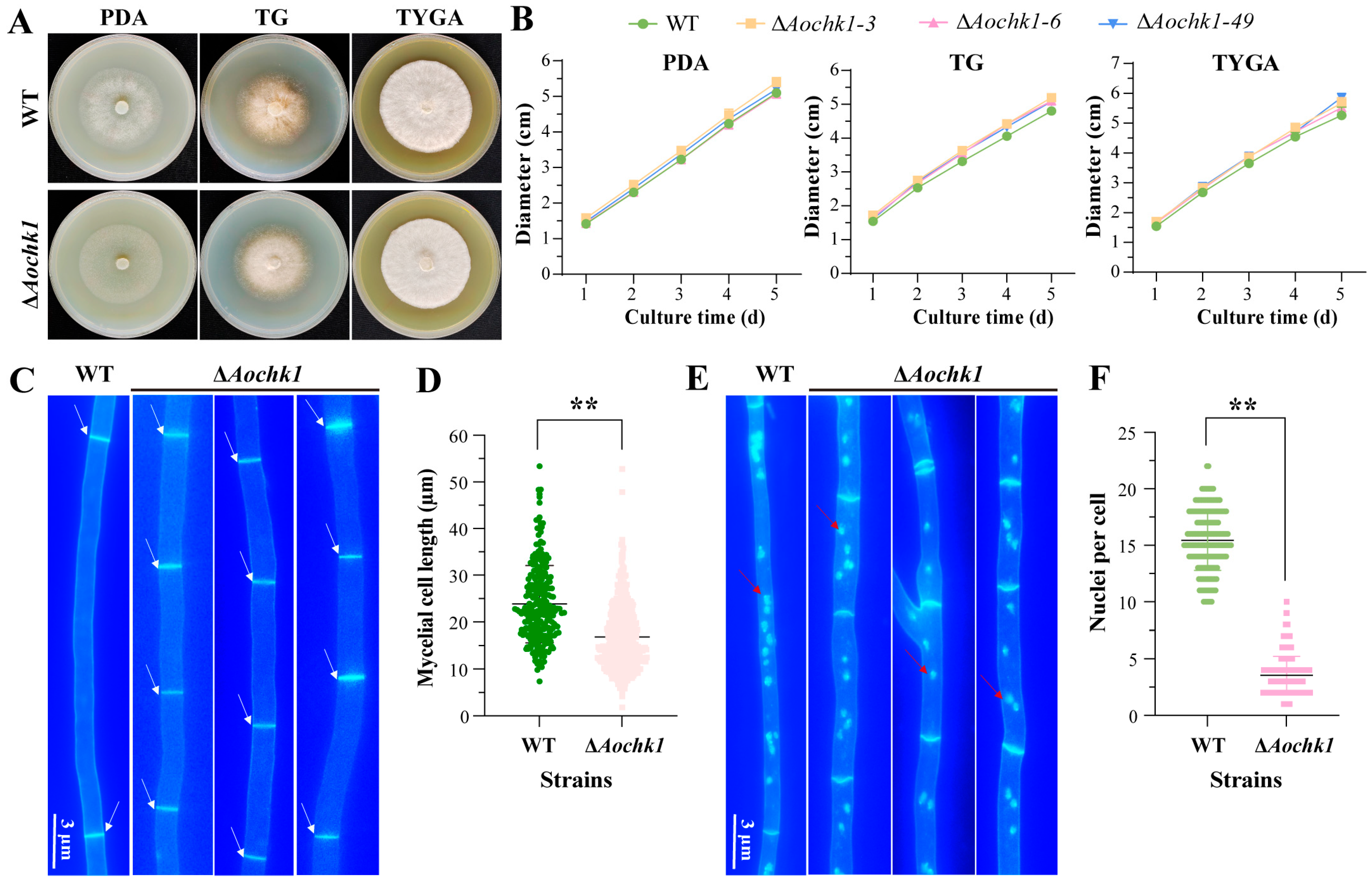
Figure 1. Comparison of hyphal growth rate and nuclear number between WT and ΔAochk1 mutant strains. (A) Colony morphology of WT and ΔAochk1 strains on different media. (B) Growth rates of WT and ΔAochk1 strains on PDA, TG, and TYGA media. (C) Hyphal septa in WT and ΔAochk1 strains, Bar = 3 μm, (white arrows indicate septa). (D) Hyphal compartment length of WT and ΔAochk1 strains. (E) Nuclear staining in hyphae of WT and ΔAochk1 strains, Bar = 3 μm, (red arrows indicate nuclei). (F) Quantitative comparison of nuclear number per hyphal compartment between strains. (Tukey’s HSD test: ** p < 0.01).
Jof 11 00602 g001
Figure 2. Comparative analysis of spore-related phenotypes between WT and ΔAochk1 mutant strains. (A) Representative images of spores produced by WT and ΔAochk1 strains after 3-day incubation on PDA medium, Bar = 100 μm. (B) Quantitative comparison of spore production. (C) Comparison of spore germination rates. (D) Relative expression levels of sporulation-related genes in WT and ΔAochk1 strains. (Tukey’s HSD test: * p < 0.05, ** p < 0.01).
Jof 11 00602 g002
Figure 3. Comparative analysis of trap formation and predation ability. (A) Representative images of nematode predation by traps following nematode induction, Bar = 50 μm, (arrows indicate traps). (B) Comparison of trap formation in WT and ΔAochk1 strains. (C) Pathogenicity assay of WT and ΔAochk1 strains against nematodes. (Tukey’s HSD test: * p < 0.05, ** p < 0.01, *** p < 0.001).
Jof 11 00602 g003
Figure 4. Transcriptomic profiling of the ΔAochk1 mutant strain. (A) Principal component analysis of transcriptomes from WT and ΔAochk1 mutant strains. (B) Bar plot showing the number of differentially expressed genes (DEGs). (C) Venn diagram of consistently downregulated DEGs shared at both 0 h and 24 h post-induction. (D) Venn diagram of consistently upregulated DEGs shared at both 0 h and 24 h post-induction. (E,F) Top 20 significantly enriched GO terms for DEGs identified at 0 h (E) and 24 h (F) (ΔAochk1 vs. WT).
Jof 11 00602 g004
Figure 5. Comparison of lipid droplets (LDs) and autophagy between WT and ΔAochk1 mutant strains. (A) Staining of LDs in hyphae of WT and ΔAochk1 strains. Red arrows: lipid droplets. Bar = 3 µm. (B) Transmission electron microscopy (TEM) of intracellular LDs in WT and ΔAochk1 strains, Bar = 2 μm. (C) Cluster analysis of the expression levels of lipid metabolism-related genes in WT and ΔAochk1 mutant strains. (D) Autophagosomes in hyphae of WT and ΔAochk1 strains. Yellow arrows: autophagosomes. Bar = 3 µm. (E) Observation of autophagosomes by TEM, Bar = 2 μm. (F) Cluster analysis of the expression levels of autophagy-related genes in WT and ΔAochk1 mutant strains.
Jof 11 00602 g005
Figure 6. Comparative secondary metabolomic profiling of WT and ΔAochk1 mutant strains. (A) HPLC chromatograms of secondary metabolites extracted from WT and ΔAochk1 strains. (B) Clustering analysis of differential metabolites between strains. (C) Volcano plot of upregulated and downregulated metabolites. (D) Comparison of arthrobotrisin relative abundance between WT and mutant strain. (E) KEGG enrichment analysis of differential metabolites. (Tukey’s HSD test: *** p < 0.001).
Jof 11 00602 g006
Disclaimer/Publisher’s Note: The statements, opinions and data contained in all publications are solely those of the individual author(s) and contributor(s) and not of MDPI and/or the editor(s). MDPI and/or the editor(s) disclaim responsibility for any injury to people or property resulting from any ideas, methods, instructions or products referred to in the content.