Abstract
Several Pleurocordyceps species have been reported as hyperparasitic fungi. A new species, Pleurocordyceps fusiformispora, and a known species, Perennicordyceps elaphomyceticola, are described here based on morphology and phylogenetic evidence from six genes (ITS, SSU, LSU, TET1-α, RPB1, and RPB2). Pl. fusiformispora differed from the other Pleurocordyceps species by producing flaky colonies, ovoid or elliptic α-conidia, and fusiform or long fusiform β-conidia. Both full genomes of Pe. elaphomyceticola and Pl. fusiformispora were sequenced, annotated, and compared. The antiSMASH and local BLAST analyses revealed significant differences in the number and types of putative secondary metabolite biosynthetic gene clusters, i.e., NPPS, PKS, and hybrid PKS–NRPS domains, between the two species. In addition, the putative BGCs of six compounds, namely ε-poly lysine, 4-epi-15-epi-brefeldin A, Monorden D/monocillin IV/monocillin VII/pochonin M/monocillin V/monocillin II, Tolypyridone, Piperazine, and Triticone DABFC, were excavated in the present study. This study motivates the use of heterologous expression and gene knockout methods to discover novel biologically active SMs from Polycephalomycetaceae.
1. Introduction
The classification system of Cordyceps sensu lato is widely accepted, with Perennicordyceps and Pleurocordyceps being sister genera within the family Polycephalomycetaceae [1]. Polycephalomycetaceae is a kind of fungus group with important application value and broad development and application prospects in medicine, agriculture, and other fields [1]. Xiao et al. [2] noted that Pleurocordyceps sp. exhibits significant potential for the production of a diverse range of secondary metabolites. Recent studies showed that Pleurocordyceps nipponicus has antibacterial [3,4] and antitumor activities [5]. Surapong et al. [6] isolated two compounds, Cordytropolone and Leptosphaerone A, from liquid cultures of Pl. nipponicus, and experimental studies showed that Cordytropolone has some antifungal activity.
Natural products, also known as secondary metabolites (SMs), have played an important role in the history of drug discovery and development [7]. As more and more microbial genomes are sequenced, bioinformatic analysis has revealed a vast resource of novel SMs. Genome mining is a new strategy of SM discovery based on gene cluster sequences and biosynthetic pathways. At the same time, it can directly associate the structures of SMs with synthetic pathways and facilitate the study of SM biosynthesis and combinatorial biosynthesis [8]. Some analysis tools and procedures (e.g., antibiotics and Secondary Metabolites Analysis Shell, antiSMASH; ClusterFinder; Antibiotic Resistant Target Seeker, ARTS) allow for the rapid and direct detection of biosynthetic gene clusters (BGCs), as well as the diversity of gene cluster families, such as polyketide synthases (PKSs), non-ribosomal peptide synthetases (NRPSs), and terpene synthases (TSs) [9]. More SM BGCs can be discovered through gene mining [10]. For instance, the sequence of amino acids of NpPKS3 in the lichenized fungus Nephrmopsis pallescens was 53% consistent with the type III PKS (CYSBs) of Beauveria bassiana [11]. Sayari et al. [12] found that all 20 genomes of Ceratocysti daceae showed extremely conserved PKS-III gene clusters containing homologous genes encoding the CHS. Wang et al. [9] discovered beauveriolide BGC in the Cordyceps militaris genome using bioinformatic analysis and then produced the compound via heterologous expression. Fumosorinone is a new 2-pyridone alkaloid isolated from Cordyceps fumosorosea [10]. The BGC of fumosorinone consisted of a hybrid PKS–NRPS, two cytochrome P450, a trans-enoyl reductase gene, and two other transcription regulatory genes [13]. In recent years, there has been little work on secondary metabolite gene clusters in Perennicordyceps and Pleurocordyceps.
In this study, the species Pl. fusiformispora Hong Yu bis and Z.H. Liu, D.X. Tang, Y.L. Lu, sp. nov. was first introduced. In order to discover more potential gene clusters of SMs, the whole genomes of Pl. fusiformispora and Pe. elaphomyceticola were sequenced and annotated and were used in gene mining studies. The potential of the Polycephalomycetaceae fungi to produce SMs was further analyzed.
2. Materials and Methods
2.1. Test Materials
Pe. elaphomyceticola was collected on 12 June 2022, suburb of Menghai County, Yunnan Province, China (21°58′10.09″ N, 100°27′30.89″ E, altitude: 1160.79 m). Pl. fusiformispora was collected on 23 July 2022, Wild Duck Lake Forest Park, Kunming City, Yunnan Province, China (25°6′24.98″ N, 102°50′12.28″ E, altitude: 2039.05 m). The voucher specimens were stored in Yunnan Herbal Herbarium (YHH) of Yunnan University, and the isolated strains were stored in Yunnan Fungal Culture Collection (YFCC) of Yunnan University.
2.2. Culture and Morphological Observations
Sexual morph observation was performed by photographing and measuring ascomata using an Olympus SZ61 stereomicroscope (Olympus Corporation, Tokyo, Japan). Freehand or frozen sections of the fruit body structure were placed in a solution of lactophenol cotton blue for microscopic study and photomicrography. The frozen sections were prepared using a freezing Microtome HM525NX (Thermo Fisher Scientific, Waltham, MA, USA). The micro-morphological characteristics of fungi (perithecia, asci, apical caps, and ascospores) were examined using Olympus CX40 and BX53 microscopes. The PDA solid media (20 g/L potato powder, 20 g/L glucose, 18 g/L agar powder, 1 L H2O (all chemicals and reagents were from Kunming City, China)) in which the colonies were cultivated were kept at room temperature (25 °C) for 40 days, and then the specimens were photographed and recorded using a Canon 750D camera (Canon Inc., Tokyo City, Japan) to observe the morphological properties of the colonies. The necessary microscope slide cultures were made according to [14], leaving them at 25 °C for 10 days. The colony was photographed and measured every fourth day. To characterize the strain, microscope slide cultures were generated by inoculating a small portion of the mycelium onto a 25 mm2 area of PDA medium (20 g/L potato powder, 20 g/L glucose, 18 g/L agar powder, 1 L H2O (all chemicals and reagents were from Kunming City, China)) block overlaid by a cover slip. Specimens were photographed and measured with an Olympus SZ61 stereomicroscope (Hamburg, Germany). Morphological observations and measurements were carried out using an Olympus CX40 microscope and a FEI QUANTA200 scanning electron microscope (Valley City, ND, USA). Fifty measurements were taken to collect the necessary information about the hypha, synnema, conidial mass, phialide, and conidium.
2.3. DNA Extraction, Polymerase Chain Reaction (PCR), and Sequencing
Specimens and axenic living cultures were prepared for DNA extraction. Total DNA was extracted using the CTAB method described by [15]. Polymerase chain reaction (PCR) was used to amplify genetic markers using the following primer pairs: LR0R/LR5 for small subunit nuclear ribosomal DNA (LSU) [16,17], EF1α-EF/EF1α-ER for translation elongation factor 1-α (TEF1-α) [18], RPB1-5′F/RPB1-5′R for partial RNA polymerase II largest subunit gene region (RPB1), RPB2-5′F/RPB2-5′R for partial RNA polymerase II second largest subunit gene region (RPB2) [19,20]. The PCR assay was completed with a final volume of 25 μL. Each reaction was composed of 2.5 μL of 10× Ex Taq Buffer (containing 2 mM MgCl2; TaKaRa, Kusatsu, Japan), 2 μL of 2.5 mM of each of the four dNTPs (TaKaRa), 1 μL of 10 μM of each primer, 0.25 μL of 5U Ex Taq DNA polymerase (TaKaRa), 2 μL of template DNA, and 17.25 μL ultrapure water. The LSU was amplified using the following PCR conditions: initial denaturation at 95 °C for 4 min and then 30 cycles of denaturation (each cycle at 95 °C for 1 min), annealing at 50 °C for 1 min, polymerization at 72 °C for 2 min, and a final extension at 72 °C for 8 min. PCR conditions for TEF1-a, RPB1, and RPB2 were performed as previously described [21]. PCR products were separated by electrophoresis in 1.0% agarose gels, purified using the Gel Band Purification Kit (Bio Teke Co., Ltd., Beijing, China), and then sequenced on an automatic sequence analyzer (BGI Co., Ltd., Yantian, Shenzhen, China). When PCR products could not be sequenced directly, cloning was performed by the TaKaRa PMD™18-T vector system (TaKaRa Biotechnology Co., Ltd., Dalian, China).
2.4. Genome Sequencing and Assembly
Pe. elaphomyceticola and Pl. fusiformispora strains were cultured on PPDA solid medium (20 g/L potato powder, 10 g/L yeast powder, 20 g/L glucose, 18 g/L agar powder, 1 L H2O) at 25 °C for 60 days. The mycelium was transferred to PPA liquid medium (20 g/L potato powder, 10 g/L yeast powder, 20 g/L glucose, 1 L H2O) at 25 °C static cultivation for 2–3 months. Appropriate amounts of Pe. elaphomyceticola and Pl. fusiformispora mycelium were scraped, and total genomic DNA was extracted using the Plant DNA Isolation Kit (Foregene Co., Ltd., Chengdu, China), and then sequenced on an automatic sequence analyzer (BGI Co., Ltd., Wuhan, China) using the same primers as used in amplification. Sequencing data contain some low-quality reads with joints, which can cause significant interference in subsequent information analysis. Illumina NovaSeq 2000 (Nanopore, Wuhan, China) high-throughput sequencing platform, used for sequencing a gene library with 400 bp insertion fragment, was used with a sequencing mode of paired-end and 2 × 150 bp. The fastp ([https://github.com/OpenGene/fastp], accessed on 14 July 2023) was used to filter the raw reads, discard low-quality reads, and obtain clean data. In order to ensure the quality of subsequent information analysis, it was necessary to further filter the raw data to generate high-quality sequences. The standards for data filtering mainly include the following points: joint contamination removal, using AdapterRemoval (v2.0) [22] to remove joint contamination from the 3 ‘end, and quality correction, using SOAPec (v2.0) software to perform quality correction on all reads based on Kmer frequency, with a Kmer setting of 17 used for correction. A5-MiSeq and SPAdes were used to construct contig and scaffold. Finally, the assembly effects of contigs and scaffolds were evaluated using pilon v1.18 [23] software.
2.5. Gene Prediction and Annotation
A combination of de novo gene prediction, transcript mapping, and homologous searches were used for gene prediction. Based on the existing database on gene function and metabolic pathways, the predicted genes were annotated by BLAST search, including Kyoto Encyclopedia of Genes and Genomes (KEGG), NCBI non-redundant protein sequences (NR), Gene Ontology (GO), Cluster of Orthologous Groups of eukaryotic complete genomes (KOG), Pfam, and Interpro.
2.6. Analysis of Secondary Metabolite Biosynthesis Gene Cluster
The antiSMASH (https://antismash.secondarymetabolites.org/, accessed on 1 September 2023) online program was used to perform gene cluster prediction at the level of genomic scaffolds of Pe. elaphomyceticola and Pl. fusiformispora complex. Based on antiSMASH-detected scaffolds with gene clusters, the online program FGENESH (www.softberry.com/, accessed on 26 September 2023) was used to predict gene structures using Ophiocordyceps sinensis as a parameter. To obtain the domain, the PKS/NRPS online program (https://nrps.igs.umaryland.edu/, accessed on 11 October 2023) was used to predict gene clusters in contigs where NRPS/PKS genes were located. At the same time, the online program Protein BLAST (https://blast.ncbi.nlm.nih.gov/, accessed on 25 October 2023) was used for NRPS/PKS genes of contig protein ratio analysis.
2.7. Cluster Analysis
The polygene nucleotide sequences (ITS, SSU, LSU, TEF-1a, RPB1, RPB2) were downloaded from NCBI (https://www.ncbi.nlm.nih.gov/, accessed on 25 August 2023) and compared with these sequences using the Clustal W program in the MEGA5.0 software for multi-sequence comparison [24,25]. Based on a six-gene dataset, the software PhyloSuite (v1.2.2 Win) was used to construct phylogenetic tree of maximum likelihood (ML) and Bayesian inference (BI). In addition, we used the method from the online program IQ-TREE web server (http://iqtree.cibiv.univie.ac.at/, accessed on 15 July 2023) since the report 1000 and the rest of the default parameter to construct an ML cluster analysis tree of NRPS or hybrid PKS–NRPS proteins of Pe. elaphomyceticola, Pl. fusiformispora, and other fungi.
3. Results
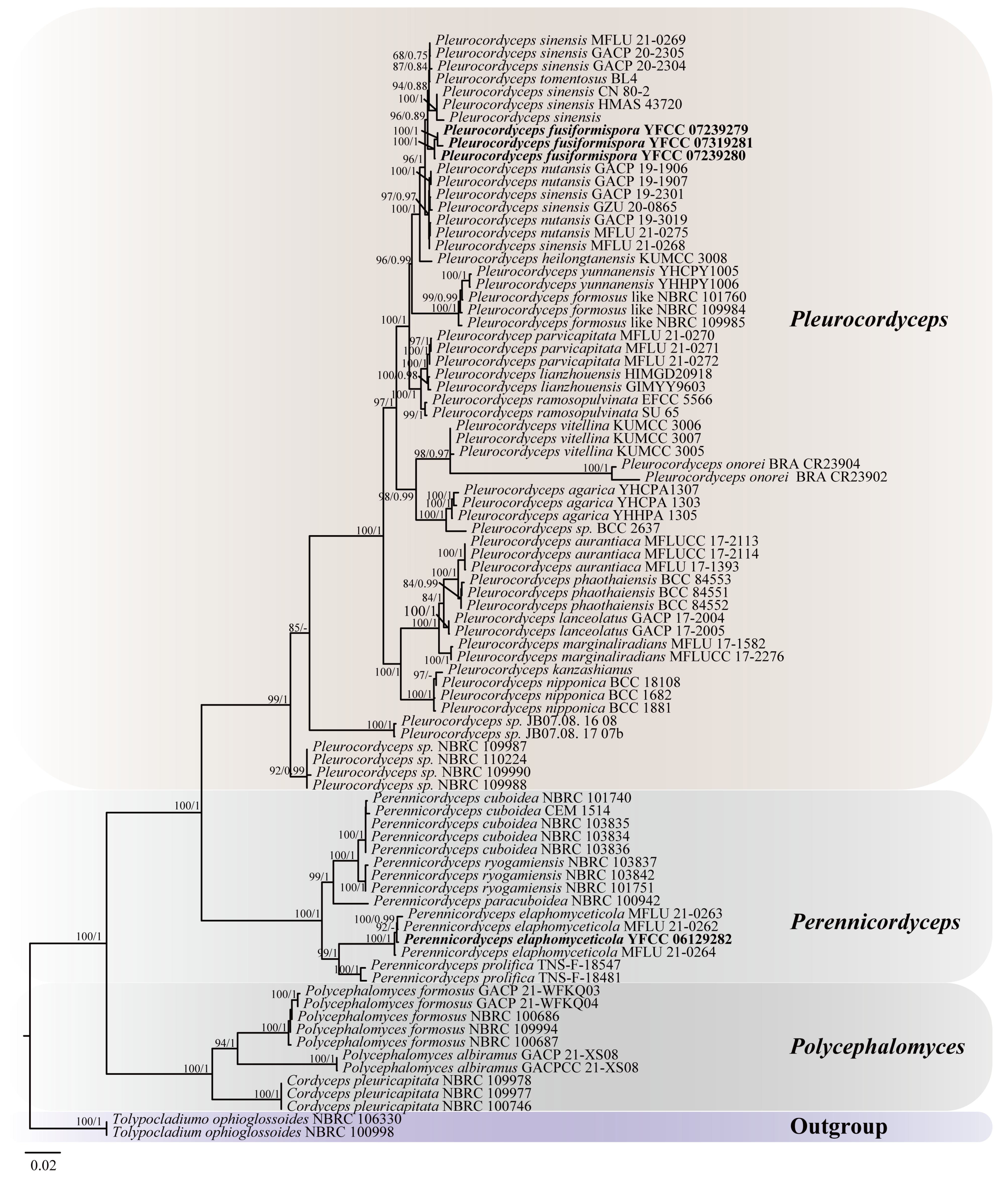
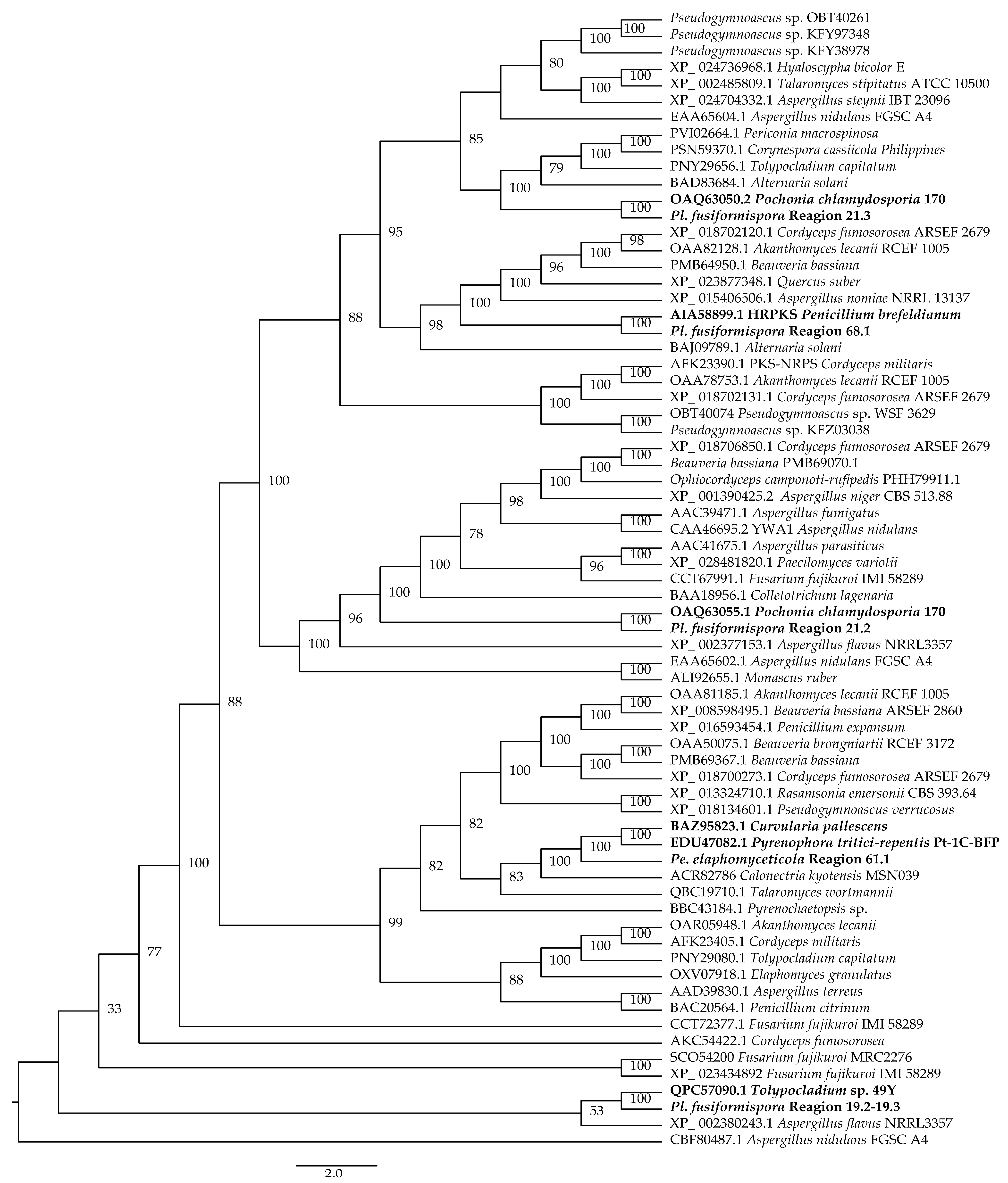
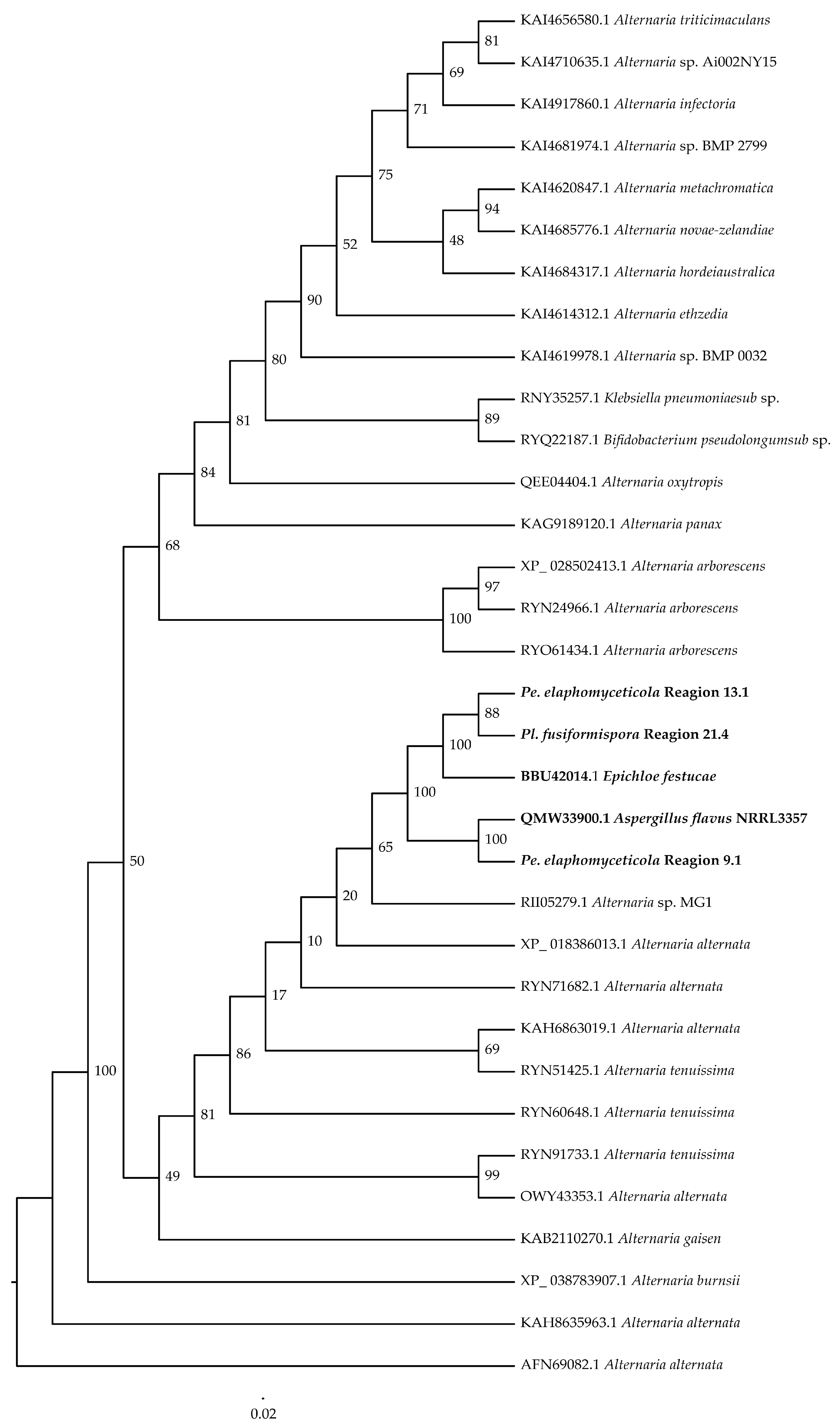
3.1. Phylogenetic Analysis
Based on the joint matrix of nucleotide sequences of ITS, SSU, LSU, TEF1-α, RPB1, and RPB2, the molecular phylogenetic tree of Polycephalomycetaceae was reconstructed by ML and BI. The total length of the concatenated dataset of six genes across the 86 samples was 5303 bp, including 554 bp for ITS, 920 bp for SSU, 911 bp for LSU, 975 bp for TEF-1α, 739 bp for RPB1, and 1,204 bp for RPB2. The phylogenetic tree, represented by Tolypocladium ophioglossoides NBRC 106330 and T. ophioglossoides NBRC 100998 as the outgroup taxa, consisted of three clades, i.e., Polycephalomyces clade, Perennicordyceps clade, and Pleurocordyceps clade, with a total of 86 sequences (Table 1), of which 4 were self-detected. The matrix had 2646 distinct patterns, 1669 parsimony-informative sites, 666 singleton sites, and 13,982 constant sites. ModelFinder was used to select the best-fitting likelihood model (GTR+F+I+G4) for ML analyses and BI analyses according to the Akaike information criterion (AIC). The ML and BI tree with the best score found was −40,864.213, and the total tree length was 1.098. The generic-level relationships of ML and BI trees were topologically similar. The tree was visualized with its maximum likelihood bootstrap proportions (ML-BS) and Bayesian posterior probability (BI-BPP) in Figtree v.1.4.3 (Figure 1).
Table 1.
Sources of selected isolates and GenBank accession number for ITS and five genes of three genera within Polycephalomycetaceae in this study.
Figure 1.
Phylogenetic tree of Polycephalomycetaceae was constructed, based on the concatenation of ITS, SSU, LSU, TEF1-α, RPB1, and RPB2 sequence data. The phylogeny was inferred using the IQ-tree. The maximum likelihood bootstrap valued greater than 60% (on the left) and the Bayesian posterior probabilities over 0.6 (on the right) were indicated above the nodes. The new species and known ones were indicated in back bold font.
From the perspective of a six-gene phylogenetic tree, the three branches of the Polycephalomycetaceae receive high support rates. The cluster of Pl. fusiformispora collected and described in this study was in the adjacent branches of Pl. sinensis. Pl. fusiformispora and Pl. sinensis were sister species to each other and formed a separate clade (BS = 96%, BPP = 0.89). Pe. elaphomyceticola and Pe. prolifica were sister species to each other and formed a separate clade (BS = 99%, BPP = 1). This was consistent with the results [1].
3.2. Taxonomy
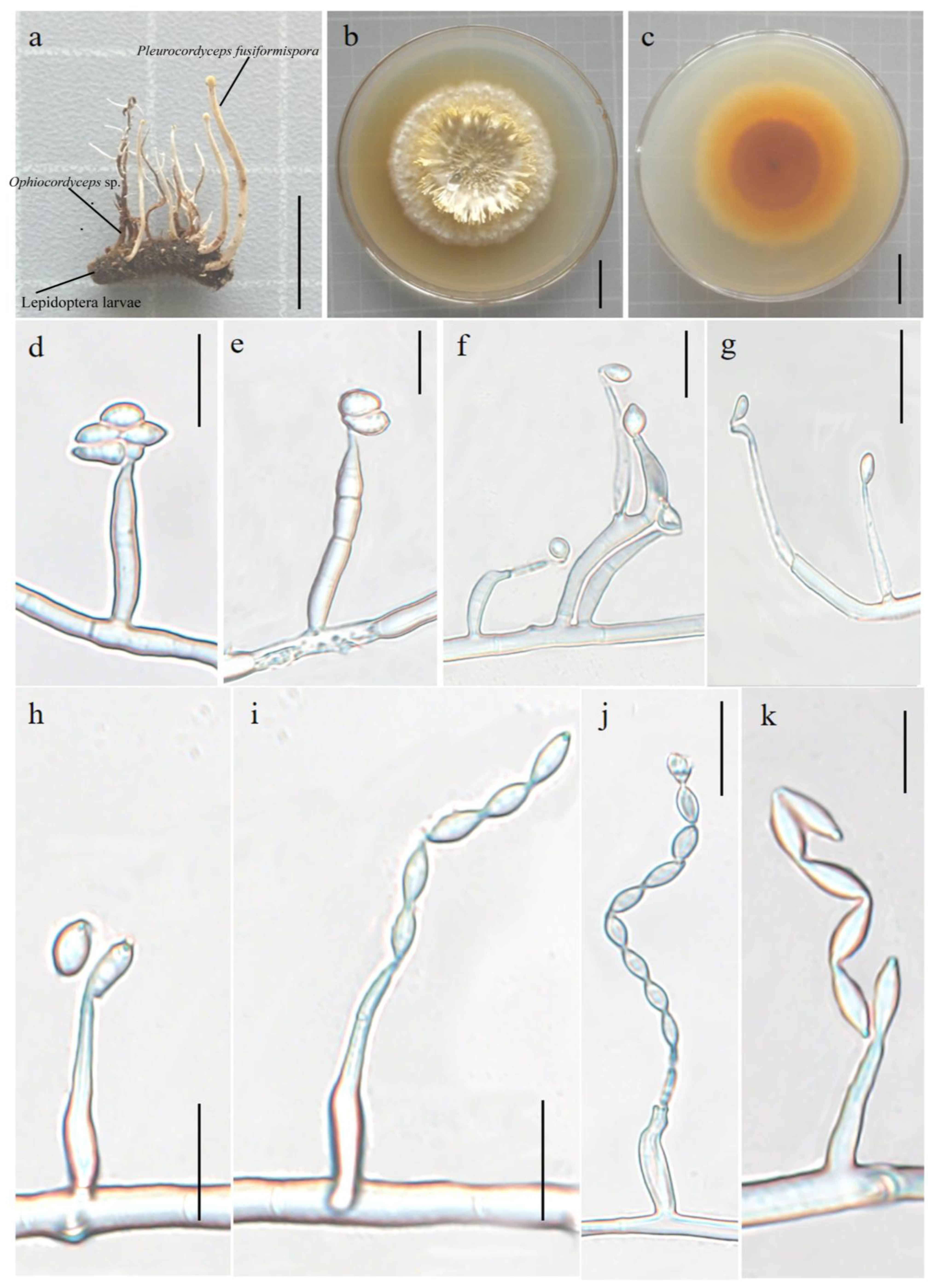
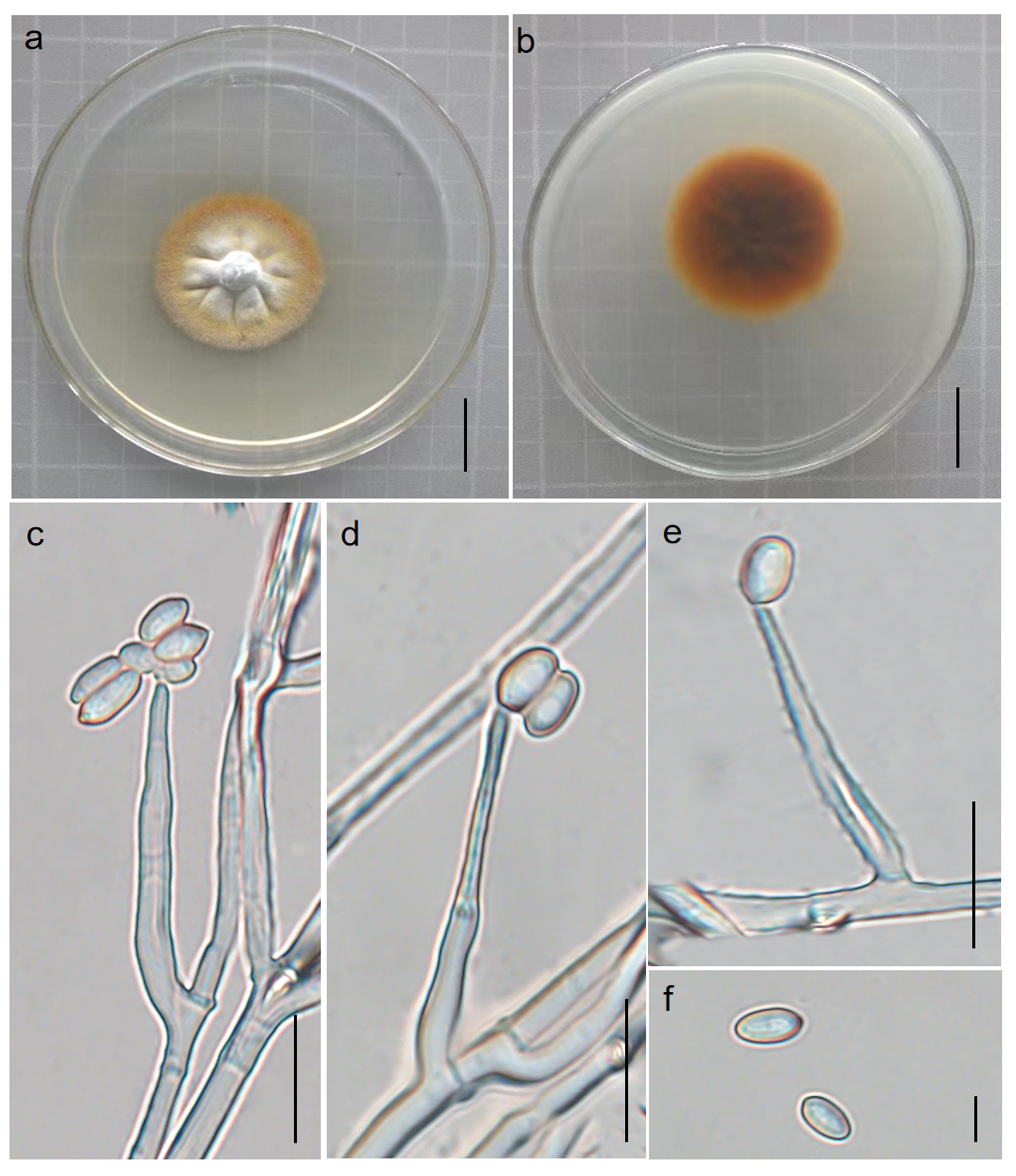
3.2.1. Pleurocordyceps fusiformispora Hong Yu bis and Z.H. Liu, D.X. Tang, Y.L. Lu, sp. nov. Figure 2
MycoBank: MB 851478
Etymology: The species name refers to the production of fusiform or long fusiform conidia during the asexual phase. Hyperparasitic on Ophiocordyceps sp. (Ophiocordycipitaceae) and on insects buried in soil. Sexual morph: undetermined. Asexual morph: Synnemata measure 1.2–1.5 cm long by 0.1–0.5 mm wide, clavate, capitate, crowd on the insect body and Ophiocordyceps sp., unbranched, white to yellowish, with or without fertile head at the apex. Stipes 2.1–2.8 cm long, 0.7–1.0 mm wide. Colonies on PDA grow slowly, attaining a diameter of 5.1–5.5 cm in 40 days at 25 °C, clustered, white–yellow, and reverse dry yellow. Synnemata emerge after 20 days, flaky, branched, 1.4–1.9 cm long and 0.3–0.5 cm wide, showing radiating distributions. Phialides exist in two types: α- and β-phialides. Both types of phialide often produce new phialides at their own apices or yield catenulate β-conidia, collarettes not flared, periclinal thickening not visible. The α-phialides are acropleurogenous on conidiophores and solitary on hyphae, narrow lageniform or subulate, taper abruptly from the base to the apex, 8.42–20.9 μm long, 1.3–2.9 μm wide at the base, and 0.6–1.6 μm wide at the apex. The β-phialides are solitary on hyphae, lanceolate, taper gradually from the base to the apex, 8.7–14.8 μm long, 2.5–3.1 μm wide at the base, and 0.9–1.1 μm wide at the apex. α-conidia are ovoid or elliptic and occur in the conidial mass on the agar or on the final portion of synnema, 2.9–4.8 × 1.3–3.1 μm. β-conidia are fusiform or long fusiform and are produced on the surface mycelium of colony, usually in chains on a phialide, 2.8–4.4 × 1.7–2.8 μm.
Material examined: Wild Duck Lake Forest Park, Kunming City, Yunnan Province, China, hyperparasitic on Ophiocordyceps sp. (Ophiocordycipitaceae) and on insects buried in soil. Collected on 23 July 2022, holotype: YFCC 07239279, paratype: YFCC 07239280, other collections: YFCC 07319281.
Notes: Pleurocordyceps fusiformispora was sister to the clade formed by Pl. sinensis (Figure 1: 94% ML/0.88 PP). Pl. fusiformispora differs from the other Pleurocordyceps species by producing flaky colonies, their α-conidia are ovoid or elliptic, and their β-conidia are fusiform or long fusiform (Table S4).
Figure 2.
Pleurocordyceps fusiformispora (holotype: YFCC 07239279). (a) Overview of Pleurocordyceps fusiformispora and its host. (b,c) Colony, obverse and reverse. (d–h) α-phialides and α-conidia. (i–k) β-phialides and β-conidia. Scale bars: (a) = 1cm; (b,c) = 2 cm; (d–f,h–j) = 10 µm; (g) = 20 µm; (k) = 5 µm.
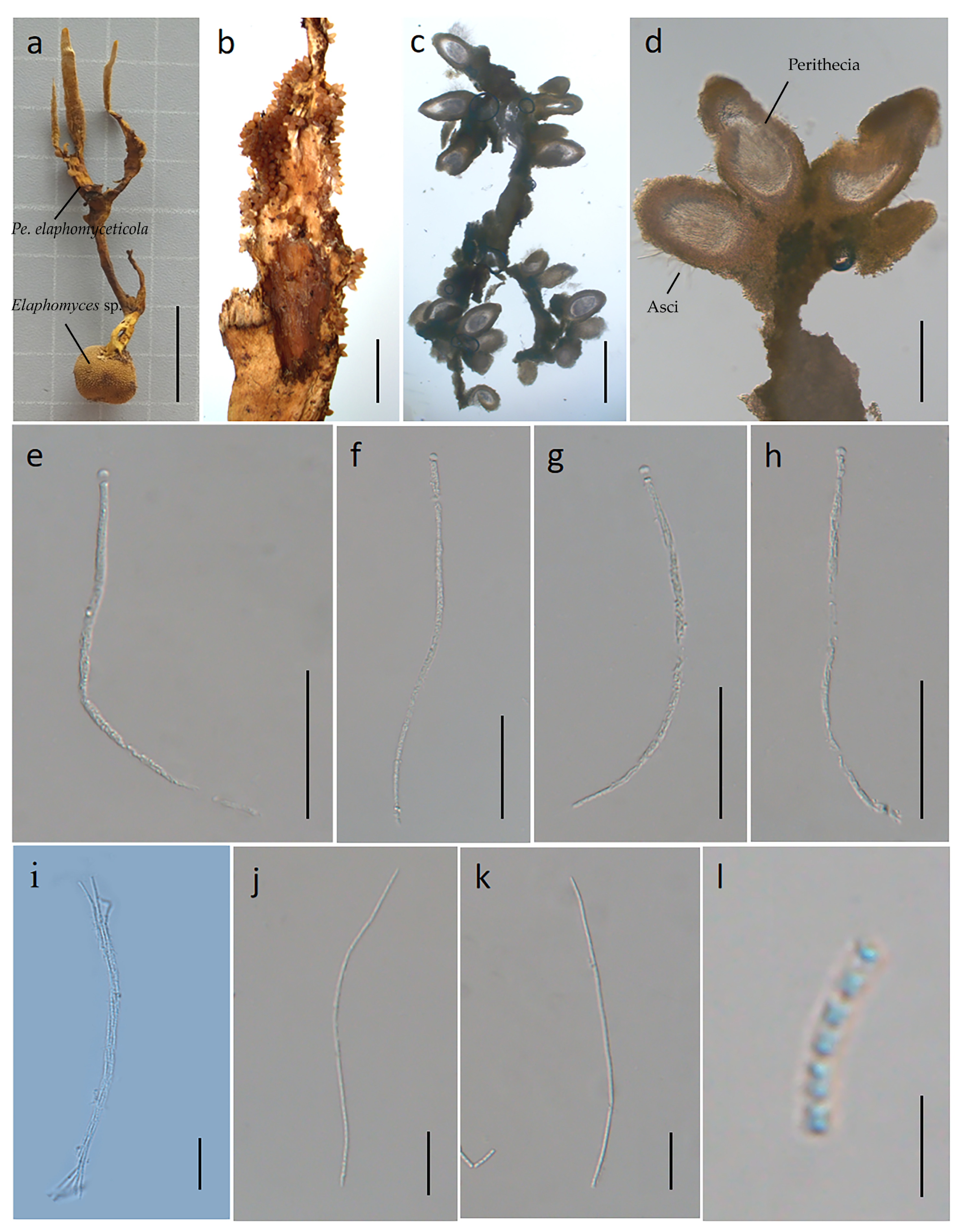
3.2.2. Perennicordyceps elaphomyceticola (WY Chuang, Ariyawansa, Jiuein Yang and Stadler) Y.P. Xiao and K.D. Hyde, comb. nov [1], Figure 3 and Figure 4
Synonymy: Polycephalomyces elaphomyceticola W.Y. Chuang, H.A. Ariyaw., J.I. Yang and Stadler, in Yang, Stadler, Chuang and Ariyawansa, Mycol. Progr. 2020, 19(1): 102.
Etymology: The specific epithet elaphomyceticola is based on the host genus from which the fungus was isolated. Parasitic on Elaphomyces sp. (Elaphomycetaceae) from soil. Sexual morph: stromata 5.1–6.2 cm long, 0.5–0.7 cm wide, cylindrical, solitary or several, branched, the color gradually becomes lighter towards the apex, yellow to dark yellow to light yellow, hard. Fertile heads: 1.5–2 cm long, 0.1–0.3 cm wide, branched, dark yellow to light yellow, upper surface roughened. Perithecia: 259–519 × 152–291 μm, superficial, ovoid to ellipsoid. Asci: 164–173 × 3.1–5.5 μm, hyaline, cylindrical. Apical cap: 2.1–3.5 × 3.6–4.2 μm, thin, hyaline. Ascospores: 55.1–105 × 0.8–1.2 μm, irregular multiseptate. Secondary spores: 0.8–1.1 × 0.6–0.8 μm globose to cylindrical, one-celled, hyaline, smooth-walled. Asexual morph: (see Figure 4) colonies on PDA 3.7–4.0 cm in diameter after 40 days at 25 °C, usually verrucose, white to orange-yellow. On the reverse appear vague concentric rings, black–brown in the center and maple-colored at the edge. Phialides develop from the edge of the colony and conidial mass of the synnema. Phialides: cylindrical to subulate at the base, occur directly on the aerial hyphae, 16.8–31.9 µm in length, taper gradually from 2.0–3.8 µm at the base to 0.4–1.1 µm at the apex, generating a single or lumpy conidia. Conidia: oval, 3.2–5.1 × 0.4–1.2 µm.
Material examined: Suburb of Menghai County, Yunnan Province, China. Parasitic on Elaphomyces sp. in soil, 12 June 2022, YFCC 06129282, Hong Yu.
Figure 3.
Sexual morph of Perennicordyceps elaphomyceticola (YFCC 06129282). (a) Stromata emerging from infected Elaphomyces sp. (b) Fertile head of ascostroma. (c) Vertical section of stroma. (d) Perithecia. (e–h) Asci. (i) Asci and ascospore. (j,k) Ascospore. (l) Secondary ascospores. Scale bars: (a) = 2 cm; (b) = 5000 µm; (c) = 500 µm; (d) = 200 µm; (e–h) = 50 µm; (i–k) = 20 µm; (l) = 5 µm.
Figure 4.
Asexual morph of Perennicordyceps elaphomyceticola (YFCC 06129282). (a,b) Colony obverse and reverse. (c–e) Phialide and conidia. (f) Conidia. Scale bars: (a,b) = 2 cm; (c–e) = 10 µm; (f) = 5 µm.
3.3. Basic Genomic Characteristics of Perennicordyceps elaphomyceticola and Pleurocordyceps fusiformispora
3.3.1. Genome Sequencing and Assembly
The Illumina sequencing produced 24,006,990 raw reads and 3,600,527,330 high-quality reads of Pl. fusiformispora (Table S1). The size of the genome Pe. elaphomyceticola was 31.51 Mb, containing 48.79% GC content and 7794 protein-coding genes along with 103 tRNA, 18 rRNA, and 52 ncRNA. Among the genome size of Pl. fusiformispora was 33.02 Mb, containing GC content, protein-coding genes, tRNA, rRNA, and ncRNA, which were 50.08%, 8804, 100, 23 and 3, respectively. The results showed that the genome assembly of these two species was of good quality. In phylogeny, Pleurocordyceps and Perennicordyceps were sister genera (Figure 1). According to the above data, these results suggested that the closer the relationship between genera, the probable smaller the differences in genome size, GC content, and total gene number.
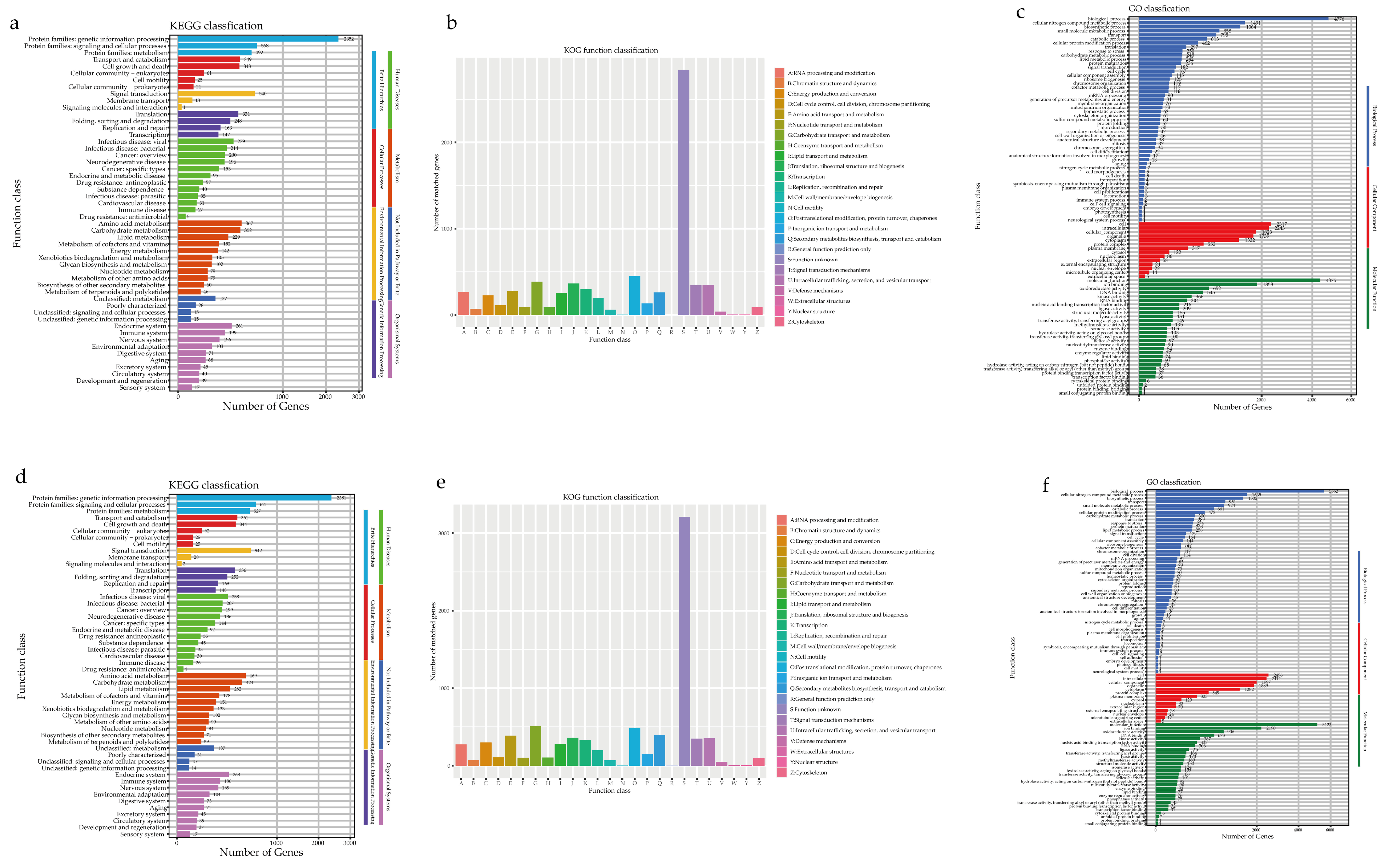
3.3.2. Genome Annotation
The Pe. elaphomyceticola species were identified using the KEGG databases (3509 genes/45.02%), the EggNOG databases (7196 genes/92.32%), the NR databases (7536 genes/96.68%), the GO database (5302 genes/68.02%), and the Pfam databases (5927 genes/76.04%). However, an analysis of 6706 non-redundant Pl. fusiformispora genes in a publicly available protein sequence database produced mixed results; these were from the KEGG databases (3723 genes/42.28%), the EggNOG databases (8183 genes/92.94%), the NR databases (8566 genes/97.29%), the GO database (5112 genes/69.50%), and the Pfam databases (6726 genes/76.39%) (Table S1). The KEGG functional classification of Pe. elaphomyceticola and Pl. fusiformispora indicated protein families for genetic information processing, signaling and cellular processing, and signal transduction (respectively, Figure 5b,e). In addition, the rich genetic information and diversity of signaling proteins may facilitate more efficient information exchange and secondary metabolism.
Figure 5.
Functional annotation of Pe. elaphomyceticola and Pl. fusiformispora genes encoding the proteins. (a–c) Pe. elaphomyceticola (a) KEGG, (b) KOG, (c) GO; (d–f) Pl. fusiformispora (d) KEGG, (e) KOG, (f) GO).
According to the EggNOG database, Pl. fusiformispora and Pe. elaphomyceticola captured more predicted genes as “Function unknown”, “Posttranslational modification, protein turnover, chaperones”, and “Carbohydrate transport and metabolism” (Figure 5a,d). However, a wide variety of carbohydrate metabolism and post-translational events suggested that they enhanced the bioactivity and energy conversion efficiency of regulatory proteins. GO annotation of the genomes of both species showed that translation, protein transport, and glucose metabolism were abundant genes in biological processes (Figure 5c,f). Furthermore, the GO annotation revealed integral components of the membrane, nucleus, and cytoplasm from the cellular component and ATP binding, metal ion binding, and zinc ion binding from the molecular function. The two species of Pe. elaphomyceticola and Pl. fusiformispora were wild strain, in which many metabolic genes might be involved in signal transduction.
3.3.3. Additional Annotation
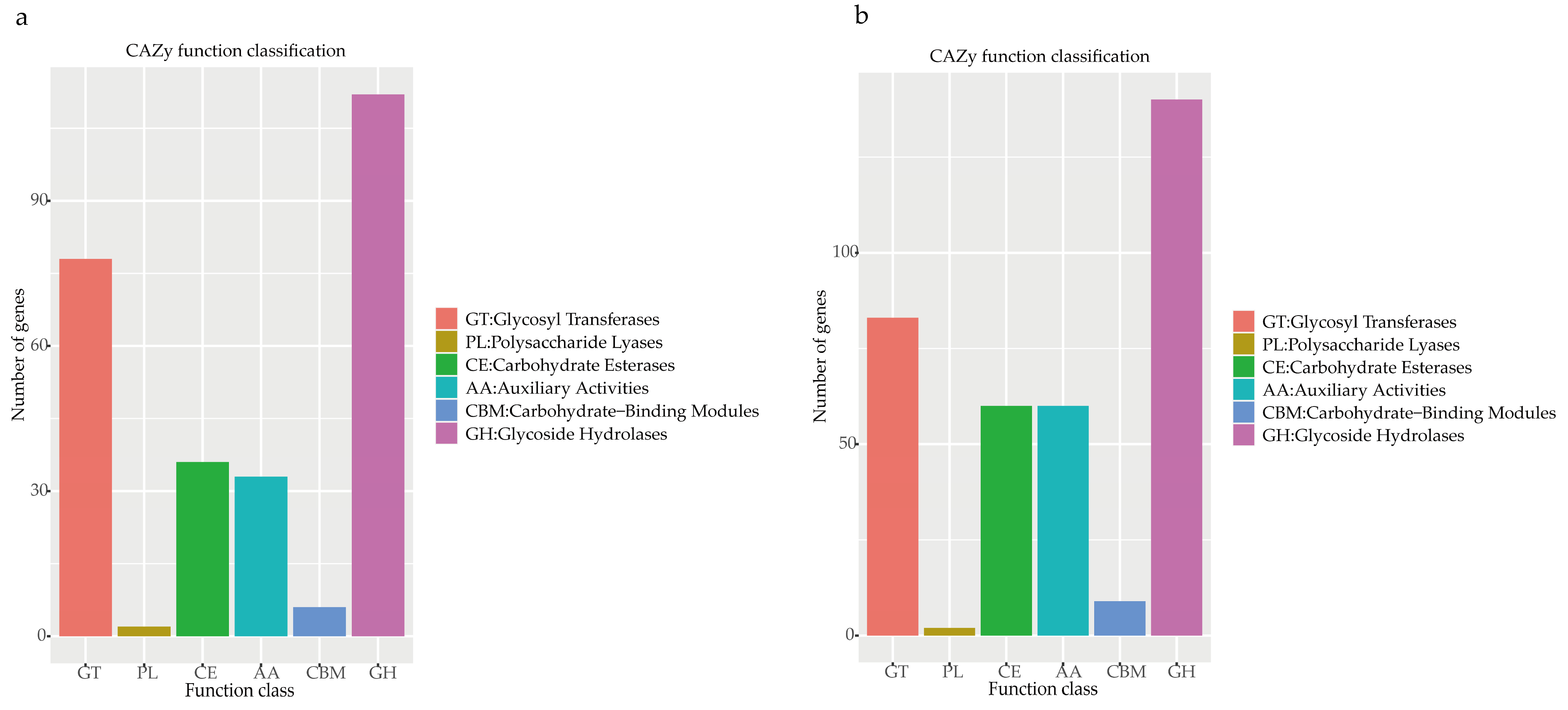
Carbohydrate-Active Enzymes (CAZy)
Enzymes that played an important role in carbohydrate modification, biosynthesis, and the degradation of fungi were carbohydrate-active enzymes (CAZy) [36], which are present in a database of carbohydrate-active enzymes and a special database of carbohydrate enzymes [37]. It was shown that Pe. elaphomyceticola (Figure 6a) and Pl. fusiformispora (Figure 6b) had many glycoside hydrolases (GHs), glycosyl transferases (GTs), and auxiliary activities (AAs), leading us to speculate that Pe. elaphomyceticola and Pl. fusiformispora possibly breakdown complex carbohydrates and capture more energy.
Figure 6.
CAZy functional classification chart of Pe. elaphomyceticola and Pl. fusiformispora. (a) Pe. elaphomyceticola (b) Pl. fusiformispora.
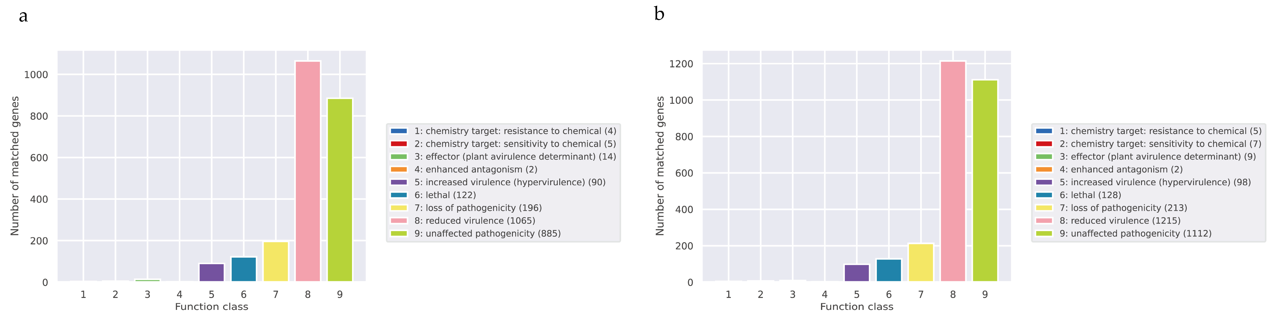
Pathogen–Host Interactions (PHI)
The full name of PHI is Pathogen Host Interactions Database, which is a database of pathogen–host interactions, mainly derived from fungi, oomycetes, and bacterial pathogens. The infected hosts include animals, plants, fungi, and insects [38]. This database plays an important role in searching for target genes for drug intervention, and it also includes antifungal compounds and corresponding target genes. The results showed that Pe. elaphomyceticola (Figure 7a) and Pl. fusiformispora (Figure 7b) had reduced virulence and the loss of pathogenicity genes. The primary annotated genes of Pe. elaphomyceticola and Pl. fusiformispora from the PHI database were used to reduce virulence and do not affect pathogenicity, indicating that Pe. elaphomyceticola and Pl. fusiformispora are not highly pathogenic strains.
Figure 7.
Distribution of the mutation types in the PHI phenotype of Pe. elaphomyceticola and Pl. fusiformispora. (a) Pe. elaphomyceticola (b) Pl. fusiformispora.
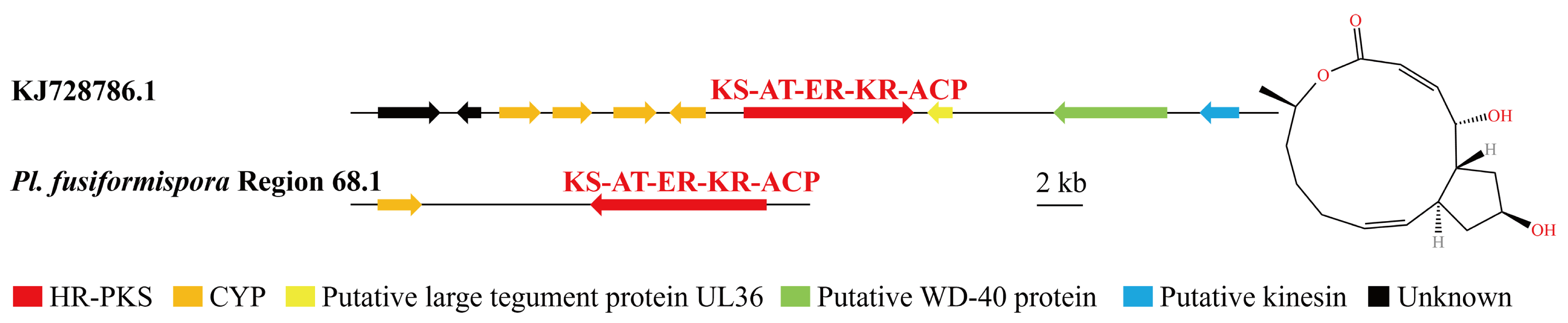
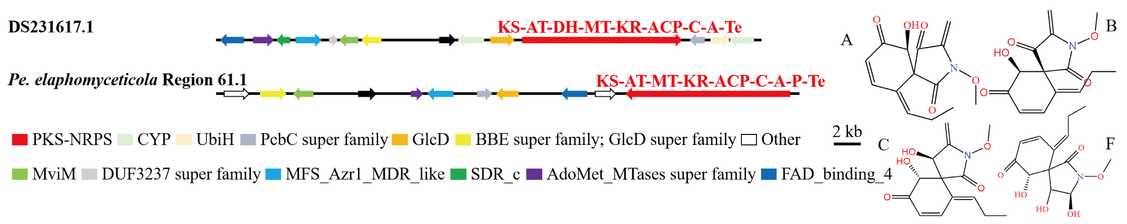
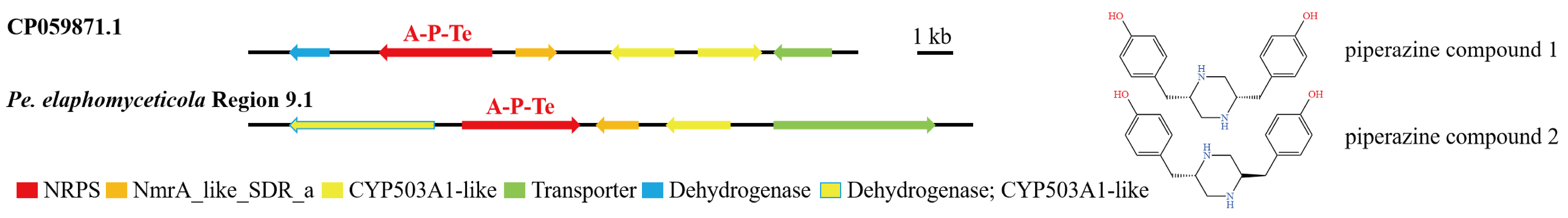
3.4. Analysis of Secondary Metabolite Biosynthesis Gene Cluster
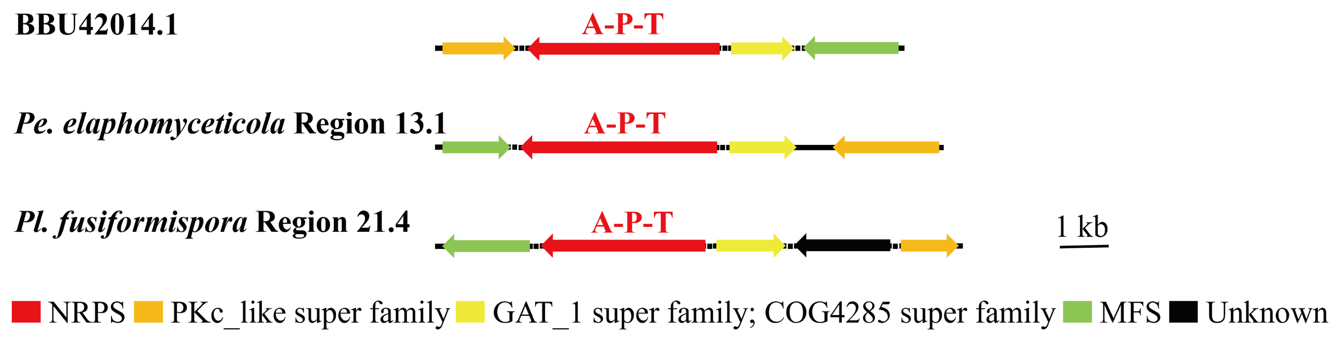
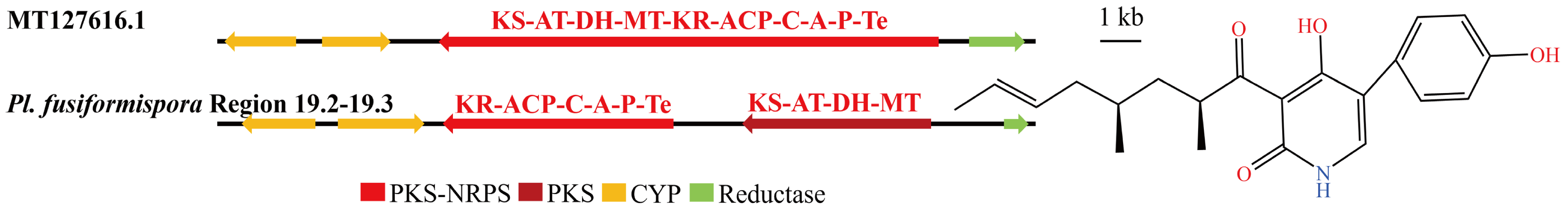
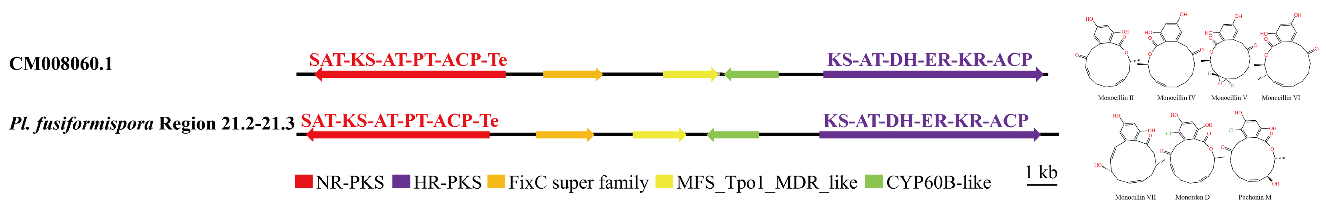
Overview of Twelve Genomic BGCs of Pe. elaphomyceticola and Pl. fusiformispora
AntiSMASH and local BLAST analyses revealed that Pe. elaphomyceticola (39) and Pl. fusiformispora (50) had different putative SM BGCs (Table S3). Pl. fusiformispora and Pe. elaphomyceticola predicted that the largest number of SM BGC types were NRPS, with 16 and 15, respectively. Pl. fusiformispora had five hybrids, PKS + NRPS, and Pe. elaphomyceticola had two. Both Pl. fusiformispora and Pe. elaphomyceticola had five terpenes and two hybrids, NRPS + other genes. Pl. fusiformispora and Pe. elaphomyceticola had seven and six other genes, respectively. For the predicted PKSs, Pl. fusiformispora had fifteen PKSs, while Pe. elaphomyceticola had nine. The Pl. fusiformispora genome had fifteen PKSs, including ten HR–PKSs, three non-reducing (NR) PKSs, and two partially reducing (PR) PKSs. The genome of Pe. elaphomyceticola had nine PKSs, including four NR-PKSs, four HR-PKSs, and one PR-NRPS. These results indicated that the number and type of SM BGCs obtained in different species vary.
The predicted BGCs had different levels of genetic homology compared by known cluster in the MIBiG database, with Pl. fusiformispora having the highest homology (42%), followed by Pe. elaphomyceticola (41.02%). Both Pe. elaphomyceticola and Pl. fusiformispora were predicted to catalyze the synthesis of ε-poly-lysine and Squalestatin S1 gene clusters. Pl. fusiformispora was predicted to catalyze the synthesis of Fusaric acid, Choline, Cyclopiazonic acid, IlicicolinH, Lucilactaene, Ascochlorin, YWA1, Tolypyridone, Monorden D/monocillin IV/monocillin VII/pochonin M/monocillin V/monocillin II, Fusarin C, AKML A-C, UNLL-YC2Q1O94PT, 4-epi-15-epi-brefeldin A, and Ergotamine (Table S2). In contrast, the BGCs Pe. elaphomyceticola predicted biosynthesis genes that catalyzed the synthesis of piperazine compound 1 and piperazine compound 2, Triticone DABFC, and Viriditixin. Further, the types and amounts of compounds predicted by catalytic synthesis differ between the two species.
Several BGCs of Pe. elaphomyceticola and Pl. fusiformispora were 100% similar to MIBiG sequences. Purev et al. [39] found that ε-poly-lysine had antifungal activity. The predicted Pe. elaphomyceticola Region 13.1 and Pl. fusiformispora Region 21.4 were responsible for ε-poly-lysine (Figure 8). The local BLAST comparison demonstrated that ε-poly-lysinehas requires enzymes with the A-P-T structural domain, PKc-like super family, GAT_1 super family, and MFS. Both species have the potential to synthesize ε-poly-lysinehas. Zhang et al. [40] studied genome mining of in-house strains using hybrid PKS–NRPS as a query and identified an endophyte Tolypocladium sp. 49Y, which possesses a potential 4-hydroxy pyridone biosynthetic gene cluster. Moreover, heterologous expression in Aspergillus oryzae NSAR1 revealed that this gene cluster was functional and able to produce a rare type of 4-hydroxy pyridones called Tolypyridone. The predicted Region 19.2 and Region 19.3 of Pl. fusiformispora might be responsible for Tolypyridone biosynthesis (Figure 9). Qin et al. [41] proved that Monorden D/monocillin IV/monocillin VII/pochonin M/monocillin V/monocillin II compounds have modest antibacterial activities. By comparison, Pl. fusiformispora Region 21.2 and Region 21.3 had highly homologous regions, being highly similar to the synthetic Monorden D/monocillin IV/monocillin VII/pochonin M/monocillin V/monocillin II gene cluster (Figure 10). These results indicated that the catalytic synthesis of NR-PKS (SAT-KS-AT-PT-ACP-Te), HR-PKS (KS-AT-DH-ER-KR-ACP), FixC super family, MFS_Tpol_MDR_like, and CYP60B-like were necessary genes. The results showed that (Figure 11) Pe. elaphomyceticola Region 61.1 was like the gene cluster DS231617.1 that catalyzed the synthesis of Triticone ABFC. However, Pl. fusiformispora did not have the cluster of Triticone ABFC. Pe. elaphomyceticola Region 9.1 was highly similar to the known gene cluster that catalyzed the synthesis of piperazine (Figure 12). The slight difference was that one gene in Pe. elaphomyceticola Region 9.1 had dehydrogenase and CYP503A1-like functions. Moreover, KJ728786.1 synthesized 4-epi-15-epi-brefeldin A (Figure 13). The core gene structural domain (KS-AT-ER-KR-ACP) in Pl. fusiformispora Region 68.1 was similar to KJ728786.1. However, Pl. fusiformispora lost some of its modified genes during evolution. It was speculated that the position direction of key enzymes and modified genes was different when different genera were in the compound. At the same time, there will be gene fusion and gene loss in the process of evolution.
Figure 8.
Comparison of putative BGC of ε-poly lysine in Pe. elaphomyceticola and Pl. fusiformispora. The number after the region and the number before the decimal point represent the scaffold, and the number after the decimal point represents the gene cluster.
Figure 9.
Comparison of biosynthesis of putative Tolypyridone in Pl. fusiformispora. The number after the region and the number before the decimal point represent the scaffold, and the number after the decimal point represents the gene cluster.
Figure 10.
Comparison of biosynthesis of putative Monorden D/monocillin IV/monocillin VII/pochonin M/monocillin V/monocillin II in Pl. fusiformispora. The number after the region and the number before the decimal point represent the scaffold, and the number after the decimal point represents the gene cluster.
Figure 11.
Comparison of biosynthesis of putative Triticone DABFC in Pe. elaphomyceticola. The number after the region and the number before the decimal point represent the scaffold, and the number after the decimal point represents the gene cluster.
Figure 12.
Comparison of biosynthesis of putative piperazine compound 1 and piperazine compound 2 in Pe. elaphomyceticola. The number after the region and the number before the decimal point represent the scaffold, and the number after the decimal point represents the gene cluster.
Figure 13.
Comparison of biosynthesis of putative 4-epi-15-epi-brefeldin A in Pl. fusiformispora. The number after the region and the number before the decimal point represent the scaffold, and the number after the decimal point represents the gene cluster.
3.5. Cluster Analysis
The protein clustering results of NRPS and hybrid PKS–NRPS from Pe. elaphomyceticola and Pl. fusiformispora were compared to other fungal NRPS and hybrid PKS–NRPS. It was showed that Pe. elaphomyceticola Region 13.1 and Pl. fusiformispora Region 21.4 aggregated with Epichloe festucae (BBU42014.1), with being catalyzed the biosynthesis of ε-poly-lysine or its analogs (Figure 14). Moreover, Pe. elaphomyceticola Region 9.1 might catalyze the biosynthesis of piperazine compound 1 and piperazine compound 2 or its analogs. Pl. fusiformispora Region 19.2–19.3 and Tolypocladium sp. 49Y (QPC57090.1) were clustered on a branch that produced Tolypyridone. It was speculated that Pl. fusiformispora Region 19.2–19.3 presumably catalyzed the synthesis of Tolypyridone or its analogs. Pl. fusiformispora Region 21.2 and Region 21.3 clustered with Chlamydosporia 170 (OAQ63055.1) and (OAQ63050.2) catalyzed Monorden D/monocillin IV/monocillin VII/pochonin M/monocillin V/monocillin II and probably produced Monorden D/monocillin IV/monocillin VII/pochonin M/monocillin V/monocillin II or its analogs. The Pl. fusiformispora Region 68.1 converged with the Penicillium brefeldianum (KJ728786.1) to form an independent branch, which might synthesize 4-epi-15-epi-brefeldin A or its analogs. Pe. elaphomyceticola Region 61.1 clustered with the hybrid PKS-NRPS protein and possibly catalyzed Triticone DABFC synthesis in Pyrenophora tritici-repentis Pt-1C-BFP (DS231617.1) (Figure 15). Moreover, Pe. elaphomyceticola Region 61.1 possibly catalyzed Triticone DABFC or its analogs.
Figure 14.
Clustering tree of NRPS proteins between Pe. elaphomyceticola and Pl. fusiformispora species and other fungi. Values at the nodes represent bootstrap values. Bold lines are shown at the nodes for 100 support. The scale bar 2.0 indicates the number of expected mutations per site. Bold indicates their clustering situation.
Figure 15.
Clustering tree of hybrid PKS–NRPS proteins between Pe. elaphomyceticola and Pl. fusiformispora species and other fungi. Values at the nodes represent bootstrap values. Bold lines are shown at the nodes for 100 support. The scale bar 2.0 indicates the number of expected mutations per site. Bold indicates their clustering situation.
3.6. Synteny Analysis
The scaffolds containing the SM BGC in the genomes of Pe. elaphomyceticola (39) and Pl. fusiformispora (50) were subjected to synteny analysis. The scaffolds where the SM BGC are located are divided into more than 10 collinear blocks, and there may be rearrangement (Figure 16). From bottom to top, they are Pl. fusiformispora and Pe. elaphomyceticola, respectively.
Figure 16.
Scaffold synteny analysis of biosynthetic gene clusters containing secondary metabolites in the genomes of Pe. elaphomyceticola and Pl. fusiformispora. Numerical representation of gene length (bp). Different colored lines represent collinear regions between different genomes.
4. Discussion
Herein, we describe a new species of Pl. fusiformispora using a combination of morphology and phylogeny. The newly established species distinctly form independent clades in the phylogenetic tree (Figure 1). Morphologically, the new species Pl. fusiformispora is similar to Pl. aurantiacus, Pl. agarica, Pl. heilongtanensis, Pl.lanceolatus, Pl. marginaliradians, Pl. nutansis, Pl. vitellina, and Pl. yunnanensis in that they have two types of phialides and conidia. However, the species Pl. fusiformispora has colonies that are flaky, α-conidia that are ovoid or elliptic, and β-conidia that are fusiform or long fusiform and differ from those of other species of Pleurocordyceps [1,24,25,42]. The discovery of the new species of Pl. fusiformispora adds to the diversity of the genus Pleurocordyceps.
Xiao et al. [1] noted five new samples (Pl. sinensis GACP 19-2301, Pl. sinensis MFLU 21-0269, Pl. sinensis MFLU 21-0268, Pl. sinensis GACP 20-0865, and Pl. sinensis GACP 20-2304), each of which is parasitic on a different host clustered with Pl. sinensis. These hosts were clavicipitoid fungi and constituted new hosts for Pl. sinensis. In addition, Xiao et al. [1] introduced Pl. nutansis as a new species under Pleurocordyceps and identified the host as Ophiocordyceps nutans. However, in the phylogenetic tree, there was a disorder in the systematic position of these two different hosts, which might be due to genetic differences caused by different strains.
Some species of Polycephalomycetaceae have also been reported as hyperparasitic fungi; these species were Cordyceps, Elaphomyces, Hirsutella, Myxomycetes, and Ophiocordyceps [24,25,32,43,44]. Through this investigation, we have observed that Pl. fusiformispora can parasitize both Lepidoptera larvae and Ophiocordyceps sp. We speculate that Pl. fusiformispora may also exhibit a hyperparasitism phenomenon. In addition, Xiao et al. [1] identified that Pe. elaphomyceticola can parasitize Ophiocordyceps sp., suggesting that Pe. elaphomyceticola probably also exhibits a hyperparasitism phenomenon. Most species in the genus Pleurocordyceps and Perennicordyceps exhibit a hyperparasitism phenomenon [1]. At present, little is known about the hyperparasitic mechanisms of the Pleurocordyceps and Perennicordyceps groups. Further research is needed on the relationship between parasitic species and their hosts.
In this paper, we present the basic genomic characteristics of Pe. elaphomyceticola and Pl. fusiformispora. The results showed that there was a certain difference in genome size, GC content, N50, and total number of genes. The BGCs of SMs that might be associated with hyperparasitism were analyzed through a gene mining analysis of Pe. elaphomyceticola and Pl. fusiformispora complexes. The estimated number of SM BGCs in the genomes of Pe. elaphomyceticola and Pl. fusiformispora were 39 and 50, respectively. However, we found that Pl. fusiformispora has a large number of PKS and HR-PKS. The number of NRPS, other and PKS, and other in each species was small or even absent. It was also unknown which functions the existing Pe. elaphomyceticola and Pl. fusiformispora might perform in their life activities.
Through genome mining, Cordyceps militaris was found likely to have existing NRPS and PKS [45]. In this study, many putative NRPS, PKS, and hybrid NRPS–PKS have been obtained. Many of the compounds were more similar to known gene clusters, such as Ochrindole A, Tubulysin A, Viriditixin, Cryptosporioptide BAC, Pyripyropene A, Leucinostatin A/Leucinostatin B, Emericellamide AB, Squalestatin S1, Sirodesmin PL, Pyripyropene, Ansaseomycin AB, Fusaric acid, Choline, Cyclopiazonic acid, Ilicicolin H, Lucilactaene, Ascochlorin, YWA1, Fusarin C, Betaenone C probetaenone I stemphyloxin II, AKML BD AC, Patulin, UNLL-YC2Q1O94PT, Scytophycin, Mangicol A, Ergotamine, and Oxaleimide C, could not be analyzed further because there were no modified genes, known gene clusters, key enzymes, indoles, or terpenes. The unique NR-PKS domains of Pl. fusiformispora include SAT-KS-AT-PT-ACP-Te, KS-AT-DH-MT, and PT-ACP-Te. At the same time, Pe. elaphomyceticola also had the unique NR-PKS domain of KS-AT-DH. They also had a common domain, NR-PKS (PKS-AT). The unique and common domains play an important role in the life cycle of each species. To exert biological functions, corresponding compounds need to be catalyzed, reflecting genetic differences between species.
Purev et al. [39] isolated the fungal gene “epls” encoding ε-poly lysine synthetase and confirmed that overexpression of epls in the different strain Epichloë festucae Fl1 resulted in the production of shorter ε-PL with 8–20 lysine, which exhibited a comparable antifungal activity to the longer one. In this study, the whole genome sequences of two species (Pe. elaphomyceticola Region 13.1 and Pl. fusiformispora Region 21.4) were present in homologous regions and were as high as 100% similar to the ε-PL gene sequence produced in MIBiG database. Therefore, the two species of Pe. elaphomyceticola and Pl. fusiformispora might produce ε-PL synthetase compounds, and their antifungal activities affect the growth of fungal hosts.
Zhang et al. [40] enacted genome mining of in-house strains using polyketide synthase-nonribosomal peptide synthase as a query and identified an endophyte, Tolypocladium sp. 49Y, which possessed a potential Tolypyridone biosynthetic gene cluster. Heterologous expression in Aspergillus oryzae NSAR1 revealed that this gene cluster is functional and able to produce a rare type of 4-hydroxy pyridones called Tolypyridone. And it was also found that Tolypyridone had antifungal activity. The whole genome sequences of Pl. fusiformispora were present in homologous Regions and were as high as 100% similar to the Tolypyridone gene sequence produced in the MIBiG database. Pl. fusiformispora may produce Tolypyridones compounds during infection with a parasite (Ophiocordyceps sp.), thereby inhibiting the normal development of Ophiocordyceps sp.
Qin et al. [41] discovered that the compounds monocillin VI/monocillin VII/monocillin II/monorden D/monocillin IV/monocillin V/pochonin M had moderate antibacterial activity. It had also been proven that these compounds had the potential to control bacterial diseases. The whole genome sequences of Pl. fusiformispora were present in homologous regions and were as high as 100% similar to the monocillin VI/monocillin VII/monocillin II/monorden D/monocillin IV/monocillin V/pochonin M gene sequence produced in MIBiG database. As a result, Pl. fusiformispora might produce monocillin VI/monocillin VII/monocillin II/monorden D/monocillin IV/monocillin V/pochonin M. Further investigation of the compounds presented in this study is needed to understand the functionality of Pl. fusiformispora.
The triticones, also known as spirostaphylotrichins, were characterized by a spirocyclic γ-lactam core structure. The triticones A to F were first purified from Ptr culture filtrates but were then found, along with an additional 18 other triticone compounds, in five other ascomycete fungi, Staphylotrichum coccosporum, Curvularia pallescens, Pyrenophora seminiperda, Cochliobolus lunatus, and Bipolaris spp. [46,47,48,49,50,51,52]. Triticone A and B are known to be phytotoxic, producing yellowish-brown lesions following leaf puncture assays on a range of hosts, including wheat. Triticones C and D were described as weakly active, and triticones E and F as inactive [49]. Rawlinson et al. [53] found that PKS–NRPS (KS-AT-DH-KR-C-A-T-R) was the key enzyme catalyzing triticones. AntiSMASH and local BLAST analyses showed that Pe. elaphomyceticola Region 61.1 and similar homologous regions also exist. The whole genome sequences of Pe. elaphomyceticola were present in homologous Regions and were as high as Triticone DABFC 57% similar to the Triticone DABFC gene sequence produced in MIBiG database. Consequently, Pe. elaphomyceticola might produce Triticone DABFC compounds. During the process of infecting the host, the Pe. elaphomyceticola species might synthesize Triticone DABFC compounds, which inhibit the growth of the host.
Forseth et al. [54] discovered the structural formula of the piperazine compound 1 and piperazine compound 2. The whole genome sequences of Pe. elaphomyceticola were present in homologous regions as high as 50% similar to the piperazine compound 1, piperazine compound 2 gene sequence produced in MIBiG database. Consequently, Pe. elaphomyceticola might produce piperazine compound 1 and piperazine compound 2. The presence of this compound in Pe. elaphomyceticola requires further study of its functionality.
Brefeldin A is a unique fungal metabolite of a 13-membered macrocyclic lactone ring [55] and shows a wide range of interesting biological activities, including an inhibitory effect on virus multiplication [56,57]. Zabala et al. [58] found that HR-PKSs (KS-AT-DH-ER-KR-ACP) were the key enzymes catalyzing Brefeldin A (BFA). Similar homologous Regions 68.1 of Pl. fusiformispora were also present in both the antiSMASH and local BLAST analyses. Only the CYP-modified gene was present, and the other modified genes were lost. Consequently, Pl. fusiformispora might produce Brefeldin A (BFA) compounds. The presence of this compound in Pl. fusiformispora requires further investigation of its functionality.
In this study, six compounds—ε-poly lysine, 4-epi-15-epi-brefeldin A, Monorden D/monocillin IV/monocillin VII/pochonin M/monocillin V/monocillin II, Tolypyridone, Piperazine, and Triticone DABFC—were discovered from the two species of Pe. elaphomyceticola and Pl. fusiformispora. In these species, the discovery of novel compound BGCs could lead to the development of new applicable antifungals. In addition, the clusters of genes that different species catalyzed to synthesize the same compound were different. It was conjectured that there might be some degree of horizontal gene transfer among these species, that the direction and location of these gene sequences might be variable, and that gene mosaicism, gene loss, or addition might occur among different species.
Supplementary Materials
The following supporting information can be downloaded at: https://www.mdpi.com/article/10.3390/jof10040297/s1, Table S1: Genomic assembly and functional annotation of Pe. elaphomyceticola and Pl. fusiformispora genome; Table S2: The biosynthesis gene cluster of putative secondary metabolites in two species, Pe. elaphomyceticola and Pl. fusiformispora; Table S3: Overview of biosynthetic gene clusters in the genomes of the two studied fungi; Table S4: Morphological comparison of asexual morph species of Pleurocordyceps [1,24,25,32,42,59,60].
Author Contributions
Z.L.: analysis, writing—original draft, data curation, resources, supervision, and project administration. Y.L. and D.T.: analysis, software, resources, and data curation. J.Z.: software and data curation. L.L. and Y.C.: software and data curation. H.Y.: writing—investigation, review and editing and supervision. All authors have read and agreed to the published version of the manuscript.
Funding
This work was supported by the National Natural Science Foundation of China (No. 31760011) and the participation and sponsorship of the Yunnan University Professional Degree Graduate Practice Innovation Fund Program (ZC-22222937).
Institutional Review Board Statement
Not applicable.
Informed Consent Statement
Not applicable.
Data Availability Statement
Data are contained within the article and Supplementary Materials.
Conflicts of Interest
The authors declare no conflicts of interest.
References
- Xiao, Y.-P.; Wang, Y.B.; Hyde, K.D.; Eleni, G.; Sun, J.; Yang, Y.; Meng, J.; Yu, H.; Wen, T.-C. Polycephalomycetaceae, a New Family of Clavicipitoid Fungi Segregates from Ophiocordycipitaceae. Fungal. Diver. 2023, 120, 1–76. [Google Scholar] [CrossRef]
- Xiao, Y.-P.; Yang, Y.; Jayawardena, R.S.; Gentekaki, E.; Peng, X.-C.; Luo, Z.-L.; Lu, Y.-Z. Four Novel Pleurocordyceps (Polycephalomycetaceae) Species from China. Front. Microbiol. 2024, 14, 1256967. [Google Scholar] [CrossRef] [PubMed]
- Thammawat, S.; Sangdee, K.; Sangdee, A. Time-Kill Profiles and Cell-Surface Morphological Effects of Crude Polycephalomyces nipponicus Cod-MK1201 Mycelial Extract against Antibiotic-Sensitive and -Resistant Staphylococcus aureus. Trop. J. Pharm Res 2017, 16, 407. [Google Scholar] [CrossRef]
- Sangdee, K.; Nakbanpote, W.; Sangdee, A. Isolation of the Entomopathogenic Fungal Strain Cod-MK1201 from a Cicada Nymph and Assessment of Its Antibacterial Activities. Int. J. Med. Mushrooms 2015, 17, 51–63. [Google Scholar] [CrossRef] [PubMed]
- Sangdee, K.; Seephonkai, P.; Buranrat, B.; Surapong, N.; Sangdee, A. Effects of Ethyl Acetate Extracts from the Polycephalomyces Nipponicus Isolate Cod-MK1201 (Ascomycetes) against Human Pathogenic Bacteria and a Breast Cancer Cell Line. Int. J. Med. Mushrooms 2016, 18, 733–743. [Google Scholar] [CrossRef] [PubMed]
- Surapong, N.; Sangdee, A.; Chainok, K.; Pyne, S.G.; Seephonkai, P. Production and Antifungal Activity of Cordytropolone and (-)-Leptosphaerone A From the Fungus Polycephalomyces nipponicus. Nat. Prod. Commun. 2019, 14, 1934578X1984412. [Google Scholar] [CrossRef]
- Chu, L.; Huang, J.; Muhammad, M.; Deng, Z.; Gao, J. Genome Mining as a Biotechnological Tool for the Discovery of Novel Marine Natural Products. Crit. Rev. Biotechnol. 2020, 40, 571–589. [Google Scholar] [CrossRef] [PubMed]
- Li, Z.; Zhu, D.; Shen, Y. Discovery of Novel Bioactive Natural Products Driven by Genome Mining. Drug. Discov. Ther. 2018, 12, 318–328. [Google Scholar] [CrossRef] [PubMed]
- Wang, X.; Gao, Y.L.; Zhang, M.L.; Zhang, H.D.; Huang, J.Z.; Li, L. Genome mining and biosynthesis of the Acyl-CoA: Cholesterol acyltransferase inhibitor beauveriolide I and III in Cordyceps militaris. J. Biotechnol. 2020, 309, 85–91. [Google Scholar] [CrossRef]
- Lu, Y.; Wang, Z.; Wang, Y.; Chen, Y.; Tang, D.; Yu, H. Genomic Comparison of Two Species of Samsoniella with Other Genera in the Family Cordycipitaceae. J. Fungi 2023, 9, 1146. [Google Scholar] [CrossRef]
- Yuan, X.L.; Hua, M.; Chen, J.; Wang, J.; Wang, Y. Identification and cloning of a polyketide synthase III gene in the Lichenized-fungi Nephrmopsis pallescens (Chinese). Acta Bot. Boreal. Occodent. Sin. 2017, 37, 2146–2152. [Google Scholar]
- Sayari, M.; Steenkamp, E.T.; van der Nest, M.A.; Wingfield, B.D. Diversity and evolution of polyketide biosynthesis gene clusters in the Ceratocystidaceae. Fungal. Biol. 2018, 122, 856–866. [Google Scholar] [CrossRef]
- Liu, L.; Zhang, J.; Chen, C.; Teng, J.; Wang, C.; Luo, D. Structure and biosynthesis of fumosorinone, a new protein tyrosine phosphatase 1B inhibitor firstly isolated from the entomogenous fungus Isaria fumosorosea. Fungal. Genet. Biol. 2015, 81, 191–200. [Google Scholar] [CrossRef]
- Wang, Y.-B.; Wang, Y.; Fan, Q.; Duan, D.-E.; Zhang, G.-D.; Dai, R.-Q.; Dai, Y.-D.; Zeng, W.-B.; Chen, Z.-H.; Li, D.-D.; et al. Multigene Phylogeny of the Family Cordycipitaceae (Hypocreales): New Taxa and the New Systematic Position of the Chinese Cordycipitoid Fungus Paecilomyces Hepiali. Fungal. Diver. 2020, 103, 1–46. [Google Scholar] [CrossRef]
- Yi Liu, Z.; Liang, Z.Q.; Whalley, A.J.S.; Yao, Y.-J.; Liu, A.Y. Cordyceps brittlebankisoides, a New Pathogen of Grubs and Its Anamorph, Metarhizium anisopliae Var. Majus. J. Insect. Pathol. 2001, 78, 178–182. [Google Scholar] [CrossRef] [PubMed]
- Vilgalys, R.; Hester, M. Rapid Genetic Identification and Mapping of Enzymatically Amplified Ribosomal DNA from Several Cryptococcus Species. J. Bacteriol. 1990, 172, 4238–4246. [Google Scholar] [CrossRef] [PubMed]
- Hopple, J.S. Phylogenetic Investigations in the Genus Coprinus Based on Morphological Andmolecular Characters; Duke University: Durham, NC, USA, 1994. [Google Scholar]
- Rehner, S.A.; Buckley, E. A Beauveria Phylogeny Inferred from Nuclear ITS and EF1- Sequences: Evidence for Cryptic Diversification and Links to Cordyceps Teleomorphs. Mycologia 2005, 97, 84–98. [Google Scholar] [CrossRef]
- Castlebury, L.A.; Rossman, A.Y.; Sung, G.-H.; Hyten, A.S.; Spatafora, J.W. Multigene Phylogeny Reveals New Lineage for Stachybotrys chartarum, the Indoor Air Fungus. Mycol. Res. 2004, 108, 864–872. [Google Scholar] [CrossRef]
- Araújo, J.P.M.; Evans, H.C.; Kepler, R.; Hughes, D.P. Zombie-Ant Fungi across Continents: 15 New Species and New Combinations within Ophiocordyceps. I. Myrmecophilous hirsutelloid Species. Stud. Mycol. 2018, 90, 119–160. [Google Scholar] [CrossRef]
- Bischoff, J.F.; Rehner, S.A.; Humber, R.A. Metarhizium frigidum sp. Nov.: A Cryptic Species of M. anisopliae and a Member of the M. flavoviride Complex. Mycologia 2006, 98, 737–745. [Google Scholar] [CrossRef]
- Schubert, M.; Lindgreen, S.; Orlando, L. AdapterRemoval v2: Rapid Adapter Trimming, Identification, and Read Merging. BMC Res. Notes 2016, 9, 88. [Google Scholar] [CrossRef]
- Walker, B.J.; Abeel, T.; Shea, T.; Priest, M.; Abouelliel, A.; Sakthikumar, S.; Cuomo, C.A.; Zeng, Q.; Wortman, J.; Young, S.K. Earl AM: Pilon: An integrated tool for comprehensive microbial variant detection and genome assembly improvement. PLoS ONE 2014, 9, e112963. [Google Scholar] [CrossRef]
- Wang, Y.; Yu, H.; Dai, Y.-D.; Chen, Z.-H.; Zeng, W.-B.; Yuan, F.; Liang, Z.-Q. Polycephalomyces Yunnanensis (Hypocreales), a New Species of Polycephalomyces Parasitizing Ophiocordyceps Nutans. Phytotaxa 2015, 208, 34. [Google Scholar] [CrossRef]
- Wang, Y.-B.; Yu, H.; Dai, Y.-D.; Wu, C.-K.; Zeng, W.-B.; Yuan, F.; Liang, Z.-Q. Polycephalomyces Agaricus, a New Hyperparasite of Ophiocordyceps Sp. Infecting Melolonthid Larvae in Southwestern China. Mycol. Progress 2015, 14, 70. [Google Scholar] [CrossRef]
- Kepler, R.; Ban, S.; Nakagiri, A.; Bischoff, J.; Hywel-Jones, N.; Owensby, C.A.; Spatafora, J.W. The Phylogenetic Placement of Hypocrealean Insect Pathogens in the Genus Polycephalomyces: An Application of One Fungus One Name. Fungal Biol. 2013, 117, 611–622. [Google Scholar] [CrossRef] [PubMed]
- Xiao, Y.-P.; Hongsanan, S.; Hyde, K.D.; Brooks, S.; Xie, N.; Long, F.-Y.; Wen, T.-C. Two New Entomopathogenic Species of Ophiocordyceps in Thailand. MycoKeys 2019, 47, 53–74. [Google Scholar] [CrossRef]
- Nikoh, N.; Fukatsu, T. Interkingdom Host Jumping Underground: Phylogenetic Analysis of Entomoparasitic Fungi of the Genus Cordyceps. Mol. Biol. Evol. 2000, 17, 629–638. [Google Scholar] [CrossRef] [PubMed]
- Zhang, W.M.; Wang, L.; Tao, M.H.; Chen, Y.Q.; Qu, L.H. Two species of Cordyceps simultaneously parasitic on a larva of Lepidoptera. Mycosystema 2007, 26, 7–21. [Google Scholar]
- Crous, P.W.; Wingfield, M.J.; Burgess, T.I.; Carnegie, A.J.; Hardy, G.E.S.J.; Smith, D.; Summerell, B.A.; Cano-Lira, J.F.; Guarro, J.; Houbraken, J.; et al. Fungal Planet Description Sheets: 625–715. Persoonia 2017, 39, 270–467. [Google Scholar] [CrossRef]
- Chaverri, P.; Bischoff, J.F.; Evans, H.C.; Hodge, K.T. Regiocrella, a New Entomopathogenic Genus with a Pycnidial anamorph and Its Phylogenetic Placement in the Clavicipitaceae. Mycologia 2005, 97, 1225–1237. [Google Scholar] [CrossRef]
- Wang, W.-J.; Wang, X.-L.; Li, Y.; Xiao, S.-R.; Kepler, R.M.; Yao, Y.-J. Molecular and Morphological Studies of Paecilomyces Sinensis Reveal a New Clade in Clavicipitaceous Fungi and Its New Systematic Position. Syst. Biodivers. 2012, 10, 221–232. [Google Scholar] [CrossRef]
- Zhu, J.; Gao, L.; Yao, Y.; Zhou, Y. Maturational Alteration of Differential Expressions of GC:AT-biased Genotypes of Cordyceps sinensis Fungi and Paecilomyces hepiali in Cordyceps sinensis. FASEB J. 2010, 24, 877.4. [Google Scholar] [CrossRef]
- Schoch, C.L.; Seifert, K.A.; Huhndorf, S.; Robert, V.; Spouge, J.L.; Levesque, C.A.; Chen, W.; Fungal Barcoding Consortium; Fungal Barcoding Consortium Author List; Bolchacova, E.; et al. Nuclear Ribosomal Internal Transcribed Spacer (ITS) Region as a Universal DNA Barcode Marker for Fungi. Proc. Natl. Acad. Sci. USA 2012, 109, 6241–6246. [Google Scholar] [CrossRef]
- Ban, S.; Sakane, T.; Nakagiri, A. Three New Species of Ophiocordyceps and Overview of Anamorph Types in the Genus and the Family Ophiocordyceptaceae. Mycol. Progress 2015, 14, 1017. [Google Scholar] [CrossRef]
- Garron, M.-L.; Henrissat, B. The Continuing Expansion of CAZymes and Their Families. Curr. Opin. Chem. Biol. 2019, 53, 82–87. [Google Scholar] [CrossRef] [PubMed]
- Drula, E.; Garron, M.-L.; Dogan, S.; Lombard, V.; Henrissat, B.; Terrapon, N. The Carbohydrate-Active Enzyme Database: Functions and Literature. Nucleic Acids Res. 2022, 50, D571–D577. [Google Scholar] [CrossRef]
- Urban, M.; Cuzick, A.; Seager, J.; Wood, V.; Rutherford, K.; Venkatesh, S.Y.; Sahu, J.; Iyer, S.V.; Khamari, L.; De Silva, N.; et al. PHI-Base in 2022: A Multi-Species Phenotype Database for Pathogen–Host Interactions. Nucleic Acids Res. 2022, 50, D837–D847. [Google Scholar] [CrossRef] [PubMed]
- Purev, E.; Kondo, T.; Takemoto, D.; Niones, J.T.; Ojika, M. Identification of ε-Poly-L-Lysine as an Antimicrobial Product from an Epichloë Endophyte and Isolation of Fungal ε-PL Synthetase Gene. Molecules 2020, 25, 1032. [Google Scholar] [CrossRef]
- Zhang, W.-Y.; Zhong, Y.; Yu, Y.; Shi, D.-F.; Huang, H.-Y.; Tang, X.-L.; Wang, Y.-H.; Chen, G.-D.; Zhang, H.-P.; Liu, C.-L.; et al. 4-Hydroxy Pyridones from Heterologous Expression and Cultivation of the Native Host. J. Nat. Prod. 2020, 83, 3338–3346. [Google Scholar] [CrossRef]
- Qin, F.; Li, Y.; Lin, R.; Zhang, X.; Mao, Z.; Ling, J.; Yang, Y.; Zhuang, X.; Du, S.; Cheng, X.; et al. Antibacterial Radicicol Analogues from Pochonia chlamydosporia and Their Biosynthetic Gene Cluster. J. Agric. Food Chem. 2019, 67, 7266–7273. [Google Scholar] [CrossRef]
- Xiao, Y.-P.; Wen, T.-C.; Hongsanan, S.; Jeewon, R.; Luangsa-ard, J.J.; Brooks, S.; Wanasinghe, D.N.; Long, F.-Y.; Hyde, K.D. Multigene Phylogenetics of Polycephalomyces (Ophiocordycipitaceae, Hypocreales), with Two New Species from Thailand. Sci. Rep. 2018, 8, 18087. [Google Scholar] [CrossRef]
- Goos, R.D.; Seifert, K.A. A Monograph of Stilbella and Some Allied Hyphomycetes. Mycologia 1986, 78, 980. [Google Scholar] [CrossRef]
- Bischoff, J.F.; Sullivan, R.F.; Struwe, L.; Hywel-Jones, N.L.; White, J.F. Resurrection of Blistum tomentosum and its exclusion from Polycephalomyces (Hyphomycetes, Deuteromycota) based on 28S rDNA sequence data. Mycotaxon 2003, 86, 433–444. [Google Scholar]
- Yuan, X.L.; Li, Y.Q.; Yi, W. The Diversity of Polyketide Synthase (PKS) Gene in Cordyceps militaris. J. West China For. Sci. 2019, 48, 97–103+113. [Google Scholar]
- Sugawara, F.; Takahashi, N.; Strobel, G.A.; Strobel, S.A.; Lu, H.S.M.; Clardy, J. ChemInform Abstract: Triticones A and B, Novel Phytotoxins from the Plant Pathogenic Fungus Drechslera Tritici-repentis. ChemInform 1988, 19, chin.198839319. [Google Scholar] [CrossRef]
- Sandmeier, P.; Tamm, C. New Spirostaphyiotrichins from the Mutant Strain P 84 of Staphylotrichum Coccosporum. Helv. Chim. Acta 1989, 72, 1107–1120. [Google Scholar] [CrossRef]
- Sandmeier, P.; Tamm, C. New Spirostaphylotrichins from the Mutant Strain P 649 of Staphylotrichum Coccosporum: The Biogenetic Interrelationship of the Known Spirostaphylotrichins. Helv. Chim. Acta 1990, 73, 975–984. [Google Scholar] [CrossRef]
- Hallock, Y.F.; Lu, H.S.M.; Clardy, J.; Strobel, G.A.; Sugawara, F.; Samsoedin, R.; Yoshida, S. Triticones, Spirocyclic Lactams from the Fungal Plant Pathogen Drechslera Tritici-Repentis. J. Nat. Prod. 1993, 56, 747–754. [Google Scholar] [CrossRef]
- Abraham, W.-R.; Hanssen, H.-P.; Arfmann, H.-A. Spirostaphylotrichins U and V from Curvularia Pallescens. Phytochemistry 1995, 38, 843–845. [Google Scholar] [CrossRef]
- De Almeida, T.T.; Ribeiro, M.A.D.S.; Polonio, J.C.; Garcia, F.P.; Nakamura, C.V.; Meurer, E.C.; Sarragiotto, M.H.; Baldoqui, D.C.; Azevedo, J.L.; Pamphile, J.A. Curvulin and Spirostaphylotrichins R and U from Extracts Produced by Two Endophytic Bipolaris Sp. Associated to Aquatic Macrophytes with Antileishmanial Activity. Nat. Prod. Res. 2018, 32, 2783–2790. [Google Scholar] [CrossRef]
- Wang, J.; Chen, F.; Liu, Y.; Liu, Y.; Li, K.; Yang, X.; Liu, S.; Zhou, X.; Yang, J. Spirostaphylotrichin X from a Marine-Derived Fungus as an Anti-Influenza Agent Targeting RNA Polymerase PB2. J. Nat. Prod. 2018, 81, 2722–2730. [Google Scholar] [CrossRef] [PubMed]
- Rawlinson, C.; See, P.T.; Moolhuijzen, P.; Li, H.; Moffat, C.S.; Chooi, Y.; Oliver, R.P. The Identification and Deletion of the Polyketide Synthase-nonribosomal Peptide Synthase Gene Responsible for the Production of the Phytotoxic Triticone A/B in the Wheat Fungal Pathogen Pyrenophora Tritici-repentis. Environ. Microbiol. 2019, 21, 4875–4886. [Google Scholar] [CrossRef]
- Forseth, R.R.; Amaike, S.; Schwenk, D.; Affeldt, K.J.; Hoffmeister, D.; Schroeder, F.C.; Keller, N.P. Homologous NRPS-like Gene Clusters Mediate Redundant Small-Molecule Biosynthesis in Aspergillus Flavus. Angew. Chem. Int. Ed. 2013, 52, 1590–1594. [Google Scholar] [CrossRef] [PubMed]
- Härri, E.; Loeffler, W.; Sigg, H.P.; Stähelin, H.; Tamm, C. Über Die Isolierung Neuer Stoffwechselprodukte Aus Penicillium Brefeldianum DODGE. Helv. Chim. Acta 1963, 46, 1235–1243. [Google Scholar] [CrossRef]
- Betina, V.; Drobnica, L.; Nemec, P.; Zemanova, M.J. Study of the antifungal activity of the antibiotic, cyanein. J. Antibrot. 1964, 17, 93–95. [Google Scholar]
- Tamura, G.; Ando, K.; Suzuki, S.; Takatsuki, A.; Arima, K.J. Antiviral Activity of Brefeldin A and Verrucarin A. J. Antibrot. 1968, 21, 160–161. [Google Scholar] [CrossRef]
- Zabala, A.O.; Chooi, Y.-H.; Choi, M.S.; Lin, H.-C.; Tang, Y. Fungal Polyketide Synthase Product Chain-Length Control by Partnering Thiohydrolase. ACS Chem. Biol. 2014, 9, 1576–1586. [Google Scholar] [CrossRef] [PubMed]
- Wang, L.; Li, H.-H.; Chen, Y.-Q.; Zhang, W.-M.; Qu, L.-H. Polycephalomyces lianzhouensis sp. Nov., a New Species, Co-Occurs with Ophiocordyceps crinalis. Mycol. Progress 2014, 13, 996. [Google Scholar] [CrossRef]
- Chen, Q.T.; Xiao, S.R.; Shi, Z.Y. Paecilomyces sinensis sp. nov. and its connection with Cordyceps sinensis. Acta Mycol. Sin. 1984, 3, 24–28. [Google Scholar]
Disclaimer/Publisher’s Note: The statements, opinions and data contained in all publications are solely those of the individual author(s) and contributor(s) and not of MDPI and/or the editor(s). MDPI and/or the editor(s) disclaim responsibility for any injury to people or property resulting from any ideas, methods, instructions or products referred to in the content. |
© 2024 by the authors. Licensee MDPI, Basel, Switzerland. This article is an open access article distributed under the terms and conditions of the Creative Commons Attribution (CC BY) license (https://creativecommons.org/licenses/by/4.0/).