
Medical Therapies for Heart Failure in Hypoplastic Left Heart Syndrome

 ,
,  ,
, 

Abstract
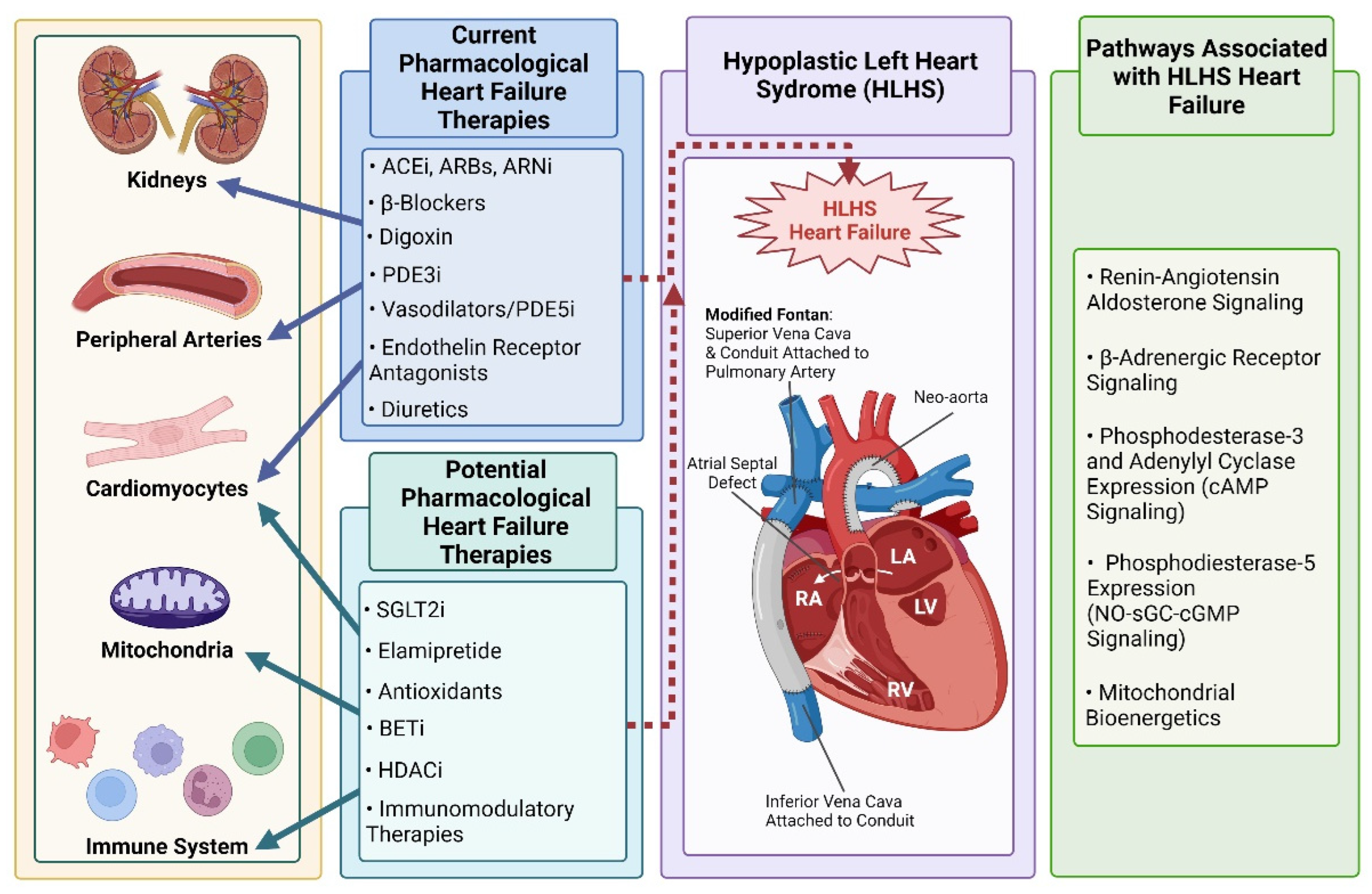
:1. Introduction
2. Current HLHS Heart Failure Therapies
2.1. ACE Inhibitors, ARBs, and ARNis
2.2. β-Blockers
2.3. Digoxin
2.4. PDE3 Inhibitors
2.5. Vasodilators
2.5.1. Endothelin Receptor Antagonists
2.5.2. Prostanoids
2.5.3. PDE5 Inhibitors
2.5.4. Guanylate Cyclase Inhibitors
2.6. Diuretics
3. Potential Future HLHS Medical Therapies
3.1. Mitochondrial Targeted Therapies
3.1.1. Elamipretide (Bendavia, MTP-131, SS-31)
3.1.2. Antioxidants
3.2. SGLT2 Inhibitors
3.3. BET Inhibitors
3.4. HDAC Inhibitors
3.5. Immunomodulatory Therapies
4. Conclusions
Author Contributions
Funding
Institutional Review Board Statement
Informed Consent Statement
Data Availability Statement
Conflicts of Interest
References
- Wu, W.; He, J.; Shao, X. Incidence and Mortality Trend of Congenital Heart Disease at the Global, Regional, and National Level, 1990-2017. Medicine 2020, 99, e20593. [Google Scholar] [CrossRef] [PubMed]
- van der Linde, D.; Konings, E.E.M.; Slager, M.A.; Witsenburg, M.; Helbing, W.A.; Takkenberg, J.J.M.; Roos-Hesselink, J.W. Birth Prevalence of Congenital Heart Disease Worldwide: A Systematic Review and Meta-Analysis. J. Am. Coll. Cardiol. 2011, 58, 2241–2247. [Google Scholar] [CrossRef] [PubMed] [Green Version]
- Best, K.E.; Miller, N.; Draper, E.; Tucker, D.; Luyt, K.; Rankin, J. The Improved Prognosis of Hypoplastic Left Heart: A Population-Based Register Study of 343 Cases in England and Wales. Front. Pediatrics 2021, 9, 635776. [Google Scholar] [CrossRef] [PubMed]
- Liu, Y.; Chen, S.; Zühlke, L.; Black, G.C.; Choy, M.K.; Li, N.; Keavney, B.D. Global Birth Prevalence of Congenital Heart Defects 1970-2017: Updated Systematic Review and Meta-Analysis of 260 Studies. Int. J. Epidemiol. 2019, 48, 455–463. [Google Scholar] [CrossRef]
- van Praagh, R.; Plett, J.A.; van Praagh, S. Single Ventricle. Pathology, Embryology, Terminology and Classification. Herz 1979, 4, 113–150. [Google Scholar]
- Mai, C.T.; Isenburg, J.L.; Canfield, M.A.; Meyer, R.E.; Correa, A.; Alverson, C.J.; Lupo, P.J.; Riehle-Colarusso, T.; Cho, S.J.; Aggarwal, D.; et al. National Population-Based Estimates for Major Birth Defects, 2010–2014. Birth Defects Res. 2019, 111, 1420–1435. [Google Scholar] [CrossRef]
- Brida, M.; Diller, G.-P.; Gatzoulis, M.A. Systemic Right Ventricle in Adults With Congenital Heart Disease: Anatomic and Phenotypic Spectrum and Current Approach to Management. Circulation 2018, 137, 508–518. [Google Scholar] [CrossRef]
- Oster, M.E.; Knight, J.H.; Suthar, D.; Amin, O.; Kochilas, L.K. Long-Term Outcomes in Single-Ventricle Congenital Heart Disease. Circulation 2018, 138, 2718–2720. [Google Scholar] [CrossRef]
- Schwartz, I.; McCracken, C.E.; Petit, C.J.; Sachdeva, R. Late Outcomes after the Fontan Procedure in Patients with Single Ventricle: A Meta-Analysis. Heart 2018, 104, 1508–1514. [Google Scholar] [CrossRef]
- Sanchez-Quintana, D.; Anderson, R.H.; Ho, S.Y. Ventricular Myoarchitecture in Tetralogy of Fallot. Heart 1996, 76, 280–286. [Google Scholar] [CrossRef]
- Dickstein, M.L.; Yano, O.; Spotnitz, H.M.; Burkhoff, D. Assessment of Right Ventricular Contractile State with the Conductance Catheter Technique in the Pig. Cardiovasc. Res. 1995, 29, 820–826. [Google Scholar] [CrossRef]
- Redington, A.N.; Rigby, M.L.; Shinebourne, E.A.; Oldershaw, P.J. Changes in the Pressure-Volume Relation of the Right Ventricle When Its Loading Conditions Are Modified. Heart 1990, 63, 45–49. [Google Scholar] [CrossRef] [PubMed] [Green Version]
- Poels, E.M.; da Costa Martins, P.A.; van Empel, V.P.M. Adaptive Capacity of the Right Ventricle: Why Does It Fail? Am. J. Physiol.-Heart Circ. Physiol. 2015, 308, H803–H813. [Google Scholar] [CrossRef]
- Siffel, C.; Riehle-Colarusso, T.; Oster, M.E.; Correa, A. Survival of Children with Hypoplastic Left Heart Syndrome. Pediatrics 2015, 136, e864–e870. [Google Scholar] [CrossRef] [PubMed] [Green Version]
- Best, K.E.; Rankin, J. Long-Term Survival of Individuals Born with Congenital Heart Disease: A Systematic Review and Meta-Analysis. J. Am. Heart Assoc. 2016, 5, e002846. [Google Scholar] [CrossRef] [PubMed]
- Yabrodi, M.; Mastropietro, C.W. Hypoplastic Left Heart Syndrome: From Comfort Care to Long-Term Survival. Pediatric Research. 2017, 81, 142–149. [Google Scholar] [CrossRef] [PubMed] [Green Version]
- Mahle, W.T.; Spray, T.L.; Wernovsky, G.; Gaynor, J.W.; Clark, B.J. Survival After Reconstructive Surgery for Hypoplastic Left Heart Syndrome. Circulation 2000, 102 Suppl. 3, III136–III141. [Google Scholar] [CrossRef]
- Budts, W. Individual Risk Stratification in Adult Congenital Heart Disease: The Way to Go? Eur. Heart J. 2017, 38, 1242–1244. [Google Scholar] [CrossRef]
- Nakano, S.J.; Miyamoto, S.D.; Price, J.F.; Rossano, J.W.; Cabrera, A.G. Pediatric Heart Failure: An Evolving Public Health Concern. J. Pediatrics 2020, 218, 217–221. [Google Scholar] [CrossRef] [Green Version]
- Shaddy, R.E.; Boucek, M.M.; Hsu, D.T.; Boucek, R.J.; Canter, C.E.; Mahony, L.; Ross, R.D.; Pahl, E.; Blume, E.D.; Dodd, D.A.; et al. Carvedilol for Children and Adolescents With Heart Failure. JAMA 2007, 298, 1171. [Google Scholar] [CrossRef]
- Kleber, F.X.; Sabin, G.V.; Winter, U.J.; Reindl, I.; Beil, S.; Wenzel, M.; Fischer, M.; Doering, W. Angiotensin-Converting Enzyme Inhibitors in Preventing Remodeling and Development of Heart Failure After Acute Myocardial Infarction. Am. J. Cardiol. 1997, 80, 162A–167A. [Google Scholar] [CrossRef]
- Sleight, P. The HOPE Study (Heart Outcomes Prevention Evaluation). J. Renin-Angiotensin-Aldosterone Syst. 2000, 1, 18–20. [Google Scholar] [CrossRef] [PubMed]
- Hsu, D.T.; Zak, V.; Mahony, L.; Sleeper, L.A.; Atz, A.M.; Levine, J.C.; Barker, P.C.; Ravishankar, C.; McCrindle, B.W.; Williams, R.V.; et al. Enalapril in Infants With Single Ventricle. Circulation 2010, 122, 333–340. [Google Scholar] [CrossRef] [PubMed]
- van der Bom, T.; Winter, M.M.; Bouma, B.J.; Groenink, M.; Vliegen, H.W.; Pieper, P.G.; van Dijk, A.P.J.; Sieswerda, G.T.; Roos-Hesselink, J.W.; Zwinderman, A.H.; et al. Effect of Valsartan on Systemic Right Ventricular Function. Circulation 2013, 127, 322–330. [Google Scholar] [CrossRef] [PubMed] [Green Version]
- Zandstra, T.E.; Nederend, M.; Jongbloed, M.R.M.; Kiès, P.; Vliegen, H.W.; Bouma, B.J.; Tops, L.F.; Schalij, M.J.; Egorova, A.D. Sacubitril/Valsartan in the Treatment of Systemic Right Ventricular Failure. Heart 2021, 107, 1725–1730. [Google Scholar] [CrossRef]
- Maurer, S.J.; Pujol Salvador, C.; Schiele, S.; Hager, A.; Ewert, P.; Tutarel, O. Sacubitril/Valsartan for Heart Failure in Adults with Complex Congenital Heart Disease. Int. J. Cardiol. 2020, 300, 137–140. [Google Scholar] [CrossRef]
- Bristow, M.R.; Gilbert, E.M.; Abraham, W.T.; Adams, K.F.; Fowler, M.B.; Hershberger, R.E.; Kubo, S.H.; Narahara, K.A.; Ingersoll, H.; Krueger, S.; et al. Carvedilol Produces Dose-Related Improvements in Left Ventricular Function and Survival in Subjects With Chronic Heart Failure. Circulation 1996, 94, 2807–2816. [Google Scholar] [CrossRef]
- Josephson, C.B.; Howlett, J.G.; Jackson, S.D.; Finley, J.; Kells, C.M. A Case Series of Systemic Right Ventricular Dysfunction Post Atrial Switch for Simple D-Transposition of the Great Arteries: The Impact of Beta-Blockade. Can. J. Cardiol. 2006, 22, 769–772. [Google Scholar] [CrossRef] [Green Version]
- Ishibashi, N.; Park, I.-S.; Waragai, T.; Yoshikawa, T.; Murakami, Y.; Mori, K.; Mimori, S.; Ando, M.; Takahashi, Y.; Doi, S.; et al. Effect of Carvedilol on Heart Failure in Patients With a Functionally Univentricular Heart. Circ. J. 2011, 75, 1394–1399. [Google Scholar] [CrossRef] [Green Version]
- Doughan, A.R.K.; McConnell, M.E.; Book, W.M. Effect of Beta Blockers (Carvedilol or Metoprolol XL) in Patients With Transposition of Great Arteries and Dysfunction of the Systemic Right Ventricle. Am. J. Cardiol. 2007, 99, 704–706. [Google Scholar] [CrossRef]
- Garcia, A.M.; Beatty, J.-T.; Stephanie, X.; Nakano, J. Heart Failure in Single Right Ventricle Congenital Heart Disease: Physiological and Molecular Considerations. Am. J. Physiol. Heart Circ. Physiol. 2020, 318, 947–965. [Google Scholar] [CrossRef] [PubMed]
- Miyamoto, S.D.; Stauffer, B.L.; Polk, J.; Medway, A.; Friedrich, M.; Haubold, K.; Peterson, V.; Nunley, K.; Nelson, P.; Sobus, R.; et al. Gene Expression and β-Adrenergic Signaling Are Altered in Hypoplastic Left Heart Syndrome. J. Heart Lung Transpl. 2014, 33, 785–793. [Google Scholar] [CrossRef] [PubMed] [Green Version]
- Williamson, K.M.; Thrasher, K.A.; Fulton, K.B.; LaPointe, N.M.A.; Dunham, G.D.; Cooper, A.A.; Barrett, P.S.; Patterson, J.H. Digoxin Toxicity: An Evaluation in Current Clinical Practice. Arch. Intern. Med. 1998, 158, 2444. [Google Scholar] [CrossRef] [PubMed] [Green Version]
- Jain, S.; Vaidyanathan, B. Digoxin in Management of Heart Failure in Children: Should It Be Continued or Relegated to the History Books? Ann. Pediatr. Cardiol. 2009, 2, 149–152. [Google Scholar] [CrossRef] [PubMed]
- Cañas, F.; Milenko, T.J.; Ma’luf, N.; Bates, D.W. Evaluating the Appropriateness of Digoxin Level Monitoring. Arch. Intern. Med. 1999, 159, 363–368. [Google Scholar] [CrossRef] [Green Version]
- Brown, D.W.; Mangeot, C.; Anderson, J.B.; Peterson, L.E.; King, E.C.; Lihn, S.L.; Neish, S.R.; Fleishman, C.; Phelps, C.; Hanke, S.; et al. National Pediatric Cardiology Quality Improvement Collaborative. Digoxin Use Is Associated With Reduced Interstage Mortality in Patients With No History of Arrhythmia After Stage I Palliation for Single Ventricle Heart Disease. J. Am. Heart Assoc. 2016, 5, e002376. [Google Scholar] [CrossRef] [Green Version]
- Packer, M.; Carver, J.R.; Rodeheffer, R.J.; Ivanhoe, R.J.; DiBianco, R.; Zeldis, S.M.; Hendrix, G.H.; Bommer, W.J.; Elkayam, U.; Kukin, M.L.; et al. Effect of Oral Milrinone on Mortality in Severe Chronic Heart Failure. N. Engl. J. Med. 1991, 325, 1468–1475. [Google Scholar] [CrossRef]
- Sucharov, C.C.; Nakano, S.J.; Slavov, D.; Schwisow, J.A.; Rodriguez, E.; Nunley, K.; Medway, A.; Stafford, N.; Nelson, P.; McKinsey, T.A.; et al. A PDE3A Promoter Polymorphism Regulates CAMP-Induced Transcriptional Activity in Failing Human Myocardium. J. Am. Coll. Cardiol. 2019, 73, 1173–1184. [Google Scholar] [CrossRef]
- Hoffman, T.M.; Wernovsky, G.; Atz, A.M.; Kulik, T.J.; Nelson, D.P.; Chang, A.C.; Bailey, J.M.; Akbary, A.; Kocsis, J.F.; Kaczmarek, R.; et al. Efficacy and Safety of Milrinone in Preventing Low Cardiac Output Syndrome in Infants and Children after Corrective Surgery for Congenital Heart Disease. Circulation 2003, 107, 996–1002. [Google Scholar] [CrossRef] [Green Version]
- Fredholm, M.; Jörgensen, K.; Houltz, E.; Ricksten, S.-E. Inotropic and Lusitropic Effects of Levosimendan and Milrinone Assessed by Strain Echocardiography-A Randomised Trial. Acta. Anaesthesiologica. Scand. 2018, 62, 1246–1254. [Google Scholar] [CrossRef]
- Abramov, D.; Haglund, N.A.; di Salvo, T.G. Effect of Milrinone Infusion on Pulmonary Vasculature and Stroke Work Indices: A Single-Center Retrospective Analysis in 69 Patients Awaiting Cardiac Transplantation. Am. J. Cardiovasc. Drugs 2017, 17, 335–342. [Google Scholar] [CrossRef] [PubMed]
- Lannemyr, L.; Bragadottir, G.; Redfors, B.; Ricksten, S.-E. Effects of Milrinone on Renal Perfusion, Filtration and Oxygenation in Patients with Acute Heart Failure and Low Cardiac Output Early after Cardiac Surgery. J. Crit. Care 2020, 57, 225–230. [Google Scholar] [CrossRef] [PubMed]
- Kanazawa, T.; Shimizu, K.; Iwasaki, T.; Baba, K.; Otsuki, S.; Kotani, Y.; Kasahara, S.; Morimatsu, H. Perioperative Milrinone Infusion Improves One-Year Survival After the Norwood-Sano Procedure. J. Cardiothorac. Vasc. Anesth. 2021, 35, 2073–2078. [Google Scholar] [CrossRef] [PubMed]
- Berg, A.M.; Snell, L.; Mahle, W.T. Home Inotropic Therapy in Children. J. Heart Lung Transplant. 2007, 26, 453–457. [Google Scholar] [CrossRef]
- Briston, S.J.; Dibb, K.M.; Solaro, R.J.; Eisner, D.A.; Trafford, A.W. Balanced Changes in Ca Buffering by SERCA and Troponin Contribute to Ca Handling during β-Adrenergic Stimulation in Cardiac Myocytes. Cardiovasc. Res. 2014, 104, 347–354. [Google Scholar] [CrossRef] [Green Version]
- Nakano, S.J.; Nelson, P.; Sucharov, C.C.; Miyamoto, S.D. Myocardial Response to Milrinone in Single Right Ventricle Heart Disease. J. Pediatrics 2016, 174, 199–203.e5. [Google Scholar] [CrossRef] [Green Version]
- Yamagishi, M.; Kurosawa, H.; Hashimoto, K.; Nomura, K.; Kitamura, N. The Role of Plasma Endothelin in the Fontan Circulation. J. Cardiovasc. Surg. 2002, 43, 793–797. [Google Scholar]
- Bowater, S.E.; Weaver, R.A.; Thorne, S.A.; Clift, P.F. The Safety and Effects of Bosentan in Patients with a Fontan Circulation. Congenit. Heart Dis. 2012, 7, 243–249. [Google Scholar] [CrossRef]
- Derk, G.; Houser, L.; Miner, P.; Williams, R.; Moriarty, J.; Finn, P.; Alejos, J.; Aboulhosn, J. Efficacy of Endothelin Blockade in Adults with Fontan Physiology. Congenit. Heart Dis. 2015, 10, E11–E16. [Google Scholar] [CrossRef]
- Hebert, A.; Mikkelsen, U.R.; Thilen, U.; Idorn, L.; Jensen, A.S.; Nagy, E.; Hanseus, K.; Sørensen, K.E.; Søndergaard, L. Bosentan Improves Exercise Capacity in Adolescents and Adults After Fontan Operation. Circulation 2014, 130, 2021–2030. [Google Scholar] [CrossRef]
- Saxena, A.; Sharma, M.; Kothari, S.S.; Juneja, R.; Reddy, S.C.; Sharma, R.; Bhan, A.; Venugopal, P. Prostaglandin E1 in Infants with Congenital Heart Disease: Indian Experience. Indian Pediatr. 1998, 35, 1063–1069. [Google Scholar] [PubMed]
- Rhodes, J.; Ubeda-Tikkanen, A.; Clair, M.; Fernandes, S.M.; Graham, D.A.; Milliren, C.E.; Daly, K.P.; Mullen, M.P.; Landzberg, M.J. Effect of Inhaled Iloprost on the Exercise Function of Fontan Patients: A Demonstration of Concept. Int. J. Cardiol. 2013, 168, 2435–2440. [Google Scholar] [CrossRef] [PubMed] [Green Version]
- Kim, Y.H.; Chae, M.H.; Choi, D.Y. Inhaled Iloprost for the Treatment of Patient with Fontan Circulation. Korean J. Pediatrics 2014, 57, 461. [Google Scholar] [CrossRef] [PubMed] [Green Version]
- Unegbu, C.; Noje, C.; Coulson, J.D.; Segal, J.B.; Romer, L. Pulmonary Hypertension Therapy and a Systematic Review of Efficacy and Safety of PDE-5 Inhibitors. Pediatrics 2017, 139, e20161450. [Google Scholar] [CrossRef] [Green Version]
- Garcia, A.M.; Nakano, S.J.; Karimpour-Fard, A.; Nunley, K.; Blain-Nelson, P.; Stafford, N.M.; Stauffer, B.L.; Sucharov, C.C.; Miyamoto, S.D. Phosphodiesterase-5 Is Elevated in Failing Single Ventricle Myocardium and Affects Cardiomyocyte Remodeling In Vitro. Circ. Heart Fail. 2018, 11. [Google Scholar] [CrossRef]
- Nakano, S.J.; Sucharov, J.; van Dusen, R.; Cecil, M.; Nunley, K.; Wickers, S.; Karimpur-Fard, A.; Stauffer, B.L.; Miyamoto, S.D.; Sucharov, C.C. Cardiac Adenylyl Cyclase and Phosphodiesterase Expression Profiles Vary by Age, Disease, and Chronic Phosphodiesterase Inhibitor Treatment. J. Card. Fail. 2017, 23, 72–80. [Google Scholar] [CrossRef] [Green Version]
- Zhang, M.; Takimoto, E.; Hsu, S.; Lee, D.I.; Nagayama, T.; Danner, T.; Koitabashi, N.; Barth, A.S.; Bedja, D.; Gabrielson, K.L.; et al. Myocardial Remodeling Is Controlled by Myocyte-Targeted Gene Regulation of Phosphodiesterase Type 5. J. Am. Coll. Cardiol. 2010, 56, 2021–2030. [Google Scholar] [CrossRef] [Green Version]
- Boolell, M.; Allen, M.J.; Ballard, S.A.; Gepi-Attee, S.; Muirhead, G.J.; Naylor, A.M.; Osterloh, I.H.; Gingell, C. Sildenafil: An Orally Active Type 5 Cyclic GMP-Specific Phosphodiesterase Inhibitor for the Treatment of Penile Erectile Dysfunction. Int. J. Impot. Res. 1996, 8, 47–52. [Google Scholar]
- Wang, H.; Anstrom, K.; Ilkayeva, O.; Muehlbauer, M.J.; Bain, J.R.; McNulty, S.; Newgard, C.B.; Kraus, W.E.; Hernandez, A.; Felker, G.M.; et al. Sildenafil Treatment in Heart Failure With Preserved Ejection Fraction. JAMA Cardiol. 2017, 2, 896. [Google Scholar] [CrossRef]
- Goldberg, D.J.; Zak, V.; Goldstein, B.H.; Schumacher, K.R.; Rhodes, J.; Penny, D.J.; Petit, C.J.; Ginde, S.; Menon, S.C.; Kim, S.-H.; et al. Results of the FUEL Trial. Circulation 2020, 141, 641–651. [Google Scholar] [CrossRef]
- Zhu, G.; Ueda, K.; Hashimoto, M.; Zhang, M.; Sasaki, M.; Kariya, T.; Sasaki, H.; Kaludercic, N.; Lee, D.-I.; Bedja, D.; et al. The Mitochondrial Regulator PGC1α Is Induced by CGMP-PKG Signaling and Mediates the Protective Effects of Phosphodiesterase 5 Inhibition in Heart Failure. FEBS Lett. 2022, 596, 17–28. [Google Scholar] [CrossRef] [PubMed]
- Yu, H.M.; Chung, H.K.; Park, K.S. The PDE5 Inhibitor Udenafil Ameliorates Nonalcoholic Fatty Liver Disease by Improving Mitochondrial Function. Biochem. Biophys. Res. Commun. 2021, 558, 57–63. [Google Scholar] [CrossRef] [PubMed]
- Yu, H.M.; Chung, H.K.; Kim, K.S.; Lee, J.M.; Hong, J.H.; Park, K.S. PDE 5 Inhibitor Improves Insulin Sensitivity by Enhancing Mitochondrial Function in Adipocytes. Biochem. Biophys. Res. Commun. 2017, 493, 631–636. [Google Scholar] [CrossRef] [PubMed]
- Hill, K.D.; Tunks, R.D.; Barker, P.C.A.; Benjamin, D.K.; Cohen-Wolkowiez, M.; Fleming, G.A.; Laughon, M.; Li, J.S. Sildenafil Exposure and Hemodynamic Effect After Stage II Single-Ventricle Surgery. Pediatric Crit. Care Med. 2013, 14, 593–600. [Google Scholar] [CrossRef] [PubMed] [Green Version]
- Bayer Pharma, AG. Adempas (Riociguat) EU Summary of Product Characteristics. 2014. Available online: http://www.ema.europa.eu/docs/en_GB/document_library/EPAR_-_Product_Information/human/002737/WC500165034.pdf (accessed on 7 March 2022).
- Bayer Pharma, AG. Adempas® US Prescribing Information. 2013. Available online: http://labeling.bayerhealthcare.com/html/products/pi/Adempas_PI.pdf (accessed on 7 March 2022).
- Rosenkranz, S.; Ghofrani, H.-A.; Beghetti, M.; Ivy, D.; Frey, R.; Fritsch, A.; Weimann, G.; Saleh, S.; Apitz, C. Riociguat for Pulmonary Arterial Hypertension Associated with Congenital Heart Disease. Heart 2015, 101, 1792–1799. [Google Scholar] [CrossRef] [PubMed]
- Felker, G.M.; Ellison, D.H.; Mullens, W.; Cox, Z.L.; Testani, J.M. Diuretic Therapy for Patients With Heart Failure. J. Am. Coll. Cardiol. 2020, 75, 1178–1195. [Google Scholar] [CrossRef]
- Heo, J.H.; Rascati, K.L.; Lopez, K.N.; Moffett, B.S. Increased Fracture Risk with Furosemide Use in Children with Congenital Heart Disease. J. Pediatrics 2018, 199, 92–98.e10. [Google Scholar] [CrossRef]
- Metra, M.; Teerlink, J.R. Heart Failure. Lancet 2017, 390, 1981–1995. [Google Scholar] [CrossRef]
- Brown, D.A.; Perry, J.B.; Allen, M.E.; Sabbah, H.N.; Stauffer, B.L.; Shaikh, S.R.; Cleland, J.G.F.; Colucci, W.S.; Butler, J.; Voors, A.A.; et al. Expert Consensus Document: Mitochondrial Function as a Therapeutic Target in Heart Failure. Nat. Rev. Cardiol. 2017, 14, 238–250. [Google Scholar] [CrossRef]
- Xu, X.; Lin, J.-H.I.; Bais, A.S.; Reynolds, M.J.; Tan, T.; Gabriel, G.C.; Kondos, Z.; Liu, X.; Shiva, S.S.; Lo, C.W. Mitochondrial Respiration Defects in Single-Ventricle Congenital Heart Disease. Front. Cardiovasc. Med. 2021, 8, 734388. [Google Scholar] [CrossRef]
- Liu, X.; Yagi, H.; Saeed, S.; Bais, A.S.; Gabriel, G.C.; Chen, Z.; Peterson, K.A.; Li, Y.; Schwartz, M.C.; Reynolds, W.T.; et al. The Complex Genetics of Hypoplastic Left Heart Syndrome. Nat. Genet. 2017, 49, 1152–1159. [Google Scholar] [CrossRef] [PubMed]
- Zhao, D.; Liu, Y.; Xu, Z.; Shen, H.; Chen, S.; Zhang, S.; Li, Y.; Zhang, H.; Zou, C.; Ma, X. Integrative Bioinformatics Analysis Revealed Mitochondrial Defects Underlying Hypoplastic Left Heart Syndrome. Int. J. Gen. Med. 2021, 14, 9747–9760. [Google Scholar] [CrossRef] [PubMed]
- Nakagawa, Y. Metabolism and Biological Function of Cardiolipin. Yakugaku Zasshi 2013, 133, 561–574. [Google Scholar] [CrossRef] [PubMed] [Green Version]
- Sabbah, H.N.; Gupta, R.C.; Kohli, S.; Wang, M.; Hachem, S.; Zhang, K. Chronic Therapy With Elamipretide (MTP-131), a Novel Mitochondria-Targeting Peptide, Improves Left Ventricular and Mitochondrial Function in Dogs With Advanced Heart Failure. Circ. Heart Fail. 2016, 9, e002206. [Google Scholar] [CrossRef] [PubMed] [Green Version]
- Dai, W.; Shi, J.; Gupta, R.C.; Sabbah, H.N.; Hale, S.L.; Kloner, R.A. Bendavia, a Mitochondria-Targeting Peptide, Improves Postinfarction Cardiac Function, Prevents Adverse Left Ventricular Remodeling, and Restores Mitochondria-Related Gene Expression in Rats. J. Cardiovasc. Pharmacol. 2014, 64, 543–553. [Google Scholar] [CrossRef] [PubMed]
- Chatfield, K.C.; Sparagna, G.C.; Chau, S.; Phillips, E.K.; Ambardekar, A.V.; Aftab, M.; Mitchell, M.B.; Sucharov, C.C.; Miyamoto, S.D.; Stauffer, B.L. Elamipretide Improves Mitochondrial Function in the Failing Human Heart. JACC Basic Transl. Sci. 2019, 4, 147–157. [Google Scholar] [CrossRef] [PubMed]
- Daubert, M.A.; Yow, E.; Dunn, G.; Marchev, S.; Barnhart, H.; Douglas, P.S.; O’Connor, C.; Goldstein, S.; Udelson, J.E.; Sabbah, H.N. Novel Mitochondria-Targeting Peptide in Heart Failure Treatment. Circ. Heart Fail. 2017, 10. [Google Scholar] [CrossRef]
- Butler, J.; Khan, M.S.; Anker, S.D.; Fonarow, G.C.; Kim, R.J.; Nodari, S.; O’Connor, C.M.; Pieske, B.; Pieske-Kraigher, E.; Sabbah, H.N.; et al. Effects of Elamipretide on Left Ventricular Function in Patients With Heart Failure With Reduced Ejection Fraction: The PROGRESS-HF Phase 2 Trial: Effects of Elamipretide in Heart Failure. J. Card. Fail. 2020, 26, 429–437. [Google Scholar] [CrossRef]
- Garcia, A.M.; McPhaul, J.C.; Sparagna, G.C.; Jeffrey, D.A.; Jonscher, R.; Patel, S.S.; Sucharov, C.C.; Stauffer, B.L.; Miyamoto, S.D.; Chatfield, K.C. Alteration of Cardiolipin Biosynthesis and Remodeling in Single Right Ventricle Congenital Heart Disease. Am. J. Physiol. Heart. Circ. Physiol. 2020, 318, H787–H800. [Google Scholar] [CrossRef]
- Magyar, K.; Halmosi, R.; Palfi, A.; Feher, G.; Czopf, L.; Fulop, A.; Battyany, I.; Sumegi, B.; Toth, K.; Szabados, E. Cardioprotection by Resveratrol: A Human Clinical Trial in Patients with Stable Coronary Artery Disease. Clin. Hemorheol. Microcirc. 2012, 50, 179–187. [Google Scholar] [CrossRef]
- van der Pol, A.; van Gilst, W.H.; Voors, A.A.; van der Meer, P. Treating Oxidative Stress in Heart Failure: Past, Present and Future. Eur. J. Heart Fail. 2019, 21, 425–435. [Google Scholar] [CrossRef] [PubMed]
- Schwemmlein, J.; Maack, C.; Bertero, E. Mitochondria as Therapeutic Targets in Heart Failure. Curr. Heart Fail. Rep. 2022, 19, 27–37. [Google Scholar] [CrossRef] [PubMed]
- Yusuf, S.; Dagenais, G.; Pogue, J.; Bosch, J.; Sleight, P. Vitamin E Supplementation and Cardiovascular Events in High-Risk Patients. N. Engl. J. Med. 2000, 342, 154–160. [Google Scholar] [CrossRef]
- Park, S.-Y.; Pekas, E.J.; Headid, R.J.; Son, W.-M.; Wooden, T.K.; Song, J.; Layec, G.; Yadav, S.K.; Mishra, P.K.; Pipinos, I.I. Acute Mitochondrial Antioxidant Intake Improves Endothelial Function, Antioxidant Enzyme Activity, and Exercise Tolerance in Patients with Peripheral Artery Disease. Am. J. Physiol. Heart Circ. Physiol. 2020, 319, H456–H467. [Google Scholar] [CrossRef]
- Turk, E.; Wright, E.M. The Sodium/Glucose Cotransport Family SLC5. Pflügers Archiv. Eur. J. Physiol. 2004, 447, 510–518. [Google Scholar] [CrossRef] [PubMed]
- Hsia, D.S.; Grove, O.; Cefalu, W.T. An Update on SGLT2 Inhibitors for the Treatment of Diabetes Mellitus. Curr. Opin. Endocrinol. Diabetes Obes. 2017, 24, 73–79. [Google Scholar] [CrossRef]
- Rosano, G.; Quek, D.; Martínez, F. Sodium–Glucose Co-Transporter 2 Inhibitors in Heart Failure: Recent Data and Implications for Practice. Card. Fail. Rev. 2020, 6, e31. [Google Scholar] [CrossRef]
- Salah, H.M.; Al’Aref, S.J.; Khan, M.S.; Al-Hawwas, M.; Vallurupalli, S.; Mehta, J.L.; Mounsey, J.P.; Greene, S.J.; McGuire, D.K.; Lopes, R.D.; et al. Efficacy and Safety of Sodium-Glucose Cotransporter 2 Inhibitors Initiation in Patients with Acute Heart Failure, with and without Type 2 Diabetes: A Systematic Review and Meta-Analysis. Cardiovasc. Diabetol. 2022, 21, 20. [Google Scholar] [CrossRef]
- Baker, W.L.; Smyth, L.R.; Riche, D.M.; Bourret, E.M.; Chamberlin, K.W.; White, W.B. Effects of Sodium-Glucose Co-Transporter 2 Inhibitors on Blood Pressure: A Systematic Review and Meta-Analysis. J. Am. Soc. Hypertens. 2014, 8, 262–275.e9. [Google Scholar] [CrossRef]
- Cardoso, R.; Graffunder, F.P.; Ternes, C.M.P.; Fernandes, A.; Rocha, A.V.; Fernandes, G.; Bhatt, D.L. SGLT2 Inhibitors Decrease Cardiovascular Death and Heart Failure Hospitalizations in Patients with Heart Failure: A Systematic Review and Meta-Analysis. EClinicalMedicine 2021, 36, 100933. [Google Scholar] [CrossRef]
- Meloche, J.; Potus, F.; Vaillancourt, M.; Bourgeois, A.; Johnson, I.; Deschamps, L.; Chabot, S.; Ruffenach, G.; Henry, S.; Breuils-Bonnet, S.; et al. Bromodomain-Containing Protein 4. Circ. Res. 2015, 117, 525–535. [Google Scholar] [CrossRef] [PubMed] [Green Version]
- Doroshow, D.B.; Eder, J.P.; LoRusso, P.M. BET Inhibitors: A Novel Epigenetic Approach. Ann. Oncol. 2017, 28, 1776–1787. [Google Scholar] [CrossRef] [PubMed]
- Chabert, C.; Khochbin, S.; Rousseaux, S.; Veyrenc, S.; Furze, R.; Smithers, N.; Prinjha, R.K.; Schlattner, U.; Pison, C.; Dubouchaud, H. Inhibition of BET Proteins Reduces Right Ventricle Hypertrophy and Pulmonary Hypertension Resulting from Combined Hypoxia and Pulmonary Inflammation. Int. J. Mol. Sci. 2018, 19, 2224. [Google Scholar] [CrossRef] [Green Version]
- Kannan, B.R.J.; Sivasankaran, S.; Tharakan, J.A.; Titus, T.; Ajith Kumar, V.K.; Francis, B.; Krishnamoorthy, K.M.; Harikrishnan, S.; Padmakumar, R.; Nair, K. Long-Term Outcome of Patients Operated for Large Ventricular Septal Defects with Increased Pulmonary Vascular Resistance. Indian Heart J. 2003, 55, 161–166. [Google Scholar]
- Seto, E.; Yoshida, M. Erasers of Histone Acetylation: The Histone Deacetylase Enzymes. Cold Spring Harb. Perspect. Biol. 2014, 6, a018713. [Google Scholar] [CrossRef] [Green Version]
- Blakeslee, W.W.; Demos-Davies, K.M.; Lemon, D.D.; Lutter, K.M.; Cavasin, M.A.; Payne, S.; Nunley, K.; Long, C.S.; McKinsey, T.A.; Miyamoto, S.D. Histone Deacetylase Adaptation in Single Ventricle Heart Disease and a Young Animal Model of Right Ventricular Hypertrophy. Pediatric Res. 2017, 82, 642–649. [Google Scholar] [CrossRef] [Green Version]
- Nicolás-Ávila, J.A.; Lechuga-Vieco, A.V.; Esteban-Martínez, L.; Sánchez-Díaz, M.; Díaz-García, E.; Santiago, D.J.; Rubio-Ponce, A.; Li, J.L.; Balachander, A.; Quintana, J.A.; et al. A Network of Macrophages Supports Mitochondrial Homeostasis in the Heart. Cell 2020, 183, 94–109.e23. [Google Scholar] [CrossRef]
- Vagnozzi, R.J.; Molkentin, J.D. Resident Macrophages Keep Mitochondria Running in the Heart. Cell Res. 2020, 30, 1057–1058. [Google Scholar] [CrossRef]
- Vredevoe, D.L.; Widawski, M.; Fonarow, G.C.; Hamilton, M.; Martínez-Maza, O.; Gage, J.R. Interleukin-6 (IL-6) Expression and Natural Killer (NK) Cell Dysfunction and Anergy in Heart Failure. Am. J. Cardiol. 2004, 93, 1007–1011. [Google Scholar] [CrossRef]
- Serra, M.B.; Barroso, W.A.; da Silva, N.N.; do Nascimento Silva, S.; Borges, A.C.R.; Abreu, I.C.; da Rocha Borges, M.O. From Inflammation to Current and Alternative Therapies Involved in Wound Healing. Int. J. Inflam. 2017, 2017, 3406215. [Google Scholar] [CrossRef] [Green Version]
- Gudmundsdottir, J.; Óskarsdóttir, S.; Skogberg, G.; Lindgren, S.; Lundberg, V.; Berglund, M.; Lundell, A.-C.; Berggren, H.; Fasth, A.; Telemo, E.; et al. Early Thymectomy Leads to Premature Immunologic Ageing: An 18-Year Follow-Up. J. Allergy Clin. Immunol. 2016, 138, 1439–1443.e10. [Google Scholar] [CrossRef] [PubMed] [Green Version]
- Kurobe, H.; Tominaga, T.; Sugano, M.; Hayabuchi, Y.; Egawa, Y.; Takahama, Y.; Kitagawa, T. Complete but Not Partial Thymectomy in Early Infancy Reduces T-Cell-Mediated Immune Response: Three-Year Tracing Study after Pediatric Cardiac Surgery. J. Thorac. Cardiovasc. Surg. 2013, 145, 656–662. [Google Scholar] [CrossRef] [PubMed] [Green Version]
- Deya-Martinez, A.; Flinn, A.M.; Gennery, A.R. Neonatal Thymectomy in Children—Accelerating the Immunologic Clock? J. Allergy Clin. Immunol. 2020, 146, 236–243. [Google Scholar] [CrossRef] [PubMed]
Publisher’s Note: MDPI stays neutral with regard to jurisdictional claims in published maps and institutional affiliations. |
© 2022 by the authors. Licensee MDPI, Basel, Switzerland. This article is an open access article distributed under the terms and conditions of the Creative Commons Attribution (CC BY) license (https://creativecommons.org/licenses/by/4.0/).
Share and Cite
Baybayon-Grandgeorge, A.N.; Pietra, A.E.; Miyamoto, S.D.; Garcia, A.M. Medical Therapies for Heart Failure in Hypoplastic Left Heart Syndrome. J. Cardiovasc. Dev. Dis. 2022, 9, 152. https://doi.org/10.3390/jcdd9050152
Baybayon-Grandgeorge AN, Pietra AE, Miyamoto SD, Garcia AM. Medical Therapies for Heart Failure in Hypoplastic Left Heart Syndrome. Journal of Cardiovascular Development and Disease. 2022; 9(5):152. https://doi.org/10.3390/jcdd9050152
Chicago/Turabian StyleBaybayon-Grandgeorge, Angela N., Ashley E. Pietra, Shelley D. Miyamoto, and Anastacia M. Garcia. 2022. "Medical Therapies for Heart Failure in Hypoplastic Left Heart Syndrome" Journal of Cardiovascular Development and Disease 9, no. 5: 152. https://doi.org/10.3390/jcdd9050152
APA StyleBaybayon-Grandgeorge, A. N., Pietra, A. E., Miyamoto, S. D., & Garcia, A. M. (2022). Medical Therapies for Heart Failure in Hypoplastic Left Heart Syndrome. Journal of Cardiovascular Development and Disease, 9(5), 152. https://doi.org/10.3390/jcdd9050152