

Identification of Hub Genes in the Remodeling of Non-Infarcted Myocardium Following Acute Myocardial Infarction


Abstract
1. Introduction
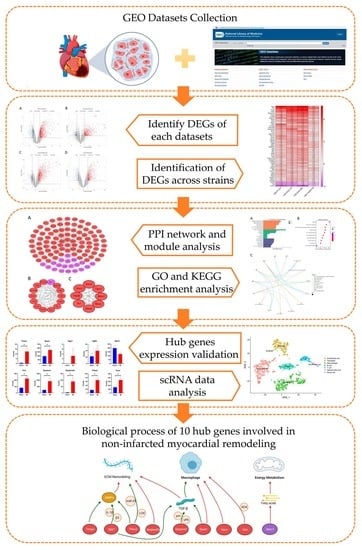
2. Materials and Methods
2.1. Gene Expression Profile Data
2.2. Screening of Differentially Expressed Genes (DEGs)
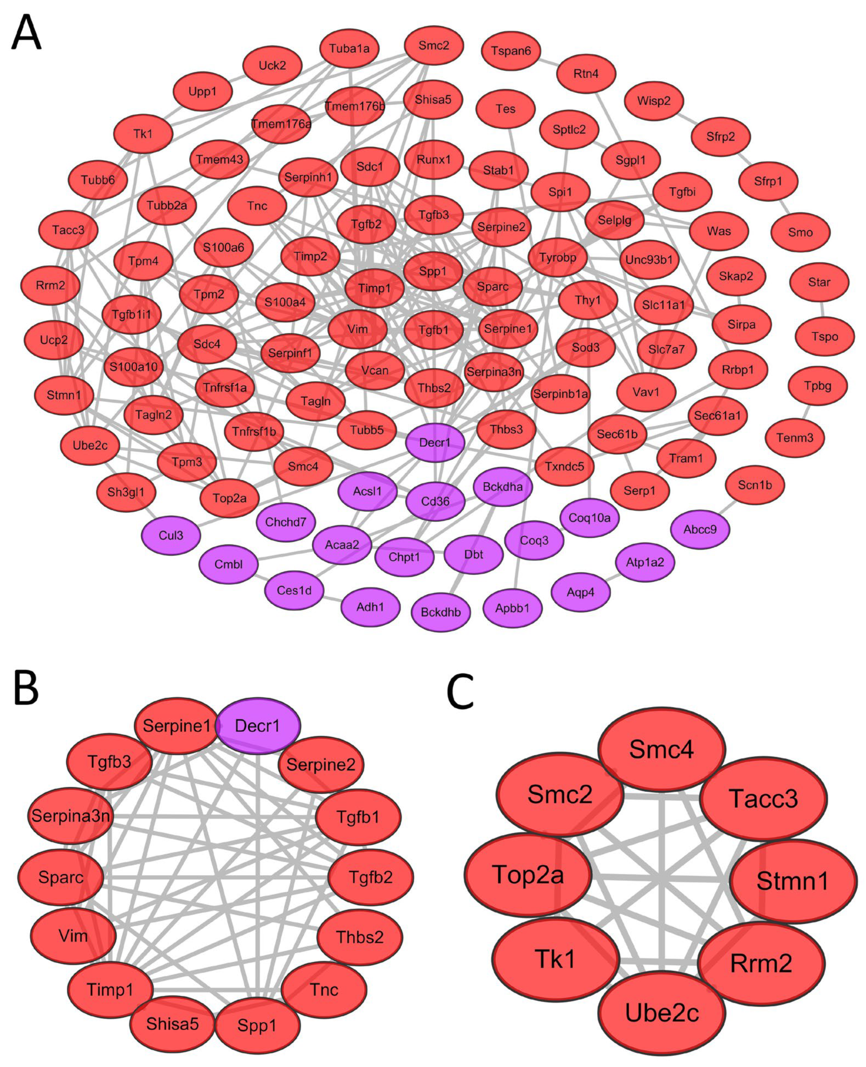
2.3. PPI Network Construction and Screening of Modules
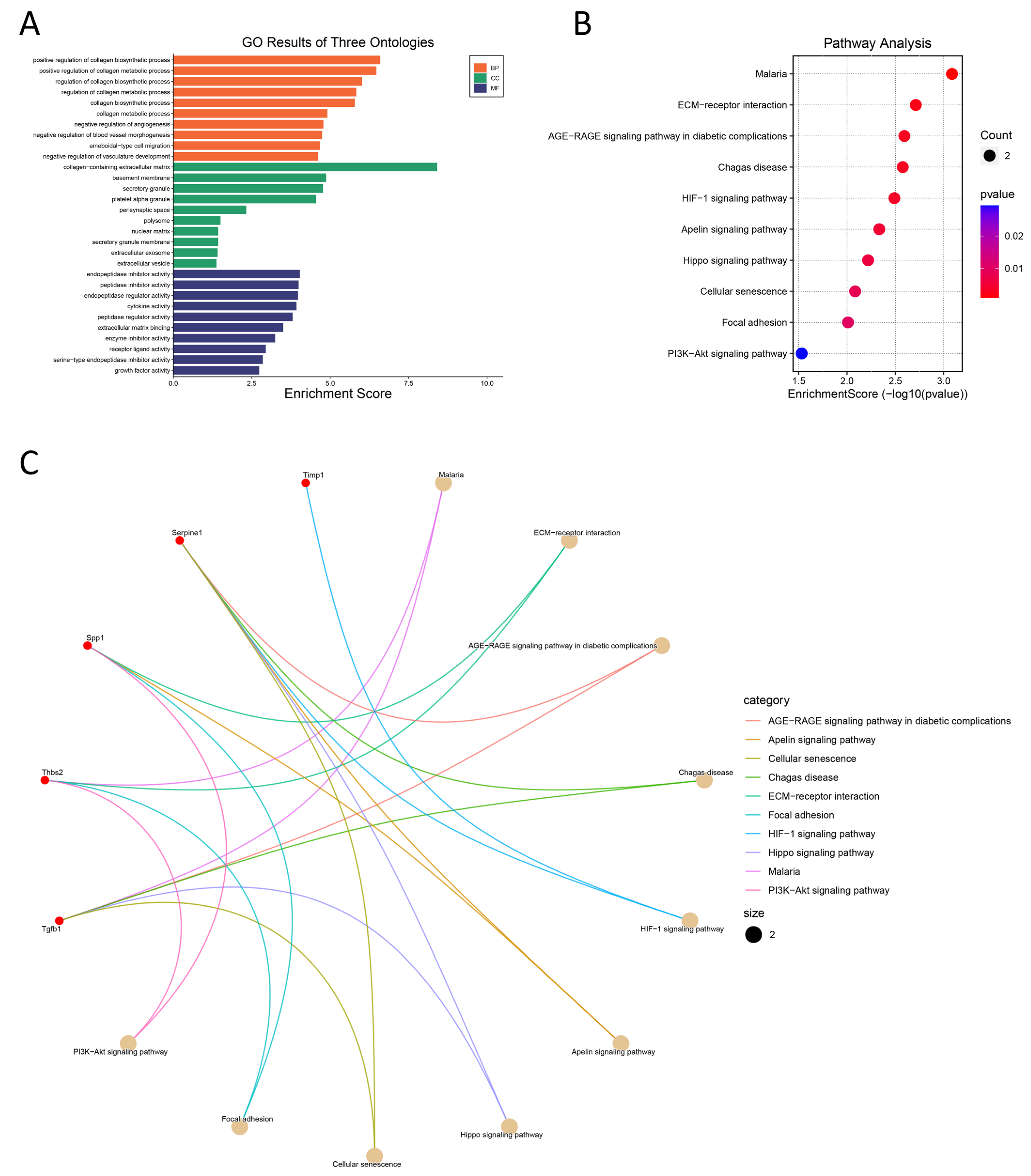
2.4. GO and KEGG Pathway Enrichment Analyses
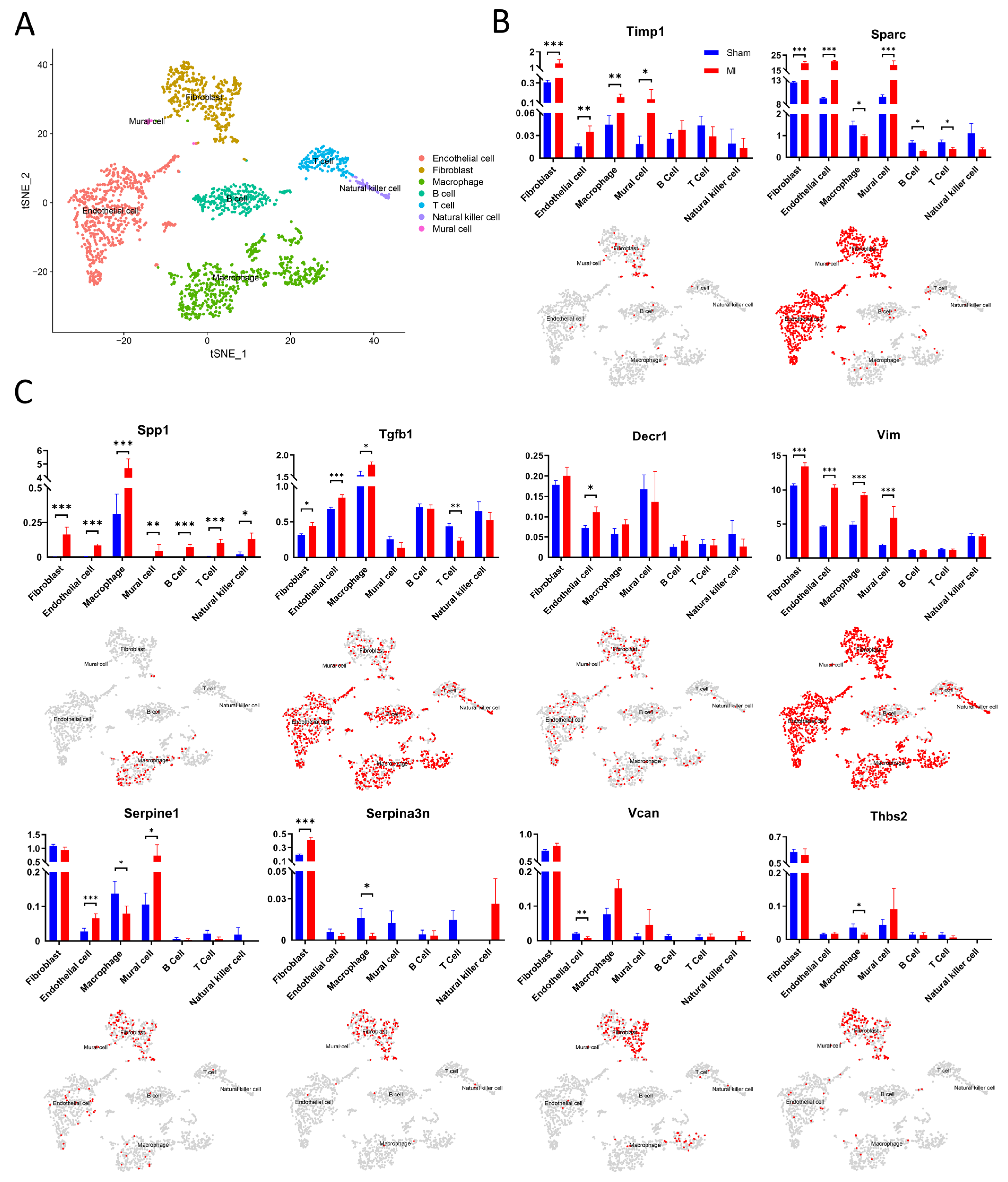
2.5. Processing and Clustering scRNA-seq Data
3. Results
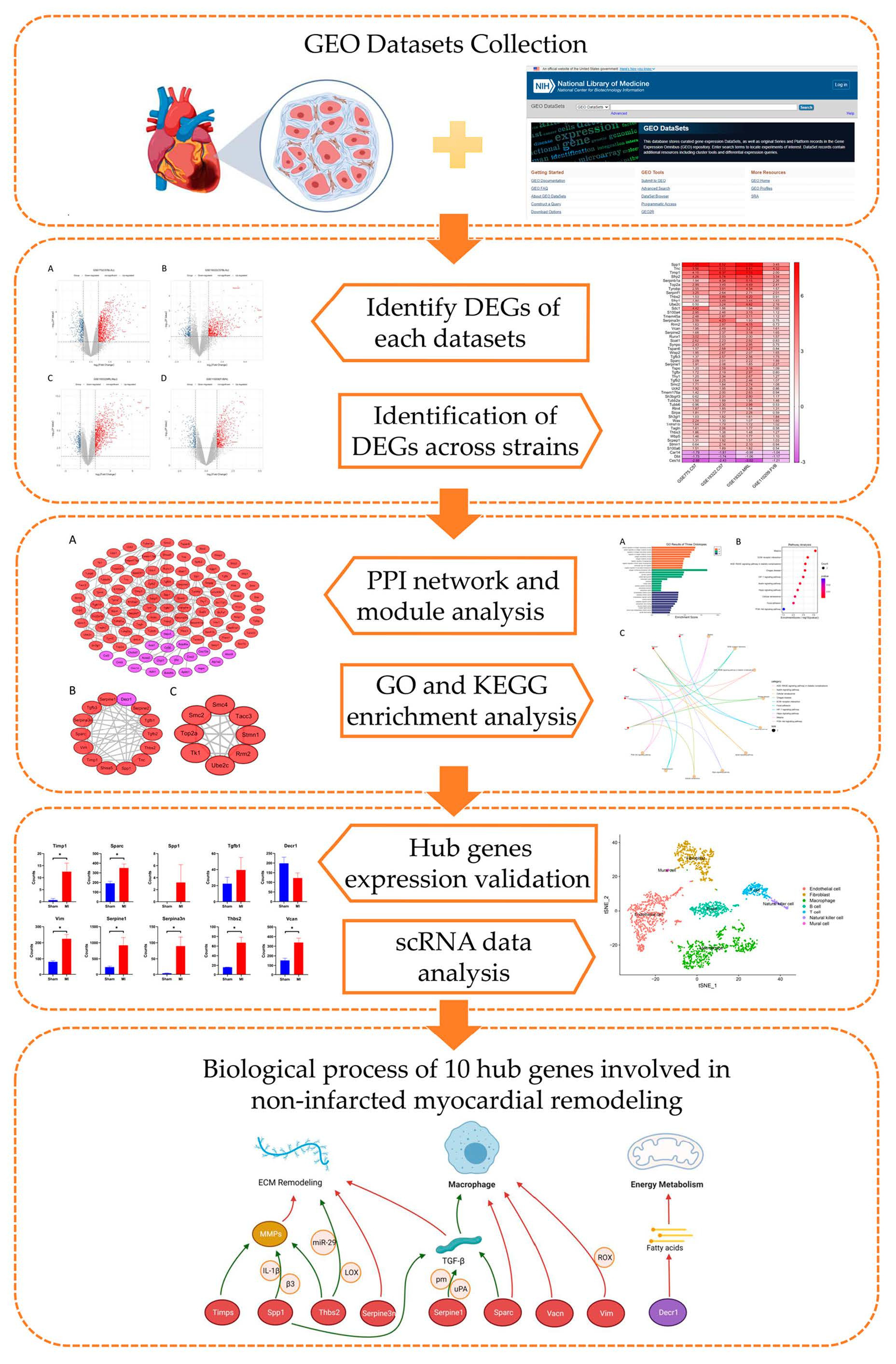
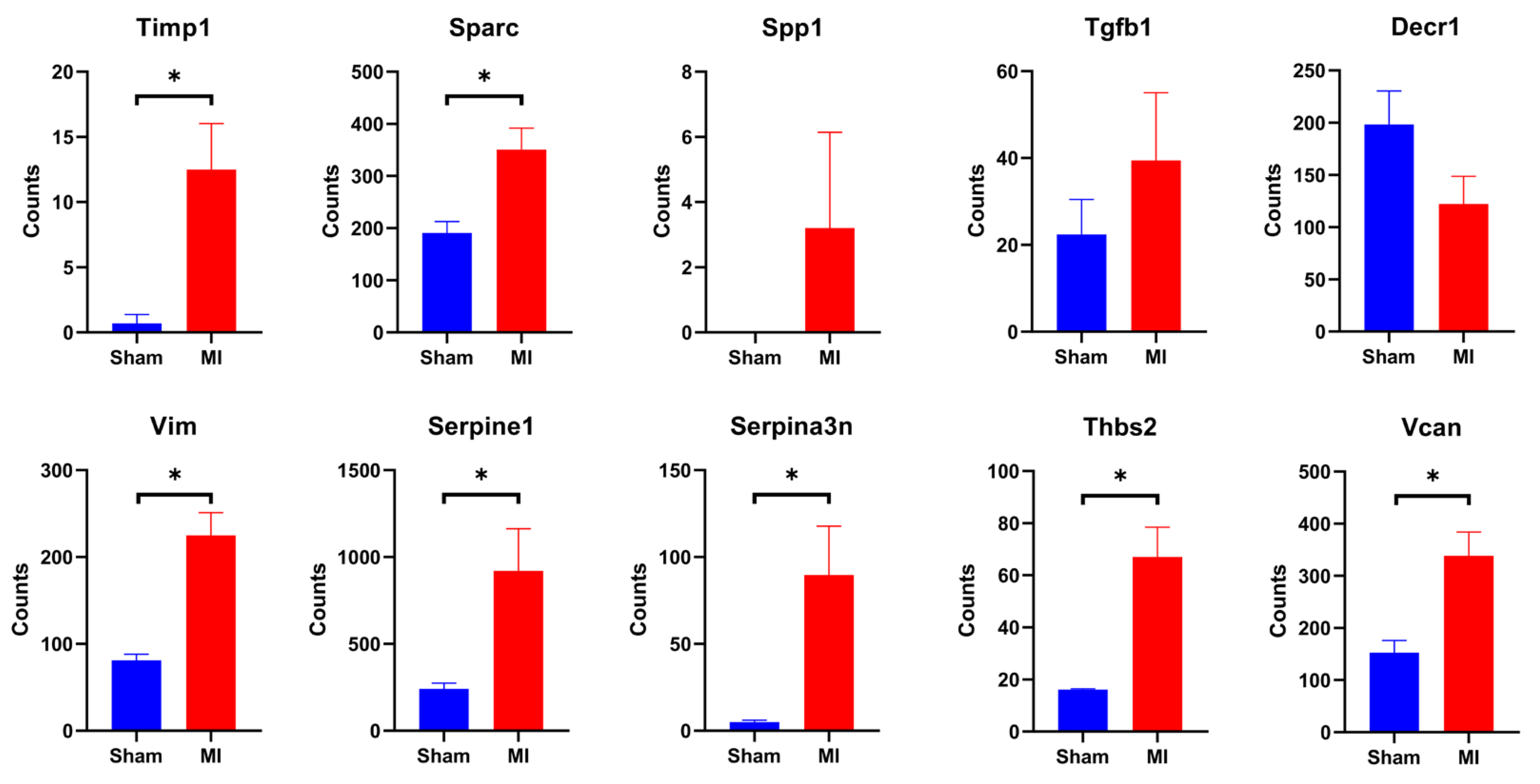
3.1. Identification of DEGs in Non-Infarcted Myocardia Following AMI
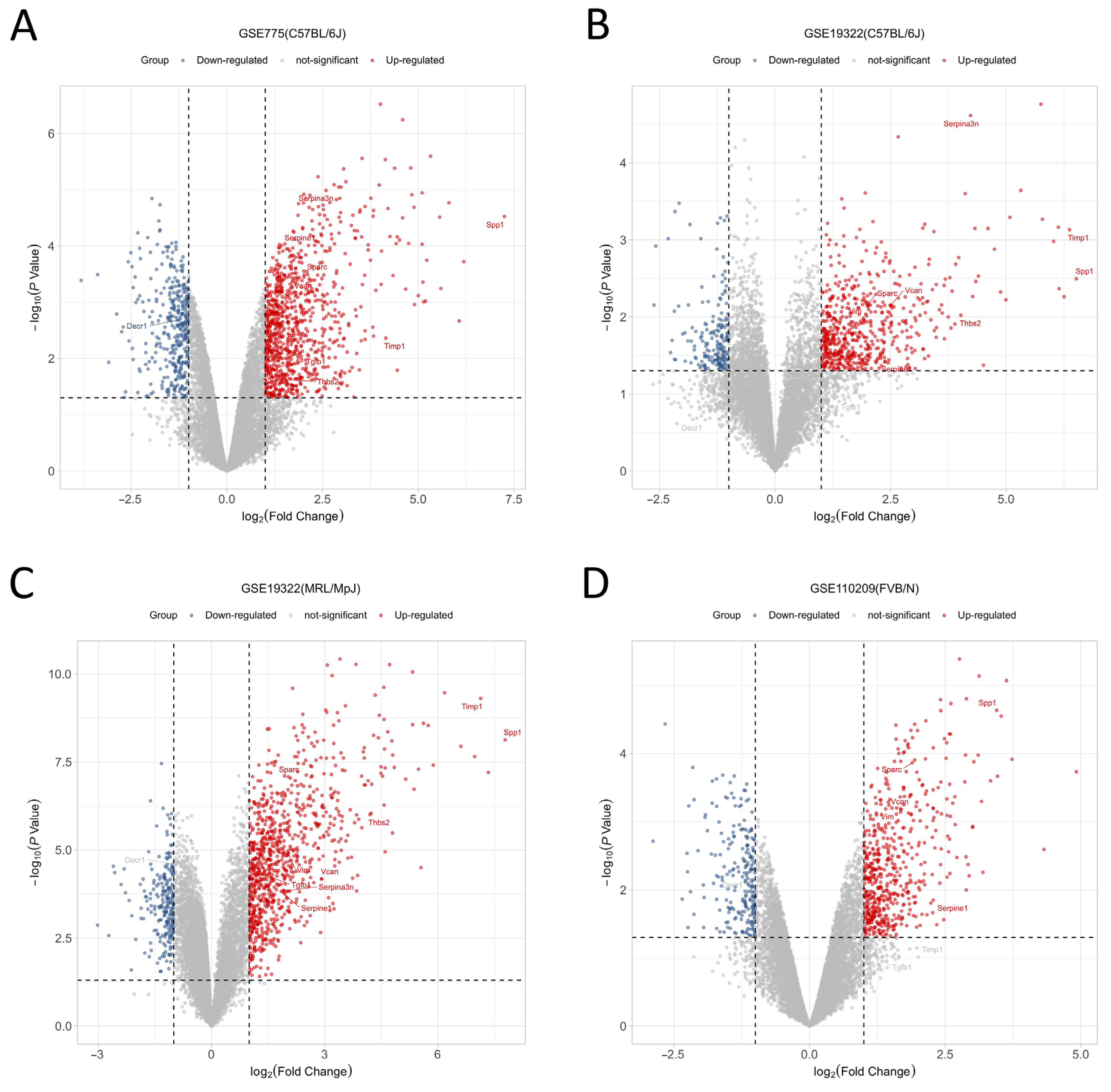
3.2. Identification of Genes Uniformly Up- or Down-Regulated across Strains
3.3. Construction of a PPI Network, Module Analysis, and Screening of Key Genes
3.4. GO and KEGG Enrichment Analyses
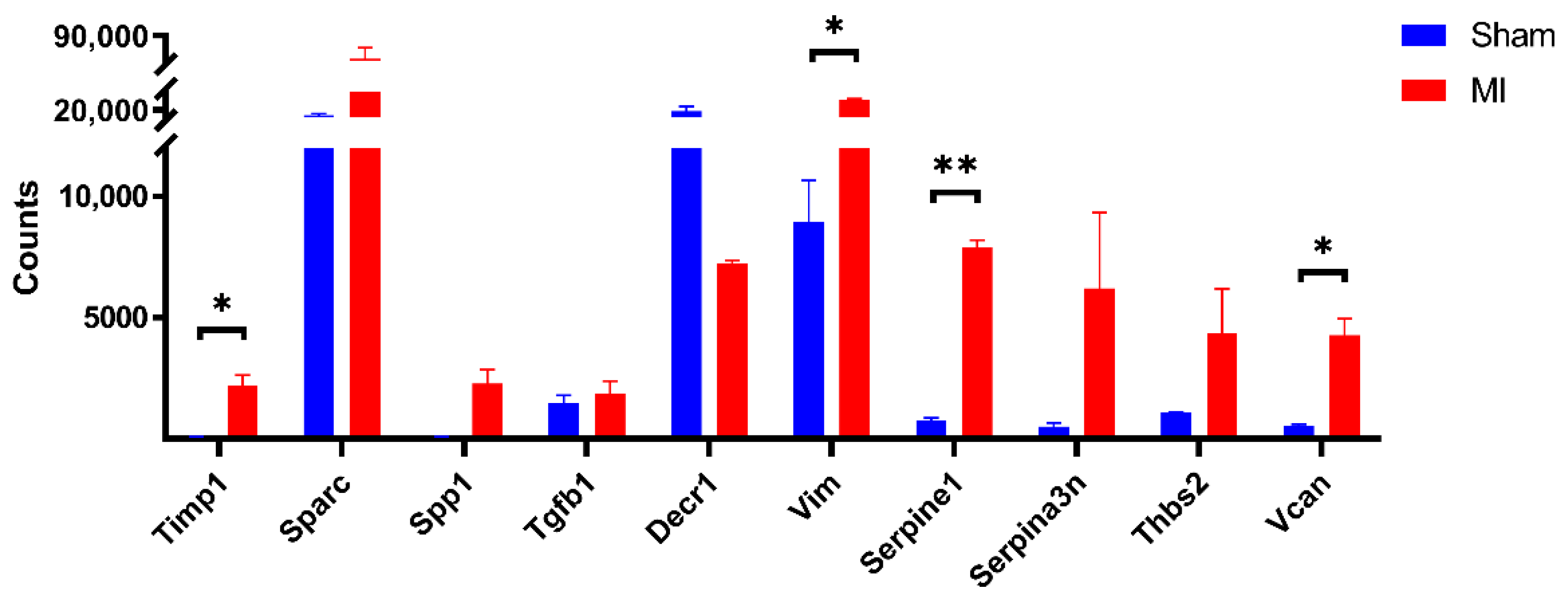
3.5. Expression of Hub Genes in Different Cell Populations
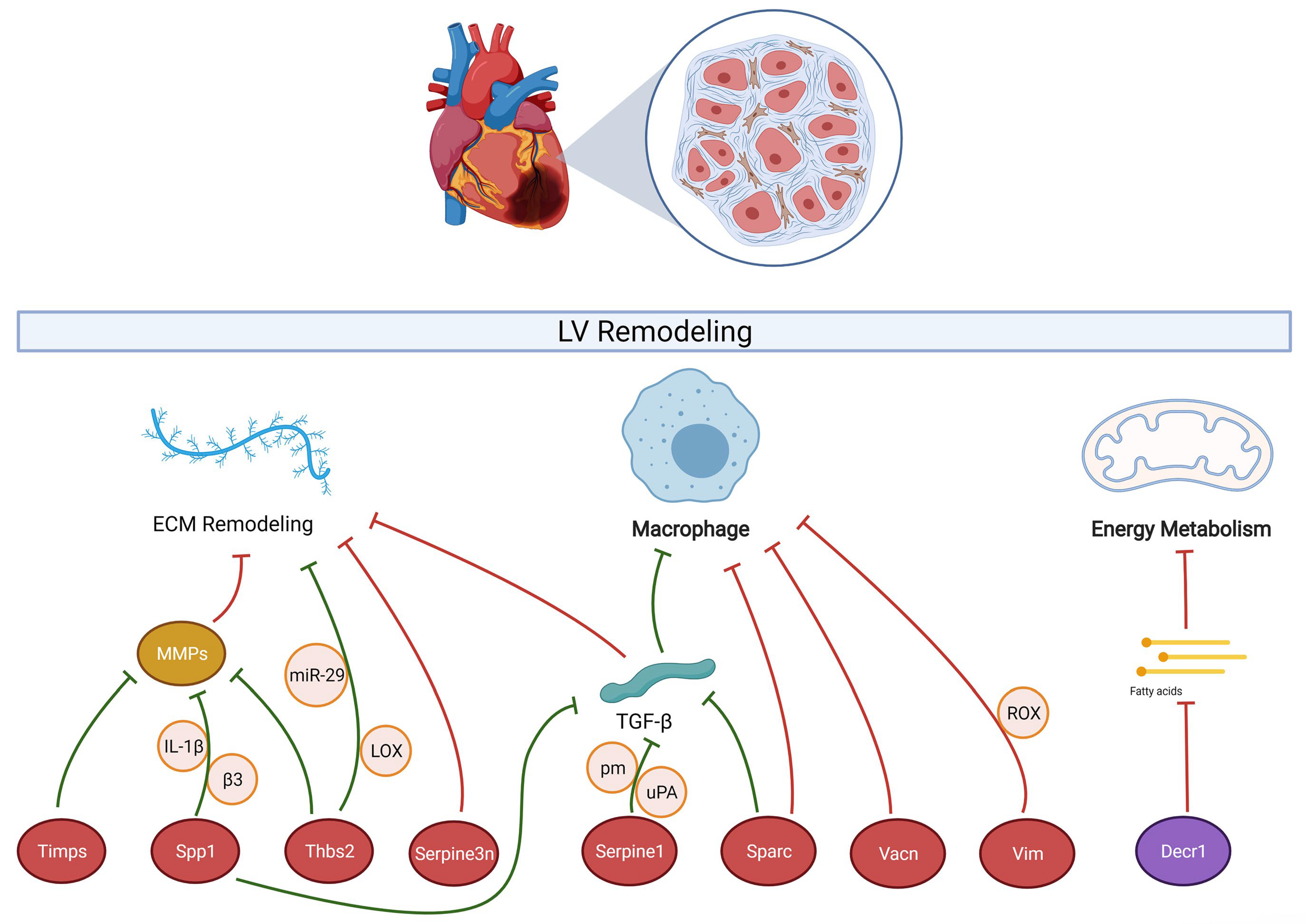
4. Discussion
4.1. Extracellular Matrix (ECM)-Mediated Myocardial Fibrosis
4.2. Macrophage-Driven Inflammation
4.3. Fatty Acid Metabolic Pathways
5. Conclusions
Supplementary Materials
Author Contributions
Funding
Institutional Review Board Statement
Informed Consent Statement
Data Availability Statement
Acknowledgments
Conflicts of Interest
Abbreviations
| AMI | Acute myocardial infarction |
| GEO | gene expression omnibus |
| DEG | differentially expressed gene |
| PPI | protein–protein interaction |
| GO | gene ontology |
| KEGG | Kyoto Encyclopedia of Genes and Genomes |
| ECM | extracellular matrix |
| TIP | total cardiac interstitial cell population |
| LV | left ventricle |
References
- Lopez, A.D.; Murray, C.C. The global burden of disease, 1990–2020. Nat. Med. 1998, 4, 1241–1243. [Google Scholar] [CrossRef] [PubMed]
- Le, T.Y.L.; Pickett, H.A.; Yang, A.; Ho, J.W.K.; Thavapalachandran, S.; Igoor, S.; Yang, S.F.; Farraha, M.; Voges, H.K.; Hudson, J.E.; et al. Enhanced cardiac repair by telomerase reverse transcriptase over-expression in human cardiac mesenchymal stromal cells. Sci. Rep. 2019, 9, 10579. [Google Scholar] [CrossRef] [PubMed]
- Bajaj, A.; Sethi, A.; Rathor, P.; Suppogu, N.; Sethi, A. Acute Complications of Myocardial Infarction in the Current Era. J. Investig. Med. 2015, 63, 844–855. [Google Scholar] [CrossRef] [PubMed]
- Ruparelia, N.; Godec, J.; Lee, R.; Chai, J.T.; Dall’Armellina, E.; McAndrew, D.; Digby, J.E.; Forfar, J.C.; Prendergast, B.D.; Kharbanda, R.; et al. Acute myocardial infarction activates distinct inflammation and proliferation pathways in circulating monocytes, prior to recruitment, and identified through conserved transcriptional responses in mice and humans. Eur. Heart J. 2015, 36, 1923–1934. [Google Scholar] [CrossRef] [PubMed]
- Cao, N.; Liang, H.; Huang, J.; Wang, J.; Chen, Y.; Chen, Z.; Yang, H.-T. Highly efficient induction and long-term maintenance of multipotent cardiovascular progenitors from human pluripotent stem cells under defined conditions. Cell Res. 2013, 23, 1119–1132. [Google Scholar] [CrossRef]
- Reed, G.W.; Rossi, J.E.; Cannon, C.P. Acute myocardial infarction. Lancet 2016, 389, 197–210. [Google Scholar] [CrossRef]
- Park, T.-J.; Park, J.H.; Lee, G.S.; Lee, J.-Y.; Shin, J.H.; Kim, M.W.; Kim, Y.S.; Kim, J.-Y.; Oh, K.-J.; Han, B.-S.; et al. Quantitative proteomic analyses reveal that GPX4 downregulation during myocardial infarction contributes to ferroptosis in cardiomyocytes. Cell Death Dis. 2019, 10, 835. [Google Scholar] [CrossRef]
- Chen, D.-Q.; Kong, X.-S.; Shen, X.-B.; Huang, M.-Z.; Zheng, J.-P.; Sun, J.; Xu, S.-H. Identification of Differentially Expressed Genes and Signaling Pathways in Acute Myocardial Infarction Based on Integrated Bioinformatics Analysis. Cardiovasc. Ther. 2019, 2019, 8490707. [Google Scholar] [CrossRef]
- Van Duijvenboden, K.; de Bakker, D.E.; Man, J.C.; Janssen, R.; Günthel, M.; Hill, M.C.; Hooijkaas, I.B.; van der Made, I.; van der Kraak, P.H.; Vink, A.; et al. Conserved NPPB + Border Zone Switches from MEF2- to AP-1–Driven Gene Program. Circulation 2019, 140, 864–879. [Google Scholar] [CrossRef]
- Stuart, S.D.F.; De Jesus, N.M.; Lindsey, M.L.; Ripplinger, C.M. The crossroads of inflammation, fibrosis, and arrhythmia following myocardial infarction. J. Mol. Cell. Cardiol. 2016, 91, 114–122. [Google Scholar] [CrossRef]
- Barrett, T.; Wilhite, S.E.; Ledoux, P.; Evangelista, C.; Kim, I.F.; Tomashevsky, M.; Marshall, K.A.; Phillippy, K.H.; Sherman, P.M.; Holko, M.; et al. NCBI GEO: Archive for functional genomics data sets—Update. Nucleic Acids Res. 2013, 41, D991–D995. [Google Scholar] [CrossRef]
- Ritchie, M.E.; Belinda, P.; Wu, D.; Hu, Y.; Law, C.W.; Shi, W.; Smyth, G.K. Limma powers differential expression analyses for RNA-sequencing and microarray studies. Nucleic Acids Res. 2015, 43, e47. [Google Scholar] [CrossRef]
- Yu, G.; Wang, L.-G.; Han, Y.; He, Q.-Y. clusterProfiler: An R Package for Comparing Biological Themes Among Gene Clusters. OMICS J. Integr. Biol. 2012, 16, 284–287. [Google Scholar] [CrossRef]
- Luo, W.; Brouwer, C. Pathview: An R/Bioconductor package for pathway-based data integration and visualization. Bioinformatics 2013, 29, 1830–1831. [Google Scholar] [CrossRef]
- Butler, A.; Hoffman, P.; Smibert, P.; Papalexi, E.; Satija, R. Integrating single-cell transcriptomic data across different conditions, technologies, and species. Nat. Biotechnol. 2018, 36, 411–420. [Google Scholar] [CrossRef]
- The Gene Ontology Consortium. Expansion of the Gene Ontology knowledgebase and resources. Nucleic Acids Res. 2017, 45, D331–D338. [Google Scholar] [CrossRef]
- Pinto, A.R.; Ilinykh, A.; Ivey, M.J.; Kuwabara, J.T.; D’Antoni, M.L.; Debuque, R.; Chandran, A.; Wang, L.; Arora, K.; Rosenthal, N.; et al. Revisiting Cardiac Cellular Composition. Circ. Res. 2016, 118, 400–409. [Google Scholar] [CrossRef]
- Shinde, A.V.; Frangogiannis, N.G. Fibroblasts in myocardial infarction: A role in inflammation and repair. J. Mol. Cell. Cardiol. 2013, 70, 74–82. [Google Scholar] [CrossRef]
- Gourdie, R.G.; Dimmeler, S.; Kohl, P. Novel therapeutic strategies targeting fibroblasts and fibrosis in heart disease. Nat. Rev. Drug Discov. 2016, 15, 620–638. [Google Scholar] [CrossRef]
- Williams, J.W.; Giannarelli, C.; Rahman, A.; Randolph, G.J.; Kovacic, J.C. Macrophage Biology, Classification, and Phenotype in Cardiovascular Disease. J. Am. Coll. Cardiol. 2018, 72, 2166–2180. [Google Scholar] [CrossRef]
- Mescher, A.L. Macrophages and fibroblasts during inflammation and tissue repair in models of organ regeneration. Regeneration 2017, 4, 39–53. [Google Scholar] [CrossRef] [PubMed]
- Gao, Y.; Qi, G.; Guo, L.; Sun, Y. Bioinformatics Analyses of Differentially Expressed Genes Associated with Acute Myocardial Infarction. Cardiovasc. Ther. 2016, 34, 67–75. [Google Scholar] [CrossRef] [PubMed]
- Zhang, T.; Zhao, L.-L.; Cao, X.; Qi, L.-C.; Wei, G.-Q.; Liu, J.-Y.; Yan, S.-J.; Liu, J.-G.; Li, X.-Q. Bioinformatics analysis of time series gene expression in left ventricle (LV) with acute myocardial infarction (AMI). Gene 2014, 543, 259–267. [Google Scholar] [CrossRef] [PubMed]
- Cleutjens, J.P.; Verluyten, M.J.; Smiths, J.F.; Daemen, M.J. Collagen remodeling after myocardial infarction in the rat heart. Am. J. Pathol. 1995, 147, 325–338. [Google Scholar] [PubMed]
- Roussel, E.; Drolet, M.-C.; Lavigne, A.-M.; Arsenault, M.; Couet, J. Multiple short-chain dehydrogenases/reductases are regulated in pathological cardiac hypertrophy. FEBS Open Bio 2018, 8, 1624–1635. [Google Scholar] [CrossRef] [PubMed]
- Lee, J.-E.; Kim, N.; Jung, M.; Mun, J.-Y.; Yoo, J.-Y. SHISA5/SCOTIN restrains spontaneous autophagy induction by blocking contact between the ERES and phagophores. Autophagy 2021, 18, 1613–1628. [Google Scholar] [CrossRef]
- Wang, X.; Guo, Z.; Ding, Z.; Mehta, J.L. Inflammation, Autophagy, and Apoptosis After Myocardial Infarction. J. Am. Heart Assoc. 2018, 7, e008024. [Google Scholar] [CrossRef]
- Ma, Y.; Brás, L.E.D.C.; Toba, H.; Iyer, R.P.; Hall, M.E.; Winniford, M.D.; Lange, R.A.; Tyagi, S.C.; Lindsey, M.L. Myofibroblasts and the extracellular matrix network in post-myocardial infarction cardiac remodeling. Pflug. Arch. Eur. J. Physiol. 2014, 466, 1113–1127. [Google Scholar] [CrossRef]
- Frangogiannis, N.G. The extracellular matrix in myocardial injury, repair, and remodeling. J. Clin. Investig. 2017, 127, 1600–1612. [Google Scholar] [CrossRef]
- Singh, M.; Foster, C.R.; Dalal, S.; Singh, K. Osteopontin: Role in extracellular matrix deposition and myocardial remodeling post-MI. J. Mol. Cell. Cardiol. 2010, 48, 538–543. [Google Scholar] [CrossRef]
- Spinale, F.G. Myocardial Matrix Remodeling and the Matrix Metalloproteinases: Influence on Cardiac Form and Function. Physiol. Rev. 2007, 87, 1285–1342. [Google Scholar] [CrossRef]
- Ma, Y.; Halade, G.V.; Lindsey, M.L. Extracellular matrix and fibroblast communication following myocardial infarction. J. Cardiovasc. Transl. Res. 2012, 5, 848–857. [Google Scholar] [CrossRef]
- Frangogiannis, N.G. Matricellular Proteins in Cardiac Adaptation and Disease. Physiol. Rev. 2012, 92, 635–688. [Google Scholar] [CrossRef]
- Hsu, I.; Parkinson, L.G.; Shen, Y.; Toro, A.A.D.C.; Brown, T.A.; Zhao, H.; Bleackley, R.C.; Granville, D.J. Serpina3n accelerates tissue repair in a diabetic mouse model of delayed wound healing. Cell Death Dis. 2014, 5, e1458. [Google Scholar] [CrossRef]
- Brunton-O’Sullivan, M.M.; Holley, A.S.; Hally, K.E.; Kristono, G.A.; Harding, S.A.; Larsen, P.D. A combined biomarker approach for characterising extracellular matrix profiles in acute myocardial infarction. Sci. Rep. 2021, 11, 12705. [Google Scholar] [CrossRef]
- Ma, Y.; Yabluchanskiy, A.; Lindsey, M.L. Neutrophil roles in left ventricular remodeling following myocardial infarction. Fibrogenesis Tissue Repair 2013, 6, 11. [Google Scholar] [CrossRef]
- Creemers, E.; Davis, J.N.; Parkhurst, A.M.; Leenders, P.; Dowdy, K.B.; Hapke, E.; Hauet, A.M.; Escobar, P.G.; Cleutjens, J.P.M.; Smits, J.F.M.; et al. Deficiency of TIMP-1 exacerbates LV remodeling after myocardial infarction in mice. Am. J. Physiol. Circ. Physiol. 2003, 284, H364–H371. [Google Scholar] [CrossRef]
- Kelly, D.; Khan, S.Q.; Thompson, M.; Cockerill, G.; Ng, L.; Samani, N.; Squire, I.B. Plasma tissue inhibitor of metalloproteinase-1 and matrix metalloproteinase-9: Novel indicators of left ventricular remodelling and prognosis after acute myocardial infarction. Eur. Heart J. 2008, 29, 2116–2124. [Google Scholar] [CrossRef]
- Shirakawa, K.; Sano, M. Osteopontin in Cardiovascular Diseases. Biomolecules 2021, 11, 1047. [Google Scholar] [CrossRef]
- Schroen, B.; Heymans, S.; Sharma, U.; Blankesteijn, W.M.; Pokharel, S.; Cleutjens, J.P.; Porter, J.G.; Evelo, C.T.; Duisters, R.; van Leeuwen, R.E.; et al. Thrombospondin-2 Is Essential for Myocardial Matrix Integrity. Circ. Res. 2004, 95, 515–522. [Google Scholar] [CrossRef]
- Pardo, A.; Gibson, K.; Cisneros, J.; Richards, T.J.; Yang, Y.; Becerril, C.; Yousem, S.; Herrera, I.; Ruiz, V.; Selman, M.; et al. Up-Regulation and Profibrotic Role of Osteopontin in Human Idiopathic Pulmonary Fibrosis. PLoS Med. 2005, 2, e251. [Google Scholar] [CrossRef] [PubMed]
- Xie, Z.; Singh, M.; Siwik, D.A.; Joyner, W.L.; Singh, K. Osteopontin Inhibits Interleukin-1β-stimulated Increases in Matrix Metalloproteinase Activity in Adult Rat Cardiac Fibroblasts. J. Biol. Chem. 2003, 278, 48546–48552. [Google Scholar] [CrossRef] [PubMed]
- Mujumdar, V.S.; Smiley, L.M.; Tyagi, S.C. Activation of matrix metalloproteinase dilates and decreases cardiac tensile strength. Int. J. Cardiol. 2001, 79, 277–286. [Google Scholar] [CrossRef]
- Calabro, N.; Barrett, A.; Chamorro-Jorganes, A.; Tam, S.; Kristofik, N.; Xing, H.; Loye, A.M.; Sessa, W.; Hansen, K.; Kyriakides, T. Thrombospondin-2 regulates extracellular matrix production, LOX levels, and cross-linking via downregulation of miR-29. Matrix Biol. 2019, 82, 71–85. [Google Scholar] [CrossRef] [PubMed]
- Bujak, M.; Frangogiannis, N. The role of TGF-β signaling in myocardial infarction and cardiac remodeling. Cardiovasc. Res. 2007, 74, 184–195. [Google Scholar] [CrossRef]
- Hanna, A.; Frangogiannis, N.G. The Role of the TGF-β Superfamily in Myocardial Infarction. Front. Cardiovasc. Med. 2019, 6, 140. [Google Scholar] [CrossRef]
- Schultz, J.E.J.; Witt, S.A.; Glascock, B.J.; Nieman, M.L.; Reiser, P.J.; Nix, S.L.; Kimball, T.R.; Doetschman, T. TGF-β1 mediates the hypertrophic cardiomyocyte growth induced by angiotensin II. J. Clin. Investig. 2002, 109, 787–796. [Google Scholar] [CrossRef]
- Kupfahl, C.; Pink, D.; Friedrich, K.; Zurbrügg, H.R.; Neuss, M.; Warnecke, C.; Fielitz, J.; Graf, K.; Fleck, E.; Regitz-Zagrosek, V. Angiotensin II directly increases transforming growth factor β1 and osteopontin and indirectly affects collagen mRNA expression in the human heart. Cardiovasc. Res. 2000, 46, 463–475. [Google Scholar] [CrossRef]
- Ichihara, S.; Senbonmatsu, T.; Price, E., Jr.; Ichiki, T.; Gaffney, F.A.; Inagami, T. Angiotensin II type 2 receptor is essential for left ventricular hypertrophy and cardiac fibrosis in chronic angiotensin II-induced hypertension. Circulation 2001, 104, 346–351. [Google Scholar] [CrossRef]
- Lijnen, P.J.; Petrov, V.V.; Fagard, R. Induction of Cardiac Fibrosis by Transforming Growth Factor-β1. Mol. Genet. Metab. 2000, 71, 418–435. [Google Scholar] [CrossRef]
- Kramerova, I.; Kumagai-Cresse, C.; Ermolova, N.; Mokhonova, E.; Marinov, M.; Capote, J.; Becerra, D.; Quattrocelli, M.; Crosbie, R.H.; Welch, E.; et al. Spp1 (osteopontin) promotes TGFβ processing in fibroblasts of dystrophin-deficient muscles through matrix metalloproteinases. Hum. Mol. Genet. 2019, 28, 3431–3442. [Google Scholar] [CrossRef]
- Vetrone, S.A.; Montecino-Rodriguez, E.; Kudryashova, E.; Kramerova, I.; Hoffman, E.P.; Liu, S.D.; Miceli, M.C.; Spencer, M.J. Osteopontin promotes fibrosis in dystrophic mouse muscle by modulating immune cell subsets and intramuscular TGF-β. J. Clin. Investig. 2009, 119, 1583–1594. [Google Scholar] [CrossRef]
- Gupta, K.K.; Donahue, D.L.; Sandoval-Cooper, M.J.; Castellino, F.J.; Ploplis, V.A. Plasminogen Activator Inhibitor-1 Protects Mice Against Cardiac Fibrosis by Inhibiting Urokinase-type Plasminogen Activator-mediated Plasminogen Activation. Sci. Rep. 2017, 7, 365. [Google Scholar] [CrossRef]
- Flevaris, P.; Khan, S.; Eren, M.; Schuldt, A.J.T.; Shah, S.; Lee, D.; Gupta, S.; Shapiro, A.D.; Burridge, P.W.; Ghosh, A.K.; et al. Plasminogen Activator Inhibitor Type I Controls Cardiomyocyte Transforming Growth Factor-β and Cardiac Fibrosis. Circulation 2017, 136, 664–679. [Google Scholar] [CrossRef]
- Schellings, M.W.M.; Vanhoutte, D.; Swinnen, M.; Cleutjens, J.P.; Debets, J.; Van Leeuwen, R.E.W.; D’Hooge, J.; Van de Werf, F.; Carmeliet, P.; Pinto, Y.M.; et al. Absence of SPARC results in increased cardiac rupture and dysfunction after acute myocardial infarction. J. Exp. Med. 2008, 206, 113–123. [Google Scholar] [CrossRef]
- McCurdy, S.; Baicu, C.F.; Heymans, S.; Bradshaw, A.D. Cardiac extracellular matrix remodeling: Fibrillar collagens and Secreted Protein Acidic and Rich in Cysteine (SPARC). J. Mol. Cell. Cardiol. 2010, 48, 544–549. [Google Scholar] [CrossRef]
- Taylor, A.W. Review of the activation of TGF- in immunity. J. Leukoc. Biol. 2008, 85, 29–33. [Google Scholar] [CrossRef]
- Ter Horst, E.N.; Hakimzadeh, N.; Van Der Laan, A.M.; Krijnen, P.A.J.; Niessen, H.W.M.; Piek, J.J. Modulators of Macrophage Polarization Influence Healing of the Infarcted Myocardium. Int. J. Mol. Sci. 2015, 16, 29583–29591. [Google Scholar] [CrossRef]
- Dutta, P.; Nahrendorf, M. Monocytes in Myocardial Infarction. Arterioscler. Thromb. Vasc. Biol. 2015, 35, 1066–1070. [Google Scholar] [CrossRef]
- Peet, C.; Ivetic, A.; Bromage, D.I.; Shah, A.M. Cardiac monocytes and macrophages after myocardial infarction. Cardiovasc. Res. 2021, 116, 1101–1112. [Google Scholar] [CrossRef]
- Dick, S.A.; Macklin, J.A.; Nejat, S.; Momen, A.; Clemente-Casares, X.; AlThagafi, M.G.; Chen, J.; Kantores, C.; Hosseinzadeh, S.; Aronoff, L.; et al. Self-renewing resident cardiac macrophages limit adverse remodeling following myocardial infarction. Nat. Immunol. 2018, 20, 29–39. [Google Scholar] [CrossRef] [PubMed]
- Frangogiannis, N.G. Inflammation in cardiac injury, repair and regeneration. Curr. Opin. Cardiol. 2015, 30, 240–245. [Google Scholar] [CrossRef] [PubMed]
- Wight, T.N. Provisional matrix: A role for versican and hyaluronan. Matrix Biol. 2017, 60–61, 38–56. [Google Scholar] [CrossRef] [PubMed]
- Mor-Vaknin, N.; Legendre, M.; Yu, Y.; Serezani, C.H.C.; Garg, S.K.; Jatzek, A.; Swanson, M.D.; Gonzalez-Hernandez, M.J.; Teitz-Tennenbaum, S.; Punturieri, A.; et al. Murine Colitis is Mediated by Vimentin. Sci. Rep. 2013, 3, 1045. [Google Scholar] [CrossRef] [PubMed]
- Slauch, J.M. How does the oxidative burst of macrophages kill bacteria? Still an open question. Mol. Microbiol. 2011, 80, 580–583. [Google Scholar] [CrossRef]
- Håversen, L.; Sundelin, J.P.; Mardinoglu, A.; Rutberg, M.; Ståhlman, M.; Wilhelmsson, U.; Hultén, L.M.; Pekny, M.; Fogelstrand, P.; Bentzon, J.F.; et al. Vimentin deficiency in macrophages induces increased oxidative stress and vascular inflammation but attenuates atherosclerosis in mice. Sci. Rep. 2018, 8, 16973. [Google Scholar] [CrossRef]
- McCurdy, S.M.; Dai, Q.; Zhang, J.; Zamilpa, R.; Ramirez, T.A.; Dayah, T.; Nguyen, N.; Jin, Y.-F.; Bradshaw, A.D.; Lindsey, M.L. SPARC mediates early extracellular matrix remodeling following myocardial infarction. Am. J. Physiol. Circ. Physiol. 2011, 301, H497–H505. [Google Scholar] [CrossRef]
- Kumar, V.; Prabhu, S.D.; Bansal, S.S. CD4+ T-lymphocytes exhibit biphasic kinetics post-myocardial infarction. Front. Cardiovasc. Med. 2022, 9, 992653. [Google Scholar] [CrossRef]
- Rosenzweig, R.; Kumar, V.; Gupta, S.; Bermeo-Blanco, O.; Stratton, M.S.; Gumina, R.J.; Bansal, S.S. Estrogen Receptor-β Agonists Modulate T-Lymphocyte Activation and Ameliorate Left Ventricular Remodeling During Chronic Heart Failure. Circ. Heart Fail. 2022, 15, e008997. [Google Scholar] [CrossRef]
- Baci, D.; Bosi, A.; Parisi, L.; Buono, G.; Mortara, L.; Ambrosio, G.; Bruno, A. Innate Immunity Effector Cells as Inflammatory Drivers of Cardiac Fibrosis. Int. J. Mol. Sci. 2020, 21, 7165. [Google Scholar] [CrossRef]
- Van Bilsen, M.; van Nieuwenhoven, F.; Van Der Vusse, G.J. Metabolic remodelling of the failing heart: Beneficial or detrimental? Cardiovasc. Res. 2008, 81, 420–428. [Google Scholar] [CrossRef]
- Forini, F.; Ucciferri, N.; Kusmic, C.; Nicolini, G.; Cecchettini, A.; Rocchiccioli, S.; Citti, L.; Iervasi, G. Low T3 State Is Correlated with Cardiac Mitochondrial Impairments after Ischemia Reperfusion Injury: Evidence from a Proteomic Approach. Int. J. Mol. Sci. 2015, 16, 26687–26705. [Google Scholar] [CrossRef]
- Fillgrove, K.L.; Anderson, V.E. The Mechanism of Dienoyl-CoA Reduction by 2,4-Dienoyl-CoA Reductase Is Stepwise: Observation of a Dienolate Intermediate. Biochemistry 2001, 40, 12412–12421. [Google Scholar] [CrossRef]
- Sanchez-Ruderisch, H.; Queirós, A.M.; Fliegner, D.; Eschen, C.; Kararigas, G.; Regitz-Zagrosek, V. Sex-specific regulation of cardiac microRNAs targeting mitochondrial proteins in pressure overload. Biol. Sex Differ. 2019, 10, 8. [Google Scholar] [CrossRef]
- Friedenberg, S.G.; Chdid, L.; Keene, B.; Sherry, B.; Motsinger-Reif, A.; Meurs, K.M. Use of RNA-seq to identify cardiac genes and gene pathways differentially expressed between dogs with and without dilated cardiomyopathy. Am. J. Vet.-Res. 2016, 77, 693–699. [Google Scholar] [CrossRef]
- Hattori, T.; Shimokawa, H.; Higashi, M.; Hiroki, J.; Mukai, Y.; Tsutsui, H.; Kaibuchi, K.; Takeshita, A. Long-Term Inhibition of Rho-Kinase Suppresses Left Ventricular Remodeling After Myocardial Infarction in Mice. Circulation 2004, 109, 2234–2239. [Google Scholar] [CrossRef]
- Dai, W.; Sun, Y.; Jiang, Z.; Du, K.; Xia, N.; Zhong, G. Key Genes Associated with Non-Alcoholic Fatty Liver Disease and Acute Myocardial Infarction. Med. Sci. Monit. 2020, 26, e922492. [Google Scholar] [CrossRef]
- Echtermeyer, F.; Harendza, T.; Hubrich, S.; Lorenz, A.; Herzog, C.; Mueller, M.; Schmitz, M.; Grund, A.; Larmann, J.; Stypmann, J.; et al. Syndecan-4 Signalling Inhibits Apoptosis and Controls NFAT Activity during Myocardial Damage and Remodelling. Cardiovasc. Res. 2011, 92, 123–131. [Google Scholar] [CrossRef]
- Kolwicz, S.C.; Odom, G.L.; Nowakowski, S.G.; Moussavi-Harami, F.; Chen, X.; Reinecke, H.; Hauschka, S.D.; Murry, C.E.; Mahairas, G.G.; Regnier, M. AAV6-Mediated Cardiac-Specific Overexpression of Ribonucleotide Reductase Enhances Myocardial Contractility. Mol. Ther. 2016, 24, 240–250. [Google Scholar] [CrossRef]
- Qi, Z.; Hu, L.; Zhang, J.; Yang, W.; Liu, X.; Jia, D.; Yao, Z.; Chang, L.; Pan, G.; Zhong, H.; et al. PCSK9 (Proprotein Convertase Subtilisin/Kexin 9) Enhances Platelet Activation, Thrombosis, and Myocardial Infarct Expansion by Binding to Platelet CD36. Circulation 2021, 143, 45–61. [Google Scholar] [CrossRef]
- Lindsey, M.L.; Jung, M.; Yabluchanskiy, A.; Cannon, P.L.; Iyer, R.P.; Flynn, E.R.; DeLeon-Pennell, K.Y.; Valerio, F.M.; Harrison, C.L.; Ripplinger, C.M.; et al. Exogenous CXCL4 Infusion Inhibits Macrophage Phagocytosis by Limiting CD36 Signalling to Enhance Post-Myocardial Infarction Cardiac Dilation and Mortality. Cardiovasc. Res. 2019, 115, 395–408. [Google Scholar] [CrossRef] [PubMed]
- Li, X.; Wang, G.; QiLi, M.; Liang, H.; Li, T.; E, X.; Feng, Y.; Zhang, Y.; Liu, X.; Qian, M.; et al. Aspirin Reduces Cardiac Interstitial Fibrosis by Inhibiting Erk1/2-Serpine2 and P-Akt Signalling Pathways. Cell Physiol. Biochem. 2018, 45, 1955–1965. [Google Scholar] [CrossRef]
- Chen, L.; Ashraf, M.; Wang, Y.; Zhou, M.; Zhang, J.; Qin, G.; Rubinstein, J.; Weintraub, N.L.; Tang, Y. The Role of Notch 1 Activation in Cardiosphere Derived Cell Differentiation. Stem Cells Dev. 2012, 21, 2122–2129. [Google Scholar] [CrossRef] [PubMed]
- Liu, Z.; Huang, S. Upregulation of SPI1 during Myocardial Infarction Aggravates Cardiac Tissue Injury and Disease Progression through Activation of the TLR4/NFκB Axis. Am. J. Transl. Res. 2022, 14, 2709–2727. [Google Scholar] [PubMed]
- Kandalam, V.; Basu, R.; Abraham, T.; Wang, X.; Soloway, P.D.; Jaworski, D.M.; Oudit, G.Y.; Kassiri, Z. TIMP2 Deficiency Accelerates Adverse Post-Myocardial Infarction Remodeling Because of Enhanced MT1-MMP Activity despite Lack of MMP2 Activation. Circ. Res. 2010, 106, 796–808. [Google Scholar] [CrossRef]
- Tillmanns, J.; Hoffmann, D.; Habbaba, Y.; Schmitto, J.D.; Sedding, D.; Fraccarollo, D.; Galuppo, P.; Bauersachs, J. Fibroblast Activation Protein Alpha Expression Identifies Activated Fibroblasts after Myocardial Infarction. J. Mol. Cell Cardiol. 2015, 87, 194–203. [Google Scholar] [CrossRef]
- Lei, J.; Xue, S.N.; Wu, W.; Zhou, S.X.; Zhang, Y.L.; Yuan, G.Y.; Wang, J.F. Increased Level of Soluble Syndecan-1 in Serum Correlates with Myocardial Expression in a Rat Model of Myocardial Infarction. Mol. Cell Biochem. 2012, 359, 177–182. [Google Scholar] [CrossRef]
- Li, Z.; Ye, Z.; Ma, J.; Gu, Q.; Teng, J.; Gong, X. MicroRNA-133b Alleviates Doxorubicin-induced Cardiomyocyte Apoptosis and Cardiac Fibrosis by Targeting PTBP1 and TAGLN2. Int. J. Mol. Med. 2021, 48, 125. [Google Scholar] [CrossRef]
- Kolur, V.; Vastrad, B.; Vastrad, C.; Kotturshetti, S.; Tengli, A. Identification of Candidate Biomarkers and Therapeutic Agents for Heart Failure by Bioinformatics Analysis. BMC Cardiovasc. Disord. 2021, 21, 329. [Google Scholar] [CrossRef]
- Klotz, L.; Norman, S.; Vieira, J.M.; Masters, M.; Rohling, M.; Dubé, K.N.; Bollini, S.; Matsuzaki, F.; Carr, C.A.; Riley, P.R. Cardiac Lymphatics Are Heterogeneous in Origin and Respond to Injury. Nature 2015, 522, 62–67. [Google Scholar] [CrossRef]
- Wu, N.; Xu, J.; Du, W.W.; Li, X.; Awan, F.M.; Li, F.; Misir, S.; Eshaghi, E.; Lyu, J.; Zhou, L.; et al. YAP Circular RNA, CircYap, Attenuates Cardiac Fibrosis via Binding with Tropomyosin-4 and Gamma-Actin Decreasing Actin Polymerization. Mol. Ther. 2021, 29, 1138–1150. [Google Scholar] [CrossRef]
- Zhao, X.; Zhang, F.; Wang, Y. Proteomic Analysis Reveals Xuesaitong Injection Attenuates Myocardial Ischemia/Reperfusion Injury by Elevating Pyruvate Dehydrogenase-Mediated Aerobic Metabolism. Mol. Biosyst. 2017, 13, 1504–1511. [Google Scholar] [CrossRef]
- Qian, L.; Zhang, Y.; Zhu, M.; Xie, F.; Porter, T.R.; Xu, D. Improvements in Left Ventricular Regional and Global Systolic Function Following Treatment with S100A4-ShRNA after Myocardial Infarction in Mice. Quant. Imaging Med. Surg. 2019, 9, 1066–1075. [Google Scholar] [CrossRef]
- Kolanowski, T.J.; Wargocka-Matuszewska, W.; Zimna, A.; Cheda, L.; Zyprych-Walczak, J.; Rugowska, A.; Drabik, M.; Fiedorowicz, M.; Krajewski, S.; Steczek, Ł.; et al. Multiparametric Evaluation of Post-MI Small Animal Models Using Metabolic ([18F]FDG) and Perfusion-Based (SYN1) Heart Viability Tracers. Int. J. Mol. Sci. 2021, 22, 12591. [Google Scholar] [CrossRef]
- Mofid, A.; Newman, N.S.; Lee, P.J.H.; Abbasi, C.; Matkar, P.N.; Rudenko, D.; Kuliszewski, M.A.; Chen, H.H.; Afrasiabi, K.; Tsoporis, J.N.; et al. Cardiac Overexpression of S100A6 Attenuates Cardiomyocyte Apoptosis and Reduces Infarct Size After Myocardial Ischemia-Reperfusion. J. Am. Heart Assoc. 2017, 6, e004738. [Google Scholar] [CrossRef]
- Zhao, M.-F.; Sun, L.-F.; Xie, X.-L.; An, D.-Q. Proteomic Study of Tianxiangdan Intervention in Rats with Myocardial Ischemia. J. Physiol. Pharmacol. 2022, 73. [Google Scholar] [CrossRef]
- Imanaka-Yoshida, K. Tenascin-C in Heart Diseases-The Role of Inflammation. Int. J. Mol. Sci. 2021, 22, 5828. [Google Scholar] [CrossRef]
- Weng, L.; Jia, S.; Xu, C.; Ye, J.; Cao, Y.; Liu, Y.; Zheng, M. Nogo-C Regulates Post Myocardial Infarction Fibrosis through the Interaction with ER Ca2+ Leakage Channel Sec61α in Mouse Hearts. Cell Death Dis. 2018, 9, 612. [Google Scholar] [CrossRef]
- Ashton, K.J.; Tupicoff, A.; Williams-Pritchard, G.; Kiessling, C.J.; See Hoe, L.E.; Headrick, J.P.; Peart, J.N. Unique Transcriptional Profile of Sustained Ligand-Activated Preconditioning in Pre- and Post-Ischemic Myocardium. PLoS ONE 2013, 8, e72278. [Google Scholar] [CrossRef]
- Schwanekamp, J.A.; Lorts, A.; Sargent, M.A.; York, A.J.; Grimes, K.M.; Fischesser, D.M.; Gokey, J.J.; Whitsett, J.A.; Conway, S.J.; Molkentin, J.D. TGFBI Functions Similar to Periostin but Is Uniquely Dispensable during Cardiac Injury. PLoS ONE 2017, 12, e0181945. [Google Scholar] [CrossRef]
- Chen, Y.; Meng, H.; Meng, X.; Yan, Z.; Wang, J.; Meng, F. Correlation Between Low THBS3 Expression in Peripheral Blood and Acute Myocardial Infarction. Front. Biosci. 2022, 27, 291. [Google Scholar] [CrossRef] [PubMed]


| Group Name | Datasets | Strain | Sample | AMI | Sham |
|---|---|---|---|---|---|
| GSE775-C57 | GSE775 | C57BL/6J | niLV | 3 | 3 |
| GSE19322-C57 | GSE19322 | C57BL/6J | niLV | 4 | 3 |
| GSE19322-MRL | GSE19322 | MRL/MpJ | niLV | 4 | 4 |
| GSE110209-FVB | GSE110209 | FVB/N | niLV | 3 | 3 |
| Data Group Name | Total DEGs | Up-Regulated | Down-Regulated |
|---|---|---|---|
| GSE775-C57 | 1286 | 933 | 353 |
| GSE19322-C57 | 782 | 593 | 189 |
| GSE19322-MRL | 1113 | 886 | 227 |
| GSE110209-FVB | 1002 | 698 | 304 |
| Expression Level | Gene Name |
|---|---|
| Up-regulated | Spp1, Tnc, Timp1, Sfrp2, Serpinb1a, Top2a, Tyrobp, Serpinf1, Thbs2, Sfrp1, Ube2c, Sdc1, S100a4, Tmem45a, Serpina3n, Rrm2, Vcan, Serpine2, Runx1, Soat1, Synpo, Tspan6, Wisp2, Tgfb3, Sparc, Serpine1, Tspo, Tgfbi, Thy1, Tgfb2, Smc2, Uck2, Tmem176a, Sh3bgrl3, Tubb2a, Tubb6, Rtn4, Sirpa, Sh3gl1, Was, Tnfrsf1b, Tagln, Thbs3, Wbp5, Scpep1, Stmn1, S100a6, Sec61a1, Stab1, Slc39a6, Tgfb1, Tacc3, Tubb5, S100a10, Tagln2, Slc11a1, Tmem43, Slc20a1, Tenm3, Tmed3, Vim, Star, Skap2, Timp2, Tmem176b, Ucp2, Sgpl1, Ugdh, Txndc5, Trpv2, Tnfrsf1a, Spi1, Slc7a7, Sod3, Trim47, Selplg, Shisa5, Sat1, Tgif1, Smo, Sec61b, Snx10, Sdc4, Serp1, Tpm3, Scd2, Tpm4, Serpinh1, Vav1, Scn1b, Tgfb1i1, Sptlc2, Smc4, Sntb2, Tes, Rrbp1, Tpbg, Unc93b1, Tpm2, Zmat3, Zfp36l2, S100pbp, Srsf9, Tram1, St3gal2, Usp18, Upp1, Tk1, Tuba1a, Sema3f |
| Down-regulated | Crhr2, Akap8, Coq3, Adi1, Cul3, Chpt1, Cmtm8, Chchd7, Bckdha, Cmbl, Adh1, Acsl1, Bckdhb, Cd36, Acaa2, Coq10a, Apbb1, Decr1, Atp1a2, Abcc9, Aqp4, Dbp, Car14, Dbt, Ces1d |
| Gene | Protein Name | Expression Change | Degree | Module (1 or 2) |
|---|---|---|---|---|
| Timp1 | Tissue inhibitor of metalloproteinase 1 | Up | 18 | 1 |
| Sparc | Secreted acidic cysteine-rich glycoprotein | Up | 16 | 1 |
| Spp1 | Osteopontin | Up | 16 | 1 |
| Tgfb1 | Transforming growth factor, beta 1 | Up | 16 | 1 |
| Decr1 | 2,4-dienoyl CoA reductase 1 | Down | 15 | 1 |
| Vim | Vimentin | Up | 14 | 1 |
| Serpine1 | Serine (or cysteine) peptidase inhibitor, clade E, member 1 | Up | 13 | 1 |
| Serpina3n | Serine (or cysteine) peptidase inhibitor, clade A, member 3N | Up | 11 | 1 |
| Thbs2 | Thrombospondin 2 | Up | 11 | 1 |
| Vcan | Versican | Up | 10 | Neither |
Publisher’s Note: MDPI stays neutral with regard to jurisdictional claims in published maps and institutional affiliations. |
© 2022 by the authors. Licensee MDPI, Basel, Switzerland. This article is an open access article distributed under the terms and conditions of the Creative Commons Attribution (CC BY) license (https://creativecommons.org/licenses/by/4.0/).
Share and Cite
Wang, L.; Zhang, Y.; Yu, M.; Yuan, W. Identification of Hub Genes in the Remodeling of Non-Infarcted Myocardium Following Acute Myocardial Infarction. J. Cardiovasc. Dev. Dis. 2022, 9, 409. https://doi.org/10.3390/jcdd9120409
Wang L, Zhang Y, Yu M, Yuan W. Identification of Hub Genes in the Remodeling of Non-Infarcted Myocardium Following Acute Myocardial Infarction. Journal of Cardiovascular Development and Disease. 2022; 9(12):409. https://doi.org/10.3390/jcdd9120409
Chicago/Turabian StyleWang, Lingxiao, Yan Zhang, Mengjie Yu, and Wuzhou Yuan. 2022. "Identification of Hub Genes in the Remodeling of Non-Infarcted Myocardium Following Acute Myocardial Infarction" Journal of Cardiovascular Development and Disease 9, no. 12: 409. https://doi.org/10.3390/jcdd9120409
APA StyleWang, L., Zhang, Y., Yu, M., & Yuan, W. (2022). Identification of Hub Genes in the Remodeling of Non-Infarcted Myocardium Following Acute Myocardial Infarction. Journal of Cardiovascular Development and Disease, 9(12), 409. https://doi.org/10.3390/jcdd9120409