The Role of Ceramides in Metabolic and Cardiovascular Diseases

 ,
,  ,
,  and
and 
Abstract
1. Introduction
2. Methods
3. Biochemical and Physiological Bases
3.1. Background
3.2. Chemical Structure of Ceramides
3.3. Nomenclature and Classification of Ceramides
3.4. Synthesis
3.4.1. De Novo Pathway
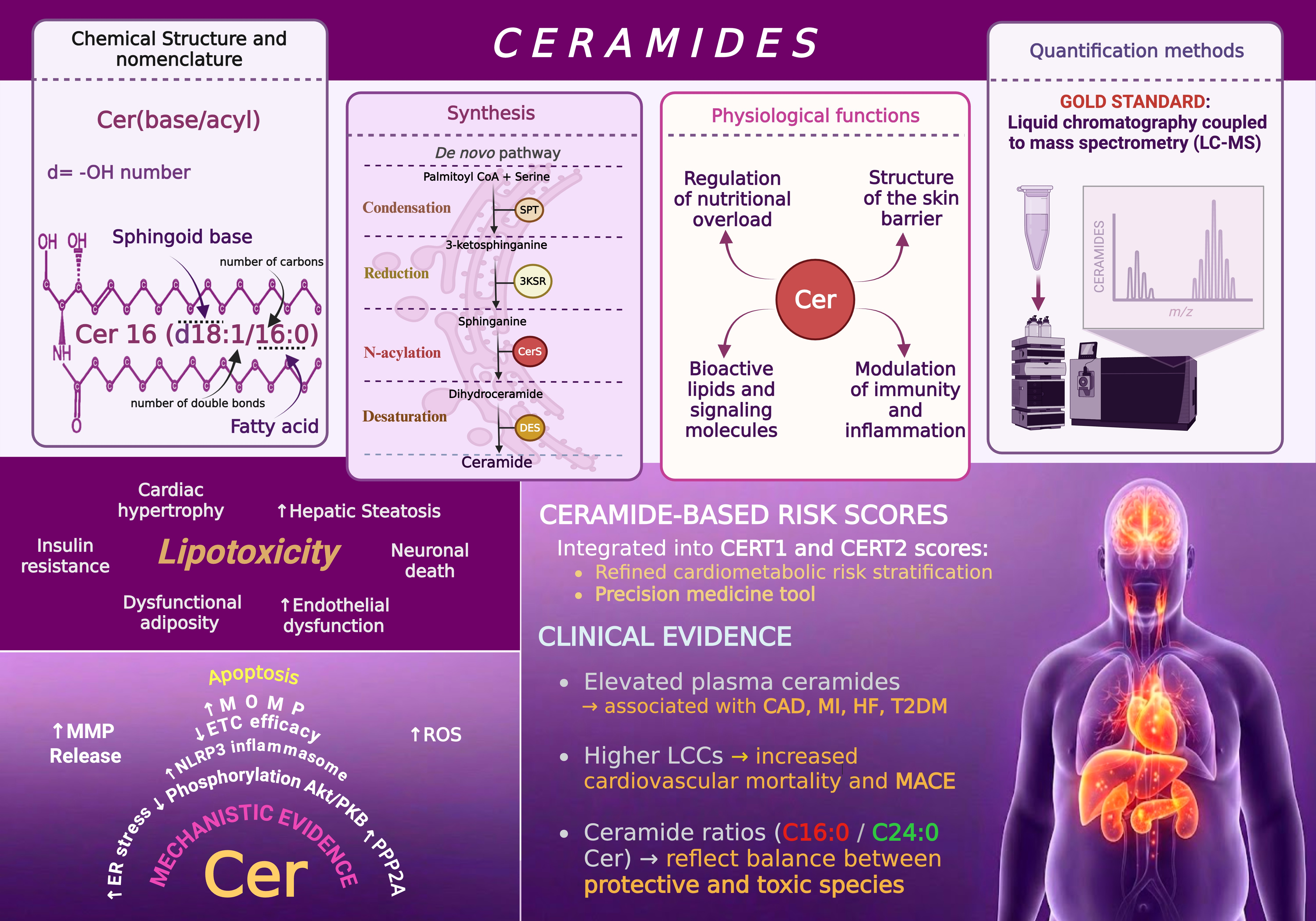
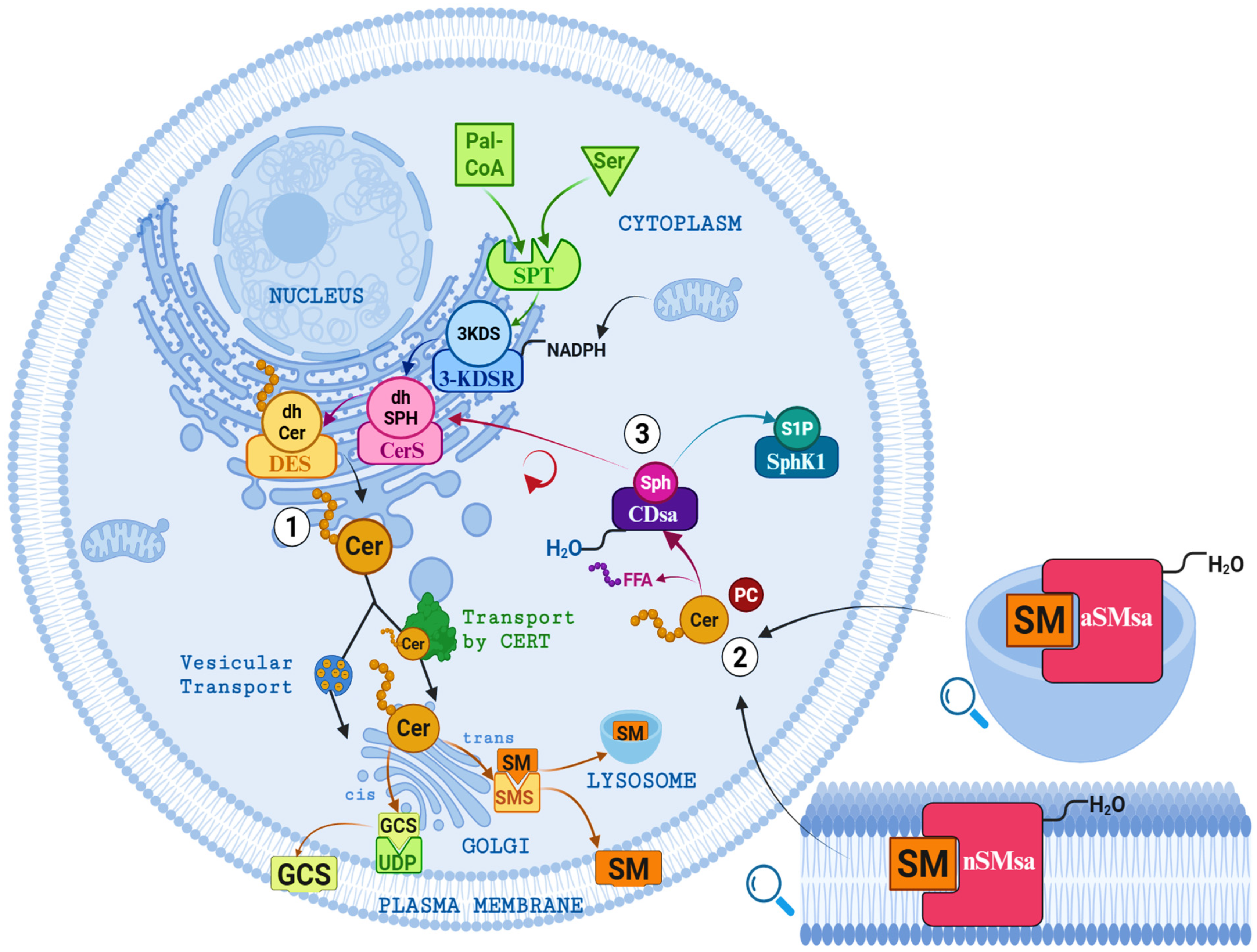
- Condensation: the initial and rate-limiting step is catalyzed by serine palmitoyltransferase (SPT), a multi-protein enzyme complex regulated by the SPTLC1–3 subunits and the small proteins ssSPTa/b. Under physiological conditions, SPT preferentially uses palmitoyl-CoA (C16:0) and serine as substrates. However, the presence of SPTLC3 allows for the utilization of alternative fatty acyl-CoAs or amino acids, leading to the formation of atypical sphingolipid species, including 1-deoxysphingolipids [6,21,47]. The reaction produces 3-ketosphinganine (3-KDS) and establishes a direct link between saturated fatty acid availability and ceramide biosynthesis [48].
- N-acylation: Subsequently, ceramide synthases (CerS1–6) catalyze the attachment of fatty acyl chains of defined lengths, ranging from 14 to 34 carbons atoms, generating dihydroceramides (dhCer). Each CerS isoform displays substrate specificity and tissue-dependent expression, thereby shaping the characteristic ceramide profile of individual organs and contributing to the functional diversity of ceramide species [1,6].
- Desaturation: In the final step, dihydroceramide desaturases (DES1 and DES2) introduce a trans double bond at the C4–C5 position of the sphingoid backbone, converting dihydroceramides (dhCer) into ceramides. This reaction determines the saturation state of the sphingoid base and regulates the intracellular balance between dhCer and ceramide [49]. DES1 is present broadly across tissues, whereas DES2 shows a more restricted distribution, particularly in the skin and intestine, where it also contributes to phytoceramides synthesis. This desaturation step is functionally critical, as ceramides—unlike their dhCer precursors—exert potent signaling and pro-apoptotic activities that influence multiple cellular processes [6,24].
3.4.2. Hydrolysis of Sphingomyelin
3.4.3. Sphingosine Recycling
3.5. Ceramide Synthase Enzymes
3.6. Cellular Metabolism and Toxicity of Free Fatty Acid
3.6.1. Activation
3.6.2. Metabolic Destination
- High energy demand:
- Low energy demand:
3.6.3. Excess Pathway
3.7. Physiological Functions of Ceramides
3.7.1. Regulators of Nutritional Overload
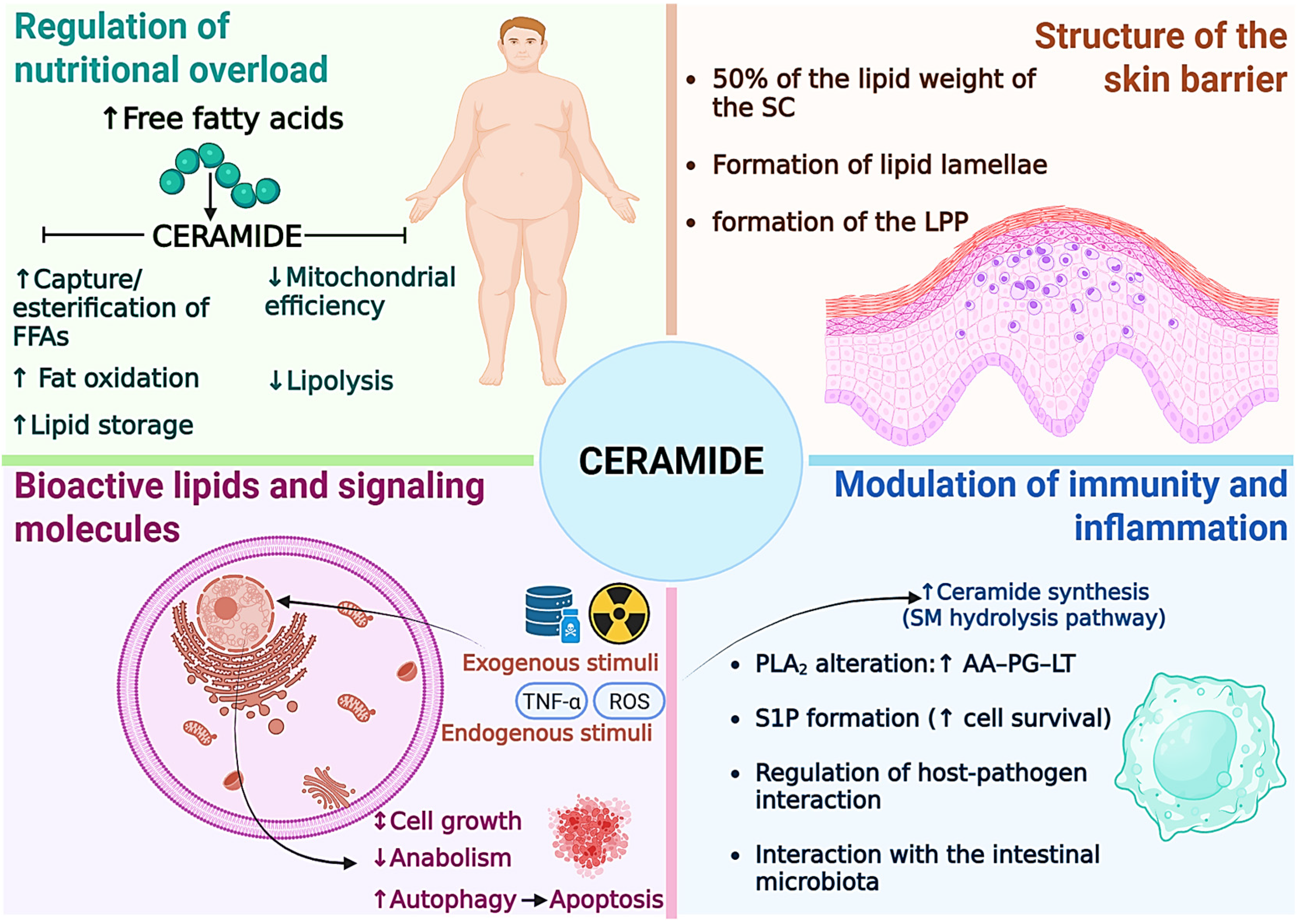
- Facilitation of safe uptake and esterification: Cer promote the translocation of the fatty acid transporter CD36 to the plasma membrane, thereby accelerating the uptake of extracellular FFAs. This process occurs in a controlled manner, allowing their immediate conversion into acyl-CoA by acyl-CoA synthetase, thus preventing the cytotoxic accumulation of FFAs [4,6,77].
- Promotion of lipid storage: The accumulation of Cer activates transcription factors such as Sterol Response Element Binding Proteins (SREBP), which increase the expression of genes involved in the synthesis of TAG and phospholipids [4,6,77]. As a result, cells channel excess fat into lipid droplets, promoting metabolically inert storage and reducing lipotoxicity.
- Induction of preferential fat utilization: Ceramides inhibit the uptake of glucose and amino acids, shifting energy metabolism toward fatty acid oxidation [4,6,77]. This change in fuel preference limits the availability of glycolytic substrates and imposes a lipid dependency that constitutes a molecular basis for the insulin resistance observed in obesity and T2DM [9,78].
- Reduction of mitochondrial efficiency: Cer interferes with the mitochondrial electron transport chain, reducing oxidative phosphorylation efficiency of and ATP production [4,6,77,79]. This phenomenon compels the cell to oxidize more fatty acids to meet its energy demands, generating an effect of dissipating excess energy as heat. Structurally, they promote DRP1-dependent mitochondrial fission, which reduces metabolic efficiency but helps alleviate lipid overload.
3.7.2. Structural Components of Membranes and the Skin Barrier
3.7.3. Bioactive Lipids and Signaling Molecules
3.7.4. Modulation of Immunity and Inflammation
3.8. Detection and Quantification of Ceramides
4. Evidence of the Role of Ceramides in Metabolic and Cardiovascular Disease
4.1. Ceramide 14
4.2. Ceramide 16

| Study Type (Reference) | Biological Matrix | Organelle Involved | Observed Pathophysiological Mechanism |
|---|---|---|---|
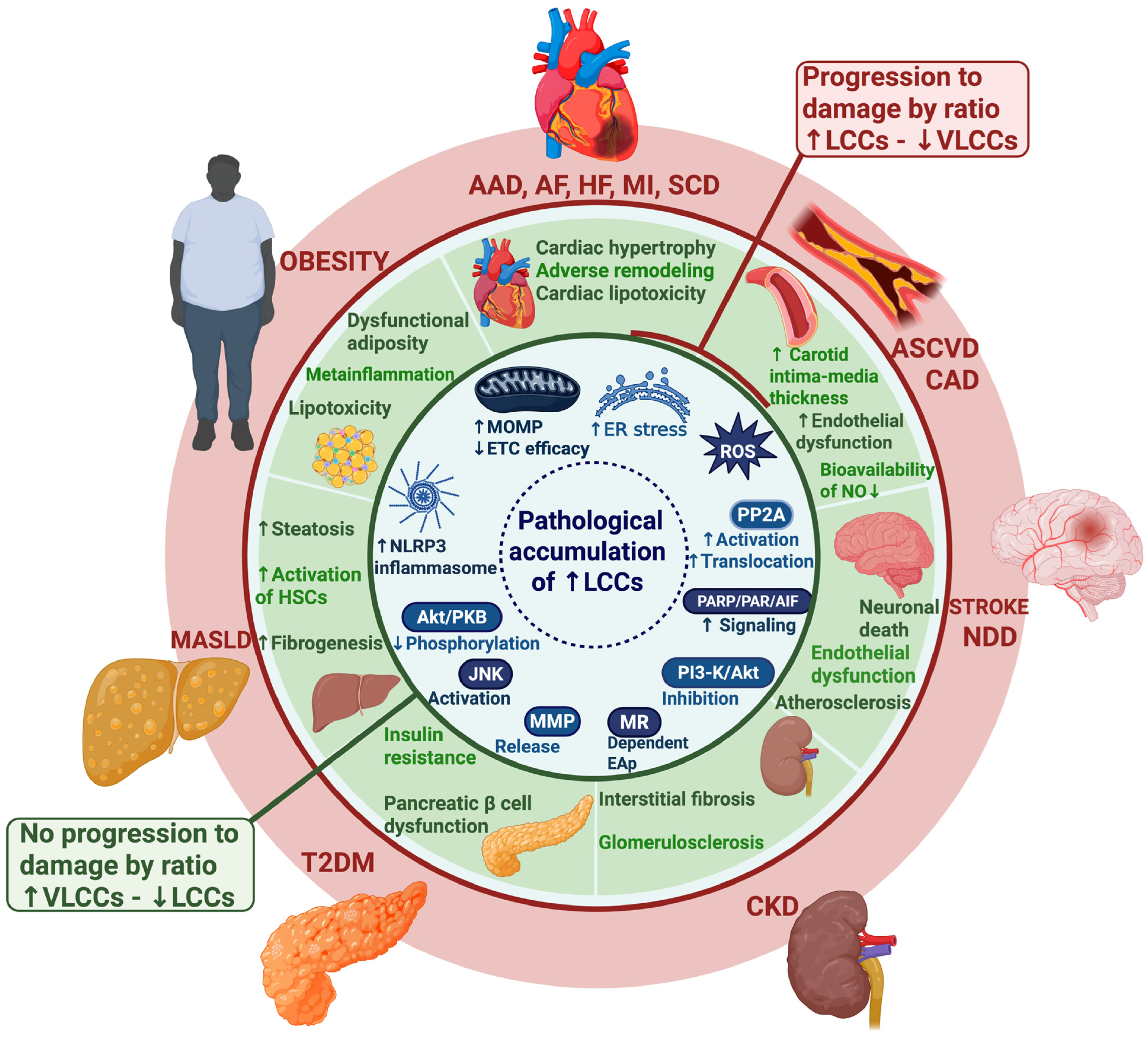
| In vitro [126] | Hepatocytes (primary) | ER + Mitoc + PM | PA → Palmitoyl-CoA → de novo synthesis → ↑ C16:0 Cer → accumulates in lipid rafts → clustering of TRAIL-R2 → DISC → caspase-8 ↑ → tBid → mitoc → formation of Cer channels that cooperate with Bax/Bak → MOMP → cytochrome c ↑ → caspases 3/6/7 ↑ → apoptosis |
| ER stress (↑ CHOP) → ↑ PUMA/Bim → activates Bax/Bak → hepatocyte lipoapoptosis + release of pro-inflammatory EVs | |||
| In vitro [129] | Macrophages (primary/peritoneal, murine lines) | Lysosome + Mitoc+ ER | Signal 1: PA → ↑ C16:0 Cer → acts as DAMP → TLR4/CD36 ↑ → ↑ transcription Nlrp3, pro-IL1β, pro-IL18. |
| Signal 2: C16:0 Cer → lysosomal destabilization (cathepsin B ↑) + mitochondrial ROS ↑ + K+ efflux → NLRP3-ASC-procaspase-1 assembly → active caspase-1 ↑ → pro-IL1β/IL18 cleavage → IL1β/IL18 secretion → chronic sterile inflammation → M1 polarization, metabolic inflammation, insulin resistance | |||
| In vitro [125] | Mice hepatocytes (C57BL/6J) | RE + Mitoc + PM | Palmitate (HFD) → ↑ CerS6 → ↑ C16:0 Cer → insertion into plasma membrane and mitochondria |
| PM: C16:0 Cer forms lipid platforms → sequestration of IR/PI3K → ↓ phosphorylation of Akt/PKB → ↓ glucose uptake → Insulin resistance | |||
| adipocytes | |||
| ER: lipid stress → ↑ TAG ↑ C16:0 Cer → inhibitory signal towards Akt and ↑ WAT inflammation | |||
| BAT | |||
| Mitochondria: C16-Cer → ↓ Complexes II and IV of the ETC → ↓ β-oxidation → ↓ ATP synthesis + ↑ ROS → Mitoc dysfunction | |||
| In vivo | HFD in wild-type vs. CerS6 KO mice | RE + Mitoc | CerS6 KO → ↓ C16:0 Cer → ↑ β-oxidation (Mitoc) → ↑ energy expenditure → protection against DIO and insulin resistance |
| In vitro | Hepatocytes + BAT cells | Liver: ↑ Cd36, Fabp4, SCD1 → ↑ lipid uptake and storage → ↓ palmitate oxidation → hepatic insulin resistance | |
| Ex vivo [130] | WAT, BAT, liver explants + human adipose biopsies | WAT: ↑ body weight ↑ inflammation ↑ serum leptin ↓ glucose tolerance ↓ Akt phosphorylation (↓ insulin signaling) BAT: ↓ lipid oxidation, ↑ accumulation of lipid droplets → ↓ energy expenditure → obesity | |
| In vitro | Hepatocyte (mouse liver) | RE + Mitoc | ↑ CerS6 → ↑ C16:0 Cer ↑ → ↓ β-oxidation → ↓ ATP → ↑ TAG → mitochondrial dysfunction → impaired FA oxidation → insulin resistance (↓ Akt/PKB-P) |
| In vivo | CerS2+/− mice | CerS2+/− → compensatory ↑ C16:0 Cer (via CerS6) → ↓ FA oxidation → acylcarnitine ↑ → steatosis → hepatocellular apoptosis (↑ cleaved PARP) → insulin resistance (↓ glucose tolerance) | |
| In vitro [131] | Adenoviral CerS6 upregulation | CerS6↑ → ↑ C16:0 Cer → ↓ ETC complex II & IV→ ↓ O2 consumption → lipid droplet ↑ → insulin signaling inhibition (↓ Akt/PKB) | |
| In vitro/ex vivo [132] | Human skeletal muscle (myocytes: vastus lateralis fibers) | RE SS (PM) IMF (Mitoc) | PA uptake → de novo synthesis of C16-Cer (ER) → preferential accumulation in the SS fraction → ↑ C16:0 Cer SS ↔ insulin ↑ ↔ HOMA-IR ↑ |
| SS C16:0 Cer → lipid platforms in PM → inhibition of IR/PI3K signaling → ↓ Akt/PKB phosphorylation, ↑ Muscle insulin resistance (↓ GLUT4 translocation, ↓ glucose uptake) | |||
| IMF C16:0 Cer does not correlate with insulin resistance → localized effect in SS | |||
| In vitro/ex vivo [133] | C2C12 myotubes (murine) skeletal muscle fibers (red gastrocnemius) | Mitoc (fusion/fission dynamics) | PA → ↑ de novo Cer synthesis (SPT2) → ↑ C16:0 Cer → activates Drp1 ↑ → excessive mitochondrial fission → ↓ mitochondrial respiration (↓ O2 consumption, complex II) → ↑ ROS (H2O2) → ↓ phospho-Akt/PKB → muscle insulin resistance |
| Inhibition of Drp1 (Mdivi-1) → blocks pathological fusion → protects maintained mitochondrial respiration, ↓ ROS, preserved insulin signal | |||
| In vitro ex vivo | HUVEC Human thoracic (ThAT) vs. subcutaneous (ScAT) adipose tissue | ER + Mitoc + PM | ↑ CerS6 → ↑ C16:0 Cer secretion (via EVs) → uptake by endothelium ⟶ PERK→p-eIF2α→CHOP↑ ⟶ ER stress ⟶ ROS↑ ⟶ ↓ NO ⟶ endothelial dysfunction ⟶ insulin resistance. |
| ThAT↑ C16:0 Cer (vs. ScAT) → ↑ Cer -enriched EVs ⟶ correlated with ↑ vascular O2 and HOMA-IR → vascular oxidative stress ⟶ metabolic impairment | |||
| In vivo (mouse) [134] | Plasma and vascular tissue | ||
| Adipose-derived EVs (C16:0 Cer+) ⟶ ↑ vascular ROS ⟶ ↓ insulin-mediated vasodilation ⟶ endothelial insulin resistance and redox imbalance | |||
| Clinical [20,44,110,135,136] | Serum/plasma | Systemic | ↑ C16:0 Cer or ratios ↑ C16:0/↓ C24:0 Cer⟶ adverse ceramide remodeling (CerS imbalance) ⟶ strong independent predictor of MACE, CV mortality and HF; reflects cardiometabolic/multiorgan dysfunction (heart, liver, kidney) ⟶ correlates with ASCVD; prognostic risk ↑ with T2DM, mitigable by Mediterranean diet |
| Inferred mechanism: C16:0 Cer↑ (plasma) ⟶ vascular/myocardial uptake ⟶ ER stress + mitochondrial dysfunction (ROS↑, ATP↓) ⟶ endothelial dysfunction (NO↓) + inflammation + apoptosis ⟶ atherosclerosis progression/MI/ventricular remodeling/arrhythmia ⟶ clinical events (MACE, MI, HF, SCD) |
4.3. Ceramide 18
4.4. Ceramide 20
4.5. Ceramide 22
4.6. Ceramide 24
5. Therapeutic Strategies for the Modulation of Ceramide Metabolism
5.1. Current Lipid-Lowering Drug Therapies
5.2. Pharmacological Therapies That Modulate Ceramide Production or Degradation
Selective Inhibition of CerS Isoforms
5.3. Non-Pharmacological Therapies
5.3.1. Dietary Evidence
5.3.2. Evidence of Physical Exercise
6. Perspectives, Limitations and Future Directions
7. Conclusions
Author Contributions
Funding
Data Availability Statement
Conflicts of Interest
Abbreviations
| 3-KDS | 3-ketosphinganine |
| 3-KDSR | 3-ketosphinganine reductase |
| AA | Arachidonic acid |
| AAD | aortic aneurysm and dissection |
| ACDase | Acid ceramidase |
| ACS | Acute Coronary Syndrome |
| AFB1 | Aflatoxin B1 |
| Akt/PKB | Protein Kinase B. |
| AlkCDases | Alkaline ceramidases |
| AlkSMase | Alkaline sphingomyelinase |
| ASC | Apoptosis-associated speck-like protein |
| ASCVD | Atherosclerotic cardiovascular disease |
| ASO | Antisense oligonucleotide |
| Atg7 | Autophagy-related gene 7 |
| ATP | Adenosine triphosphate |
| AUC | Area under the curve |
| BAPN | β-aminopropionitrile |
| BAT | Brown Adipose Tissue |
| Bax/Bak | Pro-apoptotic Bcl-2 proteins |
| BECN1 | Beclin-1 |
| BMDM | Bone Marrow-Derived Macrophages |
| BMI | Body mass index |
| BNP | Brain Natriuretic Peptide |
| BP | Blood Pressure |
| CABG | Coronary Artery Bypass Grafting |
| CAD | Coronary artery disease |
| CDase | Ceramidase |
| Cer | Ceramide |
| CerS | Ceramide synthase |
| CERT | Ceramide transfer protein |
| CERT1, CERT2 | Ceramide risk scores 1 and 2 |
| CHOP | C/EBP Homologous Protein |
| CI | Confidence interval |
| CoA | Coenzyme A |
| COL1 | Collagen Type I |
| CV | Cardiovascular |
| CVD | Cardiovascular disease |
| DAMP | Damage-associated molecular pattern |
| DES | Dihydroceramide desaturase |
| DhCer | Dihydroceramide |
| DhSph | Dihydrosphingosine (sphinganine) |
| DISC | Death-inducing signaling complex |
| Drp1 | Dynamin-related protein 1 |
| ECM | Extracellular Matrix |
| EGFR | Estimated Glomerular Filtration Rate |
| EIF2α | Eukaryotic Initiation Factor 2 alpha |
| ELOVL1 | Elongation of very long chain fatty acids protein 1 |
| ELISA | Enzyme-Linked Immunosorbent Assay |
| ENOS | Endothelial nitric oxide synthase |
| ER | Endoplasmic reticulum |
| ETC | Electron Transport Chain |
| EVOO | Extra Virgin Olive Oil |
| EVs | Extracellular Vesicles |
| F1B | Fumonisin B1 |
| FA | Fatty Acid. |
| FFA | Free fatty acid |
| FN1 | Fibronectin 1 |
| FXR | Farnesoid X Receptor |
| GalNAc | N-acetylgalactosamine conjugate |
| GCS | Global circumferential strain |
| GFP | Green fluorescent protein |
| GLUT4 | Glucose transporter type 4 |
| HF | Heart failure |
| HFD | High-fat diet |
| HMG-CoA | 3-Hydroxy-3-Methylglutaryl-CoA |
| HOMA-IR | Homeostatic model assessment of insulin resistance |
| HR | Hazard ratio |
| HRmax | Maximum heart rate |
| HSL | Hormone-sensitive lipase |
| HUH7 | Human Hepatoma Cell Line |
| HUVEC | Human umbilical vein endothelial cells |
| IL | Interleukin |
| IL-1β | Interleukin 1-beta |
| IMF | Intramyofibrillar |
| IR | Insulin Resistance |
| L-aSMase | lysosomal acid sphingomyelinase |
| LAD | Left Anterior Descending artery ligation |
| LAEF | left atrial emptying fraction |
| LASS | Longevity Assurance genes |
| LAVes | left atrial end-systolic volume |
| LC3B | Microtubule-associated protein 1A/1B-light chain 3B |
| LCCs | Long Chain Ceramides |
| LDL-C | low-density lipoprotein cholesterol |
| LDLR | Low-Density Lipoprotein Receptor |
| LN | lupus nephritis |
| LOF | Loss of Function |
| LPP | long periodicity phase |
| LV | Left Ventricle |
| LVEF | left ventricular ejection fraction |
| MA | Myristic acid |
| MACE | Major adverse cardiovascular events |
| MALDI-IMS-FTICR | Matrix-Assisted Laser Desorption/Ionization—Imaging Mass Spectrometry—Fourier Transform Ion Cyclotron Resonance |
| MAPKs | Mitogen-activated protein kinases |
| MASLD | Metabolic dysfunction-associated liver disease |
| MeSH | Medical Subject Headings |
| MFF | Mitochondrial fission factor |
| MI | Myocardial infarction |
| Mitoc | Mitochondria |
| MMF | Metformin Metabolic Failure |
| MMP | Matrix metalloproteinases |
| ModRNA | Modified messenger RNA |
| MOMP | Mitochondrial outer membrane permeabilization |
| MR | Mineralocorticoid receptor |
| MRM | Multiple Reaction Monitoring |
| MRNA | Messenger RNA |
| MS | Metabolic syndrome |
| NCDase | Neutral ceramidase |
| NFκB | Nuclear factor kappa-light-chain-enhancer of activated B cells |
| NLRP3 | NOD-, LRR- and pyrin domain-containing protein 3 |
| NPLC | Normal Phase Liquid Chromatography |
| NPC1L1 | Niemann-Pick C1-Like 1 |
| NO | Nitric oxide |
| NSMase | Neutral sphingomyelinase |
| OCR | Oxygen consumption rate |
| OR | Odds ratio |
| OXPHOS | Oxidative phosphorylation |
| PA | Palmitic acid |
| Pal-CoA | Palmitoyl-CoA |
| PCSK9 | Proprotein Convertase Subtilisin/Kexin Type 9 |
| PDMP | D-threo-1-Phenyl-2-decanoylamino-3-morpholino-1-propanol (glucosylceramide synthase inhibitor) |
| PERK | Protein kinase RNA-like endoplasmic reticulum kinase |
| PG | Prostaglandins |
| PKCζ | Protein kinase C zeta isoform |
| PLA2 | Phospholipase A2 |
| PM | Plasma Membrane |
| PMA | Phorbol 12-myristate 13-acetate |
| PP2A | Protein phosphatase 2A |
| PUFA | Polyunsaturated fatty acid |
| ROC | Receiver Operating Characteristic |
| ROS | Reactive oxygen species |
| RPLC | Reverse Phase Liquid Chromatography |
| S1P | Sphingosine-1-phosphate |
| SaSMase | Secretory acid sphingomyelinase |
| SC | Stratum corneum |
| ScAT | Subcutaneous Adipose Tissue |
| SCD | Sudden cardiac death |
| SCD1 | Stearoyl-CoA Desaturase 1 |
| Ser | Serine |
| SGMS1 | Sphingomyelin synthase 1 |
| ShRNA | Short Hairpin RNA |
| SiRNA | Small Interfering RNA |
| SLE | Systemic lupus erythematosus |
| SM | Sphingomyelin |
| SMase | Sphingomyelinase |
| SNP | Single Nucleotide Polymorphism. |
| Sph | Sphingosine |
| SphK | Sphingosine kinase |
| SPP1 | S1P phosphatase 1 |
| SPR | Surface plasmon resonance |
| SPT | Serine palmitoyltransferase |
| SPT2 | Serine Palmitoyltransferase 2 |
| SPTLC2/3 | Serine Palmitoyltransferase Long Chain Base Subunits 2/3 |
| SREBP | Sterol Regulatory Element-Binding Protein |
| SS | Subsarcolemmal |
| STEMI | ST-segment elevation myocardial infarction |
| SWAT | Subcutaneous White Adipose Tissue |
| T1DM | Type 1 Diabetes Mellitus |
| T2DM | Type 2 diabetes mellitus |
| TAA | Thoracic aortic aneurysm |
| TAC | Transverse aortic constriction |
| TAD | Thoracic aortic dissection |
| TAG | Triacylglycerol |
| tBid | Truncated Bid |
| TCA | Tricarballylic acid |
| TAG | Triglycerides |
| ThAT | Thoracic Adipose Tissue |
| TLR4 | Toll-like receptor 4 |
| TNF-α | Tumor necrosis factor alpha |
| UDCA | Ursodeoxycholic Acid |
| ULCCs | Ultralong-chain ceramide |
| UV | Ultraviolet |
| VAD | Ventricular assistance device |
| VB12 | Vitamin B12 (adenosylcobalamin) |
| VDAC2 | Voltage-dependent anion channel 2 |
| VLCCs | Very Long Chain Ceramides |
| VLDL | Very Low Density Lipoprotein |
| VSMCs | Vascular Smooth Muscle Cells |
| WAT | White Adipose Tissue |
References
- Bartke, N.; Hannun, Y.A. Bioactive sphingolipids: Metabolism and function. J. Lipid Res. 2009, 50, S91–S96. [Google Scholar] [CrossRef] [Scilit]
- Guo, J.; Feng, J.; Qu, H.; Xu, H.; Zhou, H. Potential drug targets for ceramide metabolism in cardiovascular disease. J. Cardiovasc. Dev. Dis. 2022, 9, 434. [Google Scholar] [CrossRef] [Scilit] [PubMed]
- Hannun, Y.A.; Obeid, L.M. Principles of bioactive lipid signalling: Lessons from sphingolipids. Nat. Rev. Mol. Cell Biol. 2008, 9, 139–150. [Google Scholar] [CrossRef] [Scilit]
- Summers, S.A.; Chaurasia, B.; Holland, W.L. Metabolic messengers: Ceramides. Nat. Metab. 2019, 1, 1051–1058. [Google Scholar] [CrossRef] [Scilit]
- Chaurasia, B.; Talbot, C.L.; Summers, S.A. Adipocyte ceramides—The nexus of inflammation and metabolic disease. Front. Immunol. 2020, 11, 576347. [Google Scholar] [CrossRef] [Scilit]
- Chaurasia, B.; Summers, S.A. Ceramides in metabolism: Key lipotoxic players. Annu. Rev. Physiol. 2021, 83, 303–330. [Google Scholar] [CrossRef] [Scilit]
- Tippetts, T.S.; Holland, W.L.; Summers, S.A. Cholesterol–the devil you know; ceramide–the devil you don’t. Trends Pharmacol. Sci. 2021, 42, 1082–1095. [Google Scholar] [CrossRef] [Scilit] [PubMed]
- Chaurasia, B.; Summers, S.A. Ceramides–lipotoxic inducers of metabolic disorders. Trends Endocrinol. Metab. 2015, 26, 538–550. [Google Scholar] [CrossRef] [Scilit]
- Sokolowska, E.; Blachnio-Zabielska, A. The role of ceramides in insulin resistance. Front. Endocrinol. 2019, 10, 577. [Google Scholar] [CrossRef] [Scilit] [PubMed]
- Quinville, B.M.; Deschenes, N.M.; Ryckman, A.E.; Walia, J.S. A comprehensive review: Sphingolipid metabolism and implications of disruption in sphingolipid homeostasis. Int. J. Mol. Sci. 2021, 22, 5793. [Google Scholar] [CrossRef] [Scilit]
- Hajduch, E.; Lachkar, F.; Ferré, P.; Foufelle, F. Roles of ceramides in non-alcoholic fatty liver disease. J. Clin. Med. 2021, 10, 792. [Google Scholar] [CrossRef] [Scilit] [PubMed]
- Yuan, H.; Zhu, B.; Li, C.; Zhao, Z. Ceramide in cerebrovascular diseases. Front. Cell. Neurosci. 2023, 17, 1191609. [Google Scholar] [CrossRef] [Scilit]
- Ji, R.; Akashi, H.; Drosatos, K.; Liao, X.; Jiang, H.; Kennel, P.J.; Brunjes, D.L.; Castillero, E.; Zhang, X.; Deng, L.Y. Increased de novo ceramide synthesis and accumulation in failing myocardium. JCI Insight 2017, 2, e82922. [Google Scholar] [CrossRef] [Scilit]
- Mantovani, A.; Dugo, C. Ceramides and risk of major adverse cardiovascular events: A meta-analysis of longitudinal studies. J. Clin. Lipidol. 2020, 14, 176–185. [Google Scholar] [CrossRef] [Scilit]
- Global Burden of Cardiovascular Diseases and Risks 2023 Collaborators. Global, regional, and national burden of cardiovascular diseases and risk factors in 204 countries and territories, 1990–2023. J. Am. Coll. Cardiol. 2025, 86, 2167–2243.
- Tu, C.; Xie, L.; Wang, Z.; Zhang, L.; Wu, H.; Ni, W.; Li, C.; Li, L.; Zeng, Y. Association between ceramides and coronary artery stenosis in patients with coronary artery disease. Lipids Health Dis. 2020, 19, 151. [Google Scholar] [CrossRef] [Scilit] [PubMed]
- Vasile, V.C.; Meeusen, J.W.; Medina Inojosa, J.R.; Donato, L.J.; Scott, C.G.; Hyun, M.S.; Vinciguerra, M.; Rodeheffer, R.R.; Lopez-Jimenez, F.; Jaffe, A.S. Ceramide Scores Predict Cardiovascular Risk in the Community. Arter. Thromb. Vasc. Biol. 2021, 41, 1558–1569. [Google Scholar] [CrossRef] [Scilit]
- Petrocelli, J.J.; McKenzie, A.I.; Mahmassani, Z.S.; Reidy, P.T.; Stoddard, G.J.; Poss, A.M.; Holland, W.L.; Summers, S.A.; Drummond, M.J. Ceramide biomarkers predictive of cardiovascular disease risk increase in healthy older adults after bed rest. J. Gerontol. Ser. A 2020, 75, 1663–1670. [Google Scholar] [CrossRef] [Scilit] [PubMed]
- Havulinna, A.S.; Sysi-Aho, M.; Hilvo, M.; Kauhanen, D.; Hurme, R.; Ekroos, K.; Salomaa, V.; Laaksonen, R. Circulating ceramides predict cardiovascular outcomes in the population-based FINRISK 2002 cohort. Arterioscler. Thromb. Vasc. Biol. 2016, 36, 2424–2430. [Google Scholar] [CrossRef] [Scilit]
- Peterson, L.R.; Xanthakis, V.; Duncan, M.S.; Gross, S.; Friedrich, N.; Völzke, H.; Felix, S.B.; Jiang, H.; Sidhu, R.; Nauck, M. Ceramide remodeling and risk of cardiovascular events and mortality. J. Am. Heart Assoc. 2018, 7, e007931. [Google Scholar] [CrossRef] [Scilit] [PubMed]
- Ali, O.; Szabó, A. Review of eukaryote cellular membrane lipid composition, with special attention to the fatty acids. Int. J. Mol. Sci. 2023, 24, 15693. [Google Scholar] [CrossRef] [Scilit]
- Hernández-Bello, F.; Franco, M.; Pérez-Méndez, Ó.; Donis-Maturano, L.; Zarco-Olvera, G.; Bautista-Pérez, R. Metabolismo de los esfingolípidos y su relación con las enfermedades cardiovasculares, renales y metabólicas. Arch. Cardiol. México 2023, 93, 88–95. [Google Scholar] [CrossRef] [Scilit] [PubMed]
- Hannun, Y.A.; Obeid, L.M. Sphingolipids and their metabolism in physiology and disease. Nat. Rev. Mol. Cell Biol. 2018, 19, 175–191. [Google Scholar] [CrossRef] [Scilit]
- Merrill, A.H., Jr. Sphingolipid and glycosphingolipid metabolic pathways in the era of sphingolipidomics. Chem. Rev. 2011, 111, 6387–6422. [Google Scholar] [CrossRef] [Scilit]
- Futerman, A.H.; Hannun, Y.A. The complex life of simple sphingolipids. EMBO Rep. 2004, 5, 777–782. [Google Scholar] [CrossRef] [Scilit]
- Levy, M.; Futerman, A.H. Mammalian ceramide synthases. IUBMB Life 2010, 62, 347–356. [Google Scholar] [CrossRef] [Scilit] [PubMed]
- Murray, K.; Rodwell, V.; Bender, D.; Botham, K.M.; Weil, P.A.; Kennelly, P.J. Harper’s Illustrated Biochemistry; McGraw-Hill: New York, NY, USA, 2009; Volume 28, pp. 208–209. [Google Scholar]
- Fanani, M.L.; Maggio, B. The many faces (and phases) of ceramide and sphingomyelin I–single lipids. Biophys. Rev. 2017, 9, 589–600. [Google Scholar] [CrossRef] [Scilit]
- Cha, H.J.; He, C.; Zhao, H.; Dong, Y.; An, I.-S.; An, S. Intercellular and intracellular functions of ceramides and their metabolites in skin. Int. J. Mol. Med. 2016, 38, 16–22. [Google Scholar] [CrossRef] [Scilit] [PubMed]
- Gaggini, M.; Ndreu, R.; Michelucci, E.; Rocchiccioli, S.; Vassalle, C. Ceramides as mediators of oxidative stress and inflammation in cardiometabolic disease. Int. J. Mol. Sci. 2022, 23, 2719. [Google Scholar] [CrossRef] [Scilit]
- Pinto, S.N.; Silva, L.C.; Futerman, A.H.; Prieto, M. Effect of ceramide structure on membrane biophysical properties: The role of acyl chain length and unsaturation. Biochim. Biophys. Acta (BBA)-Biomembr. 2011, 1808, 2753–2760. [Google Scholar] [CrossRef] [Scilit] [PubMed]
- Coant, N.; Sakamoto, W.; Mao, C.; Hannun, Y.A. Ceramidases, roles in sphingolipid metabolism and in health and disease. Adv. Biol. Regul. 2017, 63, 122–131. [Google Scholar] [CrossRef] [Scilit]
- Mahawar, U.; Wattenberg, B. Intricate Regulation of Sphingolipid Biosynthesis: An In-Depth Look Into ORMDL-Mediated Regulation of Serine Palmitoyltransferase. BioEssays 2025, 47, e70036. [Google Scholar] [CrossRef] [Scilit]
- Gault, C.R.; Obeid, L.M.; Hannun, Y.A. An overview of sphingolipid metabolism: From synthesis to breakdown. In Sphingolipids as Signaling and Regulatory Molecules; Springer: New York, NY, USA, 2010; pp. 1–23. [Google Scholar] [CrossRef] [Scilit]
- Vietri Rudan, M.; Watt, F.M. Mammalian epidermis: A compendium of lipid functionality. Front. Physiol. 2022, 12, 804824. [Google Scholar] [CrossRef] [Scilit] [PubMed]
- Mencarelli, C.; Martinez–Martinez, P. Ceramide function in the brain: When a slight tilt is enough. Cell. Mol. Life Sci. 2013, 70, 181–203. [Google Scholar] [CrossRef] [Scilit] [PubMed]
- Suzuki, M.; Ohno, Y.; Kihara, A. Whole picture of human stratum corneum ceramides, including the chain-length diversity of long-chain bases. J. Lipid Res. 2022, 63, 100235. [Google Scholar] [CrossRef] [Scilit]
- Fahy, E.; Subramaniam, S.; Brown, H.A.; Glass, C.K.; Merrill, A.H., Jr.; Murphy, R.C.; Raetz, C.R.; Russell, D.W.; Seyama, Y.; Shaw, W. A comprehensive classification system for lipids. Eur. J. Lipid Sci. Technol. 2005, 107, 337–364. [Google Scholar] [CrossRef] [Scilit]
- Liebisch, G.; Fahy, E.; Aoki, J.; Dennis, E.A.; Durand, T.; Ejsing, C.S.; Fedorova, M.; Feussner, I.; Griffiths, W.J.; Köfeler, H. Update on LIPID MAPS classification, nomenclature, and shorthand notation for MS-derived lipid structures. J. Lipid Res. 2020, 61, 1539–1555. [Google Scholar] [CrossRef] [Scilit]
- Fahy, E.; Subramaniam, S.; Murphy, R.C.; Nishijima, M.; Raetz, C.R.; Shimizu, T.; Spener, F.; Van Meer, G.; Wakelam, M.J.; Dennis, E.A. Update of the LIPID MAPS comprehensive classification system for lipids1. J. Lipid Res. 2009, 50, S9–S14. [Google Scholar] [CrossRef] [Scilit]
- Hannun, Y.A.; Merrill, A.H., Jr.; Luberto, C. The bioactive sphingolipid playbook. A primer for the uninitiated as well as sphingolipidologists. J. Lipid Res. 2025, 66, 100813. [Google Scholar] [CrossRef] [Scilit]
- Chester, M.A. Nomenclature of glycolipids (IUPAC recommendations 1997). Pure Appl. Chem. 1997, 69, 2475–2488. [Google Scholar] [CrossRef] [Scilit]
- Bach, A.; Babayan, V. Medium-chain triglycerides: An update. Am. J. Clin. Nutr. 1982, 36, 950–962. [Google Scholar] [CrossRef] [Scilit] [PubMed]
- Laaksonen, R.; Ekroos, K.; Sysi-Aho, M.; Hilvo, M.; Vihervaara, T.; Kauhanen, D.; Suoniemi, M.; Hurme, R.; März, W.; Scharnagl, H. Plasma ceramides predict cardiovascular death in patients with stable coronary artery disease and acute coronary syndromes beyond LDL-cholesterol. Eur. Heart J. 2016, 37, 1967–1976. [Google Scholar] [CrossRef] [Scilit]
- Schild, J.; Kalvodová, A.; Zbytovská, J.; Farwick, M.; Pyko, C. The role of ceramides in skin barrier function and the importance of their correct formulation for skincare applications. Int. J. Cosmet. Sci. 2024, 46, 526–543. [Google Scholar] [CrossRef] [Scilit] [PubMed]
- Pan, Y.; Li, J.; Lin, P.; Wan, L.; Qu, Y.; Cao, L.; Wang, L. A review of the mechanisms of abnormal ceramide metabolism in type 2 diabetes mellitus, Alzheimer’s disease, and their co-morbidities. Front. Pharmacol. 2024, 15, 1348410. [Google Scholar] [CrossRef] [Scilit]
- Choi, R.H.; Tatum, S.M.; Symons, J.D.; Summers, S.A.; Holland, W.L. Ceramides and other sphingolipids as drivers of cardiovascular disease. Nat. Rev. Cardiol. 2021, 18, 701–711. [Google Scholar] [CrossRef] [Scilit]
- Gaggini, M.; Pingitore, A.; Vassalle, C. Plasma ceramides pathophysiology, measurements, challenges, and opportunities. Metabolites 2021, 11, 719. [Google Scholar] [CrossRef] [Scilit] [PubMed]
- Rahmaniyan, M.; Curley, R.W.; Obeid, L.M.; Hannun, Y.A.; Kraveka, J.M. Identification of dihydroceramide desaturase as a direct in vitro target for fenretinide. J. Biol. Chem. 2011, 286, 24754–24764. [Google Scholar] [CrossRef] [Scilit]
- Sindhu, S.; Leung, Y.H.; Arefanian, H.; Madiraju, S.M.; Al-Mulla, F.; Ahmad, R.; Prentki, M. Neutral sphingomyelinase-2 and cardiometabolic diseases. Obes. Rev. 2021, 22, e13248. [Google Scholar] [CrossRef] [Scilit]
- Jenkins, R.W.; Canals, D.; Hannun, Y.A. Roles and regulation of secretory and lysosomal acid sphingomyelinase. Cell. Signal. 2009, 21, 836–846. [Google Scholar] [CrossRef] [Scilit]
- Pavoine, C.; Pecker, F. Sphingomyelinases: Their regulation and roles in cardiovascular pathophysiology. Cardiovasc. Res. 2009, 82, 175–183. [Google Scholar] [CrossRef] [Scilit]
- Lin, C.-H.; Kornhuber, J.; Zheng, F.; Alzheimer, C. Tonic control of secretory acid sphingomyelinase over ventral hippocampal synaptic transmission and neuron excitability. Front. Cell. Neurosci. 2021, 15, 660561. [Google Scholar] [CrossRef] [Scilit]
- Wangb, C.L.A.; Wuc, Y.; Gulbinsc, E.; Grassméc, H.; Zhaoa, Z. Acid sphingomyelinase-ceramide system in bacterial infections. Cell. Physiol. Biochem 2019, 52, 280–301. [Google Scholar]
- Marchesini, N.; Hannun, Y.A. Acid and neutral sphingomyelinases: Roles and mechanisms of regulation. Biochem. Cell Biol. 2004, 82, 27–44. [Google Scholar] [CrossRef] [Scilit]
- Shamseddine, A.A.; Airola, M.V.; Hannun, Y.A. Roles and regulation of neutral sphingomyelinase-2 in cellular and pathological processes. Adv. Biol. Regul. 2015, 57, 24–41. [Google Scholar] [CrossRef] [Scilit] [PubMed]
- Mühle, C.; Kornhuber, J. Characterization of a neutral Sphingomyelinase activity in human serum and plasma. Int. J. Mol. Sci. 2023, 24, 2467. [Google Scholar] [CrossRef] [Scilit] [PubMed]
- Li, R.-Z.; Wang, X.-R.; Wang, J.; Xie, C.; Wang, X.-X.; Pan, H.-D.; Meng, W.-Y.; Liang, T.-L.; Li, J.-X.; Yan, P.-Y. The key role of sphingolipid metabolism in cancer: New therapeutic targets, diagnostic and prognostic values, and anti-tumor immunotherapy resistance. Front. Oncol. 2022, 12, 941643. [Google Scholar] [CrossRef] [Scilit]
- He, X.; Huang, Y.; Li, B.; Gong, C.-X.; Schuchman, E.H. Deregulation of sphingolipid metabolism in Alzheimer’s disease. Neurobiol. Aging 2010, 31, 398–408. [Google Scholar] [CrossRef] [Scilit]
- Lee, J.K.; Jin, H.K.; Park, M.H.; Kim, B.-r.; Lee, P.H.; Nakauchi, H.; Carter, J.E.; He, X.; Schuchman, E.H.; Bae, J.-s. Acid sphingomyelinase modulates the autophagic process by controlling lysosomal biogenesis in Alzheimer’s disease. J. Exp. Med. 2014, 211, 1551–1570. [Google Scholar] [CrossRef] [Scilit] [PubMed]
- Bismuth, J.; Lin, P.; Yao, Q.; Chen, C. Ceramide: A common pathway for atherosclerosis? Atherosclerosis 2008, 196, 497–504. [Google Scholar] [CrossRef] [Scilit]
- Stiban, J.; Tidhar, R.; Futerman, A.H. Ceramide synthases: Roles in cell physiology and signaling. In Sphingolipids as Signaling and Regulatory Molecules; Springer: New York, NY, USA, 2010; pp. 60–71. [Google Scholar] [CrossRef] [Scilit]
- Tosetti, B.; Brodesser, S.; Brunn, A.; Deckert, M.; Blüher, M.; Doehner, W.; Anker, S.D.; Wenzel, D.; Fleischmann, B.; Pongratz, C. A tissue-specific screen of ceramide expression in aged mice identifies ceramide synthase-1 and ceramide synthase-5 as potential regulators of fiber size and strength in skeletal muscle. Aging Cell 2020, 19, e13049. [Google Scholar] [CrossRef] [Scilit]
- Park, J.-W.; Pewzner-Jung, Y. Ceramide synthases: Reexamining longevity. In Sphingolipids: Basic Science and Drug Development; Springer: Vienna, Austria, 2013; pp. 89–107. [Google Scholar] [CrossRef] [Scilit]
- Dobrzyn, A.; Dobrzyn, P.; Lee, S.-H.; Miyazaki, M.; Cohen, P.; Asilmaz, E.; Hardie, D.G.; Friedman, J.M.; Ntambi, J.M. Stearoyl-CoA desaturase-1 deficiency reduces ceramide synthesis by downregulating serine palmitoyltransferase and increasing β-oxidation in skeletal muscle. Am. J. Physiol.-Endocrinol. Metab. 2005, 288, E599–E607. [Google Scholar] [CrossRef] [Scilit] [PubMed]
- Matsui, H.; Yokoyama, T.; Sekiguchi, K.; Iijima, D.; Sunaga, H.; Maniwa, M.; Ueno, M.; Iso, T.; Arai, M.; Kurabayashi, M. Stearoyl-CoA desaturase-1 (SCD1) augments saturated fatty acid-induced lipid accumulation and inhibits apoptosis in cardiac myocytes. PLoS ONE 2012, 7, e33283. [Google Scholar] [CrossRef] [Scilit] [PubMed]
- Oh, J.; Muralidharan, S.; Zhao, Q.; Scholz, J.; Zelnik, I.D.; Blumenreich, S.; Joseph, T.; Dingjan, T.; Narayanaswamy, P.; Choi, H. Deep sphingolipidomic and metabolomic analyses of ceramide synthase 2 null mice reveal complex pathway-specific effects. J. Lipid Res. 2025, 66, 100832. [Google Scholar] [CrossRef] [Scilit] [PubMed]
- Ferreira, N.S.; Engelsby, H.; Neess, D.; Kelly, S.L.; Volpert, G.; Merrill, A.H.; Futerman, A.H.; Faergeman, N.J. Regulation of very-long acyl chain ceramide synthesis by acyl-CoA-binding protein. J. Biol. Chem. 2017, 292, 7588–7597. [Google Scholar] [CrossRef] [Scilit]
- Dupuy, F.G.; Maggio, B. N-acyl chain in ceramide and sphingomyelin determines their mixing behavior, phase state, and surface topography in Langmuir films. J. Phys. Chem. B 2014, 118, 7475–7487. [Google Scholar] [CrossRef] [Scilit]
- Li, L.; Wang, H.; Jones, J.W. Sphingolipid metabolism as a marker of hepatotoxicity in drug-induced liver injury. Prostaglandins Other Lipid Mediat. 2020, 151, 106484. [Google Scholar] [CrossRef] [Scilit]
- Sassa, T.; Hirayama, T.; Kihara, A. Enzyme activities of the ceramide synthases CERS2–6 are regulated by phosphorylation in the C-terminal region. J. Biol. Chem. 2016, 291, 7477–7487. [Google Scholar] [CrossRef] [Scilit] [PubMed]
- Shalaby, Y.M.; Al Aidaros, A.; Valappil, A.; Ali, B.R.; Akawi, N. Role of ceramides in the molecular pathogenesis and potential therapeutic strategies of cardiometabolic diseases: What we know so far. Front. Cell Dev. Biol. 2022, 9, 816301. [Google Scholar] [CrossRef] [Scilit]
- Olzmann, J.A.; Carvalho, P. Dynamics and functions of lipid droplets. Nat. Rev. Mol. Cell Biol. 2019, 20, 137–155. [Google Scholar] [CrossRef] [Scilit]
- Li, L.O.; Klett, E.L.; Coleman, R.A. Acyl-CoA synthesis, lipid metabolism and lipotoxicity. Biochim. Biophys. Acta (BBA)-Mol. Cell Biol. Lipids 2010, 1801, 246–251. [Google Scholar] [CrossRef] [Scilit]
- Liang, K. Mitochondrial CPT1A: Insights into structure, function, and basis for drug development. Front. Pharmacol. 2023, 14, 1160440. [Google Scholar] [CrossRef] [Scilit]
- Yoon, H.; Shaw, J.L.; Haigis, M.C.; Greka, A. Lipid metabolism in sickness and in health: Emerging regulators of lipotoxicity. Mol. Cell 2021, 81, 3708–3730. [Google Scholar] [CrossRef] [Scilit] [PubMed]
- Summers, S.A. Ceramides: Nutrient signals that drive hepatosteatosis. J. Lipid Atheroscler. 2020, 9, 50–65. [Google Scholar] [CrossRef] [Scilit]
- Fucho, R.; Casals, N.; Serra, D.; Herrero, L. Ceramides and mitochondrial fatty acid oxidation in obesity. FASEB J. 2017, 31, 1263–1272. [Google Scholar] [CrossRef] [Scilit] [PubMed]
- Nicholson, R.J.; Pezzolesi, M.G.; Summers, S.A. Rotten to the cortex: Ceramide-mediated lipotoxicity in diabetic kidney disease. Front. Endocrinol. 2021, 11, 622692. [Google Scholar] [CrossRef] [Scilit]
- Elias, P.M. Lipid abnormalities and lipid-based repair strategies in atopic dermatitis. Biochim. Biophys. Acta (BBA)-Mol. Cell Biol. Lipids 2014, 1841, 323–330. [Google Scholar] [CrossRef] [Scilit]
- Fujii, M. The pathogenic and therapeutic implications of ceramide abnormalities in atopic dermatitis. Cells 2021, 10, 2386. [Google Scholar] [CrossRef] [Scilit]
- Del Rosso, J.Q.; Kircik, L. Skin 101: Understanding the Fundamentals of Skin Barrier Physiology—Why is This Important for Clinicians? J. Clin. Aesthetic Dermatol. 2025, 18, 7. [Google Scholar]
- Danby, S.G.; Andrew, P.V.; Kay, L.J.; Pinnock, A.; Chittock, J.; Brown, K.; Williams, S.F.; Cork, M.J. Enhancement of stratum corneum lipid structure improves skin barrier function and protects against irritation in adults with dry, eczema-prone skin. Br. J. Dermatol. 2022, 186, 875–886. [Google Scholar] [CrossRef] [Scilit]
- Uche, L.E.; Gooris, G.S.; Bouwstra, J.A.; Beddoes, C.M. Increased levels of short-chain ceramides modify the lipid organization and reduce the lipid barrier of skin model membranes. Langmuir 2021, 37, 9478–9489. [Google Scholar] [CrossRef] [Scilit] [PubMed]
- Stahlberg, S.; Školová, B.; Madhu, P.K.; Vogel, A.; Vávrová, K.; Huster, D. Probing the role of the ceramide acyl chain length and sphingosine unsaturation in model skin barrier lipid mixtures by 2H solid-state NMR spectroscopy. Langmuir 2015, 31, 4906–4915. [Google Scholar] [CrossRef] [Scilit]
- Uchida, Y.; Park, K. Ceramides in skin health and disease: An update. Am. J. Clin. Dermatol. 2021, 22, 853–866. [Google Scholar] [CrossRef] [Scilit] [PubMed]
- Huang, W.-C.; Chen, C.-L.; Lin, Y.-S.; Lin, C.-F. Apoptotic sphingolipid ceramide in cancer therapy. J. Lipids 2011, 2011, 565316. [Google Scholar] [CrossRef] [Scilit]
- Alizadeh, J.; da Silva Rosa, S.C.; Weng, X.; Jacobs, J.; Lorzadeh, S.; Ravandi, A.; Vitorino, R.; Pecic, S.; Zivkovic, A.; Stark, H. Ceramides and ceramide synthases in cancer: Focus on apoptosis and autophagy. Eur. J. Cell Biol. 2023, 102, 151337. [Google Scholar] [CrossRef] [Scilit]
- Bikman, B.T.; Summers, S.A. Ceramides as modulators of cellular and whole-body metabolism. J. Clin. Investig. 2011, 121, 4222–4230. [Google Scholar] [CrossRef] [Scilit] [PubMed]
- Pasquaré, S.J.; Gaveglio, V.L.; Giusto, N.M. Regulation of phosphatidic acid metabolism by sphingolipids in the central nervous system. J. Lipids 2011, 2011, 342576. [Google Scholar] [CrossRef] [Scilit]
- Lee, M.; Lee, S.Y.; Bae, Y.-S. Functional roles of sphingolipids in immunity and their implication in disease. Exp. Mol. Med. 2023, 55, 1110–1130. [Google Scholar] [CrossRef] [Scilit]
- Rubenzucker, S.; Manke, M.-C.; Lehmann, R.; Assinger, A.; Borst, O.; Ahrends, R. A Targeted, Bioinert LC–MS/MS Method for Sensitive, Comprehensive Analysis of Signaling Lipids. Anal. Chem. 2024, 96, 9643–9652. [Google Scholar] [CrossRef] [Scilit]
- Huang, Q.; Hao, S.; Yao, X.; You, J.; Li, X.; Lai, D.; Han, C.; Schilling, J.; Hwa, K.Y.; Thyparambil, S. High-throughput quantitation of serological ceramides/dihydroceramides by LC/MS/MS: Pregnancy baseline biomarkers and potential metabolic messengers. J. Pharm. Biomed. Anal. 2021, 192, 113639. [Google Scholar] [CrossRef] [Scilit]
- Kauhanen, D.; Sysi-Aho, M.; Koistinen, K.M.; Laaksonen, R.; Sinisalo, J.; Ekroos, K. Development and validation of a high-throughput LC–MS/MS assay for routine measurement of molecular ceramides. Anal. Bioanal. Chem. 2016, 408, 3475–3483. [Google Scholar] [CrossRef] [Scilit] [PubMed]
- Hilvo, M.; Vasile, V.C.; Donato, L.J.; Hurme, R.; Laaksonen, R. Ceramides and ceramide scores: Clinical applications for cardiometabolic risk stratification. Front. Endocrinol. 2020, 11, 570628. [Google Scholar] [CrossRef] [Scilit] [PubMed]
- Jiang, H.; Hsu, F.-F.; Farmer, M.S.; Peterson, L.R.; Schaffer, J.E.; Ory, D.S.; Jiang, X. Development and validation of LC-MS/MS method for determination of very long acyl chain (C22: 0 and C24: 0) ceramides in human plasma. Anal. Bioanal. Chem. 2013, 405, 7357–7365. [Google Scholar] [CrossRef] [Scilit] [PubMed]
- van Smeden, J.; Hoppel, L.; van der Heijden, R.; Hankemeier, T.; Vreeken, R.J.; Bouwstra, J.A. LC/MS analysis of stratum corneum lipids: Ceramide profiling and discovery. J. Lipid Res. 2011, 52, 1211–1221. [Google Scholar] [CrossRef] [Scilit]
- Jones, E.E.; Dworski, S.; Canals, D.; Casas, J.; Fabrias, G.; Schoenling, D.; Levade, T.; Denlinger, C.; Hannun, Y.A.; Medin, J.A. On-tissue localization of ceramides and other sphingolipids by MALDI mass spectrometry imaging. Anal. Chem. 2014, 86, 8303–8311. [Google Scholar] [CrossRef] [Scilit]
- Vielhaber, G.; Brade, L.; Lindner, B.; Pfeiffer, S.; Wepf, R.; Hintze, U.; Wittern, K.P.; Brade, H. Mouse anti-ceramide antiserum: A specific tool for the detection of endogenous ceramide. Glycobiology 2001, 11, 451–457. [Google Scholar] [CrossRef] [Scilit]
- Krishnamurthy, K.; Dasgupta, S.; Bieberich, E. Development and characterization of a novel anti-ceramide antibody. J. Lipid Res. 2007, 48, 968–975. [Google Scholar] [CrossRef] [Scilit]
- Singh, K.; Singh, B.; Ray, P. Anti-ceramide antibodies in leprosy: Marker for nerve damage? J. Infect. Dev. Ctries. 2010, 4, 378–381. [Google Scholar] [CrossRef] [Scilit] [PubMed]
- Wiśniewski, A.; Wisniewska, E.; Lewandowski, L.; Nowak, I.; Kosacka, M. Increased serum anti-ceramide antibodies and decreased sphingosine-1-phosphate levels in patients with obstructive sleep apnea syndrome as potential markers of endothelial dysfunction. Front. Mol. Biosci. 2025, 12, 1644828. [Google Scholar] [CrossRef] [Scilit]
- Canals, D.; Hannun, Y.A. Biological function, topology, and quantification of plasma membrane Ceramide. Adv. Biol. Regul. 2024, 91, 101009. [Google Scholar] [CrossRef] [Scilit] [PubMed]
- Green, C.D.; Maceyka, M.; Cowart, L.A.; Spiegel, S. Sphingolipids in metabolic disease: The good, the bad, and the unknown. Cell Metab. 2021, 33, 1293–1306. [Google Scholar] [CrossRef] [Scilit]
- Kenéz, Á.; Bäßler, S.C.; Jorge-Smeding, E.; Huber, K. Ceramide metabolism associated with chronic dietary nutrient surplus and diminished insulin sensitivity in the liver, muscle, and adipose tissue of cattle. Front. Physiol. 2022, 13, 958837. [Google Scholar] [CrossRef] [Scilit]
- Novgorodov, S.A.; Gudz, T.I. Ceramide and mitochondria in ischemia/reperfusion. J. Cardiovasc. Pharmacol. 2009, 53, 198–208. [Google Scholar] [CrossRef] [Scilit]
- Shu, H.; Peng, Y.; Hang, W.; Li, N.; Zhou, N.; Wang, D.W. Emerging roles of ceramide in cardiovascular diseases. Aging Dis. 2022, 13, 232. [Google Scholar] [CrossRef] [Scilit]
- Jernigan, P.L.; Makley, A.T.; Hoehn, R.S.; Edwards, M.J.; Pritts, T.A. The role of sphingolipids in endothelial barrier function. Biol. Chem. 2015, 396, 681–691. [Google Scholar] [CrossRef] [Scilit]
- Hla, T.; Dannenberg, A.J. Sphingolipid signaling in metabolic disorders. Cell Metab. 2012, 16, 420–434. [Google Scholar] [CrossRef] [Scilit] [PubMed]
- Hilvo, M.; Meikle, P.J.; Pedersen, E.R.; Tell, G.S.; Dhar, I.; Brenner, H.; Schöttker, B.; Lääperi, M.; Kauhanen, D.; Koistinen, K.M. Development and validation of a ceramide-and phospholipid-based cardiovascular risk estimation score for coronary artery disease patients. Eur. Heart J. 2020, 41, 371–380. [Google Scholar] [CrossRef] [Scilit]
- McGurk, K.A.; Keavney, B.D.; Nicolaou, A. Circulating ceramides as biomarkers of cardiovascular disease: Evidence from phenotypic and genomic studies. Atherosclerosis 2021, 327, 18–30. [Google Scholar] [CrossRef] [Scilit] [PubMed]
- Hilvo, M.; Jylhä, A.; Lääperi, M.; Jousilahti, P.; Laaksonen, R. Absolute and relative risk prediction in cardiovascular primary prevention with a modified SCORE chart incorporating ceramide-phospholipid risk score and diabetes mellitus. Eur. Heart J. Open 2021, 1, oeab010. [Google Scholar] [CrossRef] [Scilit]
- Cantalupo, A.; Sasset, L.; Gargiulo, A.; Rubinelli, L.; Del Gaudio, I.; Benvenuto, D.; Wadsack, C.; Jiang, X.-C.; Bucci, M.R.; Di Lorenzo, A. Endothelial sphingolipid de novo synthesis controls blood pressure by regulating signal transduction and NO via ceramide. Hypertension 2020, 75, 1279–1288. [Google Scholar] [CrossRef] [Scilit]
- Zietzer, A.; Düsing, P.; Reese, L.; Nickenig, G.; Jansen, F. Ceramide Metabolism in Cardiovascular Disease: A Network With High Therapeutic Potential. Arter. Thromb. Vasc. Biol. 2022, 42, 1220–1228. [Google Scholar] [CrossRef] [Scilit]
- Couttas, T.A.; Rustam, Y.H.; Song, H.; Qi, Y.; Teo, J.D.; Chen, J.; Reid, G.E.; Don, A.S. A novel function of sphingosine kinase 2 in the metabolism of sphinga-4, 14-diene lipids. Metabolites 2020, 10, 236. [Google Scholar] [CrossRef] [Scilit]
- Merrill, A.H., Jr. Don’t be surprised when these surprise you: Some infrequently studied sphingoid bases, metabolites, and factors that should be kept in mind during sphingolipidomic studies. Int. J. Mol. Sci. 2025, 26, 650. [Google Scholar] [CrossRef] [Scilit] [PubMed]
- Meeusen, J.W.; Donato, L.J.; Bryant, S.C.; Baudhuin, L.M.; Berger, P.B.; Jaffe, A.S. Plasma ceramides: A novel predictor of major adverse cardiovascular events after coronary angiography. Arterioscler. Thromb. Vasc. Biol. 2018, 38, 1933–1939. [Google Scholar] [CrossRef] [Scilit]
- Yin, W.; Li, F.; Tan, X.; Wang, H.; Jiang, W.; Wang, X.; Li, S.; Zhang, Y.; Han, Q.; Wang, Y. Plasma ceramides and cardiovascular events in hypertensive patients at high cardiovascular risk. Am. J. Hypertens. 2021, 34, 1209–1216. [Google Scholar] [CrossRef] [Scilit] [PubMed]
- You, Y.; Zeng, Q.; Hu, Z.; Chen, Y.; Zhan, M.; Wang, Y.; Duan, J. Myristic Acid Remodels Sphingolipid Metabolism via Dual Pathways: Canonical d18-Sphingolipid Regulation and Non-Canonical d16-Sphingolipid Synthesis. Nutrients 2025, 17, 2881. [Google Scholar] [CrossRef] [Scilit]
- Martínez, L.; Torres, S.; Baulies, A.; Alarcón-Vila, C.; Elena, M.; Fabriàs, G.; Casas, J.; Caballeria, J.; Fernandez-Checa, J.C.; García-Ruiz, C. Myristic acid potentiates palmitic acid-induced lipotoxicity and steatohepatitis associated with lipodystrophy by sustaning de novo ceramide synthesis. Oncotarget 2015, 6, 41479. [Google Scholar] [CrossRef] [Scilit]
- Russo, S.B.; Baicu, C.F.; Van Laer, A.; Geng, T.; Kasiganesan, H.; Zile, M.R.; Cowart, L.A. Ceramide synthase 5 mediates lipid-induced autophagy and hypertrophy in cardiomyocytes. J. Clin. Investig. 2012, 122, 3919–3930. [Google Scholar] [CrossRef] [Scilit] [PubMed]
- de Carvalho, L.P.; Tan, S.H.; Ow, G.-S.; Tang, Z.; Ching, J.; Kovalik, J.-P.; Poh, S.C.; Chin, C.-T.; Richards, A.M.; Martinez, E.C. Plasma ceramides as prognostic biomarkers and their arterial and myocardial tissue correlates in acute myocardial infarction. JACC Basic Transl. Sci. 2018, 3, 163–175. [Google Scholar] [CrossRef] [Scilit]
- Kasumov, T.; Solomon, T.P.; Hwang, C.; Huang, H.; Haus, J.M.; Zhang, R.; Kirwan, J.P. Improved insulin sensitivity after exercise training is linked to reduced plasma C 14: 0 ceramide in obesity and type 2 diabetes. Obesity 2015, 23, 1414–1421. [Google Scholar] [CrossRef] [Scilit]
- Merrill, A.H., Jr.; Sandhoff, K. Sphingolipids: Metabolism and cell signaling. In New Comprehensive Biochemistry; Elsevier: Amsterdam, The Netherlands, 2002; Volume 36, pp. 373–407. [Google Scholar]
- Hla, T.; Kolesnick, R. C16: 0-ceramide signals insulin resistance. Cell Metab. 2014, 20, 703–705. [Google Scholar] [CrossRef] [Scilit]
- Hirsova, P.; Ibrabim, S.H.; Gores, G.J.; Malhi, H. Lipotoxic lethal and sublethal stress signaling in hepatocytes: Relevance to NASH pathogenesis. J. Lipid Res. 2016, 57, 1758–1770. [Google Scholar] [CrossRef] [Scilit]
- Lin, Z.; Long, F.; Kang, R.; Klionsky, D.J.; Yang, M.; Tang, D. The lipid basis of cell death and autophagy. Autophagy 2024, 20, 469–488. [Google Scholar] [CrossRef] [Scilit]
- Javaheri, A.; Allegood, J.C.; Cowart, L.A.; Chirinos, J.A. Circulating ceramide 16: 0 in heart failure with preserved ejection fraction. J. Am. Coll. Cardiol. 2020, 75, 2273–2275. [Google Scholar] [CrossRef] [Scilit] [PubMed]
- Camell, C.; Goldberg, E.; Dixit, V.D. Regulation of Nlrp3 inflammasome by dietary metabolites. In Seminars in Immunology; Academic Press: Cambridge, MA, USA, 2015; pp. 334–342. [Google Scholar]
- Turpin, S.M.; Nicholls, H.T.; Willmes, D.M.; Mourier, A.; Brodesser, S.; Wunderlich, C.M.; Mauer, J.; Xu, E.; Hammerschmidt, P.; Brönneke, H.S. Obesity-induced CerS6-dependent C16: 0 ceramide production promotes weight gain and glucose intolerance. Cell Metab. 2014, 20, 678–686. [Google Scholar] [CrossRef] [Scilit]
- Raichur, S.; Wang, S.T.; Chan, P.W.; Li, Y.; Ching, J.; Chaurasia, B.; Dogra, S.; Öhman, M.K.; Takeda, K.; Sugii, S. CerS2 haploinsufficiency inhibits β-oxidation and confers susceptibility to diet-induced steatohepatitis and insulin resistance. Cell Metab. 2014, 20, 687–695. [Google Scholar] [CrossRef] [Scilit]
- Chung, J.O.; Koutsari, C.; Blachnio-Zabielska, A.U.; Hames, K.C.; Jensen, M.D. Intramyocellular ceramides: Subcellular concentrations and fractional de novo synthesis in postabsorptive humans. Diabetes 2017, 66, 2082–2091. [Google Scholar] [CrossRef] [Scilit] [PubMed]
- Smith, M.E.; Tippetts, T.S.; Brassfield, E.S.; Tucker, B.J.; Ockey, A.; Swensen, A.C.; Anthonymuthu, T.S.; Washburn, T.D.; Kane, D.A.; Prince, J.T. Mitochondrial fission mediates ceramide-induced metabolic disruption in skeletal muscle. Biochem. J. 2013, 456, 427–439. [Google Scholar] [CrossRef] [Scilit]
- Akawi, N.; Checa, A.; Antonopoulos, A.S.; Akoumianakis, I.; Daskalaki, E.; Kotanidis, C.P.; Kondo, H.; Lee, K.; Yesilyurt, D.; Badi, I. Fat-secreted ceramides regulate vascular redox state and influence outcomes in patients with cardiovascular disease. J. Am. Coll. Cardiol. 2021, 77, 2494–2513. [Google Scholar] [CrossRef] [Scilit]
- Leiherer, A.; Muendlein, A.; Saely, C.H.; Laaksonen, R.; Fraunberger, P.; Drexel, H. Ceramides improve cardiovascular risk prediction beyond low-density lipoprotein cholesterol. Eur. Heart J. Open 2024, 4, oeae001. [Google Scholar] [CrossRef] [Scilit] [PubMed]
- Nwabuo, C.C.; Duncan, M.; Xanthakis, V.; Peterson, L.R.; Mitchell, G.F.; McManus, D.; Cheng, S.; Vasan, R.S. Association of circulating ceramides with cardiac structure and function in the community: The framingham heart study. J. Am. Heart Assoc. 2019, 8, e013050. [Google Scholar] [CrossRef] [Scilit]
- Kucuk, S.; Niven, J.; Caamano, J.; Jones, S.W.; Camacho-Munoz, D.; Nicolaou, A.; Mauro, C. Unwrapping the mechanisms of ceramide and fatty acid-initiated signals leading to immune-inflammatory responses in obesity. Int. J. Biochem. Cell Biol. 2021, 135, 105972. [Google Scholar] [CrossRef] [Scilit] [PubMed]
- Sergi, D.; Zauli, E.; Celeghini, C.; Previati, M.; Zauli, G. Ceramides as the molecular link between impaired lipid metabolism, saturated fatty acid intake and insulin resistance: Are all saturated fatty acids to be blamed for ceramide-mediated lipotoxicity? Nutr. Res. Rev. 2024, 38, 256–266. [Google Scholar] [CrossRef] [Scilit] [PubMed]
- SenthilKumar, G.; Zirgibel, Z.; Cohen, K.E.; Katunaric, B.; Jobe, A.M.; Shult, C.G.; Limpert, R.H.; Freed, J.K. Ying and yang of ceramide in the vascular endothelium. Arterioscler. Thromb. Vasc. Biol. 2024, 44, 1725–1736. [Google Scholar] [CrossRef] [Scilit]
- Cheng, J.M.; Suoniemi, M.; Kardys, I.; Vihervaara, T.; de Boer, S.P.; Akkerhuis, K.M.; Sysi-Aho, M.; Ekroos, K.; Garcia-Garcia, H.M.; Oemrawsingh, R.M. Plasma concentrations of molecular lipid species in relation to coronary plaque characteristics and cardiovascular outcome: Results of the ATHEROREMO-IVUS study. Atherosclerosis 2015, 243, 560–566. [Google Scholar] [CrossRef] [Scilit]
- Wu, J.M.; Schulze, P.C. Cardiovascular prognosis: A new role for ceramides and other cardiometabolites. ESC Heart Fail. 2020, 7, 3285. [Google Scholar] [CrossRef] [Scilit]
- Timmerman, N.; Waissi, F.; Dekker, M.; de Borst, G.J.; van Bennekom, J.; de Winter, R.J.; Hilvo, M.; Jylhä, A.; Pasterkamp, G.; de Kleijn, D.P. Ceramides and phospholipids in plasma extracellular vesicles are associated with high risk of major cardiovascular events after carotid endarterectomy. Sci. Rep. 2022, 12, 5521. [Google Scholar] [CrossRef] [Scilit]
- Meade, R.; Chao, Y.; Harroun, N.; Li, C.; Hafezi, S.; Hsu, F.-F.; Semenkovich, C.F.; Zayed, M.A. Ceramides in peripheral arterial plaque lead to endothelial cell dysfunction. JVS-Vasc. Sci. 2023, 4, 100181. [Google Scholar] [CrossRef] [Scilit]
- Denimal, D.; Duvillard, L.; Beland-Bonenfant, S.; Terriat, B.; Pais-de-Barros, J.-P.; Simoneau, I.; Rouland, A.; Houbachi, L.; Bouillet, B.; Verges, B. Plasma 16: 0 ceramide as a marker of cardiovascular risk estimated by carotid intima-media thickness in people with type 2 diabetes. Diabetes Metab. 2024, 50, 101542. [Google Scholar] [CrossRef] [Scilit]
- Wang, D.D.; Toledo, E.; Hruby, A.; Rosner, B.A.; Willett, W.C.; Sun, Q.; Razquin, C.; Zheng, Y.; Ruiz-Canela, M.; Guasch-Ferre, M. Plasma ceramides, Mediterranean diet, and incident cardiovascular disease in the PREDIMED trial (Prevención con Dieta Mediterránea). Circulation 2017, 135, 2028–2040. [Google Scholar] [CrossRef] [Scilit]
- Zatloukal, J.; Zylla, S.; Markus, M.R.; Ewert, R.; Gläser, S.; Völzke, H.; Albrecht, D.; Friedrich, N.; Nauck, M.; Peterson, L.R. The Association Between C24: 0/C16: 0 Ceramide Ratio and Cardiorespiratory Fitness is Robust to Effect Modifications by Age and Sex. Adv. Biol. 2024, 8, 2300633. [Google Scholar] [CrossRef] [Scilit]
- Witoslawska, A.; Meessen, J.M.; Hilvo, M.; Jylhä, A.; Zannad, F.; Cerrato, M.; Rossignol, P.; Novelli, D.; Duarte, K.; Targher, G. Ceramide and phosphatidylcholine lipids-based risk score predicts major cardiovascular outcomes in patients with heart failure. Eur. J. Clin. Investig. 2025, 55, e14359. [Google Scholar] [CrossRef] [Scilit]
- Zhang, Y.; Pan, Y.; Bian, Z.; Chen, P.; Zhu, S.; Gu, H.; Guo, L.; Hu, C. Ceramide production mediates aldosterone-induced human umbilical vein endothelial cell (HUVEC) damages. PLoS ONE 2016, 11, e0146944. [Google Scholar] [CrossRef] [Scilit]
- Yang, H.; Yang, F.; Luo, M.; Chen, Q.; Liu, X.; Zhang, Y.; Zhu, G.; Chen, W.; Li, T.; Shu, C. Metabolomic profile reveals that ceramide metabolic disturbance plays an important role in thoracic aortic dissection. Front. Cardiovasc. Med. 2022, 9, 826861. [Google Scholar] [CrossRef] [Scilit] [PubMed]
- Lee, S.; Kim, S.-A.; Kim, Y.; Kim, J.; Hong, G.; Hong, J.; Choi, K.; Eom, C.-S.; Baik, S.; Lee, M.-K. Genetic variants associated with elevated plasma ceramides in individuals with metabolic syndrome. Genes 2022, 13, 1497. [Google Scholar] [CrossRef] [Scilit] [PubMed]
- Wretlind, A.; Curovic, V.R.; Suvitaival, T.; Theilade, S.; Tofte, N.; Winther, S.A.; Vilsbøll, T.; Vestergaard, H.; Rossing, P.; Legido-Quigley, C. Ceramides as risk markers for future cardiovascular events and all-cause mortality in long-standing type 1 diabetes. Diabetes 2023, 72, 1493–1501. [Google Scholar] [CrossRef] [Scilit]
- Spaggiari, R.; Angelini, S.; Di Vincenzo, A.; Scaglione, G.; Morrone, S.; Finello, V.; Fagioli, S.; Castaldo, F.; Sanz, J.M.; Sergi, D. Ceramides as emerging players in cardiovascular disease: Focus on their pathogenetic effects and regulation by diet. Adv. Nutr. 2024, 15, 100252. [Google Scholar] [CrossRef] [Scilit] [PubMed]
- Zhang, Z.; Xie, L.; Lin, X.; He, J.; Xie, Y.; Li, J.; Zhuang, X.; Tang, L.; Xie, R.; Wu, Q. Ursodeoxycholic acid alleviates aortic aneurysm and dissection through the intestinal farnesoid X receptor/ceramide synthase 2 axis. Commun. Biol. 2025, 8, 1009. [Google Scholar] [CrossRef] [Scilit]
- Hadas, Y.; Vincek, A.S.; Youssef, E.; Żak, M.M.; Chepurko, E.; Sultana, N.; Sharkar, M.T.K.; Guo, N.; Komargodski, R.; Kurian, A.A. Altering sphingolipid metabolism attenuates cell death and inflammatory response after myocardial infarction. Circulation 2020, 141, 916–930. [Google Scholar] [CrossRef] [Scilit]
- Tarasov, K.; Ekroos, K.; Suoniemi, M.; Kauhanen, D.; Sylvänne, T.; Hurme, R.; Gouni-Berthold, I.; Berthold, H.K.; Kleber, M.E.; Laaksonen, R. Molecular lipids identify cardiovascular risk and are efficiently lowered by simvastatin and PCSK9 deficiency. J. Clin. Endocrinol. Metab. 2014, 99, E45–E52. [Google Scholar] [CrossRef] [Scilit]
- Park, L.K.; Barry, V.G.; Hong, J.; Heebink, J.; Sah, R.; Peterson, L.R. Links between ceramides and cardiac function. Curr. Opin. Lipidol. 2022, 33, 47–56. [Google Scholar] [CrossRef] [Scilit]
- McCaffrey, J.M.; Ibdah, J.A. Effects of Diet and Exercise on Mitochondrial Health in Metabolic Dysfunction-Associated Steatotic Liver Disease (MASLD): Role of Ceramides. Nutrients 2025, 17, 2972. [Google Scholar] [CrossRef] [Scilit] [PubMed]
- Schmidt, S.; Gallego, S.F.; Zelnik, I.D.; Kovalchuk, S.; Albæk, N.; Sprenger, R.R.; Øverup, C.; Pewzner-Jung, Y.; Futerman, A.H.; Lindholm, M.W. Silencing of ceramide synthase 2 in hepatocytes modulates plasma ceramide biomarkers predictive of cardiovascular death. Mol. Ther. 2022, 30, 1661–1674. [Google Scholar] [CrossRef] [Scilit]
- Chen, M.; Peng, Y.; Hu, Y.; Kang, Z.; Chen, T.; Zhang, Y.; Chen, X.; Li, Q.; Yuan, Z.; Wu, Y. A critical role for Phocaeicola vulgatus in negatively impacting metformin response in diabetes. Acta Pharm. Sin. B 2025, 15, 2511–2528. [Google Scholar] [CrossRef] [Scilit]
- Nicholson, R.J.; Poss, A.M.; Maschek, J.A.; Cox, J.E.; Hopkins, P.N.; Hunt, S.C.; Playdon, M.C.; Holland, W.L.; Summers, S.A. Characterizing a common CERS2 polymorphism in a mouse model of metabolic disease and in subjects from the Utah CAD study. J. Clin. Endocrinol. Metab. 2021, 106, e3098–e3109. [Google Scholar] [CrossRef] [Scilit] [PubMed]
- Wittenbecher, C.; Cuadrat, R.; Johnston, L.; Eichelmann, F.; Jäger, S.; Kuxhaus, O.; Prada, M.; Del Greco, M.F.; Hicks, A.A.; Hoffman, P.; et al. Dihydroceramide- and ceramide-profiling provides insights into human cardiometabolic disease etiology. Nat. Commun. 2022, 13, 936. [Google Scholar] [CrossRef] [Scilit]
- Laviad, E.L.; Albee, L.; Pankova-Kholmyansky, I.; Epstein, S.; Park, H.; Merrill, A.H.; Futerman, A.H. Characterization of ceramide synthase 2: Tissue distribution, substrate specificity, and inhibition by sphingosine 1-phosphate. J. Biol. Chem. 2008, 283, 5677–5684. [Google Scholar] [CrossRef] [Scilit] [PubMed]
- Wiley, A.M.; Krueger, M.A.; Sotoodehnia, N.; Umans, J.G.; Hoofnagle, A.N.; Lemaitre, R.N.; Totah, R.A.; Gharib, S.A. Deciphering the Role of Different Ceramide Synthases in the Human Cardiomyocyte Hypertrophic Response. Metabolites 2025, 15, 635. [Google Scholar] [CrossRef] [Scilit] [PubMed]
- Hartmann, D.; Wegner, M.-S.; Wanger, R.A.; Ferreirós, N.; Schreiber, Y.; Lucks, J.; Schiffmann, S.; Geisslinger, G.; Grösch, S. The equilibrium between long and very long chain ceramides is important for the fate of the cell and can be influenced by co-expression of CerS. Int. J. Biochem. Cell Biol. 2013, 45, 1195–1203. [Google Scholar] [CrossRef] [Scilit] [PubMed]
- Camacho-Morales, A.; Noriega, L.G.; Sánchez-García, A.; Torre-Villalvazo, I.; Vázquez-Manjarrez, N.; Maldonado-Ruiz, R.; Cárdenas-Tueme, M.; Villegas-Romero, M.; Alamilla-Martínez, I.; Rodriguez-Rocha, H. Plasma C24: 0 ceramide impairs adipose tissue remodeling and promotes liver steatosis and glucose imbalance in offspring of rats. Heliyon 2024, 10, e39206. [Google Scholar] [CrossRef] [Scilit]
- Patyna, S.; Buettner, S.; Eckes, T.; Obermueller, N.; Bartel, C.; Braner, A.; Trautmann, S.; Thomas, D.; Geiger, H.; Pfeilschifter, J. Blood ceramides as novel markers for renal impairment in systemic lupus erythematosus. Prostaglandins Other Lipid Mediat. 2019, 144, 106348. [Google Scholar] [CrossRef] [Scilit]
- Buie, J.N.J.; Hammad, S.M.; Nietert, P.J.; Magwood, G.; Adams, R.J.; Bonilha, L.; Sims-Robinson, C. Differences in plasma levels of long chain and very long chain ceramides between African Americans and whites: An observational study. PLoS ONE 2019, 14, e0216213. [Google Scholar] [CrossRef] [Scilit] [PubMed]
- Kasumov, T.; Li, L.; Li, M.; Gulshan, K.; Kirwan, J.P.; Liu, X.; Previs, S.; Willard, B.; Smith, J.D.; McCullough, A. Ceramide as a mediator of non-alcoholic fatty liver disease and associated atherosclerosis. PLoS ONE 2015, 10, e0126910. [Google Scholar] [CrossRef] [Scilit]
- Han, E.; Kim, G.; Lee, J.-Y.; Lee, Y.-h.; Kim, B.S.; Lee, B.-W.; Cha, B.-S.; Kang, E.S. Comparison between atorvastatin and rosuvastatin in renal function decline among patients with diabetes. Endocrinol. Metab. 2017, 32, 274–280. [Google Scholar] [CrossRef] [Scilit]
- Ng, T.W.; Ooi, E.M.; Watts, G.F.; Chan, D.C.; Weir, J.M.; Meikle, P.J.; Barrett, P.H.R. Dose-dependent effects of rosuvastatin on the plasma sphingolipidome and phospholipidome in the metabolic syndrome. J. Clin. Endocrinol. Metab. 2014, 99, E2335–E2340. [Google Scholar] [CrossRef] [Scilit]
- Worgall, T.S.; Johnson, R.A.; Seo, T.; Gierens, H.; Deckelbaum, R.J. Unsaturated fatty acid-mediated decreases in sterol regulatory element-mediated gene transcription are linked to cellular sphingolipid metabolism. J. Biol. Chem. 2002, 277, 3878–3885. [Google Scholar] [CrossRef] [Scilit]
- Worgall, T.S.; Juliano, R.A.; Seo, T.; Deckelbaum, R.J. Ceramide synthesis correlates with the posttranscriptional regulation of the sterol-regulatory element-binding protein. Arterioscler. Thromb. Vasc. Biol. 2004, 24, 943–948. [Google Scholar] [CrossRef] [Scilit]
- Mak, B.; Lin, H.-M.; Duong, T.; Mahon, K.L.; Joshua, A.M.; Stockler, M.R.; Gurney, H.; Parnis, F.; Zhang, A.; Scheinberg, T. Modulation of plasma lipidomic profiles in metastatic castration-resistant prostate cancer by simvastatin. Cancers 2022, 14, 4792. [Google Scholar] [CrossRef] [Scilit] [PubMed]
- Bergheanu, S.C.; Reijmers, T.; Zwinderman, A.H.; Bobeldijk, I.; Ramaker, R.; Liem, A.-H.; Greef, J.v.d.; Hankemeier, T.; Wouter Jukema, J. Lipidomic approach to evaluate rosuvastatin and atorvastatin at various dosages: Investigating differential effects among statins. Curr. Med. Res. Opin. 2008, 24, 2477–2487. [Google Scholar] [CrossRef] [Scilit]
- Klassen, A.; Faccio, A.T.; Picossi, C.R.C.; Derogis, P.B.M.C.; dos Santos Ferreira, C.E.; Lopes, A.S.; Sussulini, A.; Cruz, E.C.S.; Bastos, R.T.; Fontoura, S.C. Evaluation of two highly effective lipid-lowering therapies in subjects with acute myocardial infarction. Sci. Rep. 2021, 11, 15973. [Google Scholar] [CrossRef] [Scilit] [PubMed]
- Yamanashi, Y.; Takada, T.; Yamamoto, H.; Suzuki, H. NPC1L1 facilitates sphingomyelin absorption and regulates diet-induced production of VLDL/LDL-associated S1P. Nutrients 2020, 12, 2641. [Google Scholar] [CrossRef] [Scilit] [PubMed]
- Ye, Q.; Svatikova, A.; Meeusen, J.W.; Kludtke, E.L.; Kopecky, S.L. Effect of proprotein convertase subtilisin/kexin type 9 inhibitors on plasma ceramide levels. Am. J. Cardiol. 2020, 128, 163–167. [Google Scholar] [CrossRef] [Scilit]
- Demers, A.; Samami, S.; Lauzier, B.; Des Rosiers, C.; Sock, E.T.N.; Ong, H.; Mayer, G. PCSK9 induces CD36 degradation and affects long-chain fatty acid uptake and triglyceride metabolism in adipocytes and in mouse liver. Arterioscler. Thromb. Vasc. Biol. 2015, 35, 2517–2525. [Google Scholar] [CrossRef] [Scilit]
- Zhang, X.; Xu, H.; Yu, J.; Cui, J.; Chen, Z.; Li, Y.; Niu, Y.; Wang, S.; Ran, S.; Zou, Y. Immune regulation of the liver through the PCSK9/CD36 pathway during heart transplant rejection. Circulation 2023, 148, 336–353. [Google Scholar] [CrossRef] [Scilit]
- Hilvo, M.; Simolin, H.; Metso, J.; Ruuth, M.; Öörni, K.; Jauhiainen, M.; Laaksonen, R.; Baruch, A. PCSK9 inhibition alters the lipidome of plasma and lipoprotein fractions. Atherosclerosis 2018, 269, 159–165. [Google Scholar] [CrossRef] [Scilit]
- Anesi, A.; Di Minno, A.; Calcaterra, I.; Cavalca, V.; Tripaldella, M.; Porro, B.; Di Minno, M.N.D. An untargeted lipidomic analysis reveals depletion of several phospholipid classes in patients with familial hypercholesterolemia on treatment with evolocumab. Biomedicines 2021, 9, 1941. [Google Scholar] [CrossRef] [Scilit]
- Riley, R.T.; Merrill, A.H. Ceramide synthase inhibition by fumonisins: A perfect storm of perturbed sphingolipid metabolism, signaling, and disease. J. Lipid Res. 2019, 60, 1183–1189. [Google Scholar] [CrossRef] [Scilit]
- Kurek, K.; Mikłosz, A.; Łukaszuk, B.; Chabowski, A.; Górski, J.; Żendzian-Piotrowska, M. Inhibition of ceramide de novo synthesis ameliorates diet induced skeletal muscles insulin resistance. J. Diabetes Res. 2015, 2015, 154762. [Google Scholar] [CrossRef] [Scilit]
- Morrice, N.; Mcilroy, G.D.; Tammireddy, S.R.; Reekie, J.; Shearer, K.D.; Doherty, M.K.; Delibegović, M.; Whitfield, P.D.; Mody, N. Elevated Fibroblast growth factor 21 (FGF21) in obese, insulin resistant states is normalised by the synthetic retinoid Fenretinide in mice. Sci. Rep. 2017, 7, 43782. [Google Scholar] [CrossRef] [Scilit] [PubMed]
- Draper, J.M.; Xia, Z.; Smith, R.A.; Zhuang, Y.; Wang, W.; Smith, C.D. Discovery and evaluation of inhibitors of human ceramidase. Mol. Cancer Ther. 2011, 10, 2052–2061. [Google Scholar] [CrossRef] [Scilit] [PubMed]
- Chen, J.; Goyal, N.; Dai, L.; Lin, Z.; Del Valle, L.; Zabaleta, J.; Liu, J.; Post, S.R.; Foroozesh, M.; Qin, Z. Developing new ceramide analogs and identifying novel sphingolipid-controlled genes against a virus-associated lymphoma. Blood J. Am. Soc. Hematol. 2020, 136, 2175–2187. [Google Scholar] [CrossRef] [Scilit]
- Schiffmann, S.; Hartmann, D.; Fuchs, S.; Birod, K.; Ferreiròs, N.; Schreiber, Y.; Zivkovic, A.; Geisslinger, G.; Grösch, S.; Stark, H. Inhibitors of specific ceramide synthases. Biochimie 2012, 94, 558–565. [Google Scholar] [CrossRef] [Scilit]
- Sasset, L.; Manzo, O.L.; Zhang, Y.; Marino, A.; Rubinelli, L.; Riemma, M.A.; Chalasani, M.L.S.; Dasoveanu, D.C.; Roviezzo, F.; Jankauskas, S.S. Nogo-A reduces ceramide de novo biosynthesis to protect from heart failure. Cardiovasc. Res. 2023, 119, 506–519. [Google Scholar] [CrossRef] [Scilit]
- Zhang, Y.; Huang, Y.; Cantalupo, A.; Azevedo, P.S.; Siragusa, M.; Bielawski, J.; Giordano, F.J.; Di Lorenzo, A. Endothelial Nogo-B regulates sphingolipid biosynthesis to promote pathological cardiac hypertrophy during chronic pressure overload. JCI Insight 2016, 1, e85484. [Google Scholar] [CrossRef] [Scilit]
- Wang, S.; Jin, Z.; Wu, B.; Morris, A.J.; Deng, P. Role of dietary and nutritional interventions in ceramide-associated diseases. J. Lipid Res. 2024, 66, 100726. [Google Scholar] [CrossRef] [Scilit]
- Helge, J.W.; Dobrzyn, A.; Saltin, B.; Gorski, J. Exercise and training effects on ceramide metabolism in human skeletal muscle. Exp. Physiol. 2004, 89, 119–127. [Google Scholar] [CrossRef] [Scilit] [PubMed]
- Wang, E.; Norred, W.; Bacon, C.; Riley, R.; Merrill, A.H., Jr. Inhibition of sphingolipid biosynthesis by fumonisins. Implications for diseases associated with Fusarium moniliforme. J. Biol. Chem. 1991, 266, 14486–14490. [Google Scholar] [CrossRef] [Scilit]
- Mathur, S.; Constable, P.D.; Eppley, R.M.; Waggoner, A.L.; Tumbleson, M.E.; Haschek, W.M. Fumonisin B1 is hepatotoxic and nephrotoxic in milk-fed calves. Toxicol. Sci. 2001, 60, 385–396. [Google Scholar] [CrossRef] [Scilit]
- Singh, K.; Kumari, A. Mycotoxins and Mycotoxicoses; Springer: Berlin/Heidelberg, Germany, 2022. [Google Scholar]
- Cantalupo, A.; Zhang, Y.; Kothiya, M.; Galvani, S.; Obinata, H.; Bucci, M.; Giordano, F.J.; Jiang, X.-C.; Hla, T.; Di Lorenzo, A. Nogo-B regulates endothelial sphingolipid homeostasis to control vascular function and blood pressure. Nat. Med. 2015, 21, 1028–1037. [Google Scholar] [CrossRef] [Scilit] [PubMed]
- Manzo, O.L.; Nour, J.; Sasset, L.; Marino, A.; Rubinelli, L.; Palikhe, S.; Smimmo, M.; Hu, Y.; Bucci, M.R.; Borczuk, A. Rewiring endothelial sphingolipid metabolism to favor S1P over ceramide protects from coronary atherosclerosis. Circ. Res. 2024, 134, 990–1005. [Google Scholar] [CrossRef] [Scilit]
- Tan-Chen, S.; Guitton, J.; Bourron, O.; Le Stunff, H.; Hajduch, E. Sphingolipid metabolism and signaling in skeletal muscle: From physiology to physiopathology. Front. Endocrinol. 2020, 11, 491. [Google Scholar] [CrossRef] [Scilit]
- Hammerschmidt, P.; Brüning, J.C. Contribution of specific ceramides to obesity-associated metabolic diseases. Cell. Mol. Life Sci. 2022, 79, 395. [Google Scholar] [CrossRef] [Scilit] [PubMed]
- Mathews, A.T.; Famodu, O.A.; Olfert, M.D.; Murray, P.J.; Cuff, C.F.; Downes, M.T.; Haughey, N.J.; Colby, S.E.; Chantler, P.D.; Olfert, I.M. Efficacy of nutritional interventions to lower circulating ceramides in young adults: FRUVEDomic pilot study. Physiol. Rep. 2017, 5, e13329. [Google Scholar] [CrossRef] [Scilit] [PubMed]


| CerS. | Preferential Acyl-CoA Substrate | Tissue Expression |
|---|---|---|
| CerS1 | 18 carbons | Brain, skeletal muscle and testis |
| CerS2 * | 20–26 carbons | Nervous system (oligodendrocytes, Schwann cells), kidney and liver |
| CerS3 | >26 carbons | Testicle and skin (keratinocytes) |
| CerS4 | 18–20 carbons | Most tissues, predominantly in leukocytes, liver, heart and skin |
| CerS5 | 16 carbons | Most tissues. Higher expression in the pulmonary epithelium |
| CerS6 | 14 and 16 carbons | Most tissues. Predominantly in the intestine and kidney |
| Author | Ceramides Measured | Type of Study, Population, Details | Findings |
|---|---|---|---|
| Havulinna (2016) [19] | Ceramides (d18:1/16:0, 18:0, 24:0, 24:1) and analysis of ratios of 16, 18 and 24:1 carbon ceramide versus 24:0 carbon ceramide. | Observational-prospective nested within a cohort; (n): 8101; Apparently healthy Finnish subjects. Pioneer in the creation of the CERT1 score Validated ceramides in the general population, not just in patients at high cardiovascular risk. | Relative risk increase of 2.7-fold for MACE and 4.3-fold for death due to MACE. The Cer(d18:1/18:0) species showed the most significant individual association, presenting a Hazard ratio (HR) of 1.21 (95% CI: 1.11–1.33) for incident MACE after adjustment for Framingham risk factors. The combination with C-reactive protein is proposed to identify very high-risk subjects. |
| Meeusen (2018) [117] | Ceramides of 16, 18, 24, and 24:1 carbon were analyzed individually, as well as their ratios to the 24:0 carbon ceramide. | Prospective cohort of US patients referred for coronary angiography (n): 504. Indirectly describes the development of the CERT1 score, subsequently the MAYO clinic developed the commercial version in collaboration with Zora Biosciences Oy under the leadership of Hilvo and Laaksonen. | Patients in the highest risk category (Score ≥ 10) had a more than 2-fold higher risk of 4-year MACE and 18-year mortality compared with the low-risk group. The Ceramide Risk Score (16:0)/(24:0) ratio was the most potent predictor, with an adjusted Hazard Ratio of 1.75 per SD for MACE. The prognostic utility of these molecules was further consolidated by the Ceramide Risk Score (adjusted HR of 1.58 per SD). |
| Hilvo (2020) [110] | Individually, ceramides with 16, 18, 24, and 24:1 carbon atom were analyzed, as well as their ratios relative to the 24:0 carbon ceramide, and the measurement of phosphatidylcholines (PC 16:0/22:5, PC 14:0/22:6, and PC 16:0/16:0) was also added. | The CERT2 score was developed based on three studies; the score was developed in the WECAC cohort and validated in the LIPID and KAROLA cohorts, based on its use in secondary prevention for patients with established atherosclerotic coronary artery disease. | CERT2 proved to be a highly significant predictor of cardiovascular death, surpassing traditional lipids such as LDL-C. Its combination with troponin T (CERT2-TnT) drastically improved residual risk stratification, allowing for the precise identification of 10–20% of patients at extremely high risk. |
| Hilvo (2021) [112] | The same ones included in the CERT2 score | Based on the FINRISK 2002 cohort, used to test its efficacy in primary prevention, excluding those patients with prevalent cardiovascular events. | CERT2 proved to be a significant and robust predictor of all cardiovascular outcomes, particularly for fatal events. It was demonstrated that the incidence of fatal events was more than 10 times higher in the highest CERT2 risk category compared to the lowest. |
| Wenjie Yin (2021) [118] | Seven different ceramide species were measured; however, the score only includes Cer (18:1/16:0) and Cer (18:1/24:1), along with their proportions relative to Cer (18:1/22:0) and Cer (18:1/24:0). | They developed the CERT-HBP score using a prospective analysis model within the PROSPECT study cohort of patients with essential hypertension who were classified in a high or very high cardiovascular risk category. | The CERT-HBP allows for enhanced identification of high-risk patients, whose highest category showed a 4.6-fold increase in the risk of MACE compared to the lowest category. |
| Study Type (Reference) | Biological Matrix | Organelle Involved | Observed Pathophysiological Mechanism |
|---|---|---|---|
| In vitro [120] | Human hepatocytes (HepG2) | Mitoc + RE (functional interaction) | MA ↑ C14:0-CoA incorporation → ↑ C14:0 Cer; in the presence of PA → synergistic effect → ↑ Total Cer ↓ SM → ER stress + mitochondrial dysfunction → cytochrome c ↑ → caspases ↑ → apoptosis Result: exacerbated lipotoxicity (MA potentiates PA-induced lipotoxicity) |
| In vitro [121] | Primary cardiomyocytes | Mitoc + RE | MA (C14:0) → CerS5 ↑ → ↑ C14:0 Cer → ↑ autophagy (↑ BECN1, ↑ LC3B, ↑ Atg7) → ER/mitocytic remodeling → cardiomyocyte hypertrophy |
| In vivo [121] | Mouse heart | Inhibition of CerS5 (siRNA) or de novo synthesis (myriocin) → ↓ C14:0 Cer → ↓ autophagy → ↓ hypertrophy | |
| Clinical [13,122] | Plasma | Systemic | MI → ↑ Plasma C14:0 Cer → activates mitochondrial pathways (autophagy/apoptosis) → worse prognosis (↑ MACE) |
| [123] | Aerobic exercise → ↓ Plasma C14:0 Cer → negative correlation with ↑ insulin sensitivity (↓ Akt inhibition → ↑ glucose uptake) | ||
| In vivo [13] | Myocardium | Mitoc + RE | HF/MI patients→ ↑ SPTLC2 → ↑ de novo Cer (including C14:0) in plasma and myocardium → pathological remodeling (fibrosis, inflammation, apoptosis). SPT inhibition (myriocin) or SPTLC2 deletion → ↓ Cer → ↓ remodeling and improved function |
| Study Type (Reference) | Biological Matrix | Organelle Involved | Observed Pathophysiological Mechanism |
|---|---|---|---|
| In vitro [148] | HUVEC | RE + Mitoc | Aldosterone → MR → CerS1 ↑ → C18:0 Cer ↑ (ER) ⟶ Accumulation C18:0 Cer → ↑ pro-apoptotic signaling ⟶ Mitochondria: dysfunction + amplification of apoptotic cascade ⟶ Caspase-3 activation → DNA fragmentation → endothelial apoptosis |
| Modulators: Eplerenone (MR antagonist) ⟶ ↓ C18-Cer → protection S1P (anti-ceramide lipid) ⟶ ↓ apoptosis PDMP (GCS inhibitor) or C6:0 Cer exogenous ⟶ ↑ apoptosis CerS1 shRNA ⟶ ↓ C18:0 Cer → protection CerS1 upregulation ⟶ ↑ C18:0 Cer → ↑ apoptosis | |||
| In vitro | Macrophages (RAW264.7) | RE + Mitoc | TAD → ↑ C18:0 Cer (plasma, aortic macrophages) ⟶ activation of NLRP3 inflammasome ⟶ ↑ casp-1 act ⟶ ↑ IL-1β, IL-18 ⟶ ↑ MMPs (MMP9) ⟶ ECM degradation + aortic wall weakening ⟶ ↑ inflammation + ↑ risk of dissection |
| Murine BAPN-TAD model | In vivo | Exogenous C18:0 Cer ⟶ ↑ macrophage inflammation + ↑ MMPs | |
| Clinical [149] | plasma metabolomics in TAD vs. TAA vs. controls | Myriocin (SPT inhibitor) ⟶ ↓ de novo Cer synthesis ⟶ ↓ aortic inflammation + ↓ TAD incidence in mice | |
| Clinical [150] | Plasma (patients with MS) | RE | MS → ↑ plasma C18:0 Cer ⟶ linked to SNPs in ceramide biosynthetic genes (CERS3, CERS6, SGMS1, SPTLC2/3, ACER1) ⟶ dysregulated sphingolipid metabolism ⟶ ↑ insulin resistance (HOMA-IR ↑, insulin ↑) ⟶ ↓ adiponectin, ↑ TAG, ↑ waist circumference, ↑ BP ⟶ systemic metabolic stress and cardiovascular risk. |
| Clinical [44,145] | Plasma (patients with CAD, ACS, high CV risk) | Systemic | ↑ Plasma C18:0 Cer ⟶ associated with ↑ CV death and ↑ incident CVD. |
| Inferred mechanism: C18:0 Cer ↑ → lipoprotein uptake dysregulation + vascular inflammation + apoptosis (Mitoc stress) → atherosclerotic plaque instability → MI, stroke, CV death. | |||
| Ratios (C18:0/C24:0 Cer) stronger predictors than absolute levels. Mediterranean diet (EVOO/nuts) attenuates risk despite high ceramide score. |
| Study Type (Reference) | Biological Matrix | Organelle Involved | Observed Pathophysiological Mechanism |
|---|---|---|---|
| Clinical | Plasma (AAD patients) | Systemic | Inferred mechanism: Intestinal FXR ↑ → Cers2 ↑ → C20:0 Cer ↑ (plasma) ⟶ macrophage inflammatory transformation ⟶ ↑ MMP2/MMP9 release ⟶ ECM degradation (↓ COL1, ↓ FN1) ⟶ vascular wall weakening ⟶ ↑ incidence of AAD |
| In vivo | Mouse (BAPN models) | ER + Mitoc | Myriocin (SPT inhibitor) ⟶ ↓ de novo synthesis → ↓ AAD |
| Exogenous C20:0 Cer ⟶ directly ↑ MMP2/MMP9 from macrophages, fibroblasts, VSMCs. | |||
| In vitro [153] | Enterocytes and macrophages (BMDM) | UDCA (FXR inhibitor) ⟶ ↓ FXR activation → ↓ Cers2 → ↓ C20:0 Cer → ↓ inflammation + preserved ECM → ↓ AAD incidence | |
| In vivo | Mouse MI model (LAD ligation) | ER + Mitoc + Lysosome | MI → hypoxia/ischemia → de novo Cer synthesis ↑ → C20:0 Cer ↑ (16.5-fold in LV, 24h post-MI) ⟶ MITOC stress + casp-3 dimerization/cleavage ⟶ cardiomyocyte apoptosis ↑ ⟶ LV dysfunction + scar formation |
| In vitro [154] | LV Cardiomyocytes (myocardium under hypoxia) | aCDase inhibition ⟶ ceramide accumulation ↑ → cardiomyocyte death ↑ → survival ↓/aCDase modRNA overexpression ⟶ C20:0 Cer ↓ → sphingosine ↑ → S1P ↑ (pro-survival) ⟶ ↓ apoptosis, ↓ neutrophil infiltration, ↓ inflammation ⟶ improved LV function + survival. | |
| Clinical [155] | Plasma (CAD patients) | Systemic | ↑ C20:0 Cer ⟶ associated with ↑ CV death risk in CAD Ratio C20:0/C24:0 Cer = strong predictor of fatal outcome, independent of LDL-C. |
| Simvastatin ⟶ ↓ C20:0 Cer (~25%)/Ezetimibe ⟶ LDL-C ↓ but C20-Cer unchanged/slightly ↑/PCSK9 LOF ⟶ modest LDL-C ↓ (~13%) but ↓ C20:0 Cer (~20%) → ↓ CAD risk. | |||
| Clinical | Plasma (MI patients) | Systemic | MI → ↑ plasma C20:0 Cer (part of 12-ceramide prognostic signature) ⟶ predicted 12-month MACE (death, MI, stroke) |
| In vivo | Aortic tissue (human CABG biopsies) | CABG patients with recent MI ⟶ ↑ plasma C20:0 Cer, but not in aortic wall. | |
| Rat MI model ⟶ ↑ myocardial C20:0 Cer + ↑ expression of CerS enzymes → source of circulating ceramides. | |||
| [122] | Myocardium (rat LAD ligation) | Inferred mechanism: ischemia/reperfusion → Cer ↑ → Mitochondrial stress, apoptosis, inflammation → adverse remodeling + recurrent events. |
| Scheme | Biological Matrix | Organelle Involved | Observed Pathophysiological Mechanism |
|---|---|---|---|
| In vivo | CerS2+/− mice (chow vs. HFD) | RE + Mitoc | CerS2 haploinsufficiency → ↓ C22:0 Cer (and other VLCCs) → compensatory ↑ C16:0 Cer (via CerS5/6) ⟶ Mitoc dysfunction (↓ β-oxidation, ↓ ATP, impaired ETC activity) ⟶ lipid accumulation (↑ TAG, ↑ acylcarnitines) ⟶ hepatocyte apoptosis + macrophage infiltration ⟶ steatohepatitis + insulin resistance |
| In vitro [131] | Primary hepatocytes from CerS2+/− mice | Myriocin ⟶ blocks de novo Cer synthesis ⟶ reverses phenotype CerS6 upregulation ⟶ mimics phenotype (↑ C16:0 ↓ C22:0/24:0 Cer) → impaired insulin signaling (↓ Akt-PKB) + ↑ steatosis. | |
| In vitro | Hepatocytes HEK29 and J774A.1 (mice) (cells treated with ASOs) | RE + Mitoc | ASO silencing of CerS2 → ↓ C22:0 Cer (and ↓ C24:0/24:1 Cer) → compensatory ↑ LCCs (C16:0, C18:0 Cer) ⟶ altered sphingolipid profile in hepatocytes ⟶ modulation of plasma ceramides predictive of CV death |
| Inferred mechanism: CerS2↓ → VLCCs (C22:0/24:0 Cer) ↓ → LCCs ↑ → pro-apoptotic/inflammatory signaling ↑ (via Cer16:0 Cer) → systemic ceramide imbalance → ↑ cardiometabolic risk. | |||
| In vivo [158] | Plasma (mice treated with GalNAc) | GalNAc-ASO ⟶ hepatocyte-specific delivery → potent, sustained ↓ CerS2 activity without hepatotoxicity → plasma ceramide signature shifted (↓ protective VLCCs, ↑ risk-associated LCCs) | |
| Clinical | Plasma T2DM patients, responders vs. non-responders to metformin | Systemic | Inferred mechanism: ↑ P. vulgatus → bile acid deconjugation (↓ taurine-BAs) → intestinal FXR ↑ → ceramide synthase ↑ → C22:0 Cer ↑ (plasma + hepatocytes) ⟶ C22:0 Cer binds MFF → Mitoc fragmentation → OXPHOS ↓ → ATP ↓ ⟶ impaired β-oxidation + glucose utilization ↓ ⟶ hepatic insulin resistance + ↓ thermogenesis ⟶ metformin efficacy lost (MMF). |
| In vitro | HepG2 hepatocytes | RE + Mitoc | Exogenous C22:0 Cer in HepG2 ⟶ directly triggers MITOC fragmentation + ↓ OCR (Seahorse) |
| In vivo [159] | HFD-fed mice colonized with P. vulgatus | Interventions: cefaclor (↓ P. vulgatus) or adenosyl-cobalamin (VB12, ↑ MITOC function) ⟶ rescue metformin response. | |
| In vivo | Hepatocytes (CRISPR knock-in mice harboring rs267738/E115A mutation in Cers2) | RE + Mitoc | CerS2 SNP (E115A) → ↓ CerS2 activity (~42% in liver) → ↓ synthesis of VLCCs (C22:0, C24:0 Cer) → compensatory ↑ LCCs (C16:0 Cer) ⟶ Mitoc stress + impaired β-oxidation + ↓ insulin signaling (Akt) ⟶ hepatic steatosis + glucose intolerance (in HFD-fed mice). |
| Adipose tissue (mice) | Subcutaneous white adipose tissue: ↑ C16:0 Cer accumulation | ||
| Clinical [160] | Serum (CAD cohort) | Systemic | Humans (CAD): SNP carriers showed no significant ↓ in serum VLCCs (C22:0/24:0 Cer) nor changes in CERT1 risk score, but genetic predisposition links to impaired glucose homeostasis and renal function |
| Clinical [146] | Plasma | RE + Mitoc | ↑ C22:0 Cer plasma levels showed sex- and age-specific associations with cardiorespiratory (CR) fitness: In women < 54 y → inverse association with Wattmax/kg (↓ exercise capacity). In men ≥ 54 y → positive association with Wattmax/kg (↑ exercise capacity). |
| Inferred mechanism: C22:0 Cer (VLCCs) generally linked to protective/neutral roles vs. LCCs (C16:0 Cer), but context dependent. Suggests that C22:0 Cer may modulate energy metabolism and CR fitness differently by sex/age, possibly via mitochondrial efficiency and lipid handling. Overall, ratios (C24:0/C16:0, C22:0/C16:0 Cer) were more robust predictors of cardiorespiratory fitness than single ceramides. |
| Study Type (Reference) | Biological Matrix | Organelle Involved | Observed Pathophysiological Mechanism |
|---|---|---|---|
| In vitro [163] | Cardiomyocytes (human siRNA knockdown of CERS2 vs. CERS5/6 ± PMA hypertrophy stimulus | RE + Mitoc | CerS2 KD → ↓ C24:0 Cer (and other VLCCs) → loss of protective VLCCs pool → compensatory ↑ LCCs (C16:0 Cer) ⟶ altered transcriptomic response to hypertrophic stimulus (PMA) CerS5/6 KD (↓ C16:0 Cer) ⟶ favorable hypertrophy response (attenuated maladaptive signaling) |
| Inferred mechanism: ↓ C24:0 Cer → dysregulation of lipid/cholesterol metabolism, ECM remodeling, immune/inflammatory signaling, SREBP pathways ⟶ exacerbated hypertrophic phenotype (↑ BNP, ↑ cell size, maladaptive remodeling) | |||
| In vitro [164] | Colon cancer cells (HCT-116) upregulation and co-expression of CerS2, CerS4, CerS6; ± ELOVL1 knockdown | RE + Mitoc | CerS2 alone upregulation→ no major ↑ in C24:0 Cer unless VLCCs-CoA substrates supplied CerS2 + CerS4/6 co-expression → ↑↑ C24:0/C24:1-Cer |
| ELOVL1 knockdown → ↓ C24:0 Cer synthesis (substrate limitation) | |||
| Inferred mechanism: C24:0 Cer ↑ → equilibrium LCCs/VLCCs restored → ↓ apoptosis, ↓ cell cycle arrest, ↑ survival/colony formation In contrast, LCCs ↑ (C16:0, C18:0 Cer) → apoptosis ↑ + proliferation ↓. Balance shift toward VLCCs (C24:0 Cer) ⟶ counteracts pro-apoptotic/proliferative effects of LCCs (C16:0, C18:0 Cer) | |||
| Clinical | Plasma (obese vs. obese-T2DM subjects) | Systemic | C24:0 Cer ↑ (plasma, adipose, liver) → impairs adipocyte differentiation & lipid storage ⟶ hypertrophic WAT, ↓ remodeling ⟶ ectopic lipid spillover. |
| In vitro | Adipocytes (3T3-L1, WAT) Hepatocytes (HUH7, mouse) | RE + Mitoc | Adipose storage failure + hepatic steatosis → glucose imbalance, insulin resistance, T2DM phenotype. In hepatocytes: C24:0 Cer → Mitoc dysfunction (OCR ↓, ATP ↓, ROS ↑, Ca2+ overload) + ER stress activation ⟶ steatosis + insulin resistance. |
| In vivo [165] | Offspring of HFD-diet rats; chronic C24:0 administration in C57BL/6 mice | Fetal programming (HFD diet) → early ↑ plasma C24:0 Cer in offspring ⟶ predisposition to metabolic disease. | |
| Clinical [166] | Plasma and serum (patients with SLE ± lupus nephritis) | Systemic | LN → ↑ C24:1 Cer and C24:1 dhCer in plasma/serum ⟶ correlated with proteinuria, ↓ eGFR, ↑ creatinine |
| Inferred mechanism: C24:1 Cer accumulation → imbalance of LCCs/VLCCs ceramides → enhanced inflammatory signaling (inverse correlation with complement C3/C4) → renal injury progression | |||
| ROC analysis: serum C24:1 Cer showed AUC > 0.9 to differentiate LN from SLE without renal damage ⟶ strong, stable biomarker (not influenced by glucocorticoid therapy) | |||
| Clinical [44,145,151] | Serum and Plasma (humans: T1DM, CAD/ACS patients) | Systemic | C24:0 Cer often protective—higher levels linked to ↓ all-cause mortality (T1DM), but paradoxically ↑ CVD risk in PREDIMED when isolated |
| C24:1 Cer consistently pathogenic—↑ in CAD/ACS and LN, associated with CV death | |||
| Ratios (C16:0/C24:0, C18:0/C24:0, C24:1/C24:0 Cer): strongest predictors of CV events and mortality (HR/OR up to 10) Inferred mechanism: imbalance ↑ LCCs/↓ VLCCs → ↑ endothelial dysfunction, inflammation, apoptosis, plaque instability, insulin resistance. Mediterranean diet attenuates risk despite high ceramides. |
| Intervention | Agent/Reference | Molecular Mechanism | Ceramide Species Affected | Physiological/Therapeutic Effect | Application/Experimental Model |
|---|---|---|---|---|---|
| Pharmacological | Statins [170,173,174,175] | Inhibit HMG-CoA reductase→↓ ER cholesterol→activation of SREBP-2. Indirect modulation of phospholipid and sphingolipid biosynthetic enzymes. | C16:0 Cer, C24:0 Cer, C24:1 Cer; HexCer 16:0; SM 18:1/16:0; very-long-chain ceramides (C22:0, C24:0, C24:1). | LDL-C reduction (−38% to −49%) and ceramide decrease (−23% to −52%). Partial normalization of lipid signatures; strong correlation with apoC-III. | Indirect reduction of residual cardiovascular risk. |
| Ezetimibe [155,175,176] | Selective NPC1L1 inhibitor→reduces intestinal cholesterol and SM absorption→apoM-associated S1P production. | Neutral or modest increase in C18:0 Cer and C20:0 Cer. No reduction in circulating ceramides. | LDL-C reduction (~21%) but no additional ceramide benefit. Ceramide ratios are associated with cardiovascular risk. | Clinical cohorts for cardiovascular risk assessment. | |
| PCSK9 inhibitors [155,180,181] | PCSK9→LDLR and CD36 lysosomal degradation. Inhibition (monoclonal antibodies, siRNA) increases surface LDLR/CD36→enhanced lipid uptake. | PCSK9 deficiency linked to decreased ceramides and cerebrosides (~25%). Evidence emerging for direct sphingolipid modulation. | Potent LDL-C reduction with concomitant ceramide decrease; improved cardiovascular risk prediction beyond LDL-C. | Therapies in dyslipidemia; clinical studies of cardiovascular risk, PCSK9 and CD36 regulation. | |
| Fumonisin B1 (competitive inhibitor of CerS) [182] | Structurally mimics sphingoid bases → binds CerS active site ⟶ blocks acylation with acyl-CoA. | ↓ dhCer, Cer, complex sphingolipids; ↑ dhSph, Sph, phosphorylated forms. | Experimental Tool for studying sphingolipid biosynthesis and Cer-mediated signaling. | Model to explore therapies of diseases caused by sphingolipid accumulation; limited by toxicity. | |
| Myriocin (SPT inhibitor) [183] | Blocks SPT → ↓ 3-KDS, dhSph ⟶ limits de novo dhCer/Cer synthesis. | ↓ Total dhCer and Cer (de novo pathway). | Improves glucose homeostasis and insulin sensitivity (↑ Akt/PKB phosphorylation, ↓ FFAs, TAG). | Preclinical candidate for obesity, diabetes, and CVD. | |
| Fenretinide (Des1 inhibitor) [184] | Synthetic retinoid; RAR-dependent signaling; directly inhibits Des1 ⟶ prevents dhCer→Cer conversion. | ↓ C18:0, C20:0, C22:0; ↑ dhCer species. | Improves glucose tolerance; ↓ hyperglycemia; ↓ hepatic Cer; ↑ insulin sensitivity. | Preclinical and early clinical candidate for obesity, T2DM, MASLD. | |
| Ceranib-1/2 (non-lipid CDase inhibitors) [185] | Prevents Cer→Sph hydrolysis ⟶ ↓ S1P formation. | ↑ Total Cer (Ceranib-2 > 100% vs. control). | Inhibits tumor growth, promotes apoptosis, induces cell-cycle arrest. | Cancer and creation of useful targets for metabolic/cardiovascular disease research: selective modulators to reduce harmful ceramides while preserving signaling. | |
| Synthetic ceramide analogues (315 and 403) [186]. | Activate CerS; suppress SphK1/2, glucosylceramide synthase ⟶ shift Cer/S1P balance toward apoptosis. | ↑ C16:0, C18:0, C18:1, C20:0 (2.5–4× total Cer). | Induce caspase-dependent apoptosis; G1/G2-M arrest; ↓ cyclin D1/CDK6, ↑ p21. | ||
| Antisense oligonucleotides (ASOs) [131]. | Bind mRNA of ceramide-synthesis enzymes ⟶ degradation/block translation. | ↓ C16:0 and pathological species; DES1-ASO ↑ dhCer, ↓ mature Cer. | ↑ insulin sensitivity; ↓ hepatic steatosis, inflammation, and obesity. | Obese/insulin-resistant mice; potential for T2DM, MASLD, CVD. | |
| Fingolimod (FTY720) [187] | Sph analog; S1P receptor modulator; competitively inhibits CerS via dhSph | ↓ LC and VLC Cer (isoform-dependent) | ↓ pathological Cer; ↑ mitochondrial function and insulin sensitivity | Tested in HFD mice; tool for obesity, insulin resistance, fatty liver | |
| Nogo-A [188,189] (SPT inhibiton) (endogenous mechanism with translational potential) | Direct binding to SPTLC1 at the ER → negative regulation of serine palmitoyltransferase (SPT) activity → ↓ ceramide de novo biosynthesis. | ↓ accumulation of LCCs (C16–C22); preservation of mitochondrial function and autophagy. | Protects cardiomyocytes from lipotoxicity, maladaptive hypertrophy, and contractile dysfunction; improves survival under pressure overload. | Potential therapeutic target to limit ceramide accrual in cardiomyocytes, preserving autophagy and mitochondrial function; translation relevance for preventing progression to heart failure under sustained hemodynamic stress. | |
| Non- pharmacological | Dietary intervention [190] | Fruits, vegetables, fiber, PUFAs, polyphenols ⟶ ↓ palmitate intake, modulate CerS/SMase. | ↓ C16:0, C18:0, C24:0, C24:1; ↑ C22–C24 ratio (protective) | ↑ insulin sensitivity; ↓ steatosis/inflammation; neuroprotection | Prevention/treatment: MS, T2DM, MASLD, CVD and NDD. |
| Exercise (prolonged/resistance) [191] | Modulates nSMase and de novo synthesis in skeletal muscle. | ↑ C16 and C18:0 (adaptive) | Regulates glucose uptake, improves insulin sensitivity, muscle metabolism. | Prevention of insulin resistance and muscle dysfunction. |
Disclaimer/Publisher’s Note: The statements, opinions and data contained in all publications are solely those of the individual author(s) and contributor(s) and not of MDPI and/or the editor(s). MDPI and/or the editor(s) disclaim responsibility for any injury to people or property resulting from any ideas, methods, instructions or products referred to in the content. |
© 2026 by the authors. Licensee MDPI, Basel, Switzerland. This article is an open access article distributed under the terms and conditions of the Creative Commons Attribution (CC BY) license.
Share and Cite
Gonzalez-Plascencia, M.; Garza-Veloz, I.; Flores-Morales, V.; Martinez-Fierro, M.L. The Role of Ceramides in Metabolic and Cardiovascular Diseases. J. Cardiovasc. Dev. Dis. 2026, 13, 30. https://doi.org/10.3390/jcdd13010030
Gonzalez-Plascencia M, Garza-Veloz I, Flores-Morales V, Martinez-Fierro ML. The Role of Ceramides in Metabolic and Cardiovascular Diseases. Journal of Cardiovascular Development and Disease. 2026; 13(1):30. https://doi.org/10.3390/jcdd13010030
Chicago/Turabian StyleGonzalez-Plascencia, Manuel, Idalia Garza-Veloz, Virginia Flores-Morales, and Margarita L. Martinez-Fierro. 2026. "The Role of Ceramides in Metabolic and Cardiovascular Diseases" Journal of Cardiovascular Development and Disease 13, no. 1: 30. https://doi.org/10.3390/jcdd13010030
APA StyleGonzalez-Plascencia, M., Garza-Veloz, I., Flores-Morales, V., & Martinez-Fierro, M. L. (2026). The Role of Ceramides in Metabolic and Cardiovascular Diseases. Journal of Cardiovascular Development and Disease, 13(1), 30. https://doi.org/10.3390/jcdd13010030