Review

Right Ventricular Adaptation in Congenital Heart Diseases

by
Beatrijs Bartelds
*,
Marinus A. J. Borgdorff
and
Rolf M. F. Berger
Center for Congenital Heart Diseases and Department of Experimental Cardiology, CardioVascular Center, University Medical Center Groningen, University of Groningen, Hanzeplein 1, 9700 RB Groningen, The Netherlands
*
Author to whom correspondence should be addressed.
J. Cardiovasc. Dev. Dis. 2014, 1(1), 83-97; https://doi.org/10.3390/jcdd1010083
Submission received: 7 March 2014 / Revised: 28 April 2014 / Accepted: 30 April 2014 / Published: 21 May 2014

Abstract

:
In the last four decades, enormous progress has been made in the treatment of congenital heart diseases (CHD); most patients now survive into adulthood, albeit with residual lesions. As a consequence, the focus has shifted from initial treatment to long-term morbidity and mortality. An important predictor for long-term outcome is right ventricular (RV) dysfunction, but knowledge on the mechanisms of RV adaptation and dysfunction is still scarce. This review will summarize the main features of RV adaptation to CHD, focusing on recent knowledge obtained in experimental models of the most prevalent abnormal loading conditions, i.e., pressure load and volume load. Models of increased pressure load for the RV have shown a similar pattern of responses, i.e., increased contractility, RV dilatation and hypertrophy. Evidence is accumulating that RV failure in response to increased pressure load is marked by progressive diastolic dysfunction. The mechanisms of this progressive dysfunction are insufficiently known. The RV response to pressure load shares similarities with that of the LV, but also has specific features, e.g., capillary rarefaction, oxidative stress and inflammation. The contribution of these pathways to the development of failure needs further exploration. The RV adaptation to increased volume load is an understudied area, but becomes increasingly important in the growing groups of survivors of CHD, especially with tetralogy of Fallot. Recently developed animal models may add to the investigation of the mechanisms of RV adaptation and failure, leading to the development of new RV-specific therapies.

Graphical Abstract

1. Introduction

In the last four decades, enormous progress has been made in the treatment of congenital heart diseases (CHD), and most patients now survive into adulthood, albeit with residual lesions. As a consequence, about 50% of the patients that survive into adulthood die from right ventricular (RV) failure, arrhythmias or pulmonary hypertension [1]. Therefore, the focus has shifted from initial treatment to long-term morbidity and mortality.
An important predictor for long-term outcome in patients with CHD is RV dysfunction due to abnormal (residual) loading conditions [2], e.g., patients with a corrected tetralogy of Fallot (TOF) or a systemic RV have a 40% chance of developing heart failure within 30 years [2]. The RV is unique as compared with the left ventricle (LV) for several reasons: the RV is derived from a different set of precursor cells [3], has a complex 3D morphological shape [4], is usually unloaded after birth [5] and has a quite distinct contraction pattern [6]. As a result, RV adaptation to chronic abnormal loading conditions, as occur in CHD, is a distinct entity. Since RV dysfunction is a prime determinant of outcome in CHD, as well as in pulmonary hypertension [7] and LV failure [8], research interest to study RV adaptation has increased over the last few years. This review summarizes the main features of RV adaptation to CHD, focusing on recent knowledge obtained in experimental models of the most prevalent abnormal loading conditions, i.e., pressure load and volume load.

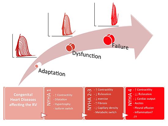
2. RV Response to Stress

2.1. Building Blocks of RV Adaptation

The RV has several unique features that may affect the adaptation to increased pressure and/or volume load. As has been known for more than a decade, the RV is derived from a different set of precursor cells than the LV, the so-called secondary heart field [3]. Despite the different inductive signals, the building blocks of the RV, i.e., the cardiomyocytes, are still the same as in the LV [9], but these are cast in a different 3D structure. The RV is a crescent-shaped organ wrapped around the LV, which complicates volumetric and functional analysis [10]. The RV contraction pattern is also different form that of the LV: the RV predominantly has a longitudinal shortening movement and presses its free wall against the septum, thereby creating a bellows effect to empty into the low-resistance pulmonary circulation [6]. These genetic, morphological and functional differences may explain the observed differences in the response to pressure or volume load between the RV and LV [11]. In addition, genetic defects, inducing the CHD, e.g., 22q11 deletion, may affect the RV response, but the effects of such a genetic defect on ventricular adaptation have rarely been addressed.
The RV is affected by abnormal loading conditions in more than 40% of the patients with CHD. Although artificial, for practical purposes, these conditions can be divided into three types: (i) increased pressure load (e.g., pulmonary stenosis); (ii) increased volume load (e.g., atrial septal defect); and (iii) a mixture of increased pressure load and volume load (Table 1). The type, the degree and the duration of the abnormal loading conditions determine the strain imposed upon the RV. Treatment strategies, i.e., corrective surgery or catheterization, aim at restoring the abnormal loading conditions, but most patients are faced with residual lesions, which affect RV function and determine the long-term outcome. For instance, patients with a TOF, the most common cyanotic heart defect, initially present with a pressure load of the RV, but after correction, face residual pulmonary insufficiency that leads to a volume load or a mixture of both [12]. To decipher the code to RV failure and to test treatment strategies, the response to increased pressure and/or volume load is studied in experimental models.

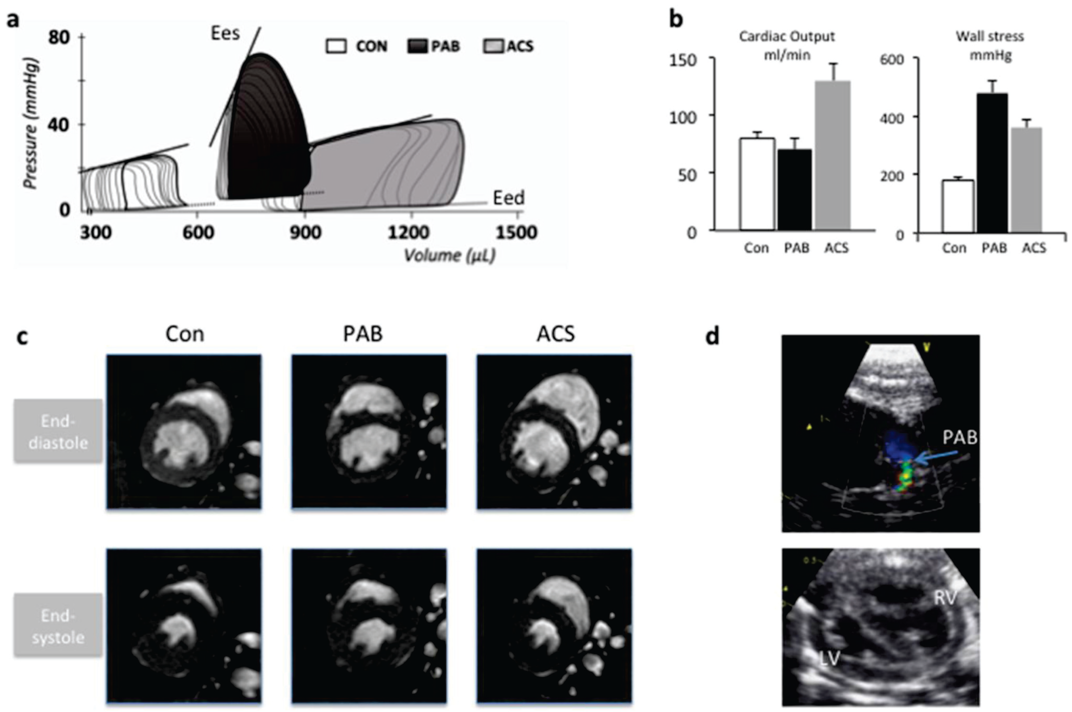
2.2. Modeling Abnormal Loading Conditions of the RV

To mimic the abnormal loading conditions of the RV, models of increased pressure load and/or volume load have been developed. Increased pressure load can be induced in mice, rat, rabbits, pigs or dogs with the use of pulmonary artery banding (PAB) [11,13,14,15,16]. The PAB induces a form of pulmonary stenosis, depending on the degree of PAB. The PAB takes the pulmonary vasculature out of the equation, in contrast to models trying to mimic pulmonary hypertension, e.g., monocrotaline (MCT) or hypoxia models [17,18,19]. Mice models of PAB are surgically challenging, but the use of transgenic mice can aid in the evaluation of signal transduction pathways in the development of RV failure. In addition, rat and pig models allow for a more sophisticated functional evaluation of the RV function using pressure-volume analysis, echocardiography and exercise performance.
To study the mechanisms of RV failure, a clinical description of the degree of failure is necessary. RV failure, as LV failure is not a disease per se, but rather a continuum from RV adaptation towards overt heart failure. Heart failure is defined as the inadequacy of the ventricle to supply the body with oxygen and substrates; in the setting of RV failure, this presents as inadequate pulmonary blood flow and RV congestion. Overt RV failure is characterized by venous congestion, ascites, pleural effusion, tachycardia and tachypnea, relating to NYHA Class IV. However, earlier stages of RV dysfunction are quite often present in the models; no signs at rest, but only a limited exercise capacity relating to NYHA Classes II–III. To distinguish adequate and beneficial RV adaptation from progressive RV failure in experimental models, it is important to classify the degree of failure, both by clinical signs at rest and by exercise capacity. RV function can be analyzed these days in mice, rats, as well as in man using cardiac MRI (CMR) [11,20] and echocardiography [21]. For a proper assessment of systolic and diastolic RV function, pressure-volume analysis is the gold standard, which has now been validated in many models of RV disease [22,23,24].
Models of increased volume load have been more difficult to develop. The aorto-caval shunt model is relatively easy to obtain in mice and rat. Although it leads to a biventricular volume load, it can be applied in mice, allowing the use of knockout or transgenic strategies [11]. Recently, also a model of pulmonary insufficiency in mice has been developed, leading to an isolated RV volume load [25], which may add additional insight into RV adaptation. Larger animals are also used to simulate TOF [26], which allow for more extensive hemodynamic assessment, but in these models, the cellular mechanisms are more difficult to study.

2.3. RV Adaptation to Pressure Load

Various models of RV pressure load have shown a similar pattern of responses. The RV adaptation to pressure load is characterized by increased contractility, assessed by end-systolic elastance using PV analysis (Figure 1) [11,14,22,27]. Furthermore, the PV loops shift to the right, indicating RV dilatation in response to pressure load. RV dilatation has earlier been seen as a sign of failure, but a summary of all experimental models suggests that it is an initial adaptation of the RV. The increased elastance is necessary to face the increased pressure load and restore RV-PA coupling. The RV dilatation induces increased wall stress, and to reduce this wall stress, RV hypertrophy develops.
Only a few studies have described animal models in which overt RV failure was reached [21,28]. These studies indicated that RV failure was characterized by progressive diastolic dysfunction, whereas intrinsic contractility was still increased in order to cope with the increased pressure load. The diastolic dysfunction, expressed as increased end-diastolic elastance, led to increased end-diastolic RV pressure, increased right atrial size, hepatic congestion and ascites. The clinical importance of diastolic dysfunction in RV failure has recently been confirmed in studies of the autopsy material of patients with RV failure in pulmonary hypertension [29].
The pressure loaded RV shows signs of ventricular remodeling similar to those observed in a stressed LV [30], such as hypertrophy, fibrosis and inflammation. Furthermore, a switch to the so-called fetal gene program has been described as in the LV; i.e., a switch in myosin heavy chain isoforms and in the metabolic profile from the use of fatty acids to that of glucose, the main energy source for the fetal myocardium [31].
Several studies have reported differences in gene expression in the RV in response to a pressure load when compared with the LV. A study comparing mice with a PAB and mice with aortic banding showed differences in expression in genes that were associated with: (i) extracellular matrix proteins; (ii) proteases and inhibitors; and (iii) developmentally regulated proteins [20]. Apart from different genes, also similar genes with differences in signal strength were found. For example, the calcineurin-NFAT pathway is involved in RV adaptation to pressure load [11], yet suppression of this pathway via an inducible transgenic approach also induced changes in the unloaded RV [32]. Another study comparing microRNA profiles in mice with a PAB and mice with an aortic stenosis found four microRNAs specific for RV adaptation [33]. These micorRNAs were all located in the non-myocyte fraction of the RV. Thus, there are different signals involved in RV adaptation to pressure load, and some of these differences are located outside the cardiomyocyte.
Intriguingly, progressive RV diastolic dysfunction seems to be associated with reduced angiogenesis, reduced energy metabolism and increased inflammation rather than with increased fibrosis [28,34]. Capillary rarefaction has been suggested to play an important role in RV dysfunction in response to pressure load. Capillary density has been shown to be reduced in several PH models [35,36,37]. Recently, two studies comparing the same trigger with different disease states, i.e., monocrotaline-induced PH [38] or PAB [28], found a slightly different pattern, i.e., increased capillary density in rats without clinical failure versus pseudo-normalized density in rats with RVF. These findings suggest that RVF is associated with reduced energy supply. Candidate pathways susceptible to reduced energy supply are substrate metabolism and mitochondrial function.
Cardiac metabolism is mainly dependent upon fatty acid oxidation, but under stress, the heart switches back to its fetal program, in which glucose and lactate are the main energy substrates [31]. A decrease in fatty acid oxidation as part of the metabolic switch has been observed in a rat model of moderate PAB [39]. Preventive inhibition via trimetazidine (which inhibits an essential step in the beta-oxidation of fatty acids) increased cardiac output measured by echocardiography; however, RV pressure in the setting of a fixed pressure load (PAB) decreased.
The metabolic switch under stress can also be described by an increase in glucose utilization. As in the LV, a switch towards glycolysis has been observed in PH-rats, Fawn-Hooded rats [19] and PAB rats [39], although many of these experimental studies were done in non-failing animals. Upregulation of pyruvate dehydrogenase kinase (PDK), which uncouples glycolysis from the Krebs cycle, has been shown in both PH and PAB models of adaptive RV remodeling [19]. Inhibition of PDK reverses mitochondrial hyperpolarization, a sign of mitochondrial dysfunction [40]. In moderate PAB, inhibition of PDK appeared to improve RV function, although the results on cardiac function, evaluated by echocardiography, are partly contradictory [19]: RV systolic pressure decreases in the setting of a fixed pressure load. Further studies in failing animals are necessary to determine the position of metabolic interventions in RV remodeling [39,41].
Mitochondria are the “power-houses” of the cardiomyocyte, but, perhaps more importantly, also regulate many processes involved in stress-response: the formation of oxygen radicals, oxygen sensing, inducers of apoptosis and inflammation [42]. Rats with MCT-induced PH showed signs of increased “oxidative stress”: increased activity of Complex II, oxygen radicals and increased production of radical scavengers [43]. Treatment with a radical scavenger (EUK-134) improved RV systolic function, but did not affect diastolic dysfunction [44], although, also, an improvement in pulmonary vascular resistance may have contributed to this effect. In another PH-model, treatment with protandim, a plant extract inducing the expression of antioxidants, mildly increased cardiac output, suggesting that at least in rats with PH, an increasing defense mechanism against oxidative stress may be beneficial [45]. These pathways have never been tested in PAB models, independent of the pulmonary circulation. Apoptosis and inflammation, both important mechanisms in PH [46,47,48], have been described in both PH and PAB models [13,49,50,51]. There is, however, no knowledge on the functional importance of these signaling pathways in the development of RV failure.

2.4. RV Adaptation to Volume Load

In contrast to the increasing number of studies in models of increased RV pressure load, the reported studies on the effects of increased volume load on the RV have been limited so far. This is surprising, as a volume overloaded RV is common in survivors of CHD. For example, patients with corrected TOF, the most common cyanotic heart defect, are threatened by residual pulmonary insufficiency [12]. TOF requires surgical relief of the RV outflow tract that often leads to residual regurgitation of the pulmonary valve. Initially, longstanding PI was thought to be innocent, maybe because there is a considerable lag-time before symptoms develop [52]. However, recent knowledge suggests that also chronic increased volume load eventually leads to RV dysfunction. At present, residual PI is treated with pulmonary valve replacement, but the timing and longstanding effects of such a replacement remain a matter of dispute in CHD. Therefore, further knowledge of the pathways involved in RV dysfunction in response to volume load, in combination with clinical parameters (such as, e.g., obtained by cardiac MRI [11,20]), may provide new ground to guide treatment strategies.
The RV is capable of adapting to increased volume load for a long period of time before RV dysfunction develops. The hemodynamic response to increased volume load is via the Frank Starling mechanism [11,22]. However, longstanding increases in volume load may lead to diastolic dysfunction, suggested by increased RV ED pressure [25]. Pigs with PI and outflow tract reconstruction have severe diastolic dysfunction in the presence of normal cardiac output [26]. There is little known about the adaptation to mixed lesions of pressure and volume load. In rats with PH and a volume load, RV contractility was less increased as compared with volume load only, inducing a pattern of pseudo-normalization [27]. Whether this holds true for other forms of mixed lesions is unknown. The mechanisms of these adaptations to mixed loading conditions are yet poorly understood.
The cardiomyocyte responds differently to increased volume load than to increased pressure load in the LV [53]. Classically, it has been described that myocytes elongate in response to increased volume load and thicken in response to increased pressure load, a mechanism for which the extracellular regulated kinase 1 and 2 pathways could be responsible [54]. The RV responds differently, as studies in volume loaded LVs show the beneficial effects of the treatment with a PDE5A-inhibitor (Sildenafil) [22], whereas studies in the volume loaded RV do not demonstrate any effects in response to Sildenafil [22]. A mouse model comparing the volume and pressure load of the RV showed different signaling intensities of calcineurin-NFAT activation and the myosin heavy chain isoform switch [11]. These differences were not present in a rat model [22], suggesting that loading conditions may influence the results. Recently, in a mouse model of PI, it was shown that mice developed RV failure after six months. The RV of mice with chronic PI showed changes in gene expression, suggesting the early dysfunction of mitochondrial energetics, enhanced TGFbeta activation, apoptosis and extracellular matrix remodeling [25]. Further studies on the contribution of these pathways to RV failure in response to volume load are necessary.

2.5. Treatment of RV Failure

At present, no RV-specific medical treatment strategies exist. The recently developed models of RV failure in response to increased pressure load can aid in the understanding of the cellular mechanisms of RV failure (Figure 2), so that RV-specific therapies may be developed. Currently, treatment strategies for LV failure (e.g., beta adrenergic blockade, RAAS inhibition) or drugs that target the pulmonary vasculature in PH (e.g., PDE5 inhibitors, endothelin receptor antagonists), have been tested in experimental models with different results.
Beta blockers are a cornerstone in the treatment of LV failure. In a rat model of MCT-induced PH, bisoprolol, a selective beta blocker, prevented fibrosis and delayed decline in cardiac output and TAPSE without effects on hypertrophy or capillary rarefaction [35]. Unfortunately, no data are published on beta blockade in the failing RV, due to stenosis-type pressure overload, which might elucidate the direct protective effects of beta blockade on the RV. Blockade of the renin-angiotensin-aldosterone-system (RAAS), the other cornerstone in the treatment of LV failure, was shown to be ineffective in a rat model of PAB [27] in accordance with the results from a study in patients with a systemic RV [55].
Inhibition of PDE5 would be an excellent therapeutic approach for RV failure in PH, as it also reduces pulmonary vasculature resistance [56]. Recently, our lab showed that preventive treatment with the PDE5 inhibitor, Sildenafil, increased contractility, reduced dilatation and attenuated the decline in exercise capacity. Diastolic function remained unchanged, and fibrosis was slightly increased [22]. In addition, Sildenafil treatment in established RV disease had slightly different effects, as it now also improved diastolic dysfunction and reduced RV fibrosis [57], supporting the concept that timing and loading severity determines cardiac response. Other pharmacological approaches to manipulate the PDE5-PKG-1 axis include stimulation of soluble guanylate cyclase by riociguat, but results so far indicate effects on the pulmonary vasculature, rather than direct beneficial RV effects [58].
Endothelin receptor antagonists have an anti-hypertrophic and anti-fibrotic effect on the RV in PH [59]. It is unclear whether this is a direct effect on the RV or a consequence of the decreased pressure load due to the effect on the pulmonary vasculature. Unfortunately, no studies address this issue. However, isolated heart studies showed that ERAs depress both contractility and relaxation in the hypertrophic RV [60].
New treatment strategies successfully applied in the failing experimental LV, such as HDAC inhibitors, appeared to be detrimental in models of RV dysfunction [61]. Hence, future studies into RV failure after prolonged increased pressure load are necessary to improve the outcome for patients with CHD.
Table 1. Summary of congenital heart defects affecting the right ventricle (RV).
RV dysfunction described?ref
Increased volume load
Atrial septal defect>25% mild RV dysfunction 30 years after correction (CMR)[62]
Pulmonary insufficiency30% symptomatic after 40 years[52]
Tricuspid insufficiencyReduces survival independent of other defects[63]
Increased pressure load
Pulmonary stenosisReduced exercise capacity[64]
Pulmonary HypertensionRV failure predicts outcome[7]
Tetralogy of FallotFatal if uncorrected
Congenitally corrected transposition of the great arteries (ccTGA)>25% RV failure after 45 y when there are no associated lesions[65]
Transposition of the great arteries after atrial switch procedure (TGA-as)Reduced RV function similar to ccTGA[66]
Mixed Lesions
Atrial Septal Defect + Pulmonary stenosis/Pulmonary HypertensionReduced survival in patients with ASD-PH[67,68]
Corrected Fallot + pulmonary insufficiencyRisk for Sudden Death, arrhythmias[69]
ccTGA + tricuspid insufficiencyRisk of RV failure increases from 25 → 60%[65]
TGA-as + tricuspid insufficiencyIdem ccTGA
Figure 1. (a) Functional adaptation of the RV to abnormal loading conditions (via pulmonary artery banding) or increased volume load (via aorto-caval shunt) in a rat model. Solid lines end-systolic elastance (Ees, reflecting contractility); dotted lines end-diastolic elastance (Eed, reflecting relaxation). (b) In rats with a moderate pulmonary artery banding (PAB), cardiac output is mildly decreased, whereas in rats with an aorto-caval shunt (ACS), cardiac output is increased. In contrast, maximal wall stress is more increased in rat with a PAB, as compared with ACS. Data are derived from [57]. (c) Typical example of cardiac MRI images obtained in mice with increased pressure load (PAB) or increased volume load (ACS). The top row shows short axis cine-images at end-diastole; the bottom row shows short-axis cine-images at end-diastole. Images were acquired using a 9.4Tesla MRI. (d) Example of echo-derived PAB-pressure gradient (top) and short axis RV/LV dimensions (bottom), obtained with a 10-MHz probe in rats with a PAB. Con, sham-operated control animals. Adapted from [11,22].
Jcdd 01 00083 g001
Figure 2. Development of RV failure in response to increased pressure load. The proposed sequence of events in response to increased pressure load: The RV adapts to increased pressure load with increased contractility and dilatation. This leads to increased wall stress, which will be balanced by RV hypertrophy. With the increasing duration of the degree of loading, relaxation becomes impaired and exercise performance reduced. This is associated (but may not be causally related to) fibrosis, reduced capillary density and a switch from the use of fatty acids to glucose. RV failure is marked by progressive diastolic dysfunction, ascites and pleural effusions and severely impaired exercise capacity and may be related to inflammatory or hypoxic phenomena. The latter are issues of current research.
Jcdd 01 00083 g002

3. Summary and Future Perspectives

The RV is frequently affected in the growing groups of survivors of CHD, and RV failure is a main determinant to the outcome in this heterogeneous population. However, knowledge about the mechanisms of RV failure in response to abnormal loading conditions, such as increased volume load and/or pressure load, is still limited.
Models of increased pressure load for the RV have shown a similar pattern of responses, i.e., increased contractility, RV dilatation and hypertrophy. Evidence is accumulating that RV failure in response to increased pressure load is marked by progressive diastolic dysfunction. The mechanisms of this progressive dysfunction are insufficiently known. The RV response to pressure load shares similarities with that of the LV, but also has specific features, e.g., capillary rarefaction, oxidative stress and inflammation. The contribution of these pathways to the development of failure needs further exploration. The RV adaptation to increased volume load is an understudied area, but becomes increasingly important in the growing groups of survivors of CHD, especially with tetralogy of Fallot.
Recently developed animal models of clinically overt RV failure due to RV abnormal loading conditions that truly represent current patient populations can aid in the search for new RV specific therapies.

Author Contributions

BB contributed to the conception and design, the drafting of the article, and critical revision for important intellectual content. MAJB contributed to the conception and design, the drafting of the article. RMFB contributed to the conception and design, and critical revision for important intellectual content.

Conflicts of Interest

The authors declare no conflict of interest.

References

  1. Zomer, A.C.; Vaartjes, I.; Uiterwaal, C.S.; van der Velde, E.T.; van den Merkhof, L.F.; Baur, L.H.; Ansink, T.J.; Cozijnsen, L.; Pieper, P.G.; Meijboom, F.J.; et al. Circumstances of death in adult congenital heart disease. Int. J. Cardiol. 2012, 154, 168–172. [Google Scholar] [CrossRef]
  2. Norozi, K.; Wessel, A.; Alpers, V.; Arnhold, J.O.; Geyer, S.; Zoege, M.; Buchhorn, R. Incidence and risk distribution of heart failure in adolescents and adults with congenital heart disease after cardiac surgery. Am. J. Cardiol. 2006, 97, 1238–1243. [Google Scholar] [CrossRef]
  3. Kelly, R.G.; Brown, N.A.; Buckingham, M.E. The arterial pole of the mouse heart forms from Fgf10-expressing cells in pharyngeal mesoderm. Dev. Cell 2001, 1, 435–440. [Google Scholar] [CrossRef]
  4. Ho, S.Y.; Nihoyannopoulos, P. Anatomy, echocardiography, and normal right ventricular dimensions. Heart 2006, 92 (Suppl. 1), i2–i13. [Google Scholar] [CrossRef]
  5. Bartelds, B.; Berger, R.M.F. The right ventricle in congenital heart diseases. In The Right Ventricle; Gaine, S., Naeije, R., Paecock, A., Eds.; Springer: Berlin/Heidelberg, Germany, 2014. [Google Scholar]
  6. Haddad, F.; Doyle, R.; Murphy, D.J.; Hunt, S.A. Right ventricular function in cardiovascular disease, part II: Pathophysiology, clinical importance, and management of right ventricular failure. Circulation 2008, 117, 1717–1731. [Google Scholar] [CrossRef]
  7. Van Wolferen, S.A.; Marcus, J.T.; Boonstra, A.; Marques, K.M.; Bronzwaer, J.G.; Spreeuwenberg, M.D.; Postmus, P.E.; Vonk-Noordegraaf, A. Prognostic value of right ventricular mass, volume, and function in idiopathic pulmonary arterial hypertension. Eur. Heart J. 2007, 28, 1250–1257. [Google Scholar] [CrossRef]
  8. Ghio, S.; Gavazzi, A.; Campana, C.; Inserra, C.; Klersy, C.; Sebastiani, R.; Arbustini, E.; Recusani, F.; Tavazzi, L. Independent and additive prognostic value of right ventricular systolic function and pulmonary artery pressure in patients with chronic heart failure. J. Am. Coll. Cardiol. 2001, 37, 183–188. [Google Scholar] [CrossRef]
  9. Olson, E.N. Gene regulatory networks in the evolution and development of the heart. Science 2006, 313, 1922–1927. [Google Scholar] [CrossRef]
  10. Freling, H.G.; Pieper, P.G.; Vermeulen, K.M.; van Swieten, J.M.; Sijens, P.E.; van Veldhuisen, D.J.; Willems, T.P. Improved cardiac MRI volume measurements in patients with tetralogy of Fallot by independent end-systolic and end-diastolic phase selection. PLoS ONE 2013, 8, e55462. [Google Scholar] [CrossRef]
  11. Bartelds, B.; Borgdorff, M.A.; Smit-van Oosten, A.; Takens, J.; Boersma, B.; Nederhoff, M.G.; Elzenga, N.J.; van Gilst, W.H.; de Windt, L.J.; Berger, R.M. Differential responses of the right ventricle to abnormal loading conditions in mice: pressure vs. volume load. Eur. J. Heart Fail. 2011, 13, 1275–1282. [Google Scholar] [CrossRef]
  12. Bouzas, B.; Kilner, P.J.; Gatzoulis, M.A. Pulmonary regurgitation: not a benign lesion. Eur. Heart J. 2005, 26, 433–439. [Google Scholar]
  13. Apitz, C.; Honjo, O.; Humpl, T.; Li, J.; Assad, R.S.; Cho, M.Y.; Hong, J.; Friedberg, M.K.; Redington, A.N. Biventricular structural and functional responses to aortic constriction in a rabbit model of chronic right ventricular pressure overload. J. Thorac. Cardiovasc. Surg. 2012, 144, 1494–1501. [Google Scholar] [CrossRef] [Green Version]
  14. Borgdorff, M.A.; Bartelds, B.; Dickinson, M.G.; Steendijk, P.; de Vroomen, M.; Berger, R.M. Distinct loading conditions reveal various patterns of right ventricular adaptation. Am. J. Physiol. Heart Circ. Physiol. 2013, 305, H354–H364. [Google Scholar] [CrossRef]
  15. Leeuwenburgh, B.P.; Helbing, W.A.; Steendijk, P.; Schoof, P.H.; Baan, J. Biventricular systolic function in young lambs subject to chronic systemic right ventricular pressure overload. Am. J. Physiol. Heart Circ. Physiol. 2001, 281, H2697–H2704. [Google Scholar]
  16. Gaynor, S.L.; Maniar, H.S.; Bloch, J.B.; Steendijk, P.; Moon, M.R. Right atrial and ventricular adaptation to chronic right ventricular pressure overload. Circulation 2005, 112, I212–I218. [Google Scholar]
  17. Hessel, M.H.; Steendijk, P.; den Adel, B.; Schutte, C.I.; van der Laarse, A. Characterization of right ventricular function after monocrotaline-induced pulmonary hypertension in the intact rat. Am. J. Physiol. Heart Circ. Physiol. 2006, 291, H2424–H2430. [Google Scholar] [CrossRef]
  18. Dickinson, M.G.; Bartelds, B.; Borgdorff, M.A.; Berger, R.M. The role of disturbed blood flow in the development of pulmonary arterial hypertension: lessons from preclinical animal models. Am. J. Physiol. Lung. Cell. Mol. Physiol. 2013, 305, L1–L14. [Google Scholar] [CrossRef]
  19. Piao, L.; Marsboom, G.; Archer, S.L. Mitochondrial metabolic adaptation in right ventricular hypertrophy and failure. J. Mol. Med. 2010, 88, 1011–1020. [Google Scholar] [CrossRef]
  20. Kreymborg, K.; Uchida, S.; Gellert, P.; Schneider, A.; Boettger, T.; Voswinckel, R.; Wietelmann, A.; Szibor, M.; Weissmann, N.; Ghofrani, A.H.; et al. Identification of right heart-enriched genes in a murine model of chronic outflow tract obstruction. J. Mol. Cell. Cardiol. 2010, 49, 598–605. [Google Scholar] [CrossRef]
  21. Urashima, T.; Zhao, M.; Wagner, R.; Fajardo, G.; Farahani, S.; Quertermous, T.; Bernstein, D. Molecular and physiological characterization of RV remodeling in a murine model of pulmonary stenosis. Am. J. Physiol. Heart Circ. Physiol. 2008, 295, H1351–H1368. [Google Scholar] [CrossRef]
  22. Borgdorff, M.A.; Bartelds, B.; Dickinson, M.G.; Boersma, B.; Weij, M.; Zandvoort, A.; Sillje, H.H.; Steendijk, P.; de Vroomen, M.; Berger, R.M. Sildenafil enhances systolic adaptation, but does not prevent diastolic dysfunction, in the pressure-loaded right ventricle. Eur. J. Heart Fail. 2012, 14, 1067–1074. [Google Scholar] [CrossRef]
  23. Faber, M.J.; Dalinghaus, M.; Lankhuizen, I.M.; Steendijk, P.; Hop, W.C.; Schoemaker, R.G.; Duncker, D.J.; Lamers, J.M.; Helbing, W.A. Right and left ventricular function after chronic pulmonary artery banding in rats assessed with biventricular pressure-volume loops. Am. J. Physiol. Heart Circ. Physiol. 2006, 291, H1580–H1586. [Google Scholar] [CrossRef]
  24. Rondelet, B.; Dewachter, L.; Kerbaul, F.; Dewachter, C.; Hubloue, I.; Fesler, P.; Franck, S.; Remmelink, M.; Brimioulle, S.; Naeije, R. Sildenafil added to sitaxsentan in overcirculation-induced pulmonary arterial hypertension. Am. J. Physiol. Heart Circ. Physiol. 2010, 299, H1118–H1123. [Google Scholar] [CrossRef]
  25. Reddy, S.; Zhao, M.; Hu, D.Q.; Fajardo, G.; Katznelson, E.; Punn, R.; Spin, J.M.; Chan, F.P.; Bernstein, D. Physiologic and molecular characterization of a murine model of right ventricular volume overload. Am. J. Physiol. Heart Circ. Physiol. 2013, 304, H1314–H1327. [Google Scholar] [CrossRef]
  26. Bove, T.; Bouchez, S.; de Hert, S.; Wouters, P.; de Somer, F.; Devos, D.; Somers, P.; van Nooten, G. Acute and chronic effects of dysfunction of right ventricular outflow tract components on right ventricular performance in a porcine model: Implications for primary repair of tetralogy of fallot. J. Am. Coll. Cardiol. 2012, 60, 64–71. [Google Scholar] [CrossRef]
  27. Borgdorff, M.A.; Bartelds, B.; Dickinson, M.G.; Steendijk, P.; Berger, R.M. A cornerstone of heart failure treatment is not effective in experimental right ventricular failure. Int. J. Cardiol. 2013, 169, 183–189. [Google Scholar] [CrossRef]
  28. Borgdorff, M.A.; Bartelds, B.; Dickinson, M.G.; Steendijk, P.; Koops, A.C.; Berger, R.M. Characterization of right ventricular failure in chronic experimental pressure load. J. Mol. Cell. Cardiol. 2014. submitted. [Google Scholar]
  29. Rain, S.; Handoko, M.L.; Trip, P.; Gan, C.T.; Westerhof, N.; Stienen, G.J.; Paulus, W.J.; Ottenheijm, C.A.; Marcus, J.T.; Dorfmuller, P.; et al. Right ventricular diastolic impairment in patients with pulmonary arterial hypertension. Circulation 2013, 128, 2016–2025. [Google Scholar] [CrossRef]
  30. Van Berlo, J.H.; Maillet, M.; Molkentin, J.D. Signaling effectors underlying pathologic growth and remodeling of the heart. J. Clin. Invest. 2013, 123, 37–45. [Google Scholar] [CrossRef]
  31. Bartelds, B.; Knoester, H.; Smid, G.B.; Takens, J.; Visser, G.H.; Penninga, L.; van der Leij, F.R.; Beaufort-Krol, G.C.; Zijlstra, W.G.; Heymans, H.S.; et al. Perinatal changes in myocardial metabolism in lambs. Circulation 2000, 102, 926–931. [Google Scholar] [CrossRef]
  32. Bartelds, B.; Borgdorff, M.A.; Boersma, B.; Takens, J.; Smit-van Oosten, A.; de WIndt, L.J.; Berger, R.M.F. Right ventricular adaptation to pressure load in mice is improved after blockade of calcineurin activation. Eur. Heart J. 2011, 31, 305. [Google Scholar]
  33. Reddy, S.; Zhao, M.; Hu, D.Q.; Fajardo, G.; Hu, S.; Ghosh, Z.; Rajagopalan, V.; Wu, J.C.; Bernstein, D. Dynamic microRNA expression during the transition from right ventricular hypertrophy to failure. Physiol. Genomics 2012, 44, 562–575. [Google Scholar] [CrossRef]
  34. Sutendra, G.; Dromparis, P.; Bonnet, S.; Haromy, A.; McMurtry, M.S.; Bleackley, R.C.; Michelakis, E.D. Pyruvate dehydrogenase inhibition by the inflammatory cytokine TNFalpha contributes to the pathogenesis of pulmonary arterial hypertension. J. Mol. Med. 2011, 89, 771–783. [Google Scholar] [CrossRef]
  35. De Man, F.S.; Handoko, M.L.; van Ballegoij, J.J.; Schalij, I.; Bogaards, S.J.; Postmus, P.E.; van der Velden, J.; Westerhof, N.; Paulus, W.J.; Vonk-Noordegraaf, A. Bisoprolol delays progression towards right heart failure in experimental pulmonary hypertension. Circ. Heart Fail. 2012, 5, 97–105. [Google Scholar]
  36. Bogaard, H.J.; Abe, K.; Vonk Noordegraaf, A.; Voelkel, N.F. The right ventricle under pressure: Cellular and molecular mechanisms of right-heart failure in pulmonary hypertension. Chest 2009, 135, 794–804. [Google Scholar] [CrossRef]
  37. Van Albada, M.E.; Berger, R.M.; Niggebrugge, M.; van Veghel, R.; Cromme-Dijkhuis, A.H.; Schoemaker, R.G. Prostacyclin therapy increases right ventricular capillarisation in a model for flow-associated pulmonary hypertension. Eur. J. Pharmacol. 2006, 549, 107–116. [Google Scholar] [CrossRef]
  38. Sutendra, G.; Dromparis, P.; Paulin, R.; Zervopoulos, S.; Haromy, A.; Nagendran, J.; Michelakis, E.D. A metabolic remodeling in right ventricular hypertrophy is associated with decreased angiogenesis and a transition from a compensated to a decompensated state in pulmonary hypertension. J. Mol. Med. 2013, 91, 1315–1327. [Google Scholar] [CrossRef]
  39. Fang, Y.H.; Piao, L.; Hong, Z.; Toth, P.T.; Marsboom, G.; Bache-Wiig, P.; Rehman, J.; Archer, S.L. Therapeutic inhibition of fatty acid oxidation in right ventricular hypertrophy: Exploiting Randle’s cycle. J. Mol. Med. 2012, 90, 31–43. [Google Scholar] [CrossRef]
  40. Nagendran, J.; Michelakis, E.D. Mitochondrial NOS is upregulated in the hypoxic heart: Implications for the function of the hypertrophied right ventricle. Am. J. Physiol. Heart Circ. Physiol. 2009, 296, H1723–H1726. [Google Scholar] [CrossRef]
  41. Piao, L.; Fang, Y.H.; Cadete, V.J.; Wietholt, C.; Urboniene, D.; Toth, P.T.; Marsboom, G.; Zhang, H.J.; Haber, I.; Rehman, J.; et al. The inhibition of pyruvate dehydrogenase kinase improves impaired cardiac function and electrical remodeling in two models of right ventricular hypertrophy: resuscitating the hibernating right ventricle. J. Mol. Med. 2010, 88, 47–60. [Google Scholar] [CrossRef]
  42. Dromparis, P.; Michelakis, E.D. Mitochondria in vascular health and disease. Annu. Rev. Physiol. 2013, 75, 95–126. [Google Scholar] [CrossRef]
  43. Redout, E.M.; Wagner, M.J.; Zuidwijk, M.J.; Boer, C.; Musters, R.J.; van Hardeveld, C.; Paulus, W.J.; Simonides, W.S. Right-ventricular failure is associated with increased mitochondrial complex II activity and production of reactive oxygen species. Cardiovasc. Res. 2007, 75, 770–781. [Google Scholar] [CrossRef]
  44. Redout, E.M.; van der Toorn, A.; Zuidwijk, M.J.; van de Kolk, C.W.; van Echteld, C.J.; Musters, R.J.; van Hardeveld, C.; Paulus, W.J.; Simonides, W.S. Antioxidant treatment attenuates pulmonary arterial hypertension-induced heart failure. Am. J. Physiol. Heart Circ. Physiol. 2010, 298, H1038–H1047. [Google Scholar] [CrossRef]
  45. Bogaard, H.J.; Natarajan, R.; Henderson, S.C.; Long, C.S.; Kraskauskas, D.; Smithson, L.; Ockaili, R.; McCord, J.M.; Voelkel, N.F. Chronic pulmonary artery pressure elevation is insufficient to explain right heart failure. Circulation 2009, 120, 1951–1960. [Google Scholar] [CrossRef]
  46. Hassoun, P.M.; Mouthon, L.; Barbera, J.A.; Eddahibi, S.; Flores, S.C.; Grimminger, F.; Jones, P.L.; Maitland, M.L.; Michelakis, E.D.; Morrell, N.W.; et al. Inflammation, growth factors, and pulmonary vascular remodeling. J. Am. Coll. Cardiol. 2009, 54, S10–S19. [Google Scholar] [CrossRef]
  47. Bartelds, B.; van Loon, R.L.; Mohaupt, S.; Wijnberg, H.; Dickinson, M.G.; Boersma, B.; Takens, J.; van Albada, M.; Berger, R.M. Mast cell inhibition improves pulmonary vascular remodeling in pulmonary hypertension. Chest 2012, 141, 651–660. [Google Scholar] [CrossRef]
  48. Van Albada, M.E.; Bartelds, B.; Wijnberg, H.; Mohaupt, S.; Dickinson, M.G.; Schoemaker, R.G.; Kooi, K.; Gerbens, F.; Berger, R.M. Gene expression profile in flow-associated pulmonary arterial hypertension with neointimal lesions. Am. J. Physiol. Lung Cell Mol. Physiol. 2010, 298, L483–L491. [Google Scholar] [CrossRef]
  49. Rajagopalan, V.; Zhao, M.; Reddy, S.; Fajardo, G.A.; Wang, X.; Dewey, S.; Gomes, A.V.; Bernstein, D. Altered Ubiquitin-Proteasome Signaling in Right Ventricular Hypertrophy and Failure. Am. J. Physiol. Heart Circ. Physiol. 2013, 305, H551–H562. [Google Scholar] [CrossRef]
  50. Dewachter, C.; Dewachter, L.; Rondelet, B.; Fesler, P.; Brimioulle, S.; Kerbaul, F.; Naeije, R. Activation of apoptotic pathways in experimental acute afterload-induced right ventricular failure. Crit. Care 2010, 38, 1405–1413. [Google Scholar] [CrossRef]
  51. Rondelet, B.; Dewachter, C.; Kerbaul, F.; Kang, X.; Fesler, P.; Brimioulle, S.; Naeije, R.; Dewachter, L. Prolonged overcirculation-induced pulmonary arterial hypertension as a cause of right ventricular failure. Eur. Heart J. 2012, 33, 1017–1026. [Google Scholar] [CrossRef]
  52. Shimazaki, Y.; Blackstone, E.H.; Kirklin, J.W. The natural history of isolated congenital pulmonaryvalve incompetence: surgical implications. Thorac. Cardiovasc. Surg. 1984, 32, 257–259. [Google Scholar] [CrossRef]
  53. Toischer, K.; Rokita, A.G.; Unsold, B.; Zhu, W.; Kararigas, G.; Sossalla, S.; Reuter, S.P.; Becker, A.; Teucher, N.; Seidler, T.; et al. Differential cardiac remodeling in preload versus afterload. Circulation 2010, 122, 993–1003. [Google Scholar] [CrossRef]
  54. Kehat, I.; Davis, J.; Tiburcy, M.; Accornero, F.; Saba-El-Leil, M.K.; Maillet, M.; York, A.J.; Lorenz, J.N.; Zimmermann, W.H.; Meloche, S.; et al. Extracellular signal-regulated kinases 1 and 2 regulate the balance between eccentric and concentric cardiac growth. Circ. Res. 2010, 108, 176–183. [Google Scholar]
  55. Van der Bom, T.; Winter, M.M.; Bouma, B.J.; Groenink, M.; Vliegen, H.W.; Pieper, P.G.; van Dijk, A.P.; Sieswerda, G.T.; Roos-Hesselink, J.W.; Zwinderman, A.H. Effect of valsartan on systemic right ventricular function: A double-blind, randomized, placebo-controlled pilot trial. Circulation 2013, 127, 322–330. [Google Scholar] [CrossRef]
  56. Archer, S.L.; Michelakis, E.D. Phosphodiesterase type 5 inhibitors for pulmonary arterial hypertension. N. Engl. J. Med. 2009, 361, 1864–1871. [Google Scholar] [CrossRef]
  57. Borgdorff, M.A.; Bartelds, B.; Dickinson, M.G.; Steendijk, P.; de Vroomen, M.; Berger, R.M. Sildenafil treatement in established right ventricular dysfunction improves diastolic function and attenuates interstitial fibrosis independent from afterload. Am. J. Physiol.-Heart Circ. Physiol. 2013. submitted. [Google Scholar]
  58. Lang, M.; Kojonazarov, B.; Tian, X.; Kalymbetov, A.; Weissmann, N.; Grimminger, F.; Kretschmer, A.; Stasch, J.P.; Seeger, W.; Ghofrani, H.A.; et al. The soluble guanylate cyclase stimulator riociguat ameliorates pulmonary hypertension induced by hypoxia and SU5416 in rats. PLoS ONE 2012, 7, e43433. [Google Scholar] [CrossRef]
  59. Choudhary, G.; Troncales, F.; Martin, D.; Harrington, E.O.; Klinger, J.R. Bosentan attenuates right ventricular hypertrophy and fibrosis in normobaric hypoxia model of pulmonary hypertension. J. Heart Lung Transplant. 2011, 30, 827–833. [Google Scholar] [CrossRef]
  60. Nagendran, J.; Sutendra, G.; Paterson, I.; Champion, H.C.; Webster, L.; Chiu, B.; Haromy, A.; Rebeyka, I.M.; Ross, D.B.; Michelakis, E.D. Endothelin axis is upregulated in human and rat right ventricular hypertrophy. Circ. Res. 2013, 112, 347–354. [Google Scholar] [CrossRef]
  61. Bogaard, H.J.; Mizuno, S.; Hussaini, A.A.; Toldo, S.; Abbate, A.; Kraskauskas, D.; Kasper, M.; Natarajan, R.; Voelkel, N.F. Suppression of histone deacetylases worsens right ventricular dysfunction after pulmonary artery banding in rats. Am. J. Respir. Crit. Care Med. 2011, 183, 1402–1410. [Google Scholar] [CrossRef]
  62. Cuypers, J.A.; Opic, P.; Menting, M.E.; Utens, E.M.; Witsenburg, M.; Helbing, W.A.; van den Bosch, A.E.; Ouhlous, M.; van Domburg, R.T.; Meijboom, F.J.; et al. The unnatural history of an atrial septal defect: Longitudinal 35 year follow up after surgical closure at young age. Heart 2013, 99, 1346–1352. [Google Scholar] [CrossRef]
  63. Nath, J.; Foster, E.; Heidenreich, P.A. Impact of tricuspid regurgitation on long-term survival. J. Am. Coll. Cardiol. 2004, 43, 405–409. [Google Scholar] [CrossRef]
  64. Luijnenburg, S.E.; de Koning, W.B.; Romeih, S.; van den Berg, J.; Vliegen, H.W.; Mulder, B.J.; Helbing, W.A. Exercise capacity and ventricular function in patients treated for isolated pulmonary valve stenosis or tetralogy of Fallot. Int. J. Cardiol. 2012, 158, 359–363. [Google Scholar] [CrossRef]
  65. Graham, T.P., Jr.; Bernard, Y.D.; Mellen, B.G.; Celermajer, D.; Baumgartner, H.; Cetta, F.; Connolly, H.M.; Davidson, W.R.; Dellborg, M.; Foster, E.; et al. Long-term outcome in congenitally corrected transposition of the great arteries: A multi-institutional study. J. Am. Coll. Cardiol. 2000, 36, 255–261. [Google Scholar] [CrossRef]
  66. Grothoff, M.; Fleischer, A.; Abdul-Khaliq, H.; Hoffmann, J.; Lehmkuhl, L.; Luecke, C.; Gutberlet, M. The systemic right ventricle in congenitally corrected transposition of the great arteries is different from the right ventricle in dextro-transposition after atrial switch: a cardiac magnetic resonance study. Cardiol. Young 2013, 23, 239–247. [Google Scholar] [CrossRef]
  67. Diller, G.P.; Dimopoulos, K.; Broberg, C.S.; Kaya, M.G.; Naghotra, U.S.; Uebing, A.; Harries, C.; Goktekin, O.; Gibbs, J.S.; Gatzoulis, M.A. Presentation, survival prospects, and predictors of death in Eisenmenger syndrome: A combined retrospective and case-control study. Eur. Heart J. 2006, 27, 1737–1742. [Google Scholar] [CrossRef]
  68. Van Loon, R.L.; Roofthooft, M.T.; Hillege, H.L.; Ten Harkel, A.D.; van Osch-Gevers, M.; Delhaas, T.; Kapusta, L.; Strengers, J.L.; Rammeloo, L.; Clur, S.A.; et al. Pediatric pulmonary hypertension in the Netherlands: Epidemiology and characterization during the period 1991 to 2005. Circulation 2011, 124, 1755–1764. [Google Scholar] [CrossRef]
  69. Gatzoulis, M.A.; Balaji, S.; Webber, S.A.; Siu, S.C.; Hokanson, J.S.; Poile, C.; Rosenthal, M.; Nakazawa, M.; Moller, J.H.; Gillette, P.C.; et al. Risk factors for arrhythmia and sudden cardiac death late after repair of tetralogy of Fallot: A multicentre study. Lancet 2000, 356, 975–981. [Google Scholar] [CrossRef]

