Review

Nutritional Mediators of Cellular Decline and Mitochondrial Dysfunction in Older Adults

1
Epidemiology & Public Health, University of Maryland School of Medicine, Baltimore, MD 21201, USA
2
Nestlé Research, CH-1015 Lausanne, Switzerland
3
Amazentis SA, CH-1015 Lausanne, Switzerland
4
Nutrition, Exercise Physiology, and Sarcopenia Laboratory, Jean Mayer USDA Human Nutrition Research Center on Aging, Tufts University, Boston, MA 02111, USA
*
Author to whom correspondence should be addressed.
Geriatrics 2021, 6(2), 37; https://doi.org/10.3390/geriatrics6020037
Submission received: 1 March 2021 / Revised: 1 April 2021 / Accepted: 2 April 2021 / Published: 6 April 2021
(This article belongs to the Section Geriatric Nutrition)

Abstract

:
Aging is a primary risk factor for the progressive loss of function, disease onset, and increased vulnerability to negative health-related outcomes. These clinical manifestations arise in part from declines in mitochondrial, metabolic, and other processes considered to be hallmarks of aging. Collectively, these changes can be defined as age-associated cellular decline (AACD) and are often associated with fatigue, reduced strength, and low physical activity. This manuscript summarizes a recent Gerontological Society of America Annual Scientific Meeting symposium that explored mechanisms, clinical signs, and emerging cellular nutrition interventions for AACD. The session opened by highlighting results of an expert consensus that developed an initial framework to identify self-reported symptoms and observable signs of AACD in adults aged >50 years. Next, findings from the multi-ethnic molecular determinants of sarcopenia study were discussed, showing impaired mitochondrial bioenergetic capacity and NAD+ metabolism in skeletal muscle of older adults with sarcopenia. Lastly, recent clinical evidence was presented linking urolithin A, a natural mitophagy activator, to improved mitochondrial and cellular health. The virtual panel discussed how stimulation of mitochondrial function via biological pathways, such as mitophagy and NAD+ augmentation, could improve cellular function and muscle health, potentially impacting clinical signs of AACD and overall healthy aging.

1. Introduction

Geroscience has proposed that the underlying biological mechanisms of aging are central to the global increase in susceptibility to disease and disability that occurs with aging. Strong correlations have been revealed between biological mechanisms and some clinical phenotypes that are typical of aging, particularly declines in autophagy, mitochondrial dysfunction, and cellular senescence [1]. Studying the common cellular declines and modifiable signs and risks serves to identify promising targets for future interventions aimed at slowing aging and preventing age-related clinical conditions.
Recently, experts convened at the virtual 2020 Annual Scientific Meeting of the Gerontological Society of America to review recent developments to target the origins of cellular decline, understand the modifiable signs and risks of accelerated aging and cellular decline, discuss data showing how mitochondrial dysfunction in older adults impairs muscle bioenergetics and function, and share emerging evidence on nutritional interventions to mitigate age-related mitochondrial dysfunction and improve health. As muscle tissues have a high abundance of easily accessible mitochondria and are of clinical significance to overall altered mitochondrial morphology, special attention was given to the common changes in skeletal muscle—from mechanisms to symptoms and performance (Figure 1)—tightly linked to well-being as adults grow older [2]. This proceedings manuscript captures the key points of the presentations and panel/audience discussion.

2. Early Detection of Age-Associated Cellular Decline: Report of an Expert Consensus (Dr. Jack M. Guralnik)

In a landmark article, López-Otín and colleagues [3] first proposed nine biological hallmarks of aging: genomic instability, telomere attrition, loss of proteostasis, deregulated nutrient sensing, altered intracellular communication, cellular senescence, stem cell exhaustion, epigenetic alterations, and mitochondrial dysfunction. Each of these nine hallmarks is considered to (1) occur during normal aging, (2) can accelerate the aging process, and (3) be potentially modifiable.
The medical model has traditionally focused on identifying and treating specific disease states and individual organ failure exhibited by older persons. This is related to but distinct from a more biologically driven approach of detecting cellular dysfunction, which often overlaps in different organs and predisposes the development of multiple age-related conditions. Moving beyond the traditional disease-specific focus to better promote healthy aging involves developing tools to better characterize the signs and symptoms of age-associated cellular decline (AACD) to facilitate early identification of those at risk of accelerated aging. To further this change in clinical approach, two interdisciplinary expert panels were recently convened to propose a novel framework to classify risk factors, early indicators, and clinical differentiators of AACD for the purpose of identifying signs, symptoms, and markers indicative of AACD [4].
A group of international experts with clinical and research expertise was convened through a virtual workshop for decision-making in this area. A consensus was established using the modified nominal group technique [5,6]. Briefly, the nominal group technique obtains views of experts on a given topic to bring about group consensus in a face-to-face setting. The preliminary theme and draft key principles were drafted by five experts from four different countries in the field of geriatrics. An extended group of thirteen experts from eleven different countries then reviewed and critically commented on the draft framework. Due to international travel and safety limitations imposed by the current COVID-19 pandemic, the face-to-face component was adapted to a virtual focus group setting [4]. Qualitative information was collected from experts on the following predetermined topics and questions:
  • What are the early signs and symptoms of AACD?
  • How could early age-associated decline be differentiated from other conditions with similar symptoms?
  • What are the risk factors for AACD?
  • Describe the profile of individuals at high risk of AACD?
  • Which of the features of AACD are clinically relevant and important triggers for intervention? [4]
Table 1 highlights prioritized physical and environmental risk factors for AACD, presented in rank order. The main framework for identifying AACD is displayed in Table 2 [4]. Overall, the group defined the following modifiable signs and symptoms of AACD after middle age: poor exercise tolerance, a chronic condition, obesity, sedentary lifestyle, high exposure to smoke and/or alcohol, fatigue, memory complaints, low mood or motivation, and poor-quality sleep [4]. The clinical indicators (Table 2), if present, should be identified and treated, irrespective of age or disease status. Using the framework for early detection of cellular decline could be pivotal to a biology-driven and function-focused approach for managing AACD. Yet, several challenges may exist. Further research will characterize the various clinical indicators’ predictive power and help improve the tools for clinical practice. In addition, AACD should be differentiated from other conditions, and biomarkers could be viable for differential diagnostic applications in the future [4].

3. Markers of Altered Muscle Mitochondrial Bioenergetics in Older Adults with Sarcopenia and the Role of Nicotinamide Adenine Dinucleotide (Dr. Jerome N. Feige)

Skeletal muscle is important for overall health, and loss of muscle mass or strength is a powerful predictor of poor health outcomes [7]. A recent systematic review of population-based studies demonstrated the overall worldwide prevalence of sarcopenia to be approximately 10% in men and women aged ≥60 years [8]. To better develop molecular markers for the identification of sarcopenia, the multi-ethnic molecular determinants of sarcopenia (MEMOSA) study recently compared genome-wide transcriptomic profiles of skeletal muscle biopsies from men aged ≥65 years diagnosed with sarcopenia with age-matched controls [9]. The study identified for the first time that the muscle of individuals with sarcopenia had reduced activity of the key energy-producing pathways and a decrease in the activity of all five mitochondrial respiratory complexes in the energy production pathway deemed critical for maintaining muscle strength and function. Mitochondrial dysfunction was the strongest molecular signature of sarcopenia, replicated across three distinct ethnic cohorts from populations in the United Kingdom, Jamaica, and Singapore. Individuals with sarcopenia have reproducibly demonstrated a strong transcriptional signature of mitochondrial bioenergetic dysfunction in skeletal muscle with low signaling of the two transcriptional regulators, estrogen-related receptor α and its coactivator peroxisome proliferator-activated receptor γ coactivator 1α, which has been shown to negatively impact oxidative phosphorylation. These demonstrated changes translate functionally to fewer mitochondria, reduced mitochondrial respiratory complex expression and activity, and low nicotinamide adenine dinucleotide (NAD+) levels through perturbed NAD+ biosynthesis and salvage in sarcopenic muscle cells [9].
A reduction in NAD+ can lead to a loss of mitochondrial homeostasis [10]. NAD+ levels have been shown to be downregulated by approximately 30% among individuals with sarcopenia [9]. Mitochondrial dysfunction can be induced by multiple factors, including energy-dense diets and aging through depletion of NAD+, whereas repletion with precursors, such as nicotinamide riboside, has shown a potential to reverse this process in experimental models [11,12,13]. NAD+ repletion through nicotinamide riboside oral supplementation in both young and aged mice has indeed shown the potential to increase muscle stem cell activity and regenerative capacity and to enhance muscle strength and performance in the context of aging [14]. Interestingly, treatment with nicotinamide riboside seems to also enhance muscle mitochondrial metabolism and exercise performance in mice in the context of metabolic dysfunction [12]. The NAD+ salvage pathway is partially impaired in sarcopenia since two rate-limiting enzymes are decreased in this condition, but de novo and exogenous pathways are intact and can likely be leveraged for therapeutic interventions. Nicotinamide riboside has been shown to be safe and efficacious to increase NAD+ levels in human blood and muscle in the context of aging [15,16,17], and there are early signals that it could contribute to muscle performance [18].

4. Urolithin A as an Emerging Nutritional Intervention for AACD (Dr. Anurag Singh)

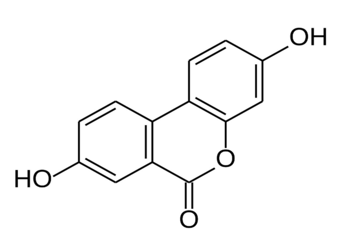
In humans, urolithins have been shown to be end-products or gut microbial metabolites of both ellagitannins and ellagic acid, which are complex polyphenols naturally contained in walnuts and fruits, such as pomegranates and red berries [19]. Urolithins A–D is measurable metabolites that have been identified in human plasma following consumption of ellagitannin- and/or ellagic acid-rich foods or extracts, with urolithin A being the predominant isoform (Figure 2) [20].
Ellagitannins are first hydrolyzed under the acidic conditions of the stomach into ellagic acid, which is then further metabolized by the microflora into urolithins via loss of one of its two lactones and by successful removal of hydroxyl groups [21]. However, the intestinal biotransformation rate of ellagic acid into urolithins varies substantially among individuals due to differences in gut microbiome composition [22]. Urolithin A has been shown to cross the basolateral membrane and has been identified in tissues of the colon, kidney, and liver in rodent models [23]. Once present in the bloodstream, urolithins undergo further metabolism by the liver as glucuronidated and sulfated metabolites. Bioavailability in skeletal muscle tissue has only recently been reported in humans [24].
With regard to safety, two rodent toxicological studies have evaluated the effects of high-dose ellagitannin supplementation at levels up to 4800 mg/kg of punicalagin (the most abundant ellagitannin in pomegranate) per day [25,26]. The safety and toxicological profile of both oral and intravenous synthetic, radiolabeled urolithin A administration also have been tested recently using a rodent model. Results from this study indicate an ingested no observed adverse effect with levels of 3451 and 3826 mg/kg per day in males and females over a 90-day period, respectively [27]. Urolithin A does not seem to demonstrate any genotoxic potential after repeated oral dosing, as shown by its null influence on hematological markers [27]. In the United States, urolithin A has been determined to be generally recognized as safe as an ingredient in food [28] and is, therefore, a legal dietary supplement in the United States per Section 201(ff) (2)-(3) of the Dietary supplement Health and Education Act of 1994 [29,30].
Research investigating the mechanism of action in regard to potential effects of urolithins on mitochondrial function and sarcopenia following dietary supplementation is emerging. In older worms (Caenorhabditis elegans), exposure to urolithin A activated mitochondrial biogenesis [31]. Mammalian C2C12 myoblasts and Mode-K intestinal cells treated with urolithin A showed dose-dependent elevation of biomarkers for both autophagy and mitophagy [31]. Chronic supplementation with urolithin A in mice fed a high-fat diet has been shown to increase muscle function measured at age 22 and 24 months. A subsequent rodent study showed supplementation to enhance average running endurance by 42% [31].
A single phase I (two-part) clinical study of urolithin A was reported in the scientific literature [32]. Part 1 of this study involved supplementing with single ascending doses of urolithin A (250–2000 mg) versus placebo in healthy elderly volunteers. The study was powered to provide sufficient information on the safety and pharmacokinetic profile of urolithin A and to allow for dose selection for future phase II efficacy trials. Part 2 consisted of supplementing multiple ascending doses of urolithin A (250, 500, and 1000 mg) versus placebo, each for 28 days. No product-related adverse events were reported at any dose of urolithin A in both parts of the study, and supplementation increased circulating levels of urolithin A in a dose-dependent manner. Several surrogate markers of mitochondrial health (e.g., mitochondrial respiratory complexes I, II, and IV abundance in vastus lateralis, mitochondrial respiratory complexes I, II, and IV activity in vastus lateralis, and relative abundance of mitochondrial DNA over nuclear DNA) were also investigated from skeletal muscle biopsies and blood samples at baseline and after 28 days of each dosing regimen. Dosing of urolithin A at 250 mg showed no significant improvement in biomarkers; however, dose-dependent decreases in acylcarnitine levels were demonstrated in the 500- and 1000-mg groups [24]. Acylcarnitines are the form in which fatty acids enter into the mitochondria to undergo oxidation. Urolithin A showed the most impact on shorter-chain acylcarnitines (C8, C10, C12, C14:1), which signifies better efficiency in the fatty acid oxidation process [33]. The direct impact of 500 and 1000 mg of urolithin A was shown through a dose-dependent upregulation of skeletal muscle mitochondrial gene expression in the human vastus lateralis. mRNA levels of autophagy/mitophagy and mitochondrial biogenesis-related biomarkers increased, as measured by quantitative PCR in the vastus lateralis, with some reaching statistical significance (GABA type A receptor-associated protein-like GABARAPL1, fatty acid-binding protein FABP3) [24]. These results were confirmed via transcriptomic profiling in the skeletal muscle that demonstrated significant upregulation of multiple mitochondrial-linked gene sets in the 500- and 1000-mg urolithin A groups compared to placebo.

5. Discussion

The scientific literature clearly demonstrates that ubiquitous cellular changes drive many age-associated chronic disease states. Targeting these specific pathways may have the potential to effectively alter the aging trajectory. Certain nutrients, including nicotinamide riboside and urolithin A, have been identified as promising ways to improve mitochondrial health, with each targeting different cellular pathways. Although the typical diet may provide low amounts of these nutrients, insufficient intakes are likely obtained due to their low abundance in commonly consumed foods and interindividual differences in their metabolization by the gut microbiome. Supplemental nicotinamide riboside has been shown to effectively boost NAD+ levels, which is important for mitochondrial biogenesis. Recent clinical data demonstrate urolithin A’s ability to activate cellular mitophagy and improve mitochondrial health in the skeletal muscle of healthy older adults. Additional research is underway on both of these cellular nutrients to understand their potential to impact cellular and clinical outcomes during aging.

Funding

These are proceedings from a programmed session held at the virtual 2020 Annual Scientific Meeting of the Gerontological Society of America. The medical writing of the symposium proceedings was funded by Nestlé Health Science.

Institutional Review Board Statement

Not applicable.

Informed Consent Statement

Not applicable.

Data Availability Statement

No new data were created or analyzed in this study. Data sharing not applicable to this article.

Conflicts of Interest

J.M.G. has received consulting fees from BioAge Labs, Pluristem Pharma, Novartis, Nestlé Health Science, and the Alliance for Aging Research. J.N.F. is an employee of Société des Produits Nestlé SA. A.S. is an employee of Amazentis SA. R.A.F. is supported in part under 58-8050-9-004 Any opinions, findings, conclusions, or recommendations expressed in this publication are those of the author(s) and do not necessarily reflect the view of the U.S. Department of Agriculture. RAF reports grants from the National Institutes of Health (National Institute on Aging), grants, personal fees and stock options from Axcella Health and Juvicell, stock options from Inside Tracker, grants and personal fees from Biophytis, grants and personal fees from Astellas, personal fees from Cytokinetics, personal fees from Amazentis, grants and personal fees from Nestle’, and personal fees from Glaxo Smith Kline and Pfizer. The funders supported a medical writing agency to summarize the symposium proceedings. All authors had significant contributions in drafting, reviewing, and final revision of the paper. Submission and publication of this paper were independent of Nestlé Health Science’s approval.

References

  1. Ferrucci, L.; Gonzalez-Freire, M.; Fabbri, E.; Simonsick, E.; Tanaka, T.; Moore, Z.; Salimi, S.; Sierra, F.; de Cabo, R. Measuring biological aging in humans: A quest. Aging Cell 2020, 19, e13080. [Google Scholar] [CrossRef] [PubMed] [Green Version]
  2. Studenski, S.; Perera, S.; Patel, K.; Rosano, C.; Faulkner, K.; Inzitari, M.; Brach, J.; Chandler, J.; Cawthon, P.; Connor, E.B.; et al. Gait speed and survival in older adults. JAMA 2011, 305, 50–58. [Google Scholar] [CrossRef] [Green Version]
  3. López-Otín, C.; Blasco, M.A.; Partridge, L.; Serrano, M.; Kroemer, G. The hallmarks of aging. Cell 2013, 153, 1194–1217. [Google Scholar] [CrossRef] [Green Version]
  4. Cesari, M.; Cherubini, A.; Guralnik, J.M.; Beresniak, A.; Rodriguez-Mañas, L.; Inzitari, M.; Walston, J. Early detection of accelerated aging and cellular decline (AACD): A consensus statement. Exp. Gerontol. 2021, 146, 111242. [Google Scholar] [CrossRef]
  5. Harvey, N.; Holmes, C.A. Nominal group technique: An effective method for obtaining group consensus. Int. J. Nurs. Pract. 2012, 18, 188–194. [Google Scholar] [CrossRef] [PubMed] [Green Version]
  6. Gallagher, M.; Hares, T.; Spencer, J.; Bradshaw, C.; Webb, I. The nominal group technique: A research tool for general practice? Fam. Pract. 1993, 10, 76–81. [Google Scholar] [CrossRef] [PubMed]
  7. Prado, C.M.; Purcell, S.A.; Alish, C.; Pereira, S.L.; Deutz, N.E.; Heyland, D.K.; Goodpaster, B.H.; Tappenden, K.A.; Heymsfield, S.B. Implications of low muscle mass across the continuum of care: A narrative review. Ann. Med. 2018, 50, 675–693. [Google Scholar] [CrossRef] [PubMed] [Green Version]
  8. Shafiee, G.; Keshtkar, A.; Soltani, A.; Ahadi, Z.; Larijani, B.; Heshmat, R. Prevalence of sarcopenia in the world: A systematic review and meta- analysis of general population studies. J. Diabetes Metab. Disord. 2017, 16, 21. [Google Scholar] [CrossRef] [Green Version]
  9. Migliavacca, E.; Tay, S.K.H.; Patel, H.P.; Sonntag, T.; Civiletto, G.; McFarlane, C.; Forrester, T.; Barton, S.J.; Leow, M.K.; Antoun, E.; et al. Mitochondrial oxidative capacity and NAD(+) biosynthesis are reduced in human sarcopenia across ethnicities. Nat. Commun. 2019, 10, 5808. [Google Scholar] [CrossRef] [PubMed]
  10. Verdin, E. NAD⁺ in aging, metabolism, and neurodegeneration. Science 2015, 350, 1208–1213. [Google Scholar] [CrossRef]
  11. Mouchiroud, L.; Houtkooper, R.H.; Moullan, N.; Katsyuba, E.; Ryu, D.; Cantó, C.; Mottis, A.; Jo, Y.S.; Viswanathan, M.; Schoonjans, K.; et al. The NAD(+)/sirtuin pathway modulates longevity through activation of mitochondrial UPR and FOXO signaling. Cell 2013, 154, 430–441. [Google Scholar] [CrossRef] [PubMed] [Green Version]
  12. Cantó, C.; Houtkooper, R.H.; Pirinen, E.; Youn, D.Y.; Oosterveer, M.H.; Cen, Y.; Fernandez-Marcos, P.J.; Yamamoto, H.; Andreux, P.A.; Cettour-Rose, P.; et al. The NAD(+) precursor nicotinamide riboside enhances oxidative metabolism and protects against high-fat diet-induced obesity. Cell Metab. 2012, 15, 838–847. [Google Scholar] [CrossRef] [Green Version]
  13. Gomes, A.P.; Price, N.L.; Ling, A.J.; Moslehi, J.J.; Montgomery, M.K.; Rajman, L.; White, J.P.; Teodoro, J.S.; Wrann, C.D.; Hubbard, B.P.; et al. Declining NAD(+) induces a pseudohypoxic state disrupting nuclear-mitochondrial communication during aging. Cell 2013, 155, 1624–1638. [Google Scholar] [CrossRef] [Green Version]
  14. Zhang, H.; Ryu, D.; Wu, Y.; Gariani, K.; Wang, X.; Luan, P.; D’Amico, D.; Ropelle, E.R.; Lutolf, M.P.; Aebersold, R.; et al. NAD⁺ repletion improves mitochondrial and stem cell function and enhances life span in mice. Science 2016, 352, 1436–1443. [Google Scholar] [CrossRef] [Green Version]
  15. Martens, C.R.; Denman, B.A.; Mazzo, M.R.; Armstrong, M.L.; Reisdorph, N.; McQueen, M.B.; Chonchol, M.; Seals, D.R. Chronic nicotinamide riboside supplementation is well-tolerated and elevates NAD(+) in healthy middle-aged and older adults. Nat. Commun. 2018, 9, 1286. [Google Scholar] [CrossRef] [PubMed]
  16. Conze, D.; Brenner, C.; Kruger, C.L. Safety and metabolism of long-term administration of NIAGEN (nicotinamide riboside chloride) in a randomized, double-blind, placebo-controlled clinical trial of healthy overweight adults. Sci. Rep. 2019, 9, 9772. [Google Scholar] [CrossRef]
  17. Elhassan, Y.S.; Kluckova, K.; Fletcher, R.S.; Schmidt, M.S.; Garten, A.; Doig, C.L.; Cartwright, D.M.; Oakey, L.; Burley, C.V.; Jenkinson, N.; et al. Nicotinamide riboside augments the aged human skeletal muscle NAD(+) metabolome and induces transcriptomic and anti-inflammatory signatures. Cell Rep. 2019, 28, 1717–1728.e6. [Google Scholar] [CrossRef] [PubMed] [Green Version]
  18. Dolopikou, C.F.; Kourtzidis, I.A.; Margaritelis, N.V.; Vrabas, I.S.; Koidou, I.; Kyparos, A.; Theodorou, A.A.; Paschalis, V.; Nikolaidis, M.G. Acute nicotinamide riboside supplementation improves redox homeostasis and exercise performance in old individuals: A double-blind cross-over study. Eur. J. Nutr. 2020, 59, 505–515. [Google Scholar] [CrossRef]
  19. Atanasov, A.G.; Waltenberger, B.; Pferschy-Wenzig, E.M.; Linder, T.; Wawrosch, C.; Uhrin, P.; Temml, V.; Wang, L.; Schwaiger, S.; Heiss, E.H.; et al. Discovery and resupply of pharmacologically active plant-derived natural products: A review. Biotechnol. Adv. 2015, 33, 1582–1614. [Google Scholar] [CrossRef] [PubMed] [Green Version]
  20. Tomás-Barberán, F.A.; González-Sarrías, A.; García-Villalba, R.; Núñez-Sánchez, M.A.; Selma, M.V.; García-Conesa, M.T.; Espín, J.C. Urolithins, the rescue of “old” metabolites to understand a “new” concept: Metabotypes as a nexus among phenolic metabolism, microbiota dysbiosis, and host health status. Mol. Nutr. Food Res. 2017, 61. [Google Scholar] [CrossRef]
  21. Espín, J.C.; Larrosa, M.; García-Conesa, M.T.; Tomás-Barberán, F. Biological significance of urolithins, the gut microbial ellagic acid-derived metabolites: The evidence so far. Evid. Based Complement. Alternat. Med. 2013, 2013, 270418. [Google Scholar] [CrossRef] [PubMed] [Green Version]
  22. Tomás-Barberán, F.A.; García-Villalba, R.; González-Sarrías, A.; Selma, M.V.; Espín, J.C. Ellagic acid metabolism by human gut microbiota: Consistent observation of three urolithin phenotypes in intervention trials, independent of food source, age, and health status. J. Agric. Food Chem. 2014, 62, 6535–6538. [Google Scholar] [CrossRef] [PubMed]
  23. Seeram, N.P.; Aronson, W.J.; Zhang, Y.; Henning, S.M.; Moro, A.; Lee, R.P.; Sartippour, M.; Harris, D.M.; Rettig, M.; Suchard, M.A.; et al. Pomegranate ellagitannin-derived metabolites inhibit prostate cancer growth and localize to the mouse prostate gland. J. Agric. Food Chem. 2007, 55, 7732–7737. [Google Scholar] [CrossRef] [PubMed]
  24. Andreux, P.A.; Blanco-Bose, W.; Ryu, D.; Burdet, F.; Ibberson, M.; Aebischer, P.; Auwerx, J.; Singh, A.; Rinsch, C. The mitophagy activator urolithin A is safe and induces a molecular signature of improved mitochondrial and cellular health in humans. Nat. Metab. 2019, 1, 595–603. [Google Scholar] [CrossRef] [PubMed]
  25. Patel, C.; Dadhaniya, P.; Hingorani, L.; Soni, M.G. Safety assessment of pomegranate fruit extract: Acute and subchronic toxicity studies. Food Chem. Toxicol. 2008, 46, 2728–2735. [Google Scholar] [CrossRef] [PubMed]
  26. Cerdá, B.; Cerón, J.J.; Tomás-Barberán, F.A.; Espín, J.C. Repeated oral administration of high doses of the pomegranate ellagitannin punicalagin to rats for 37 days is not toxic. J. Agric. Food Chem. 2003, 51, 3493–3501. [Google Scholar] [CrossRef]
  27. Heilman, J.; Andreux, P.; Tran, N.; Rinsch, C.; Blanco-Bose, W. Safety assessment of urolithin A, a metabolite produced by the human gut microbiota upon dietary intake of plant derived ellagitannins and ellagic acid. Food Chem. Toxicol. 2017, 108, 289–297. [Google Scholar] [CrossRef]
  28. Keefe, D. GRAS Notice No. GRN 000791. 2018. Available online: https://www.fda.gov/media/120300/download (accessed on 16 February 2021).
  29. National Institutes of Health Office of Dietary Supplements. Dietary Supplement Health and Education Act of 1994. 1994. Available online: https://ods.od.nih.gov/About/DSHEA_Wording.aspx (accessed on 16 February 2021).
  30. Wallace, T.C. Twenty years of the Dietary Supplement Health and Education Act--how should dietary supplements be regulated? J. Nutr. 2015, 145, 1683–1686. [Google Scholar] [CrossRef] [Green Version]
  31. Ryu, D.; Mouchiroud, L.; Andreux, P.A.; Katsyuba, E.; Moullan, N.; Nicolet-Dit-Félix, A.A.; Williams, E.G.; Jha, P.; Lo Sasso, G.; Huzard, D.; et al. Urolithin A induces mitophagy and prolongs lifespan in C. elegans and increases muscle function in rodents. Nat. Med. 2016, 22, 879–888. [Google Scholar] [CrossRef]
  32. Andreux, P.A.; van Diemen, M.P.J.; Heezen, M.R.; Auwerx, J.; Rinsch, C.; Groeneveld, G.J.; Singh, A. Mitochondrial function is impaired in the skeletal muscle of pre-frail elderly. Sci. Rep. 2018, 8, 8548. [Google Scholar] [CrossRef]
  33. Schooneman, M.G.; Vaz, F.M.; Houten, S.M.; Soeters, M.R. Acylcarnitines: Reflecting or inflicting insulin resistance? Diabetes 2013, 62, 1–8. [Google Scholar] [CrossRef] [PubMed] [Green Version]
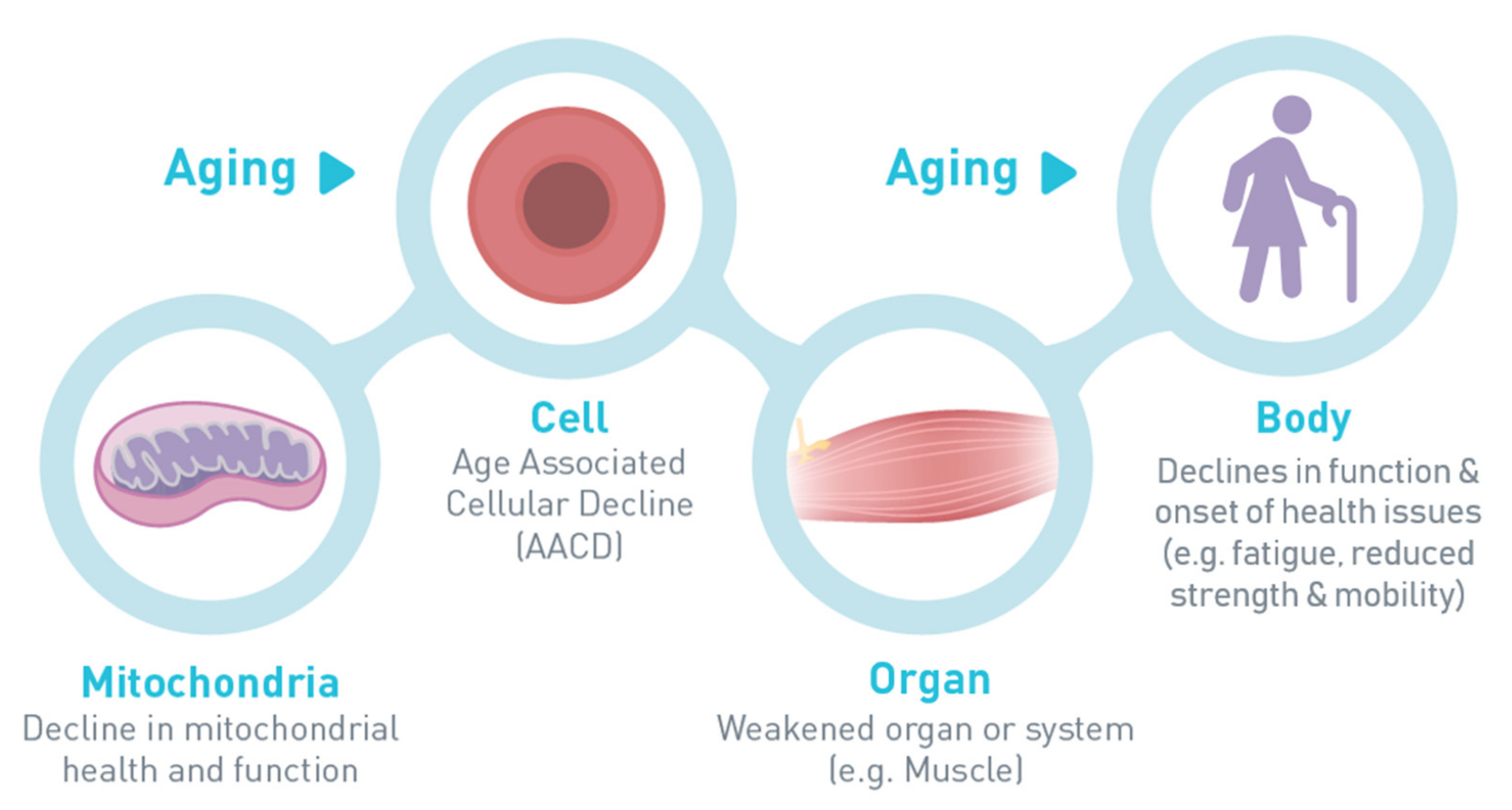
Figure 1. Mitochondrial origin of age-related decline.
Geriatrics 06 00037 g001
Figure 2. Chemical structure of urolithin A.
Geriatrics 06 00037 g002
Table 1. Prioritized physical and environmental risk factors for age-associated cellular decline (AACD) (in rank order determined by the interdisciplinary expert panel).
Demographic and Clinical Risk Factors
Clinical conditions (e.g., cardiovascular disease)
Obesity
Unfavorable genetic background
Insulin resistance
Low physical capacity (e.g., slow gait speed and muscle weakness)
Environmental and behavioral risk factors
Smoking
Sedentary lifestyle
Low physical activity
Persistent physical or psychological stress
Low socioeconomic status
Alcohol abuse
Inadequate nutrition
Air pollution
Adapted with permission from Cesari et al. [4].
Table 2. Framework connecting biological mechanisms with the clinical phenotype of AACD.
Underlying Cellular and
Subcellular Networks
DomainsClinical Indicators
Cellular senescenceEnergy metabolismFatigue
Mitochondrial abnormalitiesImmune systemLow-quality of sleep
Metabolic signalingCentral nervous systemLow mood
InflammationBody compositionLack of motivation
Autophagy/mitophagy Subjective memory complaints
Oxidant/antioxidant balance Poor exercise tolerance
Adapted with permission from Cesari et al. [4].
Publisher’s Note: MDPI stays neutral with regard to jurisdictional claims in published maps and institutional affiliations.