Article

Molecular Survey for Major Canine Enteric Viral Pathogens in Wild Carnivores, Northwestern Italy

1
Department of Veterinary Medicine, Università degli Studi di Teramo, Località Piano D’Accio, 64100 Teramo, Italy
2
Centro di Referenza Nazionale per le Malattie degli Animali Selvatici (CeRMAS), Istituto Zooprofilattico Sperimentale del Piemonte, della Liguria e della Valle d’Aosta, 11020 Quart, Italy
3
Department of Veterinary Medicine, Università Aldo Moro di Bari, S.p. per Casamassima Km3, Valenzano, 70010 Bari, Italy
4
Department of Medical Biology, Medical School, University of Pécs, 7624 Pécs, Hungary
*
Author to whom correspondence should be addressed.
Vet. Sci. 2025, 12(9), 814; https://doi.org/10.3390/vetsci12090814
Submission received: 27 July 2025 / Revised: 19 August 2025 / Accepted: 23 August 2025 / Published: 26 August 2025

Simple Summary

Wildlife animals could act as a source of viral pathogens for pets, complicating the ecology of these viruses and the plans for controlling the disease. In particular, canine protoparvovirus and canine adenovirus are considered major pathogens of dogs, and the vaccines for the prevention of canine protoparvovirus and adenovirus-associated diseases are considered core vaccines. This study examined the occurrence of these viral pathogens in wild carnivores in Northwestern Italy. The investigation included free-ranging wolves, red foxes, stone martens, and Eurasian badgers, combining consensus PCR assays and third-generation sequencing. Carnivore protoparvoviruses were identified in three wolves and one badger, while canine adenovirus type 1 was detected in one fox. The results confirm the circulation of major viral pathogens among wild canids and mustelids. The study underscores the risk of cross-species transmission at the domestic–wildlife interface and highlights the value of molecular surveillance for emerging pathogens in wildlife populations.

Abstract

Wild carnivores can harbor pathogens affecting wildlife conservation and domestic animal health. This study surveyed major viral pathogens in free-ranging wolves, red foxes, stone martens, and Eurasian badgers in Northwestern Italy. Duodenal samples from 140 carcasses were screened by consensus PCR for members of the species Protoparvovirus carnivoran1 and for canine adenoviruses (CAdV-1/2). PCR-positive samples underwent sequence-independent amplification and Oxford Nanopore sequencing. Canine parvovirus type 2 (CPV-2) and feline panleukopenia virus (FPV) DNAs were identified in three wolves (6.4%) and one badger (4.3%), whereas CAdV-1 was detected in one red fox (1.8%). Nanopore sequencing yielded near-complete genomes of two CPV-new 2a, one CPV-2c, and one FPV strains, along with partial CAdV-1 sequences. Furthermore, the complete genome of a canine circovirus (CaCV) strain in co-infection with a CPV-2c-positive wolf and partial sequences of a canine kobuvirus (CaKoV) strain were also obtained. Phylogenetic analysis placed these viruses within known European lineages and linked them to domestic and wild hosts. These findings revealed the circulation of multiple viral pathogens among wild carnivores, reflecting ongoing cross-species spillover. Continuing molecular surveillance at the wildlife–domestic interface is recommended.

1. Introduction

The order Carnivora comprises over 290 species and plays a crucial role in maintaining ecological balance by regulating populations of herbivores and smaller carnivores, facilitating seed dispersal, and preserving local vegetation dynamics [1,2]. However, these ecological contributions are increasingly threatened by several factors, including habitat degradation, environmental changes, and infectious diseases [3]. In particular, infectious diseases pose a significant threat to wild carnivores in increasingly anthropized landscapes, resulting in the growth in domestic animal populations around protected areas and greater opportunities for inter-species disease transmission [4]. Indeed, carnivores at the human–wildlife interface are known to influence the epidemiology of domestic carnivore viruses [5]. Specifically, wild carnivores face a heightened risk of viral transmission due to their close phylogenetic relationship with domestic dogs and cats [6]. Increased contact between these groups fosters pathogen exchange among related hosts, such as species within the Canidae and Felidae families [7,8].
Among these pathogens, members of the Protoparvovirus carnivoran1 species (family Parvoviridae, genus Protoparvovirus), which include canine parvovirus type 2 (CPV-2) and feline panleukopenia virus (FPV) [9], are of particular concern. CPV-2 variants (CPV-2a, CPV-2b, CPV-2c) and FPV are often associated with cross-species transmission from domestic to wild carnivores, with molecular studies confirming their widespread circulation [10,11,12]. In Europe, CPV-2 has been identified in different wild animals, like foxes (Vulpes vulpes) [13,14,15], wolves (Canis lupus) [13,14,15,16,17], stone martens (Martes foina) [13,14,18], Eurasian badgers (Meles meles) [14,15,19], and Eurasian otters (Lutra lutra) [20], in a common genet (Genetta genetta), and in a European wildcat (Felis silvestris) [13]. On the other hand, FPV has been detected in foxes [14,15,18,21], Eurasian badgers [13,14,15,22], stone martens [13,15], golden jackals (Canis aureus) [22], Eurasian lynx (Lynx lynx) [23], Iberian lynx (Lynx pardinus) [24], wolves, crested porcupines (Hystrix cristata), and Marsican brown bears (Ursus arctos marsicanus) [15].
In addition to CPV-2 and FPV, canine adenoviruses (CAdVs) are important pathogens of domestic dogs and are able to infect different species of wild carnivores, although their ecology and impact remain poorly investigated. CAdVs are classified in the genus Mastadenovirus within the family Adenoviridae [25]. Two distinct types of the virus are known, canine adenovirus type 1 (CAdV-1) and canine adenovirus type 2 (CAdV-2), causing infectious canine hepatitis (ICH) and kennel cough in dogs, respectively [26]. CAdV-1 was described in silver foxes mainly associated with encephalitis [27] and has been since then identified in diseased and apparently healthy wild carnivores [28,29,30,31,32,33,34]. Likewise, CAdV-2 has been documented in foxes [21] and wolves in Italy [35] and in wild raccoon dogs (Nyctereutes procyonoides) in Korea [36], but its epidemiological role and pathogenicity are not clear yet.
In this study, by employing a combination of molecular diagnostic approaches and third-generation sequencing with the Oxford Nanopore Technologies (ONT) platform, we investigated the presence of these major canine viral pathogens in free-ranging carnivore populations from Northwestern Italy.

2. Materials and Methods

2.1. Sampling

Molecular investigations were performed on archived duodenal samples collected from a total of 140 wild carnivores found dead, including 47 wolves (Canis lupus), 55 foxes (Vulpes vulpes), 15 stone martens (Martes foina), and 23 Eurasian badgers (Meles meles), between February 2021 and April 2022. Sampling was conducted in Northwestern Italy, specifically in the Piemonte and Valle d’Aosta regions, as part of a national passive surveillance program carried out by the National Reference Centre for Wild Animal Diseases (CeRMAS-IZS PLV).

2.2. Nucleic Acids Extraction and Molecular Investigations

Duodenal tissues (1g) were homogenized using a Mini Bead Mill (VWR International Srl, Milan, Italy) in 7 mL of hard-tissue homogenization tubes containing 4 mL of phosphate-buffered saline (PBS, 0.15 M, pH 7.2). To enhance tissue breakdown and promote virion release into the solution, 0.5 mL of proteinase K (PK) at a concentration of 100 μg/mL (1 mg/mL) was added. The samples were incubated at 37 °C for 3 h with agitation at 320 rpm. To inactivate the PK, the reaction mixture was further incubated at 60 °C for 15 min. After centrifugation at 2500 rpm for 15 min, the supernatant was collected and stored at −20 °C. RNA and DNA were extracted from 0.5 mL aliquots of the duodenal and liver samples using TRIzol LS reagent (Invitrogen, Ltd., Paisley, UK), according to the manufacturer’s protocol. Nucleic acid extracts were successively processed with the OneStep PCR Inhibitor Removal Kit (Zymo Research, Irvine, CA, USA) to remove potential PCR inhibitors. All samples were screened for the presence of members of the species Protoparvovirus carnivoran1 [37] and for CAdV-1/CAdV-2 [38].
RT-PCR and PCR assays were carried out using SuperScript™ One-Step RT-PCR (Invitrogen, Ltd., Paisley, UK) and GoTaq® Green Master Mix 1 (Promega Italia S.r.l., Milan, Italy), respectively. Amplified products were separated by electrophoresis on 1.5% agarose gels and visualized under UV illumination following ethidium bromide staining. Bands of the expected size were excised from the gels, purified using a QIAquick gel extraction kit (Qiagen GmbH, Hilden, Germany), and sequenced bidirectionally using BigDye Terminator Cycle chemistry on an Applied Biosystems 3730 DNA Analyzer (Foster, CA, USA). Resulting sequences were compared against databases using BLAST (http://www.ncbi.nlm.nih.gov) and FASTA (http://www.ebi.ac.uk/fasta33) with default parameters to identify homologous sequences.

2.3. SISPA, ONT Library Preparation and Sequencing

A sequence-independent single-primer amplification (SISPA) method [39], combined with Oxford Nanopore Technologies (ONT) sequencing, was employed to characterize the virome of the PCR-positive duodenal samples. Additionally, for each animal species, total RNA and DNA were combined into pools consisting of five samples mixed in equal volumes. These pooled RNA and DNA samples were then subjected to SISPA amplification and ONT sequencing.
Briefly, 20 µL of extracted DNA was combined with 2.5 µL of NEBuffer™ 2 (New England Biolabs, Hitchin, UK), 1 µL of 10 mM dNTPs (Invitrogen, Ltd., Paisley, UK), and 2 µL of 10 µM primer FR26RV-N [40]. The mixture was initially incubated at 94 °C for 2 min, followed by cooling on ice for an additional 2 min. Subsequently, 0.5 µL (2.5 units) of Klenow Fragment (3′→5′ exo-) (New England Biolabs, Hitchin, UK) was added, and the reaction was incubated at 37 °C for 1 h. This process was repeated, and then an enzyme inactivation step was performed at 75 °C for 10 min.
For reverse transcription, DNase-treated RNA was converted into single-stranded cDNA using SuperScript IV Reverse Transcriptase (Invitrogen Ltd., Milan, Italy) with primers FR26RV-N [40] and FR40RV-T [41]. The synthesis of the second cDNA strand was completed using Klenow Fragment (3′→5′ exo-) (New England Biolabs, Hitchin, UK). A 5 µL aliquot of the resulting reaction was used as the template in a subsequent PCR amplification. The 50 µL PCR mixture included 10 µL of 5X Q5 Reaction Buffer (New England Biolabs, Hitchin, UK), 1 µL of dNTPs, 1 µL of 10 µM primer FR20RV [40], and 1 µL of Q5 High-Fidelity DNA Polymerase (0.04 U/µL; New England Biolabs, Hitchin, UK). The thermal cycling protocol consisted of an initial activation at 94 °C for 2 min, followed by 40 cycles of amplification (94 °C for 30 s, 48 °C for 30 s, 68 °C for 8 min), and a final extension at 68 °C for 8 min.
PCR products were purified using AMPure XP beads (Beckman Coulter, Indianapolis, IN, USA) and quantified with a Qubit 4.0 fluorometer using the dsDNA HS Assay Kit (Thermo Fisher Scientific, Waltham, MA, USA). Library preparation was carried out according to the manufacturer’s protocol, utilizing the PCR Barcoding Expansion 1–12 (EXP-PBC001) Kit and the Ligation Sequencing Kit (SQK-LSK-114) to enable multiplexing of samples on an R10 flow cell (FLO-MIN110D, ONT). Sequencing was performed on a MinION Mk1C device (ONT, Oxford, UK) for six hours. Real-time basecalling and demultiplexing during sequencing were carried out using the ONT GUPPY (v3.2.8) basecaller within MinKNOW software (v3.1.5). FastQ MinION files were processed with the online tool Genome Detective (https://www.genomedetective.com/db/ui/login) [42] for de novo assembly with default settings. Additionally, reference assembly was performed using the Minimap2 plugin in Geneious Prime software v.2021.2.2 (Biomatters Ltd., Auckland, New Zealand, https://www.geneious.com). Phylogenetic analysis was conducted by using Maximum Likelihood or neighbor-joining methods, with statistical support with bootstrapping of 1000 replicates in MEGA 11 software [43].

3. Results

3.1. Molecular Screening and Sanger Sequencing

The molecular screenings revealed positivity in five of the 140 animals tested, with an overall prevalence of 3.6% (5/140) (Table 1). In detail, Protoparvovirus carnivoran1 DNA was identified in three intestinal samples of wolves (6.4%, 3/47) from the Piemonte region and in one enteric specimen of a Eurasian badger, 4.3% (1/23), collected in Valle d’Aosta. Additionally, CAdV-1 DNA was detected in one intestinal sample collected from a fox found dead in the Valle d’Aosta region (1.8%, 1/55), while all other wild carnivores tested negative. All animals tested negative for CAdV-2, and all stone marten samples tested negative.
All amplicons (Supplementary Figure S1) acquired through consensus PCR protocols were subjected to direct sequencing. For the Protoparvovirus Carnivoran1 strategy [37], sequence analysis revealed that all sequences detected shared 97.7–100.0% nucleotide (nt) identity to each other. Two wolf samples (LI6/2021/ITA and LI8/2021/ITA) exhibited the highest nt identity (99.65–100%) with CPV-2a strains detected in Canada in wild raccoons (Procyon lotor) [44] and in a wild American mink (Neovison vison) [45], respectively, as well as with strains identified in dogs in Italy [22,46,47], Hungary [48], New Zealand [49], and in cats in China [50]. The sample LI9/2022/ITA exhibited the closest relationship (99.65–100% nt identity) with CPV-2c strains identified in dogs in Italy [22,51], in India [52], in China [53], in Vietnam [54], and in Zambia [55]. The badger positive sample (TI6/2022/ITA) showed the closest match (99.47–99.65% nt) to FPV strains identified in wild animals, including strains identified in China in a giant panda (Ailuropoda melanoleuca) [50,56], in a red panda (Ailurus fulgens) [50], in a captive tiger [57], and in a captive jaguar (unpublished), in the brain of a Iberian lynx in Spain [24], in strains isolated in Italy from a crested porcupine [15] and a golden jackal [22], and in domestic cats in Italy [46], and Spain [24].
Sequence analysis of the CAdV-1 DNA amplicon (GenBank accession no. PV955937) showed the highest sequence identity (99.5–100.0%) to CAdV-1 strains previously identified in wolves in Italy [30] and in France [31], in foxes in Italy [29] and in the United Kingdom [58], in European brown bears in Spain (Ursus arctos arctos) [32], in a badger in Italy [59], in a civet in India (Civettictis civetta) (unpublished), and domestic dogs from various regions, including Australia [60] and Italy [61].

3.2. ONT Sequencing

After enrichment using a SISPA protocol, the five positive samples (Wolf/LI6/2021/ITA, Wolf/LI8/2021/ITA, Wolf/LI9/2022/ITA, Badger/TI6/2022/ITA, Fox/VI9/2021/ITA) and pooled samples were processed for high-throughput sequencing with ONT. A total of 2,816,158 raw nanopore reads were generated, and low-quality sequencing data were filtered using a quality score threshold of eight. A total of 1,807,115 (64.17%) viral reads, with an average length of 1315 nt (range 110–5588 nt), form a total of 16 contigs (>100 nt) were assembled using the NCBI reference sequence and compared with viral sequences currently available on the GenBank database (Table 2).
We generated the complete coding regions of the genomes for the parvoviral DNA-positive samples. Specifically, strains CPV-2/Wolf/LI6/2021/ITA (PV955931), CPV-2/Wolf/LI8/2021/ITA (PV955932), and CPV-2/Wolf/LI9/2022/ITA (PV955933) each formed a single contig aligned to reference sequence NC001539, with 131,239, 489,633, and 831,287 reads assembled, respectively. Similarly, the FPV strain FPV/Badger/TI6/2022/ITA (PV955934) formed a single contig with 344,382 reads assembled from 399,962 total reads. In detail, for the CPV-2/Wolf/LI6/2021/ITA, CPV-2/Wolf/LI8/2021/ITA, and CPV-2/Wolf/LI9/2022/ITA strains, a genome sequence of 4492 nt was generated, including a partial 5′ untranslated region (UTR) (128 nt), the complete NS1 sequence (648 aa), the complete NS2 sequence (165 aa), the complete VP1 sequence (727 aa), the complete VP2 sequence (584 aa), and a partial 3′ UTR (95 nt). The genome coding sequence of the FPV/Badger/TI6/2022/ITA (4591 nt) strain, excluding the terminal UTR regions, was 4258 nt and exhibited two major ORFs. The left ORF, coding for NS1 and NS2, was 2007 nt, and the right ORF, encoding for VP1 and VP2, was 2185 nt. Deduced VP2 amino acid sequences were compared with CPV-2 and FPV reference strains, along with strains identified in wolves and badgers. Strains CPV-2/Wolf/LI6/2021/ITA and CPV-2/Wolf/LI8/2021/ITA exhibited identical VP2 sequences, aligning closely with the CPV-new 2a strain and characterized by the mutation 297-Ser to Ala. Additionally, strain CPV-2/Wolf/LI9/2022/ITA, closely related to strains classified as CPV-2c, was distinguished by the substitution of 426-Asn to Glu, a hallmark of CPV-2c strains. Sequence alignment highlighted two amino acid mutations in the badger capsid sequence compared to the reference FPV strain, 101-Ile to Thr and 103-Val to Ala.
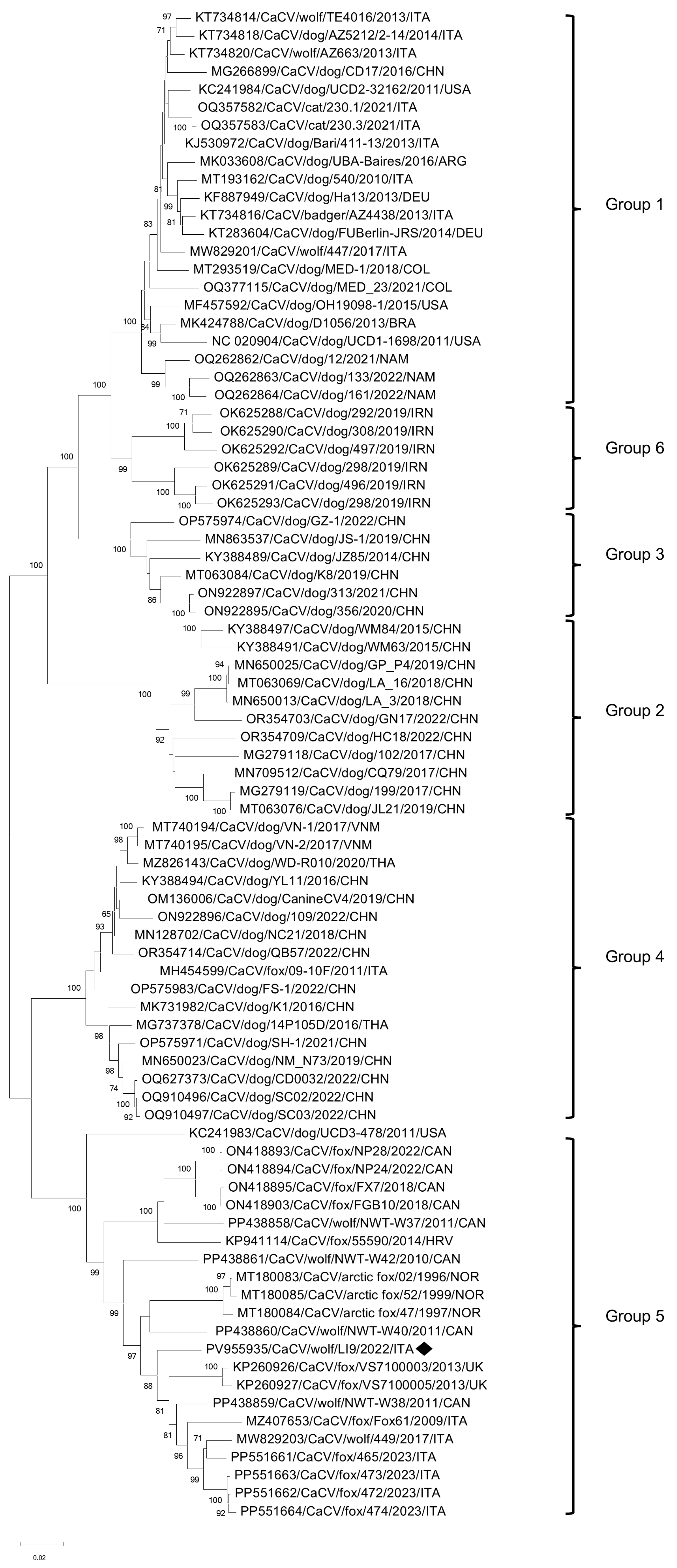
Phylogenetic analysis of the complete amino acid sequence of the VP2 gene indicated that the CPV-2 sequences from wolves were closely related to CPV-2 field isolates from Europe and America, sourced from various animal species. The FPV strain FPV/Badger/TI6/2022/ITA was segregated with the strain ITA/2023/bear/74, found in a Marsican brown bear in Central Italy [15], but separate from other FPV strains detected from different animal species (Figure 1).
In the sample LI9/2022/ITA, the combined SISPA/ONT approach generated an additional 588 reads forming a single contig of 2063 nt. This contig was assembled against the NC020904 strain, the prototype strain of the species Canine circovirus, genus Circovirus, family Circoviridae [62]. The genome corresponds to the complete genome of the strain CaCV/Wolf/LI9/2022/ITA (PV955935) and includes two main ORFs situated on complementary strands in opposite orientations. ORF1 (912 nt) is present on the virion strand, while ORF2 (813 nt) is located on the complementary strand of the replicative form. These ORFs encode the Rep (303 aa) and Cap (270 aa) proteins, respectively. Like other animal circoviruses, the genome of the Italian wolf strain features two intergenic non-coding regions, measuring 135 and 203 nt, positioned between the start and stop codons of the replicase and capsid protein genes. The 5′-intergenic region forms a thermodynamically stable stem-loop structure containing the conserved nucleotide sequence TAGTATTAC [63,64]. The nucleotide alignment between the complete genomic sequences of CaCV/Wolf/LI9/2022/ITA and the CaCV strains retrieved from the GenBank database displayed an overall nt identity ranging from 82.88% to 95.98%.
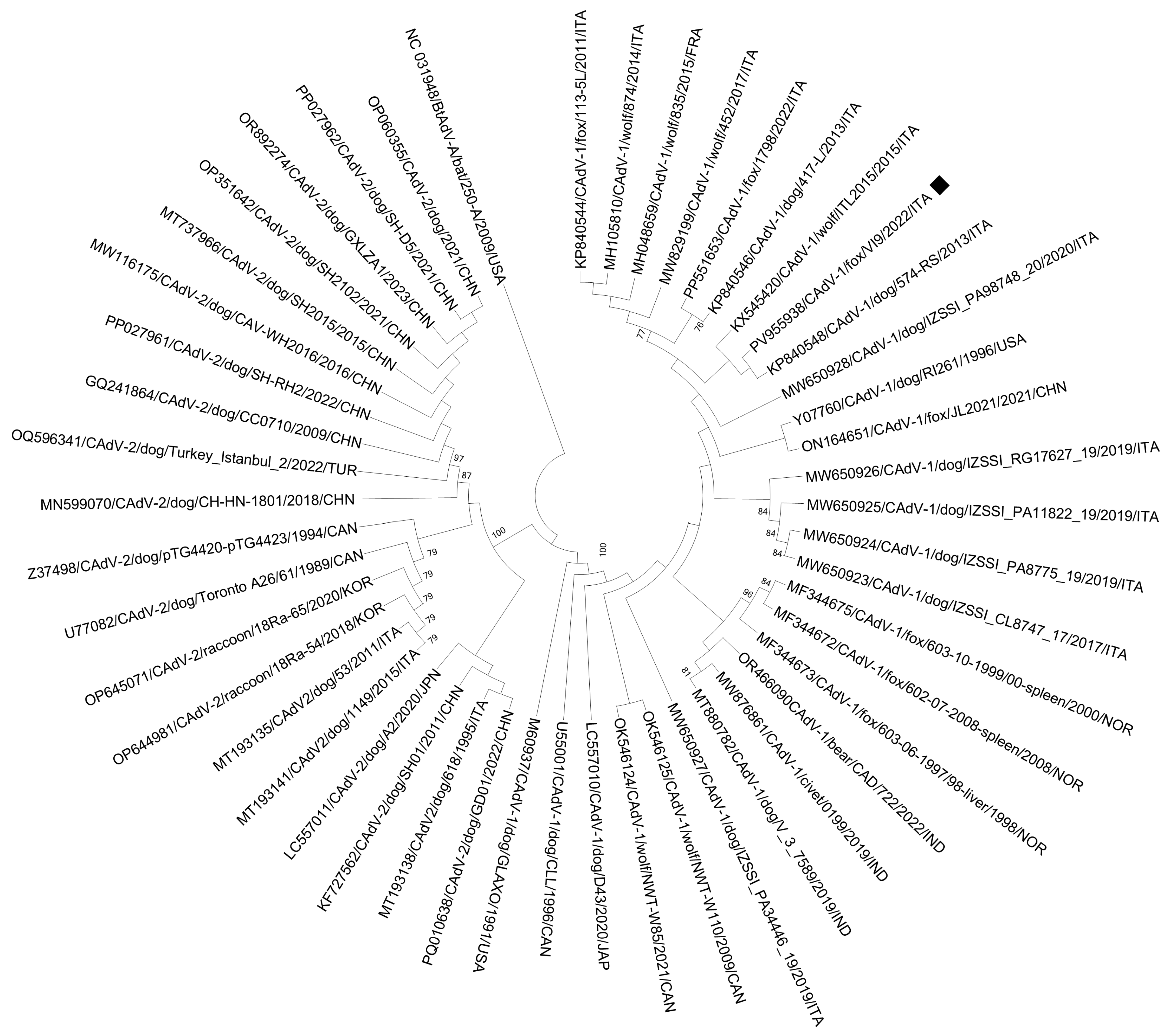
Based on the complete genome nucleotide sequences, the unrooted phylogenetic tree (Figure 2) reveals the clustering of CaCV strains into six well-defined groups. Group 1 comprises strains identified in dogs, wolves, and a badger across Europe, America, and Asia. Groups 2, 3, and 4 include CaCVs detected in dogs from Asia and a red fox in Italy. Group 6 consists of sequences detected in Iranian dogs, while Group 5 encompasses CaCVs identified in wolves, and arctic and red foxes from various countries [65,66]. The sequence from this study clusters within Group 5 alongside strains isolated from foxes and wolves in Europe and in Canada.
For the sample Fox/VI9/2022/ITA, positive for CAdV-1 DNA, SISPA, and ONT, sequencing produced eight contigs ranging in length from 102 to 7124 nt. These contigs partially covered the E1A protein gene, the E1B-55K gene, and the Iva2 coding sequence, as well as the pTP gene, pV gene, and the genes encoding hexon, protease, DNA-binding protein (DBP), and 100k protein. Furthermore, contigs fully encompassed the complete coding sequences of the E1B-19K, the polymerase (pol) gene, and the genes encoding 33K, 22K, pVIII, E3, U exon, fiber, and E4 proteins. Sequence analyses revealed nt identities ranging from 98.9% to 100.0% when compared with previously detected CAdV-1 sequences from liver, blood, lung, and fecal samples of dogs and various wild carnivores and classified within the species Mastadenovirus canidae of the genus Mastadenovirus (family Adenoviridae) [25].
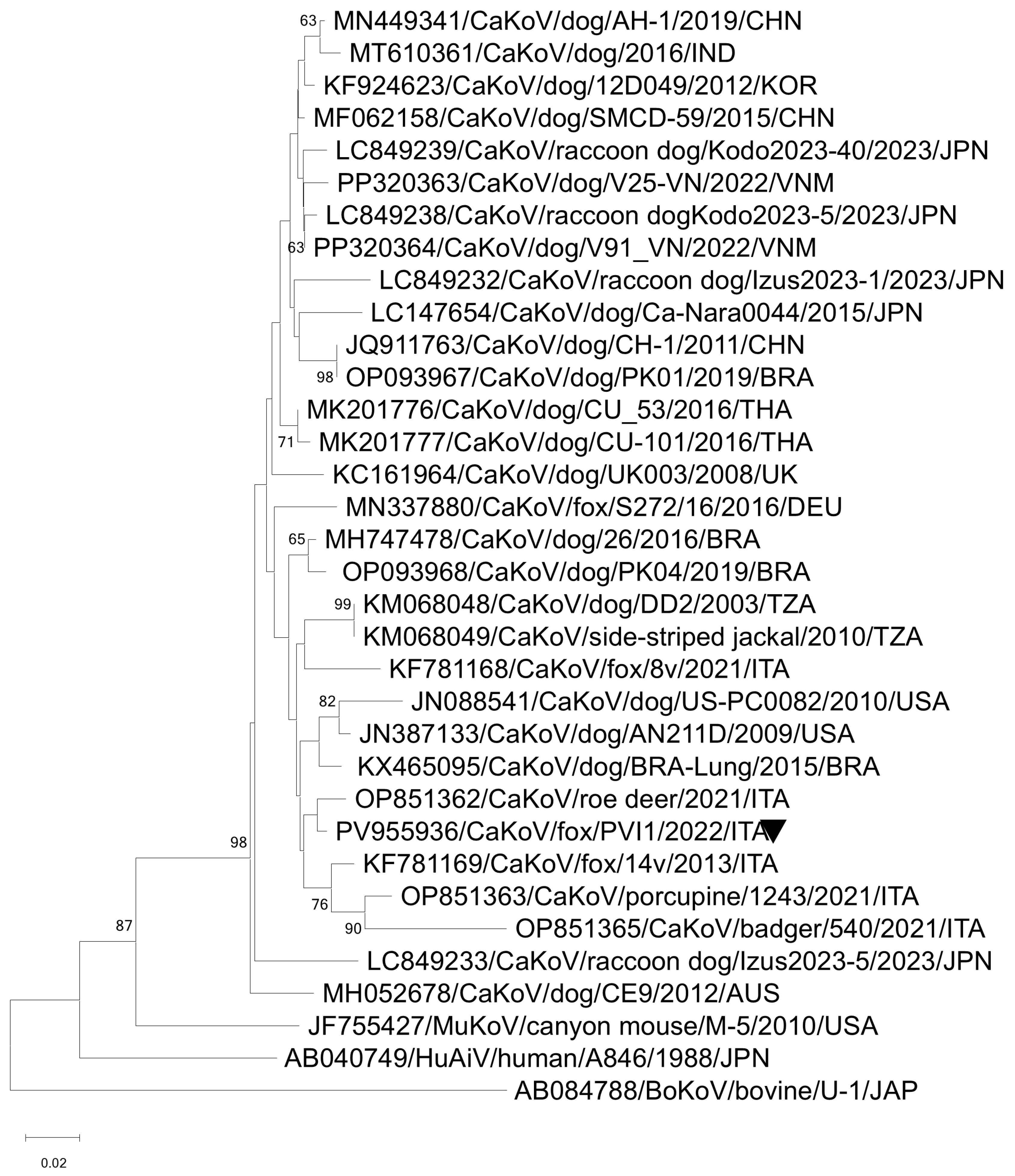
In the phylogenetic tree constructed from the complete nucleotide sequences of the fiber gene (PV955938), strain CAdV-1/VI9/ITA/2022 strictly clustered with other CAdV-1 strains identified in Italy, in a dog rectal swab [67], and in a wolf liver sample [30] (Figure 3).
In the fox pooled sample PVI1/2021/ITA, the SISPA/ONT strategy allowed the recovery of three contigs of 682, 1254, and 2823 nt in length that were assembled against the prototype strain (NC001918) of the species Kobuvirus aichi, genus Kobuvirus. The three contigs covered the partial VP0, VP1, 2B, 3D, and the complete VP3, 2C, 3A, 3B, and 3D coding regions. Upon sequence analysis, the obtained sequences showed 89.4–95.6% nt identities to the canine kobuvirus (CaKoV) sequences available in the database. In the phylogenetic tree, based on the nt sequence of the partial RdRp gene strain, CaKoV/fox/PVI1/2022/ITA was segregated within the species Kobuvirus aichi, falling into the Aichivirus A2 clades and grouped with CaKoVs previously identified in Italy in a roe deer, in a badger, and in a porcupine [59] (Figure 4).

4. Discussion

In the present study, we utilized a combination of consensus molecular diagnostics and third-generation sequencing technologies to explore the circulation of selected viral pathogens among wild carnivore populations in Northwestern Italy.
The presence of CPV-2 and FPV in free-ranging wild carnivores underscores the persistent threat that these viruses pose to wildlife health and conservation efforts. In our analyses, CPV-2 strains were identified in wolves, and an FPV strain was detected in a Eurasian badger, confirming that both viruses are present in ecosystems with multiple host species [4,68,69]. Previous studies have consistently indicated that wild canids and other carnivorous species are vulnerable to CPV-2/FPV infection [18,69], and our results support the idea that wild carnivores may not only act as spillover hosts but could also serve as supplementary reservoirs for these viruses [13,14,70]. The overall prevalence (2.9%) of CPV-2/FPV DNA detected in this survey was low, aligning with the sporadic occurrence of protoparvoviruses reported in other wildlife populations [5,71]. On sequence analysis of the VP2 gene, key amino acid markers in the viral capsids that are known to influence CPV host range and antigenicity were conserved, in particular, changes at the VP2 position 426 (Asn to Glu), defining the CPV-2c antigenic type, and at position 297 (Ser to Ala), characteristic of the CPV-new 2a lineage [46,72]. While the biological relevance of some capsid mutations remains unclear, sequencing and monitoring these changes in wild-host viruses is crucial [73,74]. In detail, strains CPV-2/Wolf/LI6/2021/ITA and CPV-2/Wolf/LI8/2021/ITA clustered with Italian CPV-new 2a strains identified in puppies illegally imported into the Friuli Venezia-Giulia region between 2018 and 2021 [22]. The strain CPV-2/Wolf/LI9/2022/ITA formed a cluster with the CPV-2c strains identified in dogs in Italy [22,51], in China (unpublished), in Nigeria [75], in Thailand in the brain of a dog [54], and in a wolf fecal sample in Italy [22]. The detection of a CPV-new 2a variant in wolves is particularly noteworthy, showcasing ongoing viral evolution that echoes patterns observed in domestic dog populations [76]. This result corroborates previous research indicating that novel CPV-2 variants can swiftly disseminate among canid populations, both domestic and wild, facilitated by shared habitats, overlapping prey ranges, and direct or indirect interactions among sympatric carnivores [22]. Furthermore, the high genetic similarity between our wolf CPV-2 isolates and those found in domestic dogs points to a persistent bidirectional flow of parvovirus strains between wild and domestic hosts [69,71].
Furthermore, the detection of an FPV strain in a badger underscores the extensive host range of the members of the Protoparvovirus carnivoran1 species. While FPV is traditionally associated with felids, it has been identified in various non-felid carnivores, including canids and mustelids [13,14]. Our discovery of FPV in a badger aligns with earlier reports, indicating that Eurasian badgers may serve as susceptible hosts and potential reservoirs for FPV [13,22,56]. Molecular analysis of the FPV/Badger/TI6/2022/ITA strain demonstrated high nucleotide identity with FPV strains circulating among diverse hosts globally, including domestic cats [46,77], as well as wild carnivores [15,22,24,50,56,57]. This suggests that the FPV strain present in the badger may have been acquired through shared ecological spaces between felids and other susceptible species [50]. Notably, the FPV VP2 sequence from the badger exhibited amino acid substitutions (101-Ile to Thr and 103-Val to Ala). While the functional impact of these changes is uncertain, their occurrence hints at adaptive variation as FPV jumps into new hosts, underscoring the genetic plasticity of FPV in wildlife [15,78]. Although the implications of these mutations remain uncertain, their occurrence emphasizes the genetic plasticity of FPV in adapting to new hosts [69,70]. The role of wild mustelids, such as badgers, as hosts for FPV merits further investigation, especially considering that badgers inhabit areas overlapping with domestic cat and wild canid territories, thereby potentially exposing them to a variety of parvoviruses in the environment [14,18,79].
We detected CAdV-1 DNA in an intestinal sample from a red fox. The detection of CAdV-1 in a fox is consistent with numerous studies indicating that red foxes serve as important reservoir hosts for this virus in wild populations. For instance, molecular surveys conducted in Italy and the UK have frequently identified CAdV-1 in free-ranging red foxes, with notable prevalence or seropositivity rates [21,29,33,58]. Conversely, wolves and other larger wild carnivores are less frequently reported to harbor CAdV-1, although cases have been documented [30,32]. In our survey, the negativity of other wild carnivores mirrors previous research in which CAdV-1 DNA was exclusively detected in foxes and not in sympatric wolf populations in southern Italy [33]. Sequence analysis of the fox strain (CAdV-1/Fox/VI9/2021/ITA) revealed the highest nucleotide identity to CAdV-1 strains previously isolated from domestic dogs and various European wild carnivores [29,30,31,32]. Phylogenetic analysis confirmed that our CAdV-1 strain clusters closely with other CAdV-1 isolates from both domestic dogs and wild carnivores, indicating that the virus circulating within fox populations is part of the globally distributed CAdV-1 lineage maintained through interspecies transmission [33]. The ongoing circulation of CAdV-1 among wild carnivores such as foxes has important implications for management. Although infections are often subclinical, these hosts can serve as reservoirs, facilitating spillover into other susceptible species, including unvaccinated domestic dogs or vulnerable wild canids [31,58]. Lethal outbreaks of CAdV-1 have been documented in species such as wolves [31] and bears [32], raising concerns that endemic CAdV-1 in reservoir hosts could pose a significant conservation threat. The absence of CAdV-2 in our samples aligns with prior studies that have sporadically detected this virus in wild carnivores. Occasional detections of CAdV-2 have been reported in foxes and wolves [21,35], but its role in wildlife epidemiology appears negligible.
The SISPA strategy, coupled with ONT metaviromic sequencing, facilitated the detection of CaCV in a wolf sample that also tested positive for CPV-2c. CaCV belongs to the family Circoviridae, genus Circovirus, species Circovirus canine [62]. CaCV was described for the first time in 2011 in six dog sera [63]. Since then, CaCV has been reported in dogs worldwide [64,80,81] and in wild carnivores, including arctic and red foxes [82,83,84], wolves and badgers [17,85,86]. CaCV has a worldwide distribution [64,80,87], although its ecology within wild animal populations remains poorly understood. Surveillance studies across Europe have identified the presence of CaCV DNA in arctic and red foxes [82,83,84], wolves, and badgers [17,85,86], as well as in other species such as jackals [88] and minks [89]. In particular, our strain is closely related to CaCV strains detected in spleen samples collected from wolves in Italy and in Canada [21,85], in liver samples from arctic foxes in Norway [82], spleen samples from foxes in Italy and Canada [85,88], and from serum samples of foxes with meningoencephalitis in the United Kingdom [83]. Prior investigations in Italy have reported the presence of CaCV DNA in wolf populations, with prevalence rates of 26.4% [86], 47.2% [17], and 50% [33], exhibiting significant regional variability. Such differences may reflect underlying ecological or sampling factors, including host density and exposure opportunities, influencing the circulation of CaCV in different geographical regions [21]. Nonetheless, our findings confirm that CaCV is capable of circulating among wild wolves in Northwestern Italy, thereby expanding the existing knowledge of its geographic distribution. In particular, CaCV DNA was identified in a wolf duodenal sample from Piemonte, an alpine region characterized by sympatric carnivore communities with possible overlap between wild and domestic canids. In a recent study, the presence of CaCV was documented in red foxes from Valle d’Aosta, one of the two regions involved in our investigation, demonstrating circulation in both alpine regions with strains clustered within the proposed Group 5 [65,66,88]. Although the pathogenic role of CaCV in free-ranging carnivores remains incomplete and most detections in wildlife occur in clinically non-evident animals, the virus has been documented in co-infections and associated with enteric or vascular syndromes in dogs [65,87]. This suggests a possible contribution of opportunistic transmission of disease under stressful conditions or exposure to multiple pathogens. Environmental transmission routes (e.g., fecal contamination of habitats or depletion of infected carcasses) are plausible in alpine ecosystems and may facilitate spread among sympatric carnivores.
We also identified CaKoV RNA in a red fox pooled from the Valle d’Aosta sample using the SISPA/ONT protocol. CaKoV is a member of the species Kobuvirus aichi in the genus Kobuvirus within the family Picornaviridae [90], first detected in dogs in the United States of America (USA) [91,92] and subsequently identified in domestic dogs worldwide [93,94,95,96,97]. CaKoV has also been reported in wild carnivore species, including foxes in Italy [98] and in Germany [99], wolves in Italy [100], golden jackal, side-striped jackal (Canis adustus), and spotted hyena (Crocuta crocuta) in Tanzania [101].
On sequence analysis, the CaKoV strain showed 94–98% nucleotide identity with strains identified in both domestic and wild hosts. Specifically, our strain exhibited close similarity to CaKoVs found in Italian roe deer and foxes, Arctic foxes in China, and domestic dogs in Asia [59,98,102,103]. Notably, CaKoV strains had previously been identified in red foxes and wolves within this geographic area [98,100], and our current findings confirm the circulation of this virus within the local wildlife population. Additionally, the high similarity to CaKoV strains of canine origin suggests that spillover from domestic dogs may serve as a potential source of infection for wild foxes, possibly via environmental contamination or scavenging of infected carcasses. Likewise, the detection of CaKoV in remote wildlife and even in non-canids, such as a roe deer and porcupine in the Toscana region, raises the possibility that CaKoV is maintained in nature through wildlife that can act as mechanical, passive carriers that could facilitate the introduction and spread of CaKoV in the environment [59]. The pathogenic significance of CaKoV in wild carnivores remains uncertain, as infections are often subclinical. The foxes included in this study were found deceased during passive surveillance, with no indication that CaKoV was the causative agent [93,94,100]. However, CaKoV could contribute to enteric disease under specific conditions or during co-infection with other enteropathogens, as suggested in domestic animals [92,104]. From a conservation and One Health perspective, the presence of CaKoV in wildlife underscores the complex microbial sharing between domestic and wild species. Including emerging pathogens such as CaKoV in wildlife, disease surveillance can enhance our understanding of their circulation and evolutionary dynamics.
Over the course of several decades, control programs have aimed to eradicate high-impact canine viruses through widespread vaccination, focusing also on CPV and CAdV-1. However, these pathogens persist within dog populations due to vaccination failures, the movement of infected animals from endemic regions, and their ongoing circulation in wildlife reservoirs [10,33,105]. Noteworthy, unvaccinated or poorly supervised dogs can serve as maintenance hosts and shed carnivore protoparvoviruses [105]. In Italian Alpine areas, field observations suggest that even pet dogs may contribute to viral persistence if they are not adequately vaccinated or allowed to roam freely [106]. Furthermore, phylogenetic and geographic data show frequent interactions between domestic and wild carnivores. FPV and CPV-2 strains have been found in wildlife, and regions with CPV-2-positive wild canids are more likely to see cases in owned dogs [13,22,105]. Similarly, European studies reveal that red foxes could carry CAdV-1 and may infect dogs, underscoring the bidirectional risk at the domestic–wildlife interface [29,107]. Collectively, these findings emphasise the need for targeted management strategies, including maintaining high vaccination coverage, restricting free-ranging behavior of local dogs, and managing feral cat colonies through population and vaccination efforts to curb transmission between wildlife and domestic animals.

5. Conclusions

Our findings provide molecular and phylogenetic evidence of the circulation of diverse viral pathogens, including CPV-2, FPV, CaCV, CAdV-1, and CaKoV, among wild carnivores in Northwestern Italy. Although the overall prevalence was low, these results emphasize the persistent risk of cross-species transmission and pathogen spillover between domestic animals and wildlife. Continued molecular surveillance and collaboration among veterinary, conservation, and public health sectors will improve our ability to detect and mitigate viral threats at the wildlife–domestic animal interface, thereby contributing to the preservation of biodiversity and animal health.

Supplementary Materials

The following supporting information can be downloaded at: https://www.mdpi.com/article/10.3390/vetsci12090814/s1, Figure S1: (a) Polymerase chain reaction (PCR) amplification for members of the species Protoparvovirus carnivoran1 [37]. Line M: BenchTop 1 kb DNA ladder; line 3, 4, 5, and 7: wolf positive samples corresponding to LI6, LI8, LI9, and TI6, respectively. (b) PCR amplification for CAdV-1/CAdV2 [38]. Line M: BenchTop 1 kb DNA ladder; line 4: fox positive sample (VI9).

Author Contributions

Conceptualization, V.S., F.D.P. and B.D.M.; methodology, V.S., F.D.P. and B.D.M.; software, V.S.; validation, B.D.M., F.M. and V.M.; formal analysis, V.S., F.D.P., B.V., F.P., R.O. and B.D.M.; investigation, V.S. and F.D.P.; resources, S.R. and B.D.M.; data curation, V.S., F.D.P., B.V., F.P., R.O. and B.D.M.; writing—original draft preparation, V.S. and B.D.M.; writing—review and editing, S.R., B.V., F.P., F.M. and V.M.; supervision, V.M. and B.D.M.; project administration, B.D.M.; funding acquisition, B.D.M. and R.O. All authors have read and agreed to the published version of the manuscript.

Funding

This research was funded partially by the Italian Ministry of University and Research (MUR) under the PRIN 2022 call (D.D. n. 104/2022), project code 2022EPP2TT, CUP C53D23000590006, “Investigating hepatotropic viruses in carnivores and humans in a One Health perspective (HVOH)” and partly by the Italian Ministry of Health by Research Project (IZS PLV 08/22 Ricerca Corrente) “Hepatotropic Virus Hunting: indagine virologica nella fauna selvatica in un contesto integrato di “One Health”.

Institutional Review Board Statement

Ethical approval was not required under Italian legislation (Legislative Decree No. 26 of 4 March 2014) because the materials analyzed were duodenal tissues obtained from wild-animal carcasses submitted to the National Reference Centre for Wild Animal Diseases (CeRMAS—IZS PLV) for routine diagnostic purposes; no procedures were performed on live animals.

Informed Consent Statement

Not applicable.

Data Availability Statement

Data supporting the results of this study are available from the corresponding author upon reasonable request. Sequence data have been deposited in GenBank and are openly accessible.

Acknowledgments

V.M. was supported by the National Laboratory for Infectious Animal Diseases, Antimicrobial Resistance, Veterinary Public Health and Food Chain Safety, RRF-2.3.1-21-2022-00001.

Conflicts of Interest

The authors declare no conflicts of interest.

References

  1. Roemer, G.W.; Gompper, M.E.; Van Valkenburgh, B. The Ecological Role of the Mammalian Mesocarnivore. BioScience 2009, 59, 165–173. [Google Scholar] [CrossRef]
  2. Ripple, W.J.; Estes, J.A.; Beschta, R.L.; Wilmers, C.C.; Ritchie, E.G.; Hebblewhite, M.; Berger, J.; Elmhagen, B.; Letnic, M.; Nelson, M.P.; et al. Status and Ecological Effects of the World’s Largest Carnivores. Science 2014, 343, 1241484. [Google Scholar] [CrossRef] [PubMed]
  3. Henle, K.; Davies, K.F.; Kleyer, M.; Margules, C.; Settele, J. Predictors of Species Sensitivity to Fragmentation. Biodivers. Conserv. 2004, 13, 207–251. [Google Scholar] [CrossRef]
  4. Daszak, P.; Cunningham, A.A.; Hyatt, A.D. Emerging Infectious Diseases of Wildlife—Threats to Biodiversity and Human Health. Science 2000, 287, 443–449. [Google Scholar] [CrossRef] [PubMed]
  5. Belsare, A.V.; Vanak, A.T.; Gompper, M.E. Epidemiology of Viral Pathogens of Free-Ranging Dogs and Indian Foxes in a Human-Dominated Landscape in Central India. Transbound. Emerg. Dis. 2014, 61, 78–86. [Google Scholar] [CrossRef]
  6. Murray, D.L.; Kapke, C.A.; Evermann, J.F.; Fuller, T.K. Infectious Disease and the Conservation of Free-Ranging Large Carnivores. Anim. Conserv. 1999, 2, 241–254. [Google Scholar] [CrossRef] [PubMed]
  7. Pedersen, A.B.; Jones, K.E.; Nunn, C.L.; Altizer, S. Infectious Diseases and Extinction Risk in Wild Mammals. Conserv. Biol. 2007, 21, 1269–1279. [Google Scholar] [CrossRef]
  8. Martins, N.B.; Neves de Almeida, J.C.; Gonçalves, M.S.S.; Gila, L.I.; Yogui, D.R.; Alves, M.H.; Desbiez, A.L.J.; Brandão, P.E.; da Hora, A.S. Occurrence of Typical Domestic Animal Viruses in Wild Carnivorans: An Emerging Threat to the Conservation of Endangered Species. Transbound. Emerg. Dis. 2024, 71, e3931047. [Google Scholar] [CrossRef]
  9. Pénzes, J.J.; Söderlund-Venermo, M.; Canut, M.; Eis-Hübinger, A.M.; Hughes, J.; Cotmore, S.F.; Harrach, B. Reorganizing the Family Parvoviridae: A Revised Taxonomy Independent of the Canonical Approach Based on Host Association. Arch. Virol. 2020, 165, 2133–2146. [Google Scholar] [CrossRef]
  10. Allison, A.B.; Harbison, C.E.; Pagan, I.; Stucker, K.M.; Kaelber, J.T.; Brown, J.D.; Ruder, M.G.; Keel, M.K.; Dubovi, E.J.; Holmes, E.C.; et al. Role of Multiple Hosts in the Cross-Species Transmission and Emergence of a Pandemic Parvovirus. J. Virol. 2012, 86, 865–872. [Google Scholar] [CrossRef]
  11. Canuti, M.; Fry, K.; Cluff, H.D.; Mira, F.; Fenton, H.; Lang, A.S. Co-Circulation of Five Species of Dog Parvoviruses and Canine Adenovirus Type 1 among Gray Wolves (Canis lupus) in Northern Canada. Transbound. Emerg. Dis. 2022, 69, e1407–e1433. [Google Scholar] [CrossRef]
  12. Kurucay, H.N.; Tamer, C.; Muftuoglu, B.; Elhag, A.E.; Gozel, S.; Cicek-Yildiz, Y.; Demirtas, S.; Ozan, E.; Albayrak, H.; Okur-Gumusova, S.; et al. First Isolation and Molecular Characterization of Canine Parvovirus Type 2b (CPV-2b) from Red Foxes (Vulpes vulpes) in Turkey. Virol. J. 2023, 20, 27. [Google Scholar] [CrossRef]
  13. Calatayud, O.; Esperón, F.; Velarde, R.; Oleaga, Á.; Llaneza, L.; Ribas, A.; Negre, N.; de la Torre, A.; Rodríguez, A.; Millán, J. Genetic Characterization of Carnivore Parvoviruses in Spanish Wildlife Reveals Domestic Dog and Cat-Related Sequences. Transbound. Emerg. Dis. 2020, 67, 626–634. [Google Scholar] [CrossRef]
  14. Ndiana, L.A.; Lanave, G.; Desario, C.; Berjaoui, S.; Alfano, F.; Puglia, I.; Fusco, G.; Colaianni, M.L.; Vincifori, G.; Camarda, A.; et al. Circulation of Diverse Protoparvoviruses in Wild Carnivores, Italy. Transbound. Emerg. Dis. 2021, 68, 2489–2502. [Google Scholar] [CrossRef] [PubMed]
  15. Diakoudi, G.; Lanave, G.; Berjaoui, S.; Desario, C.; Di Teodoro, G.; Vasinioti, V.I.; Pellegrini, F.; Defourny, S.V.P.; Salucci, S.; Cocco, A.; et al. Feline Panleukopenia Virus in a Marsican Brown Bear and Crested Porcupine, Italy, 2022-2023. Emerg. Infect. Dis. 2024, 30, 2655–2659. [Google Scholar] [CrossRef]
  16. Battilani, M.; Scagliarini, A.; Tisato, E.; Turilli, C.; Jacoboni, I.; Casadio, R.; Prosperi, S. Analysis of Canine Parvovirus Sequences from Wolves and Dogs Isolated in Italy. J. Gen. Virol. 2001, 82, 1555–1560. [Google Scholar] [CrossRef] [PubMed]
  17. Balboni, A.; Urbani, L.; Delogu, M.; Musto, C.; Fontana, M.C.; Merialdi, G.; Lucifora, G.; Terrusi, A.; Dondi, F.; Battilani, M. Integrated Use of Molecular Techniques to Detect and Genetically Characterise DNA Viruses in Italian Wolves (Canis lupus italicus). Animals 2021, 11, 2198. [Google Scholar] [CrossRef]
  18. Duarte, M.D.; Henriques, A.M.; Barros, S.C.; Fagulha, T.; Mendonça, P.; Carvalho, P.; Monteiro, M.; Fevereiro, M.; Basto, M.P.; Rosalino, L.M.; et al. Snapshot of Viral Infections in Wild Carnivores Reveals Ubiquity of Parvovirus and Susceptibility of Egyptian Mongoose to Feline Panleukopenia Virus. PLoS ONE 2013, 8, e59399. [Google Scholar] [CrossRef]
  19. Barlow, A.M.; Schock, A.; Bradshaw, J.; Mullineaux, E.; Dastjerdi, A.; Everest, D.J.; McGowan, S.; Steinbach, F.; Cowen, S. Parvovirus enteritis in Eurasian badgers (Meles meles). Vet. Rec. 2012, 170, 416. [Google Scholar] [CrossRef] [PubMed]
  20. Viscardi, M.; Santoro, M.; Clausi, M.T.; Cozzolino, L.; Decaro, N.; Colaianni, M.L.; Fusco, G. Molecular Detection and Characterization of Carnivore Parvoviruses in Free-Ranging Eurasian Otters (Lutra lutra) in Southern Italy. Transbound. Emerg. Dis. 2019, 66, 1864–1872. [Google Scholar] [CrossRef]
  21. Magliocca, M.; Taddei, R.; Urbani, L.; Bertasio, C.; Facile, V.; Gallina, L.; Sampieri, M.; Rugna, G.; Rubini, S.; Maioli, G.; et al. Molecular Detection of Viral and Bacterial Pathogens in Red Foxes (Vulpes vulpes) from Italy. Animals 2024, 14, 1969. [Google Scholar] [CrossRef]
  22. Leopardi, S.; Milani, A.; Cocchi, M.; Bregoli, M.; Schivo, A.; Leardini, S.; Festa, F.; Pastori, A.; De Zan, G.; Gobbo, F.; et al. Carnivore Protoparvovirus 1 (CPV-2 and FPV) Circulating in Wild Carnivores and in Puppies Illegally Imported into North-Eastern Italy. Viruses 2022, 14, 2612. [Google Scholar] [CrossRef] [PubMed]
  23. Wasieri, J.; Schmiedeknecht, G.; Förster, C.; König, M.; Reinacher, M. Parvovirus Infection in a Eurasian Lynx (Lynx lynx) and in a European Wildcat (Felis silvestris silvestris). J. Comp. Pathol. 2009, 140, 203–207. [Google Scholar] [CrossRef]
  24. Campoy, A.; Gomez-Lucia, E.; Garcia, T.; Crespo, E.; Olmeda, S.; Valcarcel, F.; Fandiño, S.; Domenech, A. First Description of a Carnivore Protoparvovirus Associated with a Clinical Case in the Iberian Lynx (Lynx pardinus). Animals 2025, 15, 1026. [Google Scholar] [CrossRef]
  25. Benkő, M.; Aoki, K.; Arnberg, N.; Davison, A.J.; Echavarría, M.; Hess, M.; Jones, M.S.; Kaján, G.L.; Kajon, A.E.; Mittal, S.K.; et al. ICTV Virus Taxonomy Profile: Adenoviridae 2022. J. Gen. Virol. 2022, 103, 001721. [Google Scholar] [CrossRef]
  26. Decaro, N.; Martella, V.; Buonavoglia, C. Canine Adenoviruses and Herpesvirus. Vet. Clin. N. Am. Small Anim. Pract. 2008, 38, 799–814. [Google Scholar] [CrossRef] [PubMed]
  27. Green, R.G.; Ziegler, N.R.; Breen, B.B. Epizootic Fox Encephalitis. I. General Description. Am. J. Epidemiol. 1930, 12, 109–129. [Google Scholar] [CrossRef]
  28. Junge, R.E.; Bauman, K.; King, M.; Gompper, M.E. A Serologic Assessment of Exposure to Viral Pathogens and Leptospira in an Urban Raccoon (Procyon lotor) Population Inhabiting a Large Zoological Park. J. Zoo Wildl. Med. 2007, 38, 18–26. [Google Scholar] [CrossRef]
  29. Balboni, A.; Verin, R.; Morandi, F.; Poli, A.; Prosperi, S.; Battilani, M. Molecular Epidemiology of Canine Adenovirus Type 1 and Type 2 in Free-Ranging Red Foxes (Vulpes vulpes) in Italy. Vet. Microbiol. 2013, 162, 551–557. [Google Scholar] [CrossRef]
  30. Pizzurro, F.; Marcacci, M.; Zaccaria, G.; Orsini, M.; Cito, F.; Rosamilia, A.; Di Renzo, L.; Malatesta, D.; Di Sabatino, D.; Lorusso, A. Genome Sequence of Canine Adenovirus Type 1 Isolated from a Wolf (Canis lupus) in Southern Italy. Genome Announc. 2017, 5, e00225-17. [Google Scholar] [CrossRef]
  31. Dowgier, G.; Lahoreau, J.; Lanave, G.; Losurdo, M.; Varello, K.; Lucente, M.S.; Ventriglia, G.; Bozzetta, E.; Martella, V.; Buonavoglia, C.; et al. Sequential Circulation of Canine Adenoviruses 1 and 2 in Captive Wild Carnivores, France. Vet. Microbiol. 2018, 221, 67–73. [Google Scholar] [CrossRef]
  32. García Marín, J.F.; Royo, L.J.; Oleaga, A.; Gayo, E.; Alarcia, O.; Pinto, D.; Martínez, I.Z.; González, P.; Balsera, R.; Marcos, J.L.; et al. Canine adenovirus type 1 (CAdV-1) in free-ranging European brown bear (Ursus arctos arctos): A threat for Cantabrian population? Transbound. Emerg. Dis. 2018, 65, 2049–2056. [Google Scholar] [CrossRef]
  33. Ndiana, L.A.; Lanave, G.; Vasinioti, V.; Desario, C.; Martino, C.; Colaianni, M.L.; Pellegrini, F.; Camarda, A.; Berjaoui, S.; Sgroi, G.; et al. Detection and Genetic Characterization of Canine Adenoviruses, Circoviruses, and Novel Cycloviruses from Wild Carnivores in Italy. Front. Vet. Sci. 2022, 9, 851987. [Google Scholar] [CrossRef] [PubMed]
  34. Oleaga, A.; Balseiro, A.; Espí, A.; Royo, L.J. Wolf (Canis lupus) as Canine Adenovirus Type 1 (CAdV-1) Sentinel for the Endangered Cantabrian Brown Bear (Ursus arctos arctos). Transbound. Emerg. Dis. 2022, 69, 516–523. [Google Scholar] [CrossRef] [PubMed]
  35. Di Francesco, C.E.; Smoglica, C.; Paoletti, B.; Angelucci, S.; Innocenti, M.; Antonucci, A.; Di Domenico, G.; Marsilio, F. Detection of Selected Pathogens in Apennine Wolf (Canis lupus italicus) by a Non-Invasive GPS-Based Telemetry Sampling of Two Packs from Majella National Park, Italy. Eur. J. Wildl. Res. 2019, 65, 84. [Google Scholar] [CrossRef]
  36. Kim, Y.J.; Lee, S.Y.; Kim, Y.S.; Na, E.J.; Park, J.S.; Oem, J.K. Genetic Characteristics of Canine Adenovirus Type 2 Detected in Wild Raccoon Dogs (Nyctereutes procyonoides) in Korea (2017–2020). Vet. Sci. 2022, 9, 591. [Google Scholar] [CrossRef]
  37. Buonavoglia, C.; Martella, V.; Pratelli, A.; Tempesta, M.; Cavalli, A.; Buonavoglia, D.; Bozzo, G.; Elia, G.; Decaro, N.; Carmichael, L.E. Evidence for Evolution of Canine Parvovirus Type 2 in Italy. J. Gen. Virol. 2001, 82, 3021–3025. [Google Scholar] [CrossRef] [PubMed]
  38. Hu, R.L.; Huang, G.; Qiu, W.; Zhong, Z.H.; Xia, X.Z.; Yin, Z. Detection and Differentiation of CAV-1 and CAV-2 by Polymerase Chain Reaction. Vet. Res. Commun. 2001, 25, 77–84. [Google Scholar] [CrossRef]
  39. Reyes, G.R.; Kim, J.P. Sequence-Independent Single-Primer Amplification (SISPA) of Complex DNA Populations. Mol. Cell Probes 1991, 5, 473–481. [Google Scholar] [CrossRef]
  40. Allander, T.; Tammi, M.T.; Eriksson, M.; Bjerkner, A.; Tiveljung-Lindell, A.; Andersson, B. Cloning of a Human Parvovirus by Molecular Screening of Respiratory Tract Samples. Proc. Natl. Acad. Sci. USA 2005, 102, 12891–12896. [Google Scholar] [CrossRef]
  41. Djikeng, A.; Halpin, R.; Kuzmickas, R.; Depasse, J.; Feldblyum, J.; Sengamalay, N.; Afonso, C.; Zhang, X.; Anderson, N.G.; Ghedin, E.; et al. Viral Genome Sequencing by Random Priming Methods. BMC Genom. 2008, 9, 5. [Google Scholar] [CrossRef]
  42. Vilsker, M.; Moosa, Y.; Nooij, S.; Fonseca, V.; Ghysens, Y.; Dumon, K.; Pauwels, R.; Alcantara, L.C.; Vanden Eynden, E.; Vandamme, A.-M.; et al. Genome Detective: An Automated System for Virus Identification from High-Throughput Sequencing Data. Bioinformatics 2019, 35, 871–873. [Google Scholar] [CrossRef]
  43. Tamura, K.; Stecher, G.; Kumar, S. MEGA11: Molecular Evolutionary Genetics Analysis Version 11. Mol. Biol. Evol. 2021, 38, 3022–3027. [Google Scholar] [CrossRef]
  44. Canuti, M.; Britton, A.P.; Graham, S.M.; Lang, A.S. Epidemiology and Molecular Characterization of Protoparvoviruses Infecting Wild Raccoons (Procyon lotor) in British Columbia, Canada. Virus Res. 2017, 242, 85–89. [Google Scholar] [CrossRef]
  45. Canuti, M.; McDonald, E.; Graham, S.M.; Rodrigues, B.; Bouchard, É.; Neville, R.; Pitcher, M.; Whitney, H.G.; Marshall, H.D.; Lang, A.S. Multi-Host Dispersal of Known and Novel Carnivore Amdoparvoviruses. Virus Evol. 2020, 6, veaa072. [Google Scholar] [CrossRef]
  46. Mira, F.; Canuti, M.; Purpari, G.; Cannella, V.; Di Bella, S.; Occhiogrosso, L.; Schirò, G.; Chiaramonte, G.; Barreca, S.; Pisano, P.; et al. Molecular Characterization and Evolutionary Analyses of Carnivore Protoparvovirus1 NS1 Gene. Viruses 2019, 11, 308. [Google Scholar] [CrossRef] [PubMed]
  47. Carrino, M.; Tassoni, L.; Campalto, M.; Cavicchio, L.; Mion, M.; Corrò, M.; Natale, A.; Beato, M.S. Molecular Investigation of Recent Canine Parvovirus-2 (CPV-2) in Italy Revealed Distinct Clustering. Viruses 2022, 14, 917. [Google Scholar] [CrossRef] [PubMed]
  48. Demeter, Z.; Palade, E.A.; Soós, T.; Farsang, A.; Jakab, C.; Rusvai, M. Misleading Results of the MboII-Based Identification of Type 2a Canine Parvovirus Strains from Hungary Reacting as Type 2c Strains. Virus Genes 2010, 41, 37–42. [Google Scholar] [CrossRef] [PubMed]
  49. Ohneiser, S.A.; Hills, S.F.; Cave, N.J.; Passmore, D.; Dunowska, M. Canine Parvoviruses in New Zealand Form a Monophyletic Group Distinct from the Viruses Circulating in Other Parts of the World. Vet. Microbiol. 2015, 178, 190–200. [Google Scholar] [CrossRef]
  50. Zhao, M.; Yue, C.; Yang, Z.; Li, Y.; Zhang, D.; Zhang, J.; Yang, S.; Shen, Q.; Su, X.; Qi, D.; et al. Viral Metagenomics Unveiled Extensive Communication of Viruses within Giant Pandas and Their Associated Organisms in the Same Ecosystem. Sci. Total Environ. 2022, 820, 153317. [Google Scholar] [CrossRef]
  51. Mira, F.; Purpari, G.; Lorusso, E.; Di Bella, S.; Gucciardi, F.; Desario, C.; Macaluso, G.; Decaro, N.; Guercio, A. Introduction of Asian Canine Parvovirus in Europe through Dog Importation. Transbound. Emerg. Dis. 2018, 65, 16–21. [Google Scholar] [CrossRef]
  52. Alam, S.; Chowdhury, Q.M.M.K.; Roy, S.; Chowdhury, M.S.R.; Hasan, M.; Mamun, M.A.; Uddin, M.B.; Hossain, M.M.; Rahman, M.M.; Rahman, M.M. Molecular Detection and Phylogenetic Analysis of Canine Parvovirus (CPV) in Diarrhoeic Pet Dogs in Bangladesh. Vet. Anim. Sci. 2021, 14, 100224. [Google Scholar] [CrossRef]
  53. Zhuang, Q.Y.; Qiu, Y.; Pan, Z.H.; Wang, S.C.; Wang, B.; Wu, W.K.; Yu, J.M.; Yi, Y.; Sun, F.L.; Wang, K.C. Genome Sequence Characterization of Canine Parvoviruses Prevalent in the Sichuan Province of China. Transbound. Emerg. Dis. 2019, 66, 897–907. [Google Scholar] [CrossRef]
  54. Wardhani, S.W.; Wongsakul, B.; Kasantikul, T.; Piewbang, C.; Techangamsuwan, S. Molecular and Pathological Investigations of Selected Viral Neuropathogens in Rabies-Negative Brains of Cats and Dogs Revealed Neurotropism of Carnivore Protoparvovirus1. Front. Vet. Sci. 2021, 8, 710701. [Google Scholar] [CrossRef]
  55. Kapiya, J.; Nalubamba, K.S.; Kaimoyo, E.; Changula, K.; Chidumayo, N.; Saasa, N.; Simuunza, M.C.; Takada, A.; Mweene, A.S.; Chitanga, S.; et al. First Genetic Detection and Characterization of Canine Parvovirus from Diarrheic Dogs in Zambia. Arch. Virol. 2019, 164, 303–307. [Google Scholar] [CrossRef]
  56. Yang, Y.; Geng, Y.; Peng, Q.; Li, Y.; Guo, H.; Deng, H.; Hou, R.; Lai, W.; Zhang, D.; Liu, S. Identification of a Feline Panleukopenia Virus from Captive Giant Pandas (Ailuropoda melanoleuca) and Its Phylogenetic Analysis. Transbound. Emerg. Dis. 2023, 70, e1486–e1491. [Google Scholar] [CrossRef]
  57. Wang, X.; Li, T.; Liu, H.; Du, J.; Zhou, F.; Dong, Y.; He, X.; Li, Y.; Wang, C. Recombinant Feline Parvovirus Infection of Immunized Tigers in Central China. Emerg. Microbes Infect. 2017, 6, e42. [Google Scholar] [CrossRef]
  58. Walker, D.; Fee, S.A.; Hartley, G.; Learmount, J.; O’Hagan, M.J.; Meredith, A.L.; Bronsvoort, B.M.D.C.; Porphyre, T.; Sharp, C.P.; Philbey, A.W. Serological and Molecular Epidemiology of Canine Adenovirus Type 1 in Red Foxes (Vulpes vulpes) in the United Kingdom. Sci. Rep. 2016, 6, 36051. [Google Scholar] [CrossRef]
  59. Pacini, M.I.; Mazzei, M.; Sgorbini, M.; D’Alfonso, R.; Papini, R.A. A One-Year Retrospective Analysis of Viral and Parasitological Agents in Wildlife Animals Admitted to a First Aid Hospital. Animals 2023, 13, 931. [Google Scholar] [CrossRef]
  60. Wong, M.; Woolford, L.; Hasan, N.H.; Hemmatzadeh, F. A Novel Recombinant Canine Adenovirus Type 1 Detected from Acute Lethal Cases of Infectious Canine Hepatitis. Viral Immunol. 2017, 30, 258–263. [Google Scholar] [CrossRef]
  61. Mira, F.; Puleio, R.; Schirò, G.; Condorelli, L.; Di Bella, S.; Chiaramonte, G.; Purpari, G.; Cannella, V.; Balboni, A.; Randazzo, V.; et al. Study on the Canine Adenovirus Type 1 (CAdV-1) Infection in Domestic Dogs in Southern Italy. Pathogens 2022, 11, 1254. [Google Scholar] [CrossRef] [PubMed]
  62. Breitbart, M.; Delwart, E.; Rosario, K.; Segalés, J.; Varsani, A.; Ictv Report Consortium. ICTV Virus Taxonomy Profile: Circoviridae. J. Gen. Virol. 2017, 98, 1997–1998. [Google Scholar] [CrossRef]
  63. Kapoor, A.; Dubovi, E.J.; Henriquez-Rivera, J.A.; Lipkin, W.I. Complete Genome Sequence of the First Canine Circovirus. J. Virol. 2012, 86, 7018. [Google Scholar] [CrossRef]
  64. Li, L.; McGraw, S.; Zhu, K.; Leutenegger, C.M.; Marks, S.L.; Kubiski, S.; Gaffney, P.; Dela Cruz, F.N., Jr.; Wang, C.; Delwart, E.; et al. Circovirus in Tissues of Dogs with Vasculitis and Hemorrhage. Emerg. Infect. Dis. 2013, 19, 534–541. [Google Scholar] [CrossRef]
  65. Niu, L.; Wang, Z.; Zhao, L.; Wang, Y.; Cui, X.; Shi, Y.; Chen, H.; Ge, J. Detection and Molecular Characterization of Canine Circovirus Circulating in Northeastern China during 2014-2016. Arch. Virol. 2020, 165, 137–143. [Google Scholar] [CrossRef]
  66. Beikpour, F.; Ndiana, L.A.; Sazmand, A.; Capozza, P.; Nemati, F.; Pellegrini, F.; Zafari, S.; Zolhavarieh, S.M.; Cardone, R.; Faraji, R.; et al. Detection and Genomic Characterization of Canine Circovirus in Iran. Animals 2022, 12, 507. [Google Scholar] [CrossRef]
  67. Balboni, A.; Dondi, F.; Agnoli, C.; Verin, R.; Gruarin, M.; Morini, M.; Battilani, M. Novel Sequence Variants of Viral Hexon and Fibre Genes in Two Dogs with Canine Adenovirus Type 1-Associated Disease. Vet. J. 2017, 223, 73–75. [Google Scholar] [CrossRef]
  68. Steinel, A.; Parrish, C.R.; Bloom, M.E.; Truyen, U. Parvovirus Infections in Wild Carnivores. J. Wildl. Dis. 2001, 37, 594–607. [Google Scholar] [CrossRef]
  69. Allison, A.B.; Kohler, D.J.; Fox, K.A.; Brown, J.D.; Gerhold, R.W.; Dubovi, E.J.; Parrish, C.R.; Holmes, E.C. Frequent Cross-Species Transmission of Parvoviruses among Diverse Carnivore Hosts. J. Virol. 2013, 87, 2342–2347. [Google Scholar] [CrossRef] [PubMed]
  70. Truyen, U. Emergence and Recent Evolution of Canine Parvovirus. Vet. Microbiol. 1999, 69, 47–50. [Google Scholar] [CrossRef] [PubMed]
  71. Miranda, C.; Santos, N.; Parrish, C.R.; Thompson, G. Genetic Characterization of Canine Parvovirus in Sympatric Free-Ranging Wild Carnivores in Portugal. J. Wildl. Dis. 2017, 53, 824–831. [Google Scholar] [CrossRef] [PubMed]
  72. Zhou, P.; Zeng, W.; Zhang, X.; Li, S. The Genetic Evolution of Canine Parvovirus—A New Perspective. PLoS ONE 2017, 12, e0175035. [Google Scholar] [CrossRef]
  73. Martella, V.; Decaro, N.; Elia, G.; Buonavoglia, C. Surveillance Activity for Canine Parvovirus in Italy. J. Vet. Med. B 2005, 52, 312–315. [Google Scholar] [CrossRef]
  74. Jager, M.C.; Tomlinson, J.E.; Lopez-Astacio, R.A.; Parrish, C.R.; Van de Walle, G.R. Small but Mighty: Old and New Parvoviruses of Veterinary Significance. Virol. J. 2021, 18, 210. [Google Scholar] [CrossRef]
  75. Ogbu, K.I.; Chukwudi, I.C.; Mira, F.; Eze, U.U.; Di Bella, S.; Olaolu, O.S.; Tion, M.T.; Purpari, G.; Cannella, V.; Nwosuh, I.C.; et al. Current Status and Risk Factors of Canine Parvovirus Type 2 in North Central Nigeria. Comp. Immunol. Microbiol. Infect. Dis. 2021, 74, 101578. [Google Scholar] [CrossRef]
  76. Decaro, N.; Buonavoglia, C. Canine Parvovirus: A Review of Epidemiological and Diagnostic Aspects, with Emphasis on Type 2c. Vet. Microbiol. 2012, 155, 1–12. [Google Scholar] [CrossRef]
  77. Li, Z.; Cai, J.; Feng, C.; Wang, Y.; Fang, S.; Xue, X. Two Novel Sites Determine Genetic Relationships Between CPV-2 and FPV: An Epidemiological Survey of Canine and Feline Parvoviruses in Changchun, China (2020). Front. Vet. Sci. 2024, 11, 1444984. [Google Scholar] [CrossRef]
  78. Callaway, H.M.; Welsch, K.; Weichert, W.; Allison, A.B.; Hafenstein, S.L.; Huang, K.; Iketani, S.; Parrish, C.R. Complex and Dynamic Interactions between Parvovirus Capsids, Transferrin Receptors, and Antibodies Control Cell Infection and Host Range. J. Virol. 2018, 92, e00460-18. [Google Scholar] [CrossRef]
  79. Kim, Y.J.; Yoon, S.-W.; Jang, J.H.; Jeong, D.G.; Lee, B.J.; Kim, H.K. Genetic Characterization of Feline Parvovirus Isolate Fe-P2 in Korean Cat and Serological Evidence on Its Infection in Wild Leopard Cat and Asian Badger. Front. Vet. Sci. 2021, 8, 650866. [Google Scholar] [CrossRef] [PubMed]
  80. Anderson, A.; Hartmann, K.; Leutenegger, C.M.; Proksch, A.L.; Mueller, R.S.; Unterer, S. Role of canine circovirus in dogs with acute haemorrhagic diarrhoea. Vet. Rec. 2017, 180, 542. [Google Scholar] [CrossRef] [PubMed]
  81. Dowgier, G.; Lorusso, E.; Decaro, N.; Desario, C.; Mari, V.; Lucente, M.S.; Lanave, G.; Buonavoglia, C.; Elia, G. A molecular survey for selected viral enteropathogens revealed a limited role of Canine circovirus in the development of canine acute gastroenteritis. Vet. Microbiol. 2017, 204, 54–58. [Google Scholar] [CrossRef]
  82. Urbani, L.; Tryland, M.; Ehrich, D.; Fuglei, E.; Battilani, M.; Balboni, A. Ancient Origin and Genetic Segregation of Canine Circovirus Infecting Arctic Foxes (Vulpes lagopus) in Svalbard and Red Foxes (Vulpes vulpes) in Northern Norway. Transbound. Emerg. Dis. 2021, 68, 1283–1293. [Google Scholar] [CrossRef]
  83. Bexton, S.; Wiersma, L.C.; Getu, S.; van Run, P.R.; Verjans, G.M.; Schipper, D.; Schapendonk, C.M.; Bodewes, R.; Oldroyd, L.; Haagmans, B.L.; et al. Detection of Circovirus in Foxes with Meningoencephalitis, United Kingdom, 2009-2013. Emerg. Infect Dis. 2015, 21, 1205–1208. [Google Scholar] [CrossRef]
  84. De Arcangeli, S.; Balboni, A.; Kaehler, E.; Urbani, L.; Verin, R.; Battilani, M. Genomic Characterization of Canine Circovirus Detected in Red Foxes (Vulpes vulpes) from Italy using a New Real-Time PCR Assay. J. Wildl. Dis. 2020, 56, 239–242. [Google Scholar] [CrossRef] [PubMed]
  85. Canuti, M.; King, A.V.L.; Franzo, G.; Cluff, H.D.; Larsen, L.E.; Fenton, H.; Dufour, S.C.; Lang, A.S. Diverse Fox Circovirus (Circovirus canine) Variants Circulate at High Prevalence in Grey Wolves (Canis lupus) from the Northwest Territories, Canada. Peer Community J. 2024, 4, e73. [Google Scholar] [CrossRef]
  86. Zaccaria, G.; Malatesta, D.; Scipioni, G.; Di Felice, E.; Campolo, M.; Casaccia, C.; Savini, G.; Di Sabatino, D.; Lorusso, A. Circovirus in Domestic and Wild Carnivores: An important Opportunistic Agent? Virology 2016, 490, 69–74. [Google Scholar] [CrossRef]
  87. Thaiwong, T.; Wise, A.G.; Maes, R.K.; Mullaney, T.; Kiupel, M. Canine Circovirus 1 (CaCV-1) and Canine Parvovirus 2 (CPV-2): Recurrent Dual Infections in a Papillon Breeding Colony. Vet. Pathol. 2016, 53, 1204–1209. [Google Scholar] [CrossRef] [PubMed]
  88. Franzo, G.; Menandro, M.L.; Tucciarone, C.M.; Barbierato, G.; Crovato, L.; Mondin, A.; Libanora, M.; Obber, F.; Orusa, R.; Robetto, S.; et al. Canine Circovirus in Foxes from Northern Italy: Where Did It All Begin? Pathogens 2021, 10, 1002. [Google Scholar] [CrossRef]
  89. de Villiers, L.; Molini, U.; Coetzee, L.M.; Visser, L.; Spangenberg, J.; de Villiers, M.; Berjaoui, S.; Khaiseb, S.; Lorusso, A.; Franzo, G. Molecular Epidemiology of Canine Circovirus in Domestic Dogs and Wildlife in Namibia, Africa. Infect. Genet. Evol. 2023, 112, 105458. [Google Scholar] [CrossRef]
  90. Zell, R.; Delwart, E.; Gorbalenya, A.E.; Hovi, T.; King, A.M.Q.; Knowles, N.J.; Lindberg, A.M.; Pallansch, M.A.; Palmenberg, A.C.; Reuter, G.; et al. ICTV Virus Taxonomy Profile: Picornaviridae. J. Gen. Virol. 2017, 98, 2421–2422. [Google Scholar] [CrossRef]
  91. Kapoor, A.; Simmonds, P.; Dubovi, E.J.; Qaisar, N.; Henriquez, J.A.; Medina, J.; Shields, S.; Lipkin, W.I. Characterization of a Canine Homolog of Human Aichivirus. J. Virol. 2011, 85, 11520–11525. [Google Scholar] [CrossRef]
  92. Li, L.; Pesavento, P.A.; Shan, T.; Leutenegger, C.M.; Wang, C.; Delwart, E. Viruses in Diarrhoeic Dogs Include Novel Kobuviruses and Sapoviruses. J. Gen. Virol. 2011, 92, 2534–2541. [Google Scholar] [CrossRef]
  93. Carmona-Vicente, N.; Buesa, J.; Brown, P.A.; Merga, J.Y.; Darby, A.C.; Stavisky, J.; Sadler, L.; Gaskell, R.M.; Dawson, S.; Radford, A.D. Phylogeny and Prevalence of Kobuviruses in Dogs and Cats in the UK. Vet. Microbiol. 2013, 164, 246–252. [Google Scholar] [CrossRef]
  94. Di Martino, B.; Di Felice, E.; Ceci, C.; Di Profio, F.; Marsilio, F. Canine Kobuviruses in Diarrhoeic Dogs in Italy. Vet. Microbiol. 2013, 166, 246–249. [Google Scholar] [CrossRef] [PubMed]
  95. Oem, J.K.; Choi, J.W.; Lee, M.H.; Lee, K.K.; Choi, K.S. Canine Kobuvirus Infections in Korean Dogs. Arch. Virol. 2014, 159, 2751–2755. [Google Scholar] [CrossRef] [PubMed]
  96. Miyabe, F.M.; Ribeiro, J.; Alfieri, A.F.; Alfieri, A.A. Detection of Canine Kobuvirus RNA in Diarrheic Fecal Samples of Dogs with Parvoviruses. Braz. J. Microbiol. 2019, 50, 871–874. [Google Scholar] [CrossRef]
  97. Wang, Y.; Cui, Y.; Li, Y.; Wang, X.; Yang, K.; Zhang, D.; Zhao, L.; Bai, C.; Jiang, S.; Li, Y. Identification and Full-Genome Sequencing of Canine Kobuvirus in Canine Fecal Samples Collected from Anhui Province, Eastern China. Arch. Virol. 2020, 165, 2495–2501. [Google Scholar] [CrossRef] [PubMed]
  98. Di Martino, B.; Di Profio, F.; Melegari, I.; Robetto, S.; Di Felice, E.; Orusa, R.; Marsilio, F. Molecular Evidence of Kobuviruses in Free-Ranging Red Foxes (Vulpes vulpes). Arch. Virol. 2014, 159, 1803–1806. [Google Scholar] [CrossRef]
  99. Kaiser, F.K.; van Dyck, L.; Jo, W.K.; Schreiner, T.; Pfankuche, V.M.; Wohlsein, P.; Baumann, I.; Peters, M.; Baumgärtner, W.; Osterhaus, A.D.M.E.; et al. Detection of Systemic Canine Kobuvirus Infection in Peripheral Tissues and the Central Nervous System of a Fox Infected with Canine Distemper Virus. Microorganisms 2021, 9, 2521. [Google Scholar] [CrossRef]
  100. Melegari, I.; Sarchese, V.; Di Profio, F.; Robetto, S.; Carella, E.; Bermudez Sanchez, S.; Orusa, R.; Martella, V.; Marsilio, F.; Di Martino, B. First Molecular Identification of Kobuviruses in Wolves (Canis lupus) in Italy. Arch. Virol. 2018, 163, 509–513. [Google Scholar] [CrossRef]
  101. Olarte-Castillo, X.A.; Heeger, F.; Mazzoni, C.J.; Greenwood, A.D.; Fyumagwa, R.; Moehlman, P.D.; Hofer, H.; East, M.L. Molecular Characterization of Canine Kobuvirus in Wild Carnivores and the Domestic Dog in Africa. Virology 2015, 477, 89–97. [Google Scholar] [CrossRef] [PubMed]
  102. He, W.T.; Hou, X.; Zhao, J.; Sun, J.; He, H.; Si, W.; Wang, J.; Jiang, Z.; Yan, Z.; Xing, G.; et al. Virome Characterization of Game Animals in China Reveals a Spectrum of Emerging Pathogens. Cell 2022, 185, 1117–1129.e8. [Google Scholar] [CrossRef] [PubMed]
  103. Van Nguyen, T.; Kasantikul, T.; Piewbang, C.; Techangamsuwan, S. Evolutionary Dynamics of Canine Kobuvirus in Vietnam and Thailand Reveal the Evidence of Viral Ability to Evade Host Immunity. Sci. Rep. 2024, 14, 12037. [Google Scholar] [CrossRef] [PubMed]
  104. de Deus, D.R.; Siqueira, J.A.M.; Maués, M.A.C.; de Fátima Mesquita de Figueiredo, M.J.; Júnior, E.C.S.; da Silva Bandeira, R.; da Costa Pinheiro, K.; Teixeira, D.M.; da Silva, L.D.; de Fátima Dos Santos Guerra, S.; et al. Analysis of Viral Diversity in Dogs With Acute Gastroenteritis From Brazilian Amazon. Infect. Genet. Evol. 2024, 123, 105637. [Google Scholar] [CrossRef]
  105. Decaro, N.; Buonavoglia, C.; Barrs, V.R. Canine Parvovirus Vaccination and Immunisation Failures: Are We Far from Disease Eradication? Vet. Microbiol. 2020, 247, 108760. [Google Scholar] [CrossRef]
  106. Costanzi, L.; Brambilla, A.; Di Blasio, A.; Dondo, A.; Goria, M.; Masoero, L.; Gennero, M.S.; Bassano, B. Beware of dogs! Domestic animals as a threat for wildlife conservation in Alpine protected areas. Eur. J. Wildl. Res. 2021, 67, 70. [Google Scholar] [CrossRef]
  107. Verin, R.; Forzan, M.; Schulze, C.; Rocchigiani, G.; Balboni, A.; Poli, A.; Mazzei, M. Multicentric Molecular and Pathologic Study On Canine Adenovirus Type 1 in Red Foxes (Vulpes vulpes) in Three European Countries. J. Wildl. Dis. 2019, 55, 935–939. [Google Scholar] [CrossRef]
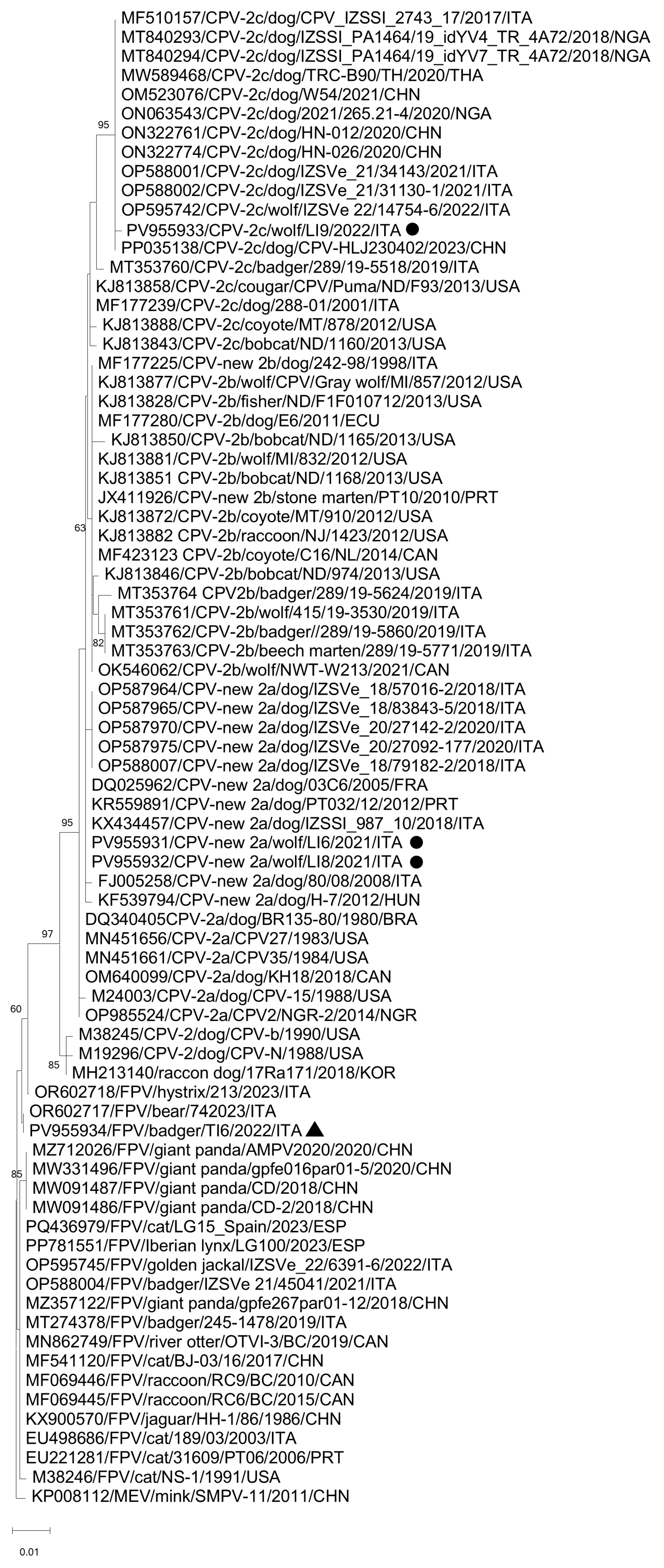
Figure 1. Phylogenetic tree based on the complete aa sequence of the VP2 gene of the CPV-2 and FPV strains detected in this study. A selection of cognate sequences of the species Protoparvovirus carnivoran1 was retrieved from GenBank. Mink enteritis virus (SMPV-11) (GenBank accession n. KP008112) was used as an outgroup. The tree was generated using the neighbour-joining method and Jones-Taylor-Thornton substitution model, supplying statistical support with bootstrapping of 1000 replicates. Bootstrap values > 60% are shown. Black triangles and black circles indicate the FPV and CPV-2 strains, respectively, identified in this survey.
Vetsci 12 00814 g001
Figure 2. Unrooted phylogenetic tree was constructed based on the complete genome nucleotide sequences of CaCV strain obtained in this study and cognate strains retrieved from the GenBank database. The phylogeny was inferred using the Maximum Likelihood method under the General Time Reversible (GTR) model with gamma distribution and invariable sites. To assess the robustness of the tree, bootstrap analysis was performed with 1000 replicates, and bootstrap values ≥ 70% are indicated on the tree. The scale bar represents the estimated number of nucleotide substitutions. Black rhombus indicates the CaCV strain identified in this study. To the right of the figure, the groups evidenced in this study are indicated and correspond to proposed clusters [65,66].
Vetsci 12 00814 g002
Figure 3. Phylogenetic analysis of CAdV-1/VI9/ITA/2022 strain. The tree was generated using the neighbor-joining method and the p-distance model, supplying statistical support with bootstrapping of 1000 replicates, and bootstrap values ≥ 70% are indicated on the tree. Black square indicates the CAdV-1 strain identified in this study. BtAdV (accession no. NC031948) was used as an outgroup.
Vetsci 12 00814 g003
Figure 4. Neighbor-joining phylogenetic tree based on the nt sequence of the partial RdRp gene of the CaKoV strain detected in this study. The tree was generated using the neighbor-joining method and p-distance correction, with statistical support provided by bootstrapping with 1000 replicates. The scale bar indicates nucleotide substitutions per site. The marker indicates the CaKoV sequence detected in this study.
Vetsci 12 00814 g004
Table 1. Viral pathogens detected in wild carnivore species assessed in this study with molecular investigations.
Animal Species
Tested
No. of
Animals
Virus %
(Positive/Total)
Total %
(Positive/Total)
FPV/CPV-2CAdV-1/CAdV-2
Wolf (Canis lupus)476.4% (3/47)0% (0/47)6.4% (3/47)
Fox (Vulpes vulpes)550% (0/55)1.8% (1/55)1.8% (1/55)
Stone marten
(Martes foina)
150% (0/15)0% (0/15)0.0% (0/15)
Eurasian badger
(Meles meles)
234.3% (1/23)0% (0/23)4.3% (1/23)
Total %
(Positive/Total)
1402.9% (4/140)0.7% (1/140)3.6% (5/140)
Table 2. Statistics of ONT sequencing data.
Strain IDVirus
Species
Virus
Name
Reads
Assembled
Total
Reads
No. of ContigsCoverage Length (nt)Reference
Sequence
CPV-2/Wolf/LI6/2021/ITAProtoparvovirus carnivoran1CPV-2a131,239188,29114492NC001539
CPV-2/Wolf/LI8/2021/ITACPV-2a489,633886,74014492NC001539
CPV-2/Wolf/LI9/2022/ITACPV-2c831,2871,191,65214492NC001539
FPV/Badger/TI6/2022/ITAFPV344,382399,96214591NC001539
CaCV/Wolf/LI9/2022/ITACircovirus
canine
CaCV5881,191,65212063NC020904
CaKoV/Fox/PVI1/2021/ITAKobuvirus aichiCaKoV263092,17834759NC001918
CAdV-1/Fox/VI9/2021/ITAMastadenovirus canidaeCAdV-1735657,335817,320NC001734
Disclaimer/Publisher’s Note: The statements, opinions and data contained in all publications are solely those of the individual author(s) and contributor(s) and not of MDPI and/or the editor(s). MDPI and/or the editor(s) disclaim responsibility for any injury to people or property resulting from any ideas, methods, instructions or products referred to in the content.