Morphological Evaluation of Transscleral Laser Retinopexy in Rabbits: Comparison of Optical Coherence Tomography and Histologic Examinations



1
Centre Hospitalier Universitaire Vétérinaire, Département de Sciences Cliniques, Faculté de Médecine Vétérinaire, Université de Montréal, 3200 Rue Sicotte, Saint-Hyacinthe, QC J2S 2M2, Canada
2
Faculté de Médecine Vétérinaire, Université de Montréal, 3200 Rue Sicotte, Saint-Hyacinthe, QC J2S 2M2, Canada
3
Groupe de Recherche sur les Maladies Infectieuses en Production Animale (GREMIP), Département de Pathologie et Microbiologie, Faculté de Médecine Vétérinaire, Université de Montréal, 3200 Rue Sicotte, Saint-Hyacinthe, QC J2S 2M2, Canada
*
Author to whom correspondence should be addressed.
Vet. Sci. 2023, 10(9), 535; https://doi.org/10.3390/vetsci10090535
Submission received: 20 June 2023
/
Revised: 2 August 2023
/
Accepted: 15 August 2023
/
Published: 23 August 2023
(This article belongs to the Special Issue Spotlight on Ophthalmologic Pathology in Animals)
Simple Summary
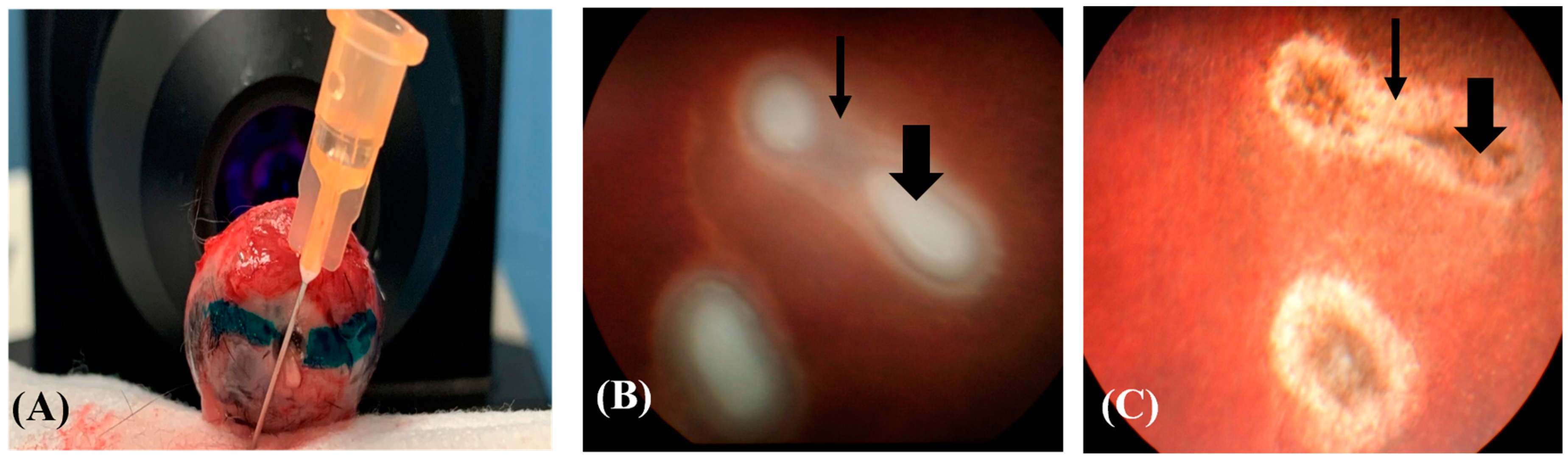
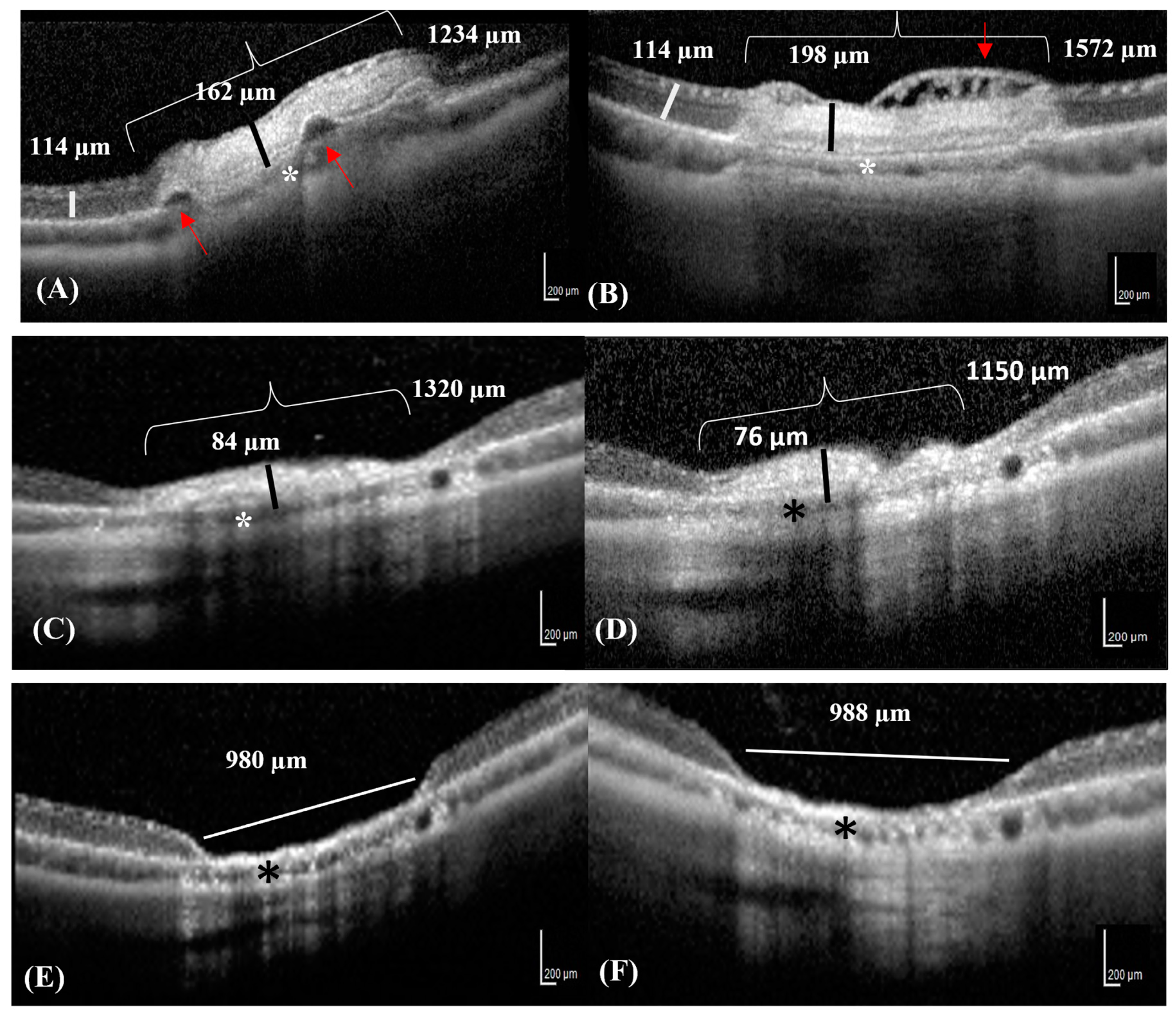
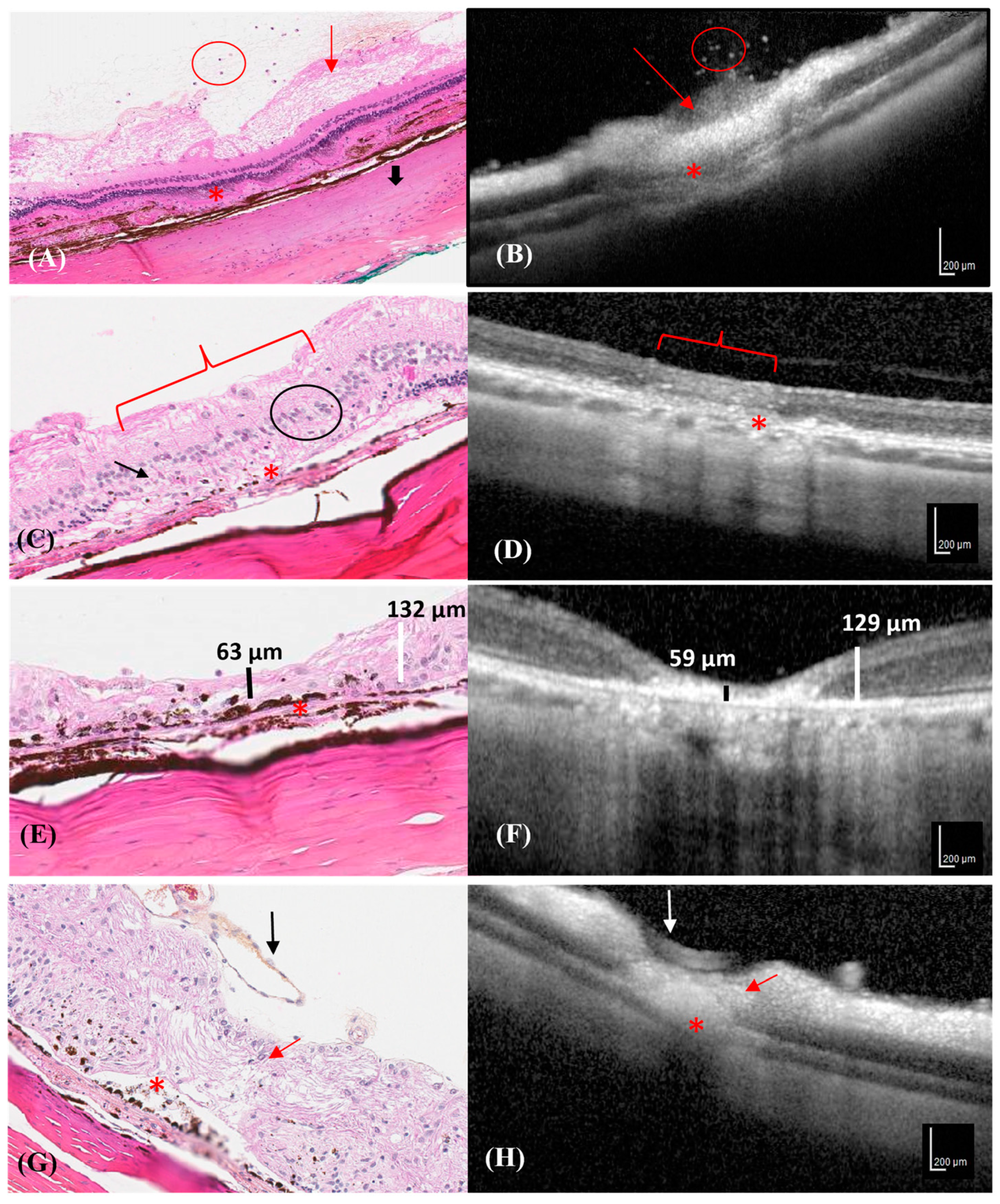
Transscleral retinopexy is performed to prevent retinal detachment. Retinal lesions induced by the laser have the appearance of retinal burns, (photocoagulation lesions), which evolve into scars (atrophic retinal lesions), inking the retina to the choroid and preventing retinal detachment. It is not known whether this procedure is effective. The current study compared retinal lesions by histology and OCT over time. Retinal damage occurred immediately during transscleral retinopexy. The follow-up of the retinal lesion was carried out 42 days after the laser procedure. The study provided OCT and histologic images that show the morphological evolution of the lesions over time until the scar. The first stage in the photocoagulation lesion is focal edema, which fades on the 7th day. On the 15th, the lesions exhibit tissue fibrosis. On the 21st day, the retinal lesion was excavated and cup-shaped, appearing as a scar. In the future, it will be possible to perform an OCT to identify scars and validate the correct transscleral laser retinopexy outcome.
Abstract
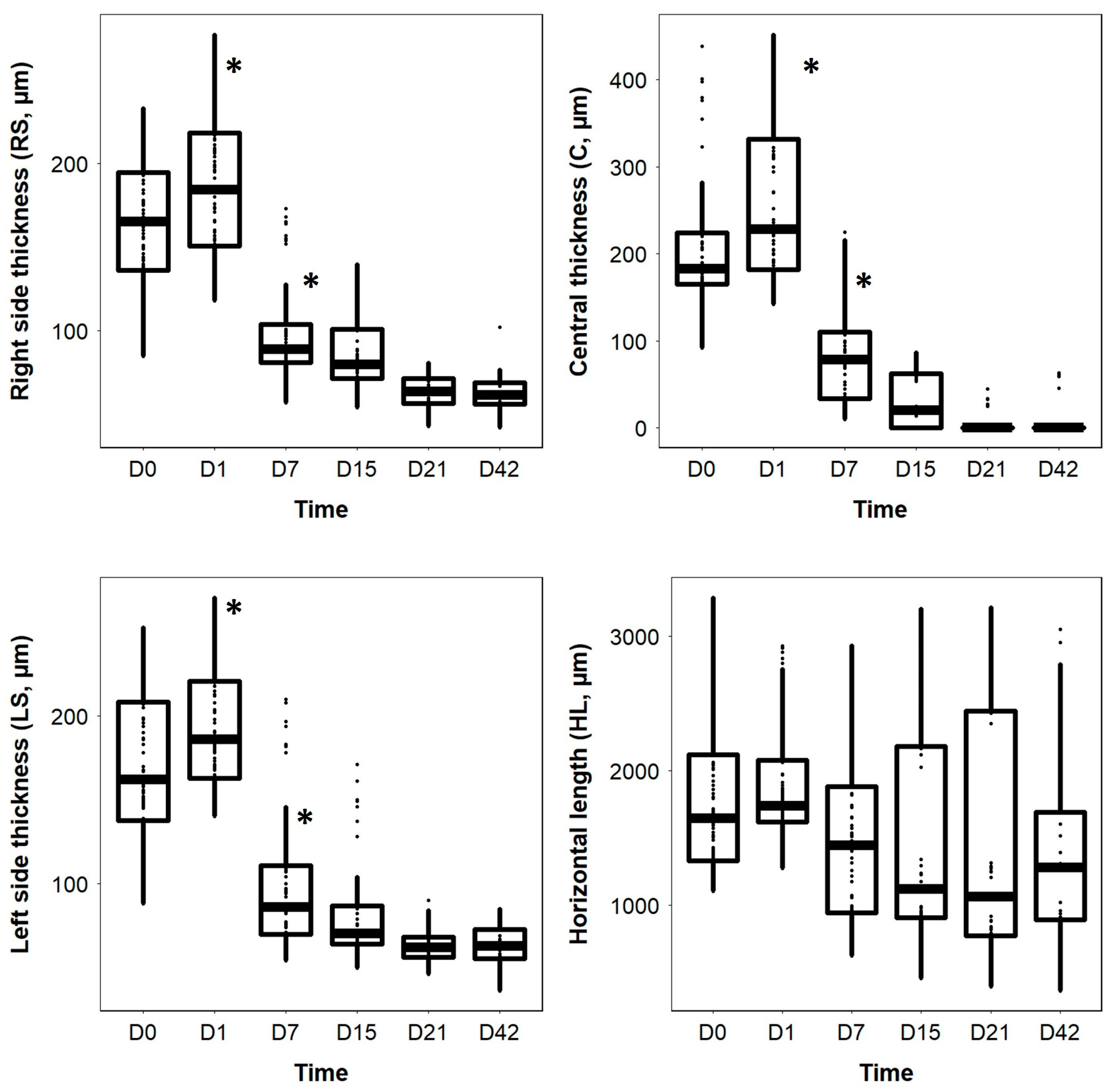
Transscleral retinopexy is a preventive technique used against retinal detachment. Fundus examination can allow the monitoring of morphological retinal changes in the progression of photocoagulation lesions, without offering details on the morphological changes by the retinal lesion. The aim of the study was to assess the progression of photocoagulation lesions induced by transscleral retinopexy (840 nm diode laser), by comparing the optical coherence tomography (OCT) and histological images over a period of six weeks on eight pigmented New Zealand healthy rabbits (four males and four females; n = 16 eyes). All rabbits underwent transscleral retinopexy on their left eye on day 0 (D0). Measurements of the photocoagulation lesions were obtained in vivo on D0, D7, D15, D21, and D42 by acquiring OCT images of both eyes from all rabbits. On D1, D7, D21, and D42, two rabbits were euthanized, and their eyes were enucleated. A significant effect by time on the decrease in the central retinal thickness of the photocoagulation lesion was observed from D1 to D7 (p = 0.001); however, no such effect was observed on the horizontal length ((HL) p = 0.584) of the lesion surface. The reliability between the OCT and histological measurements, which were evaluated using intraclass correlation coefficients, was excellent for measuring the retinal thickness at the center (ICC = 0.91, p < 0.001), moderate for the right side of the retinal lesions (ICC = 0.72, p = 0.006), and not significant for the left side and HL (p = 0.055 and 0.500, respectively). The morphological changes observed in the OCT and histopathological images of the photocoagulation lesions were qualitatively described over time. OCT is an effective tool for monitoring changes in photocoagulation lesions. Some measurements and qualitative changes showed an adequate correlation between the OCT and histological findings.