Abstract
A mangrove is a unique ecosystem with abundant resources, in which fungi are an indispensable microbial part. Numerous mangrove fungi-derived secondary metabolites are considerable sources of novel bioactive substances, such as polyketides, terpenoids, alkaloids, peptides, etc., which arouse people’s interest in the search for potential natural anti-tumor drugs. This review includes a total of 44 research publications that described 110 secondary metabolites that were all shown to be anti-tumor from 39 mangrove fungal strains belonging to 18 genera that were acquired from the South China Sea between 2016 and 2022. To identify more potential medications for clinical tumor therapy, their sources, unique structures, and cytotoxicity qualities were compiled. This review could serve as a crucial resource for the research status of mangrove fungal-derived natural products deserving of further development.
1. Introduction
A mangrove is a unique ecosystem distributed along the coastline of the tropical and subtropical regions, whose specific saline environment is what gives rise to its diverse microbial population [1]. Additionally, the biodiversity of fungi is more abundant than that of other mangrove microorganisms, which has a fairly good prospect of exploration that causes overwhelming focus [2,3]. Modern research suggests that the secondary metabolites which mangrove-derived fungi yielded are considerable sources of novel bioactive compounds [4]. The OSMAC strategy (one strain, many compounds) and co-cultures of mangrove fungus, as well as other microbes, have become the traditional methods for inducing the formation of metabolites [4,5]. The recent explosive multiomics methods wrestle with multi-trophic interactions and computational approaches based on artificial intelligence to estimate function distribution and demonstrate a paradigm change in the use of high-throughput methods for the discovery of natural products from plant-associated microorganisms or traditional Chinese medicine as the potential biomarkers for anti-cancer therapies [6,7]. Further research for these metabolites can be expected to discover their various biological features, such as enzyme inhibition, antimicrobial activity, antitumor activity, anti-inflammatory activity, antiviral activity, cytotoxicity, antioxidant activity, etc. Around the world, mangrove fungus-derived chemicals are being discovered at a progressively higher rate. Among them, many promising ones are beneficial in treating diseases [8].
Cancer, one of the leading causes of human death and the biggest impediment to extending life expectancy, is a serious worldwide health issue [9]. According to cancer statistics worldwide, there would be about 19.2 million emerging cases and 6.0 million cancer deaths occurring in 2022. In the past decade, anti-cancer research has accelerated due to rapid advances in early detection, surgical techniques, chemotherapy, radiation therapy, and targeted therapy [10]. Cytotoxic drugs are the mainstay of tumor chemotherapy, but their safety and resistance are unavoidable clinical concerns [11,12,13]. More novel anti-tumor drugs are still needed.
The South China Sea, a major mangrove region in the world, has been exploited and utilized for its rich mangrove fungal resources for many years, from which many promising potential anti-tumor drugs have been investigated for cancer treatment [8]. Xyloketal B originated from South China Sea’s mangrove fungal strain Xylaria sp. (No. 2508), and has displayed inhibition against glioblastoma cell proliferation and migration [14]. SZ-685C, an anthraquinone from another fungal strain Halorosellinia sp. (No. 1403), could suppress the reproduction of six human cancer cell lines and mice breast carcinoma xenografts [15]. Penicisulfuranol A, a new structural compound originated from Penicillium janthinellum possessing an infrequent benzofuran piperazine ring, was found to particularly suppress the C-terminal of Hsp90 (heat shock protein 90) and limit the development of HCT116, the human colon cancer cell line, not only in vitro but also in vivo [16]. These natural products, with their obvious anti-tumor effects and abundant candidate resources, have become the priority for anti-tumor drug development.
In this review, the sources, structures, and anti-tumor properties of the secondary metabolites isolated from fungal strains which were acquiredfrom the South China Sea’s mangrove ecosystem were outlined. A total of 44 research papers describing 110 anti-tumor compounds reported between 2016 and 2022 were included; the vast majority of those are novel compounds discovered in this period.
2. Summarisation of Anti-Tumor Secondary Metabolites from Mangrove-Derived Fungi
The one hundred and ten anti-tumor secondary metabolites obtained in the past seven years from fungi produced from the South China Sea’s mangrove ecosystem can be branched out into four types according to their chemical constructions and properties: polyketides (including nine groups: azaphilones, coumarins and isocoumarins, chromones, lactones, benzoates, xanthones, quinones and benzophenones, phenol, phenyl aldehydes, phenolic acids, and depsidones) (56, 51%), terpenoids (including three groups: sesquiterpenes, diterpenes, and steroids) (14, 13%), alkaloids (including three groups: amines and amides, diketopiperazines, and cytochalasins) (39, 35%) and peptides (including one group: cyclic peptides) (1, 1%) (Figure 1a).

Figure 1.
Summarization of anti-tumor secondary metabolites isolated from mangrove-derived fungi. (a) shows four types of anti-tumor secondary metabolites. (b) shows the genera of fungi producing anti-tumor secondary metabolites.
These secondary metabolites were identified from the 39 strains belonging to 18 genera (Penicillium, Aspergillus, Cladosporium, Diaporthe, Phomopsis, Fusarium, Colletotrichum, Lasiodiplodia, Mucor irregularis, Phoma, Sarocladium, Xylaria, Cytospora, Didymella, Eutypella, Pseudofusicoccum, Chaetomium, and Pseudopithomyces). Among them, the compounds derived from Penicillium (26, 23%), Aspergillus (25, 23%), and Lasiodiplodia (12, 11%) account for more than half of the total secondary metabolites, illustrating that the three genera are the major producers of the anti-tumor compounds (Figure 1b).
3. Sources, Structures, and Anti-Tumor Activities of Secondary Metabolites Originating from Fungal Strains in the Mangrove Ecosystem
One hundred and ten anti-tumor compounds obtained from fungi produced from the South China Sea’s mangrove ecosystem were reported from 2016 to 2022 and their classifications, sources, and cytotoxicities (described with IC50, the half maximal inhibitory concentration) were listed (Table 1).

Table 1.
Anti-tumor activities of secondary metabolites originating from fungal strains in the South China Sea’s mangrove ecosystem.
3.1. Polyketides
Polyketides make up more than half of the recently discovered anti-tumor secondary metabolites of mangrove-associated fungus from 2016 to 2022. They can be branched out into nine categories on the ground of the polyketide backbone, including azaphilones, coumarins and isocoumarins, chromones, lactones, benzoates, xanthones, quinones and benzophenones, phenols, phenyl aldehydes, and phenolic acids, as well as depsidones (Compounds 1–56, Figure 2 and Figure 3).
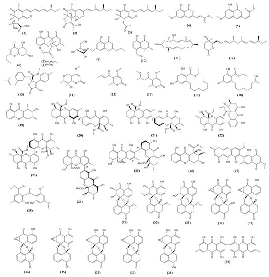
Figure 2.
The structures of compounds 1–39.
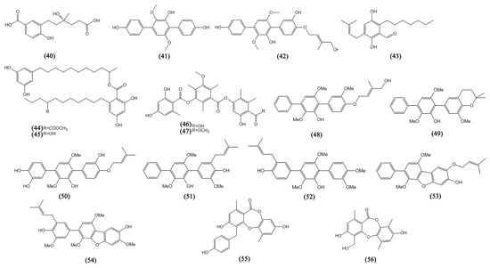
Figure 3.
The structures of compounds 40–56.
3.1.1. Azaphilones
Azaphilones, the major class of fungal polyketides, are known to possess the structural features of a highly oxidized pyranoquinone core [61]. In the past seven years, three azaphilones (1–3) with remarkable anti-tumor bioactivity were reported from the fungal genera Diaporthe sp. SCSIO 41011. They originated from the mangrove and were found to possess the specific structure and exhibit inhibition against ACHN, OS-RC-2, and 786-O human renal cancer cell lines (IC50 values from 3.0 to 38 μM) [17]. Further study showed that the new compound isochromophilone D (1) could induce cell cycle arrest and even apoptosis in 786-O cells.
3.1.2. Coumarins and Isocoumarins
Coumarins have a benzopyrene structure and their isomers are called isocoumarins [62]. Aspergisocoumrins A-B (4–5) were two novel isocoumarins derived from the endophytic fungal strain Aspergillus sp. HN15-5D, showing significant cytotoxicity against the MDA-MB-435 human breast cancer cell line (IC50 values near 5 μM) and possessing inhibitory against the MCF10A human non-cancer breast epithelial cell line (IC50 values between 11.3 and 21.4 μM), making an indication that they may have certain safety risks if used in clinical treatment. Aspergisocoumrin A (4) showed relatively weak cytotoxicity against human liver cancer cells HepG2 and human lung carcinoma cells H460 (with IC50 values > 20 μM) [18]. From another mangrove genus Fusarium sp. 2ST2, aspergisocoumrin A (4) was also derived and inhibited human lung carcinoma cells, with IC50 values of 6.2 ± 0.2 μM [19].
3.1.3. Chromones
With strategic significance in anti-tumor development, chromone and its derivatives have been recognized as the major structural component of various functional organic molecules [63]. The five new chromones were described from four different genera of mangrove associated fungi: Colletotrichum sp., Penicillium sp., Cladosporium sp., and Fusarium sp. (5R,7S)-5,7-dihydroxy-2-propyl-5,6,7,8-tetrahydro-4H-chromen-4-one (6) from C. gloeosporioides exhibited inhibitory activity against A549 cells with an IC50 value of 0.094 mM [20]. Penixanthones C-D (7–8), two chromones from Penicillium sp. SCSIO041218 with a unique 6/6/6/5 polycyclic skeleton showed a mild inhibitory effect against K562, MCF-7 (human breast cancer cells), and Huh-7 (human liver cancer cells), with IC50 values from 55.2 to 67.5 µM [21]. 7-O-α-D-ribosyl-5-hydroxy-2-propylchromone (9) from Cladosporium sp. OUCMDZ-302 was cytotoxic to human lung carcinoma cells H1975, with an IC50 value of 10.0 µM [22]. 4H-1-benzopyran-4-one-2,3-dihydro-5-hydroxy-8-(hydroxylmethyl)-2-methyl (10) was derived from Fusarium sp. 2ST2 accompanied by aspergisocoumrin A (4), showing the same inhibition against A549 and MDA-MB-435 cells (IC50 5.6 and 3.8 μM, respectively) [19].
3.1.4. Lactones
This group of lactones with six members is characterized by mangrove fungi. Macrolides are abundant in microorganisms and plants, belonging to a class of polyketides with rings of various sizes that are distinguished by the presence of lactone groups [64]. A macrolide obtained both from Penicillium sp. (HS-N-27) and (HS-N-29) was identified as brefeldin A (11) [23], which is a well-known Golgi-disruptor and the inhibitor of GEFs (the exchange factor for guanine nucleotides) for the ARFs (small GTPases that catalyze ADP-ribosylation) and possessed obvious inhibitory effects on multiple cancer cell lines in the previous study, including lung (A549), colon (HCT-116), glioma (SF-539), melanoma (UACC-62), ovarian (OVCAR-3), renal (SN12C), prostate (PC3), and breast (MCF7) cancer cells, with IC50 values lower than 0.1 μM [24]. Similarly, nafuredin B (12) obtained from P. variabile with co-culture of Talaromyces aculeatus, was also found to be a multiple cytotoxic lactones to HeLa (cervical), K562 (renal), HCT-116, HL-60 (myeloid leukemia), A549, and MCF-7 cancer cell lines with IC50 values lower than 10 μM [25]. The other four lactones (13–16) from A. sydowii #2B showed cytotoxicities against human prostate cancer VCaP cells, with IC50 values from 1.92 to 20.06 μM [26].
3.1.5. Benzoates
Lasiodiplodins are lactones possessing a 14/12-member macrocyclic ring, while 2,4-dihydroxy-6-nonylbenzoate (17) and ethyl-2,4-dihydroxy-6-(80-hydroxynonyl)-benzoate (18) were detected to be the open-ring structures of lasiodiplodins, characterized as benzoates [27,28]. They were obtained from the same strain Lasiodiplodia sp. 318#. The benzoate 2,4-Hihydroxy-6-nonylbenzoate (17) was evaluated in vitro against rat pituitary adenoma lines MMQ and GH3 (with IC50 values between 5.29 μM and 13.05 μM) [27]. Ethyl-2,4-dihydroxy-6-(80-hydroxynonyl)-benzoate (18) showed inhibition against human cancer lines THP1 (monocytic lymphoma), MDA-MB-435, A549, HepG2 (liver), and HCT-116 (with IC50 values from 10.13 to 39.74 μM) [28].
3.1.6. Xanthones
Xanthones are dibenzo-pyrone-structured aromatic oxygenated heterocyclic compounds. This molecule may accept different substituents at various places, which are known as privileged scaffolds in the quest for novel medications [65]. There are nine members characterized in this group. Penixanthone A (19) from Penicillium sp. SYFz-1 showed weak cytotoxicities against the human cancer cell lines, H1975 (lung), MCF-7, and K562, as well as human liver cells HL7702, at a concentration of 30 μM [29]. Aspergillus sp. was found to be a promising resource to obtain anti-tumor xanthone metabolites (20–25, 27). Versixanthones G-H (20–21) and L-M (22–23) from A. vericolor were detected to be cytotoxic to HL-60, K562 A549, H1975, MGC803 (stomach), HO-8910 (ovarian), and HCT-116 human cancer cells, but showed low cytotoxicity against human embryonic kidney cells HEK293 [30]. Versixanthones G-H (20–21) were discovered to be effective DNA Topo I inhibitors to arrest the cell cycle and induce necrosis in MGC803 cells. They exhibited extensive cytotoxicities against five cancer cell lines (HL-60, K562, H1975, MGC803, and HO-8910), with IC50 values ranging from 0.4 µM to 16.1 µM. Versixanthones N-O (24–25) from A. versicolor HDN1009 showed multiple cytotoxicities against these cancer cell lines, with IC50 values from 1.7 µM to 16.1 µM [31]. Xanthoradone A (27) from A. sydowii #2B showed strong cytotoxicity against VCaP cells, with IC50 values of 4.19 ± 1.02 µM. Peniophora incarnata Z4 yielded a tetrahydroxanthone, and incarxanthone B (26) was found to inhibit the growth of A375, MCF-7, and HL-60 cell lines, with IC50 values of 8.6, 6.5, and 4.9 µM, respectively [32].
3.1.7. Quinones and Benzophenones
Benzophenones may be derived from the corresponding anthraquinones. Penibenzophenones B (28), a benzophenone extracted from Diaporthe sp. SCSIO 41011 showed cytotoxicity against A549 cell lines (IC50 15.7 µg/mL) [33]. Ten preussomerins, chloropreussomerins A-B (29–30), preussomerins A, D, F-H, K, and M (31–36, 38), and Ymf 1029E (37) from L. theobromae ZJ-HQ1 were found to show the cytotoxicity activities towards A549, HepG2, HeLa, and MCF-7 cells (IC50 from 2.5 to 83 μM), which is similar to the cytotoxicity to HEK293T cells [34]. Another quinone, the (+)-3,3′-7,7′-8,8′-hexahydroxy-5,5′-dimethyl-bianthra-quinone (39) from A. sydowii #2B, showed weak cytotoxicity against VCaP cells with an IC50 value of 33.36 ± 1.42 μM [26].
3.1.8. Phenols, Phenyl Aldehydes, and Phenolic Acids
Phenyl aldehydes and phenolic acids are phenol derivatives, and there are 15 family members characterized by mangrove fungi. A new benzoic acid, cladoslide A (40) from Cladosporium sp. HNWSW-1, was discovered to be cytotoxic to the K562 cells with an IC50 value of 13.10 ± 0.08 μM [35]. Terphenyllin (41) was obtained from A. candidus (HS-Y-23), which showed weak cytotoxicity against HeLa cells with an IC50 value of 19.0 µM [23]. Asperterphenyllin G (42) from A. candidus LDJ-5, which was first isolated from a natural source, showed broad activities against the L-02 (liver), MGC-803, HCT-116, BEL-7402 (liver), A549, SH-SY5Y (neuroblastoma), Hela, U87, and HO8910 (ovarian) cancer cell lines, with IC50 values lower than 1.7 µM [36]. Aspergillus sp. SCSIO41407 yielded a benzophenone flavoglaucin (43), showing weak activity against A549 cells with an IC50 value of 22.2 μM [37]. Integracins A-B (44–45) from Cytospora sp. displayed significant cytotoxicity against HepG2 lines, with IC50 values lower than 10 µM [38]. Phoma sp. SYSU-SK-7 yielded colletotric A (46) and colletotric B (47) and also showed weak cytotoxicity against MAD-MB-435 and A549 cells, with IC50 values from 16.82 to 37.73 μM [39]. Seven newly reported prenylated p-terphenyls (48–54) derived from A. candidus LDJ-5 were discovered to possess inhibitory properties against a variety of human cancer cell lines, with IC50 values ranging from 0.9 to 29.7 μM [40].
3.1.9. Depsidones
Two depsidones, botryorhodines H and C (55–56), yielded by Trichoderma sp. 307 with the co-culture of Acinetobacter johnsonii B2 showed potent cytotoxicity against MMQ and GH3 cell lines, with IC50 values from 3.09 to 31.62 μM [41].
3.2. Terpenoids
Fungi constitute a class of organisms that are particularly appealing for the discovery of terpenoid pathways because they frequently combine their biosynthetic genes [66]. The new terpenoids, including steroids from mangrove fungi in the South China Sea, can be divided into three categories based on their chemical structures and biogenetic pathways: sesquiterpenes, diterpenes, and steroids (Compounds 57–70, Figure 4).
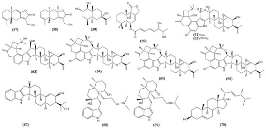
Figure 4.
The structures of compounds 57–70.
3.2.1. Sesquiterpenes
Sesquiterpenes are the largest group and an excellent source of terpenoids, having a high potential for inhibiting various cancers. A group of well-known natural sesquiterpenoids named eudesmanolides has diverse bioactivities [67]. Two eudesmanolides, 13-hydroxy-3,8,7(11)-eudesmatrien-12,8-olide (57) and 13-hydroxy-3,7(11)-eudesmatrien-12,8-olide (58), were obtained from Eutypella sp. 1–15 exhibiting potent anti-tumor activity against JEKO-1 (human mantle cell lymphoma) and HepG2 cells, with IC50 values ranging from 8.4 to 48.4 μM [42]. A novel eudesmane-type sesquiterpenoid penicieudesmol B (59) from Penicillium sp. J-54 displayed weak cytotoxicity against K-562 cells, with an IC50 value of 90.1 µM [43]. A. ustus 094102 yielded a drimane sesquiterpenoid ustusolate I (60), showing anti-proliferation against CAL-62 (human thyroid cancer) and MG-63 (human osteosarcoma) cells with IC50 values of 16.3 and 10.1 µM, respectively [44].
3.2.2. Diterpenes
Diterpenes may be one of the key chemicals in the therapy of cancer, although mangrove fungi have only sometimes produced new bioactive diterpenes [68]. Here, two fungus strains, Mucor irregularis and Eupenicillium sp. HJ002 were highlighted for the production of nine structurally diverse indole-diterpenes with anti-tumor activity (61–69), including five novel diterpenes rhizovarins A, B, and E (61–63) and penicilindoles A-B (68–69). Rhizovarins A-B (61–62) possessed a never reported 4,6,6,8,5,6,6,6,6-fused indole-diterpene ring which enduing them with unique chemical properties [45]. The seven bioactive compounds (61–67) from M. irregularis were detected to exhibit significant cytotoxicity against A549 and HL60 cells with IC50 values of about 10 µM [45]. Another two diterpenes penicilindoles A-B (68–69) from Eupenicillium sp. HJ002 displayed inhibitory activity against A549 and HepG2 cell lines, with IC50 values ranging from 1.5 to 47.2 μM [46].
3.2.3. Steroids
The most significant class of tiny biomolecules, steroids play a variety of cellular functions connected to membrane structure and signaling. Pseudofusicoccum sp. J003 yielded a steroid derivative, ergosterol (70), displaying significant inhibition effects on HL-60 and SW480 (human colon adenocarcinoma) cells for inhibition rates of 98.68 ± 0.97% and 60.40 ± 4.51% at the concentration of 40 μM [47].
3.3. Alkaloids
Alkaloids are a class of compounds containing organic nitrogenous bases that occupy a major part of the secondary metabolites of mangrove fungi. They have been found to have various biological activities [69]. The new alkaloids from mangrove fungi in the South China Sea can be divided into three categories: amines and amides, diketopiperazines, and cytochalasins (Compounds 71–109, Figure 5 and Figure 6).
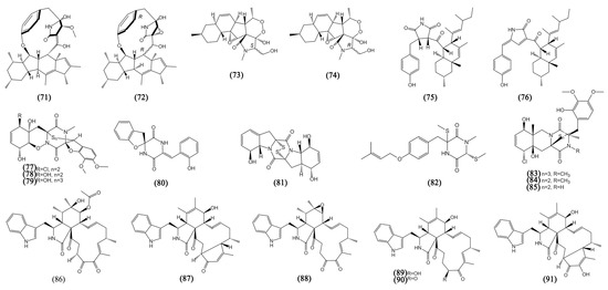
Figure 5.
The structures of compounds 71–91.
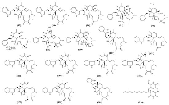
Figure 6.
The structures of compounds 92–110.
3.3.1. Amines and Amides
Ascomylactams A (71) and C (72) were two novel alkaloids from Didymella sp. CYSK-4 with a special structure of 12- or 13-membered-ring macrocyclic, displaying moderate anti-proliferative activity against MDA-MB-435, MDA-MB-231, SNB19 (glioma), HCT116, NCI-H460 (lung cancer), and PC-3 cells, with IC50 values ranging from 4.2 to 7.8 μM [49]. Further research for ascomylactams A (71) showed that it suppressed the growth of A549, NCI-H460, and NCI-H1975 mice tumor xenografts in vivo and arrested the cell cycle through the ROS/Akt/Cyclin D1/Rb pathway both in vivo and in vitro [48]. Fusarisetins E-F (73–74) were two novel 3-decalinoyltetramic acid derivatives with the structure of a peroxide bridge derived from Fusarium sp. 2ST2, showing obvious inhibitory effects on A549 cells, with IC50 values of 8.7 to 4.3 μM [19]. The mangrove-derived fungus Cladosporium sp. HNWSW-1 yielded two new derivatives containing succinimide, cladosporitin B (75) and talaroconvolutin A (76), detected to be cytotoxic to BEL-7042, K562, SGC-7901 (human stomach cancer), and Hela cells, with IC50 values from 14.9 to 41.7 µM [50].
3.3.2. Diketopiperazines
Diketopiperazine alkaloids, with a disulfide moiety connected to the diketopiperazine ring, play a major role in alkaloids with significant biological properties [70]. Penicisulfuranols A-C (77–79), three new epipolythiodioxopiperazine alkaloids consisting of sulfur atoms on both α- and β-positions of amino acid residues and a 1,2-oxazadecaline core, were obtained from P. janthinellum HDN13-309 and were cytotoxic to Hela and HL60 cells, with IC50 values ranging from 0.1 to 3.9 μM [51]. Although spirobrocazine C (80) and brocazine G (81) are two new diketopiperazine alkaloids from the same mangrove-derived fungus P. brocae MA-231. Spirobrocazine C (80) showed moderate activity against A2780 cells (IC50 59 μM) and brocazine G (81) showed strong activity not only to A2780 but also to A2780 CisR cells (IC50 664 and 661 nM) [52]. Saroclazine B (82) was a diketopiperazine derivative from Sarocladium kiliense HDN11-84 possessing the structure of a free amide found firstly in sulfur-containing aromatic diketopiperazine derivatives, and it was cytotoxic to HeLa cells, with an IC50 value of 4.2 μM [53]. A novel trithiodiketopiperazine derivative, adametizine C (83), with two dithiodiketopiperazine derivatives (84–85) from P. ludwigii SCSIO 41408 displayed cytotoxicity against human prostate cancer cell line 22Rv1, with IC50 values from 13.0 to 13.9 µM, and compound 85 showed significant inhibitory activity against PC-3 cells, with an IC50 value of 5.1 µM [54].
3.3.3. Cytochalasins
Cytochalasins are known as a vast class of fungal polyketide-non ribosomal secondary metabolites with a wide variety of biological functions. Possessing a substituted isoindole scaffold joined with a macrocyclic ring produced from a substantially reduced polyketide backbone and an amino acid makes cytochalasins distinctive [71]. Nine cytochalasins from Chaetomium globosum kz-19, including phychaetoglobins C-D (86–87), chaetoglobosins C, E, G, J, V (88–92), penochalasin J (93), and prochaetoglobosin IIIed (94) displayed moderate cytotoxicities against A549 and HeLa cells, with all the IC50 values less than 20 µM [55]. Phomopsis sp. QYM-13 yielded four cytochalasins, phomopchalasin E (95), cytochalasins U, J (96–97), and cytochalasin H (98), which all showed significant cytotoxicity against MDA-MB-435 cells, with IC50 values ranging from 0.2 to 8.2 μM [56]. Moreover, cytochalasins U (96) was detected to be cytotoxic to human glioma cell line SNB19, with an IC50 value of 6.9 ± 1.4 μM. Two cytochalasins, 12-hydroxylcytochalasin Q (99) and zygosporin D (100) from Xylaria arbuscula, possessed a tetracyclic ring system and displayed significant inhibitory effects on human colorectal adenocarcinoma cell lines HCT15, with IC50 values of 13.5 and 13.4 μM, respectively [57]. A new chaetoglobosin with the structure of an unprecedented six-cyclic 6/5/6/5/6/13 fused ring system, penochalasin I (101), together with another seven chaetoglobosins penochalasin J (102), chaetoglobosins G, F, C, A, and E, and cytoglobosin C (103–108) were derived from P. chrysogenum V11, showing cytotoxicity against MDA-MB-435, SGC-7901 and A549 cells, with IC50 values ranging from 3.35 to 38.77 μM [58]. Another new chaetoglobosin, penochalasin K (109) with the same special structure of six-cyclic 6/5/6/5/6/13 fused ring and the same fungal strain source, exhibited strong cytotoxicity against MDA-MB-435, SGC-7901, and A549 cells, with IC50 values lower than 10 μM [59].
3.4. Peptides
In many clinical medication treatments, peptides with molecular weights under 1000 Da can adapt to drug resistance and have fewer hazardous side effects, which may have implications for the ongoing development of novel therapies [72]. In the past seven years, only one cyclic peptide was reported to be an anti-tumor peptide (Compound 110, Figure 6).
Cyclic Peptides
A cyclic peptide, fusaristatin C (110), was rapidly separated from Pseudopithomyces sp. 1512101 and analyzed using a new strategy of ligand fishing based on PLA2-MNPs (Phospholipase A2-functionalized magnetic nanoparticles) with LC–MS (liquid chromatography–mass spectrometry), exerting a potent inhibitory effect on A549 cells, with IC50 values of 10.10 µM [60].
4. Discussion
Without a doubt, more and more mangrove secondary metabolites are being discovered and that may be a major source for the creation of novel anti-cancer drugs that can be applied both therapeutically and preventively. Vinca alkaloids are the most notable representatives of plant-derived natural compounds as anticancer weapons, which are frequently utilized as first-line anticancer medications in hematological malignancies [73]. Furthermore, a number of marine isolated targeted compounds, such as Brentuximab2 vedotin (AdcetrisTM), Enfortumab vedotin, and Marizomib, had been used as apoptotic inducers in different cancer types at FDA (the Food and Drug Administration in the US)-approved or treatment phases [74]. More effective natural anti-tumor drugs are expected to emerge.
This review concentrated on one hundred and ten anti-tumor secondary metabolites from thirty-nine mangrove fungus strains belonging to eighteen genera from the South China Sea that have been newly reported over the last seven years. Penicillium (23%), Aspergillus (23%), and Lasiodiplodia (11%) were their main producers and at the structural level, polyketides occupy more than half of the secondary metabolites. Seventy-eight compounds were considered to possess multiple anti-tumor properties, as they exhibited cytotoxicity against more than two tumor cell lines.
It has been proposed that mangrove fungal-derived drugs could be effective weapons for human beings to fight cancer. However, the potential regulatory mechanisms of the vast anti-tumor compounds on tumor microenvironment still need to do in-depth exploration. The bulk of them could not be employed temporarily in tumor diagnosis and treatment due to the lack of reliable clinical research data and studies confirming their biosafety. Additionally, a fungal culture is restricted by special environments or growth factors. Co-cultures of fungi are a popular way to promote the production of metabolites, such as nafuredin B (12), botryorhodine H (55), and botryorhodine C (56), and in the future, multi-omics methods will be richer than the secondary metabolites of mangrove-derived fungi and will be good for screening anti-cancer compounds. Another challenge is figuring out how to produce these secondary metabolites in large quantities.
To date, chemotherapy remains to be regarded as the cornerstone of many adjunctive therapies for cancer. Recently, the design and development of efficient anti-cancer treatment strategies have advanced, and decisions regarding treatment have taken the immunological perspective of chemotherapy into account. Because nucleotides are significant chemicals that must be generated to sustain the state of proliferation in cancer, nucleotide metabolism is regarded as the most crucial link in oncogenesis and progression [75]. On the one hand, targeted regulation of the level of nucleotide metabolism can make tumor cells more sensitive to chemotherapy drugs, mediate anti-tumor response, and enhance the efficacy of chemotherapy and immunotherapy in the treatment of tumors [75,76]. A good anti-tumor impact may also be obtained by altering the amount of nucleotide metabolism, such as by providing exogenous UDP (uridine diphosphate) to modify the tumor microenvironment [77]. This makes immunotherapy tend to have a similar outcome to chemotherapy. On the other hand, chemotherapy stimulates the immune system and triggers tumor cells to undergo immunogenic cell death (ICD) [78]. An important feature of ICD is the extracellular release of ATP from dying cells after apoptosis. For example, daunorubicin, a classic anthracycline anti-tumor drug, induced ATP release into the extracellular space of acute myeloid leukemia cells and was considered a very strong ICD inducer [79]. Another possible drug from the Agelas mauritianus sponge, KRN7000, was found to play an anti-tumor role by activating the immune system, having been put into a clinical investigation for many years [80]. In addition, the level of nucleotide release will also be one of the indicators to evaluate the efficacy of anti-tumor drugs in the near future [81].
5. Conclusions
Looking forward to the newly developed anti-cancer treatment strategies and anti-cancer metabolite discovery strategies, this review serves as a crucial resource for the research status of mangrove fungal-derived natural products deserving of further development, demonstrating the great medical benefits of mangrove fungal-derived drugs for the treatment of clinical cancers.
Author Contributions
Conceptualization, Y.L. and X.L.; data curation, T.Z., S.L. (Siyuan Li) and S.L. (Shuping Liu); writing—original draft preparation, Y.L.; writing—review and editing, X.L., J.L. and X.W.; visualization and supervision, Y.M., Z.W. and X.J.; project administration, J.L.; funding acquisition, X.W. All authors have read and agreed to the published version of the manuscript.
Funding
This research was funded by the National Natural Science Foundation of China for Young Scholars [grant number 81802678], the Guangdong Natural Science Foundation Project [grant number 2018A030310107], the Medical Scientific Research Foundation of the Guangdong Province of China [grant number A2018558], the Guangzhou Basic and Applied Basic Research Foundation [grant number 202102021116] and the National Natural Science Foundation of China [grant number 31670349].
Institutional Review Board Statement
Not applicable.
Informed Consent Statement
Not applicable.
Data Availability Statement
No new data were created or analyzed in this study. Data sharing is not applicable to this article.
Conflicts of Interest
The authors declare no conflict of interest.
References
- Jia, S.-L.; Chi, Z.; Liu, G.-L.; Hu, Z.; Chi, Z.-M. Fungi in mangrove ecosystems and their potential applications. Crit. Rev. Biotechnol. 2020, 40, 852–864. [Google Scholar] [CrossRef]
- Gozari, M.; Alborz, M.; El-Seedi, H.R.; Jassbi, A.R. Chemistry, biosynthesis and biological activity of terpenoids and meroterpenoids in bacteria and fungi isolated from different marine habitats. Eur. J. Med. Chem. 2021, 210, 112957. [Google Scholar] [CrossRef]
- Li, K.; Chen, S.; Pang, X.; Cai, J.; Zhang, X.; Liu, Y.; Zhu, Y.; Zhou, X. Natural products from mangrove sediments-derived microbes: Structural diversity, bioactivities, biosynthesis, and total synthesis. Eur. J. Med. Chem. 2022, 230, 114117. [Google Scholar] [CrossRef]
- Carroll, A.R.; Copp, B.R.; Davis, R.A.; Keyzers, R.A.; Prinsep, M.R. Marine natural products. Nat. Prod. Rep. 2021, 38, 362–413. [Google Scholar] [CrossRef] [PubMed]
- Knowles, S.L.; Raja, H.A.; Roberts, C.D.; Oberlies, N.H. Fungal–fungal co-culture: A primer for generating chemical diversity. Nat. Prod. Rep. 2022, 39, 1557–1573. [Google Scholar] [CrossRef]
- Aghdam, S.A.; Brown, A.M.V. Deep learning approaches for natural product discovery from plant endophytic microbiomes. Environ. Microbiome 2021, 16, 6. [Google Scholar] [CrossRef]
- Li, R.; Zhou, W. Multi-omics analysis to screen potential therapeutic biomarkers for anti-cancer compounds. Heliyon 2022, 8, e9616. [Google Scholar] [CrossRef]
- Chen, S.; Cai, R.; Liu, Z.; Cui, H.; She, Z. Secondary metabolites from mangrove-associated fungi: Source, chemistry and bioactivities. Nat. Prod. Rep. 2022, 39, 560–595. [Google Scholar] [CrossRef]
- Bray, F.; Ferlay, J.; Soerjomataram, I.; Siegel, R.L.; Torre, L.A.; Jemal, A. Global cancer statistics 2018: GLOBOCAN estimates of incidence and mortality worldwide for 36 cancers in 185 countries. CA Cancer J. Clin. 2018, 68, 394–424. [Google Scholar] [CrossRef]
- Siegel, R.L.; Miller, K.D.; Fuchs, H.E.; Jemal, A. Cancer statistics, 2022. CA: A Cancer J. Clin. 2022, 72, 7–33. [Google Scholar] [CrossRef] [PubMed]
- Vasan, N.; Baselga, J.; Hyman, D.M. A view on drug resistance in cancer. Nature 2019, 575, 299–309. [Google Scholar] [CrossRef]
- Kelloff, G.J.; Sigman, C.C. Assessing intraepithelial neoplasia and drug safety in cancer-preventive drug development. Nat. Rev. Cancer 2007, 7, 508–518. [Google Scholar] [CrossRef]
- Bukowski, K.; Kciuk, M.; Kontek, R. Mechanisms of Multidrug Resistance in Cancer Chemotherapy. Int. J. Mol. Sci. 2020, 21, 3233. [Google Scholar] [CrossRef]
- Gong, H.; Bandura, J.; Wang, G.-L.; Feng, Z.-P.; Sun, H.-S. Xyloketal B: A marine compound with medicinal potential. Pharmacol. Ther. 2022, 230, 107963. [Google Scholar] [CrossRef]
- Xie, G.; Zhu, X.; Li, Q.; Gu, M.; He, Z.; Wu, J.; Li, J.; Lin, Y.; Li, M.; She, Z.; et al. SZ-685C, a marine anthraquinone, is a potent inducer of apoptosis with anticancer activity by suppression of the Akt/FOXO pathway. Brit. J. Pharmacol. 2010, 159, 689–697. [Google Scholar] [CrossRef]
- Dai, J.; Chen, A.; Zhu, M.; Qi, X.; Tang, W.; Liu, M.; Li, D.; Gu, Q.; Li, J. Penicisulfuranol A, a novel C-terminal inhibitor disrupting molecular chaperone function of Hsp90 independent of ATP binding domain. Biochem. Pharmacol. 2019, 163, 404–415. [Google Scholar] [CrossRef]
- Luo, X.; Lin, X.; Tao, H.; Wang, J.; Li, J.; Yang, B.; Zhou, X.; Liu, Y. Isochromophilones A–F, Cytotoxic Chloroazaphilones from the Marine Mangrove Endophytic Fungus Diaporthe sp. SCSIO 41011. J. Nat. Prod. 2018, 81, 934–941. [Google Scholar] [CrossRef]
- Wu, Y.; Chen, S.; Liu, H.; Huang, X.; Liu, Y.; Tao, Y.; She, Z. Cytotoxic isocoumarin derivatives from the mangrove endophytic fungus Aspergillus sp. HN15-5D. Arch. Pharmacal Res. 2019, 42, 326–331. [Google Scholar] [CrossRef]
- Chen, Y.; Wang, G.; Yuan, Y.; Zou, G.; Yang, W.; Tan, Q.; Kang, W.; She, Z. Metabolites with Cytotoxic Activities from the Mangrove Endophytic Fungus Fusarium sp. 2ST2. Front. Chem. 2022, 10, 842405. [Google Scholar] [CrossRef]
- Luo, Y.; Song, X.; Zheng, C.; Chen, G.; Luo, X.; Han, J. Four New Chromone Derivatives from Colletotrichum gloeosporioides. Chem. Biodivers. 2020, 17, e1900547. [Google Scholar] [CrossRef]
- Huang, J.; She, J.; Yang, X.; Liu, J.; Zhou, X.; Yang, B. A New Macrodiolide and Two New Polycyclic Chromones from the Fungus Penicillium sp. SCSIO041218. Molecules 2019, 24, 1686. [Google Scholar] [CrossRef] [PubMed]
- Wang, L.; Han, X.; Zhu, G.; Wang, Y.; Chairoungdua, A.; Piyachaturawat, P.; Zhu, W. Polyketides from the Endophytic Fungus Cladosporium sp. Isolated from the Mangrove Plant Excoecaria agallocha. Front. Chem. 2018, 6, 344. [Google Scholar] [CrossRef] [PubMed]
- Wang, C.-F.; Ma, J.; Jing, Q.-Q.; Cao, X.-Z.; Chen, L.; Chao, R.; Zheng, J.-Y.; Shao, C.-L.; He, X.-X.; Wei, M.-Y. Integrating Activity-Guided Strategy and Fingerprint Analysis to Target Potent Cytotoxic Brefeldin A from a Fungal Library of the Medicinal Mangrove Acanthus ilicifolius. Mar. Drugs 2022, 20, 432. [Google Scholar] [CrossRef] [PubMed]
- Anadu, N.O.; Davisson, V.J.; Cushman, M. Synthesis and Anticancer Activity of Brefeldin a Ester Derivatives. J. Med. Chem. 2006, 49, 3897–3905. [Google Scholar] [CrossRef] [PubMed]
- Zhang, Z.; He, X.; Zhang, G.; Che, Q.; Zhu, T.; Gu, Q.; Li, D. Inducing Secondary Metabolite Production by Combined Culture of Talaromyces aculeatus and Penicillium variabile. J. Nat. Prod. 2017, 80, 3167–3171. [Google Scholar] [CrossRef] [PubMed]
- Wang, Y.; Zhong, Z.; Zhao, F.; Zheng, J.; Zheng, X.; Zhang, K.; Huang, H. Two new pyrone derivatives from the mangrove-derived endophytic fungus Aspergillus sydowii #2B. Nat. Prod. Res. 2021, 36, 3872–3878. [Google Scholar] [CrossRef] [PubMed]
- Huang, J.; Xu, J.; Wang, Z.; Khan, D.; Niaz, S.I.; Zhu, Y.; Lin, Y.; Li, J.; Liu, L. New lasiodiplodins from mangrove endophytic fungus Lasiodiplodia sp. 318#. Nat. Prod. Res. 2016, 31, 326–332. [Google Scholar] [CrossRef]
- Li, J.; Xue, Y.; Yuan, J.; Lu, Y.; Zhu, X.; Lin, Y.; Liu, L. Lasiodiplodins from mangrove endophytic fungus Lasiodiplodia sp. 318#. Nat. Prod. Res. 2015, 30, 755–760. [Google Scholar] [CrossRef]
- Tao, H.; Wei, X.; Lin, X.; Zhou, X.; Dong, J.; Yang, B. Penixanthones A and B, two new xanthone derivatives from fungus Penicillium sp. SYFz-1 derived of mangrove soil sample. Nat. Prod. Res. 2017, 31, 2218–2222. [Google Scholar] [CrossRef]
- Wu, G.; Qi, X.; Mo, X.; Yu, G.; Wang, Q.; Zhu, T.; Gu, Q.; Liu, M.; Li, J.; Li, D. Structure-based discovery of cytotoxic dimeric tetrahydroxanthones as potential topoisomerase I inhibitors from a marine-derived fungus. Eur. J. Med. Chem. 2018, 148, 268–278. [Google Scholar] [CrossRef]
- Yu, G.; Wu, G.; Sun, Z.; Zhang, X.; Che, Q.; Gu, Q.; Zhu, T.; Li, D.; Zhang, G. Cytotoxic Tetrahydroxanthone Dimers from the Mangrove-Associated Fungus Aspergillus versicolor HDN1009. Mar. Drugs 2018, 16, 335. [Google Scholar] [CrossRef] [PubMed]
- Li, S.J.; Jiao, F.W.; Li, W.; Zhang, X.; Yan, W.; Jiao, R.H. Cytotoxic Xanthone Derivatives from the Mangrove-Derived Endophytic Fungus Peniophora incarnata Z4. J. Nat. Prod. 2020, 83, 2976–2982. [Google Scholar] [CrossRef]
- Zheng, C.-J.; Liao, H.-X.; Mei, R.-Q.; Huang, G.-L.; Yang, L.-J.; Zhou, X.-M.; Shao, T.-M.; Chen, G.-Y.; Wang, C.-Y. Two new benzophenones and one new natural amide alkaloid isolated from a mangrove-derived Fungus Penicillium citrinum. Nat. Prod. Res. 2018, 33, 1127–1134. [Google Scholar] [CrossRef] [PubMed]
- Chen, S.; Chen, D.; Cai, R.; Cui, H.; Long, Y.; Lu, Y.; Li, C.; She, Z. Cytotoxic and Antibacterial Preussomerins from the Mangrove Endophytic Fungus Lasiodiplodia theobromae ZJ-HQ1. J. Nat. Prod. 2016, 79, 2397–2402. [Google Scholar] [CrossRef] [PubMed]
- Cao, X.; Guo, L.; Cai, C.; Kong, F.; Yuan, J.; Gai, C.; Dai, H.; Wang, P.; Mei, W. Metabolites from the Mangrove-Derived Fungus Cladosporium sp. HNWSW-1. Front. Chem. 2021, 9, 773703. [Google Scholar] [CrossRef] [PubMed]
- Zhou, G.; Chen, X.; Zhang, X.; Che, Q.; Zhang, G.; Zhu, T.; Gu, Q.; Li, D. Prenylated p-Terphenyls from a Mangrove Endophytic Fungus, Aspergillus candidus LDJ-5. J. Nat. Prod. 2020, 83, 8–13. [Google Scholar] [CrossRef]
- Cai, J.; Chen, C.; Tan, Y.; Chen, W.; Luo, X.; Luo, L.; Yang, B.; Liu, Y.; Zhou, X. Bioactive Polyketide and Diketopiperazine Derivatives from the Mangrove-Sediment-Derived Fungus Aspergillus sp. SCSIO41407. Molecules 2021, 26, 4851. [Google Scholar] [CrossRef]
- Wei, C.; Deng, Q.; Sun, M.; Xu, J. Cytospyrone and Cytospomarin: Two New Polyketides Isolated from Mangrove Endophytic Fungus, Cytospora sp. Molecules 2020, 25, 4224. [Google Scholar] [CrossRef]
- Chen, Y.; Yang, W.; Zou, G.; Chen, S.; Pang, J.; She, Z. Bioactive polyketides from the mangrove endophytic fungi Phoma sp. SYSU-SK-7. Fitoterapia 2019, 139, 104369. [Google Scholar] [CrossRef]
- Zhou, G.; Zhang, X.; Shah, M.; Che, Q.; Zhang, G.; Gu, Q.; Zhu, T.; Li, D. Polyhydroxy p-Terphenyls from a Mangrove Endophytic Fungus Aspergillus candidus LDJ-5. Mar. Drugs 2021, 19, 82. [Google Scholar] [CrossRef]
- Zhang, L.; Niaz, S.I.; Wang, Z.; Zhu, Y.; Lin, Y.; Li, J.; Liu, L. α-Glucosidase inhibitory and cytotoxic botryorhodines from mangrove endophytic fungus Trichoderma sp. 307. Nat. Prod. Res. 2017, 32, 2887–2892. [Google Scholar] [CrossRef]
- Wang, Y.; Wang, Y.; Wu, A.-A.; Zhang, L.; Hu, Z.; Huang, H.; Xu, Q.; Deng, X. New 12,8-Eudesmanolides from Eutypella sp. 1–15. J. Antibiot. 2017, 70, 1029–1032. [Google Scholar] [CrossRef]
- Qiu, L.; Wang, P.; Liao, G.; Zeng, Y.; Cai, C.; Kong, F.; Guo, Z.; Proksch, P.; Dai, H.; Mei, W. New Eudesmane-Type Sesquiterpenoids from the Mangrove-Derived Endophytic Fungus Penicillium sp. J-54. Mar. Drugs 2018, 16, 108. [Google Scholar] [CrossRef]
- Gui, P.; Fan, J.; Zhu, T.; Fu, P.; Hong, K.; Zhu, W. Sesquiterpenoids from the Mangrove-Derived Aspergillus ustus 094102. Mar. Drugs 2022, 20, 408. [Google Scholar] [CrossRef]
- Gao, S.-S.; Li, X.-M.; Williams, K.; Proksch, P.; Ji, N.-Y.; Wang, B.-G. Rhizovarins A–F, Indole-Diterpenes from the Mangrove-Derived Endophytic Fungus Mucor irregularis QEN-189. J. Nat. Prod. 2016, 79, 2066–2074. [Google Scholar] [CrossRef]
- Zheng, C.-J.; Bai, M.; Zhou, X.-M.; Huang, G.-L.; Shao, T.-M.; Luo, Y.-P.; Niu, Z.-G.; Niu, Y.-Y.; Chen, G.-Y.; Han, C.-R. Penicilindoles A–C, Cytotoxic Indole Diterpenes from the Mangrove-Derived Fungus Eupenicillium sp. HJ002. J. Nat. Prod. 2018, 81, 1045–1049. [Google Scholar] [CrossRef]
- Jia, S.; Su, X.; Yan, W.; Wu, M.; Wu, Y.; Lu, J.; He, X.; Ding, X.; Xue, Y. Acorenone C: A New Spiro-Sesquiterpene from a Mangrove-Associated Fungus, Pseudofusicoccum sp. J003. Front. Chem. 2021, 9, 780304. [Google Scholar] [CrossRef]
- Wang, L.; Huang, Y.; Huang, C.-H.; Yu, J.-C.; Zheng, Y.-C.; Chen, Y.; She, Z.-G.; Yuan, J. A Marine Alkaloid, Ascomylactam A, Suppresses Lung Tumorigenesis via Inducing Cell Cycle G1/S Arrest through ROS/Akt/Rb Pathway. Mar. Drugs 2020, 18, 494. [Google Scholar] [CrossRef]
- Chen, Y.; Liu, Z.; Huang, Y.; Liu, L.; He, J.; Wang, L.; Yuan, J.; She, Z. Ascomylactams A–C, Cytotoxic 12- or 13-Membered-Ring Macrocyclic Alkaloids Isolated from the Mangrove Endophytic Fungus Didymella sp. CYSK-4, and Structure Revisions of Phomapyrrolidones A and C. J. Nat. Prod. 2019, 82, 1752–1758. [Google Scholar] [CrossRef]
- Wang, P.; Cui, Y.; Cai, C.; Chen, H.; Dai, Y.; Chen, P.; Kong, F.; Yuan, J.; Song, X.; Mei, W.; et al. Two New Succinimide Derivatives Cladosporitins A and B from the Mangrove-derived Fungus Cladosporium sp. HNWSW-1. Mar. Drugs 2018, 17, 4. [Google Scholar] [CrossRef]
- Zhu, M.; Zhang, X.; Feng, H.; Dai, J.; Li, J.; Che, Q.; Gu, Q.; Zhu, T.; Li, D. Penicisulfuranols A–F, Alkaloids from the Mangrove Endophytic Fungus Penicillium janthinellum HDN13-309. J. Nat. Prod. 2017, 80, 71–75. [Google Scholar] [CrossRef] [PubMed]
- Meng, L.-H.; Wang, C.-Y.; Mándi, A.; Li, X.-M.; Hu, X.-Y.; Kassack, M.U.; Kurtán, T.; Wang, B.-G. Three Diketopiperazine Alkaloids with Spirocyclic Skeletons and One Bisthiodiketopiperazine Derivative from the Mangrove-Derived Endophytic Fungus Penicillium brocae MA-231. Org. Lett. 2016, 18, 5304–5307. [Google Scholar] [CrossRef] [PubMed]
- Li, F.; Guo, W.; Wu, L.; Zhu, T.; Gu, Q.; Li, D.; Che, Q. Saroclazines A–C, thio-diketopiperazines from mangrove-derived fungi Sarocladium kiliense HDN11-84. Arch. Pharmacal Res. 2017, 41, 30–34. [Google Scholar] [CrossRef]
- Cai, J.; Wang, X.; Yang, Z.; Tan, Y.; Peng, B.; Liu, Y.; Zhou, X. Thiodiketopiperazines and Alkane Derivatives Produced by the Mangrove Sediment–Derived Fungus Penicillium ludwigii SCSIO 41408. Front. Microbiol. 2022, 13, 857041. [Google Scholar] [CrossRef] [PubMed]
- Li, T.; Wang, Y.; Li, L.; Tang, M.; Meng, Q.; Zhang, C.; Hua, E.; Pei, Y.; Sun, Y. New Cytotoxic Cytochalasans from a Plant-Associated Fungus Chaetomium globosum kz-19. Mar. Drugs 2021, 19, 438. [Google Scholar] [CrossRef]
- Chen, Y.; Yang, W.; Zou, G.; Wang, G.; Kang, W.; Yuan, J.; She, Z. Cytotoxic Bromine- and Iodine-Containing Cytochalasins Produced by the Mangrove Endophytic Fungus Phomopsis sp. QYM-13 Using the OSMAC Approach. J. Nat. Prod. 2022, 85, 1229–1238. [Google Scholar] [CrossRef] [PubMed]
- Su, J.-H.; Wang, M.-Q.; Li, Y.-Z.; Lin, Y.-S.; Gu, J.-Y.; Zhu, L.-P.; Yang, W.-Q.; Jiang, S.-Q.; Zhao, Z.-X.; Sun, Z.-H. Rare cytochalasans isolated from the mangrove endophytic fungus Xylaria arbuscula. Fitoterapia 2022, 157, 105124. [Google Scholar] [CrossRef]
- Huang, S.; Chen, H.; Li, W.; Zhu, X.; Ding, W.; Li, C. Bioactive Chaetoglobosins from the Mangrove Endophytic Fungus Penicillium chrysogenum. Mar. Drugs 2016, 14, 172. [Google Scholar] [CrossRef]
- Zhu, X.; Zhou, D.; Liang, F.; Wu, Z.; She, Z.; Li, C. Penochalasin K, a new unusual chaetoglobosin from the mangrove endophytic fungus Penicillium chrysogenum V11 and its effective semi-synthesis. Fitoterapia 2017, 123, 23–28. [Google Scholar] [CrossRef]
- Wei, N.; Zhao, J.; Wu, G.; Cao, W.; Luo, P.; Zhang, Z.; Chen, G.; Wen, L. Rapid Screening and Identification of Antitumor Ingredients from the Mangrove Endophytic Fungus Using an Enzyme-Immobilized Magnetic Nanoparticulate System. Molecules 2021, 26, 2255. [Google Scholar] [CrossRef]
- Williams, K.; Greco, C.; Bailey, A.M.; Willis, C.L. Core Steps to the Azaphilone Family of Fungal Natural Products. ChemBioChem 2021, 22, 3027–3036. [Google Scholar] [CrossRef]
- Xia, D.; Liu, H.; Cheng, X.; Maraswami, M.; Chen, Y.; Lv, X. Recent Developments of Coumarin-based Hybrids in Drug Discovery. Curr. Top. Med. Chem. 2022, 22, 269–283. [Google Scholar] [CrossRef] [PubMed]
- Duan, Y.-D.; Jiang, Y.-Y.; Guo, F.-X.; Chen, L.-X.; Xu, L.-L.; Zhang, W.; Liu, B. The antitumor activity of naturally occurring chromones: A review. Fitoterapia 2019, 135, 114–129. [Google Scholar] [CrossRef] [PubMed]
- Evidente, A. Fungal bioactive macrolides. Nat. Prod. Rep. 2022, 39, 1591–1621. [Google Scholar] [CrossRef] [PubMed]
- Veríssimo, A.C.S.; Pinto, D.C.G.A.; Silva, A.M.S. Marine-Derived Xanthone from 2010 to 2021: Isolation, Bioactivities and Total Synthesis. Mar. Drugs 2022, 20, 347. [Google Scholar] [CrossRef]
- Wawrzyn, G.T.; Bloch, S.E.; Schmidt-Dannert, C. Chapter Five—Discovery and Characterization of Terpenoid Biosynthetic Pathways of Fungi. In Methods in Enzymology; Hopwood, D.A., Ed.; Academic Press: Cambridge, MA, USA, 2012; Volume 515, pp. 83–105. [Google Scholar]
- Abu-Izneid, T.; Rauf, A.; Shariati, M.A.; Khalil, A.A.; Imran, M.; Rebezov, M.; Uddin, S.; Mahomoodally, M.F.; Rengasamy, K.R. Sesquiterpenes and their derivatives-natural anticancer compounds: An update. Pharmacol. Res. 2020, 161, 105165. [Google Scholar] [CrossRef]
- Islam, M.T. Diterpenes and Their Derivatives as Potential Anticancer Agents. Phytother. Res. 2017, 31, 691–712. [Google Scholar] [CrossRef]
- Wibowo, J.T.; Ahmadi, P.; Rahmawati, S.I.; Bayu, A.; Putra, M.Y.; Kijjoa, A. Marine-Derived Indole Alkaloids and Their Biological and Pharmacological Activities. Mar. Drugs 2022, 20, 3. [Google Scholar] [CrossRef]
- Ma, Y.-M.; Liang, X.-A.; Kong, Y.; Jia, B. Structural Diversity and Biological Activities of Indole Diketopiperazine Alkaloids from Fungi. J. Agric. Food Chem. 2016, 64, 6659–6671. [Google Scholar] [CrossRef]
- Trendowski, M. Recent Advances in the Development of Antineoplastic Agents Derived from Natural Products. Drugs 2015, 75, 1993–2016. [Google Scholar] [CrossRef]
- Zhang, Q.-T.; Liu, Z.-D.; Wang, Z.; Wang, T.; Wang, N.; Wang, N.; Zhang, B.; Zhao, Y.-F. Recent Advances in Small Peptides of Marine Origin in Cancer Therapy. Mar. Drugs 2021, 19, 115. [Google Scholar] [CrossRef] [PubMed]
- Găman, A.M.; Egbuna, C.; Găman, M.-A. Chapter 6—Natural bioactive lead compounds effective against haematological malignancies. In Phytochemicals as Lead Compounds for New Drug Discovery; Elsevier: Amsterdam, The Netherlands, 2020; pp. 95–115. [Google Scholar] [CrossRef]
- Chaudhry, G.-E.; Akim, A.M.; Sung, Y.Y.; Sifzizul, T.M.T. Cancer and apoptosis: The apoptotic activity of plant and marine natural products and their potential as targeted cancer therapeutics. Front. Pharmacol. 2022, 13, 842376. [Google Scholar] [CrossRef] [PubMed]
- Wu, H.-L.; Gong, Y.; Ji, P.; Xie, Y.-F.; Jiang, Y.-Z.; Liu, G.-Y. Targeting nucleotide metabolism: A promising approach to enhance cancer immunotherapy. J. Hematol. Oncol. 2022, 15, 45. [Google Scholar] [CrossRef] [PubMed]
- Zanoni, M.; Pegoraro, A.; Adinolfi, E.; De Marchi, E. Emerging roles of purinergic signaling in anti-cancer therapy resistance. Front. Cell Dev. Biol. 2022, 10, 1006384. [Google Scholar] [CrossRef] [PubMed]
- Vecchio, E.; Caiazza, C.; Mimmi, S.; Avagliano, A.; Iaccino, E.; Brusco, T.; Nisticò, N.; Maisano, D.; Aloisio, A.; Quinto, I.; et al. Metabolites Profiling of Melanoma Interstitial Fluids Reveals Uridine Diphosphate as Potent Immune Modulator Capable of Limiting Tumor Growth. Front. Cell Dev. Biol. 2021, 9, 730726. [Google Scholar] [CrossRef]
- Solari, J.I.G.; Filippi-Chiela, E.; Pilar, E.S.; Nunes, V.; Gonzalez, E.; Figueiró, F.; Andrade, C.F.; Klamt, F. Damage-associated molecular patterns (DAMPs) related to immunogenic cell death are differentially triggered by clinically relevant chemotherapeutics in lung adenocarcinoma cells. BMC Cancer 2020, 20, 474. [Google Scholar] [CrossRef]
- Ocadlikova, D.; Iannarone, C.; Redavid, A.R.; Cavo, M.; Curti, A. A Screening of Antineoplastic Drugs for Acute Myeloid Leukemia Reveals Contrasting Immunogenic Effects of Etoposide and Fludarabine. Int. J. Mol. Sci. 2020, 21, 6802. [Google Scholar] [CrossRef]
- Schneiders, F.L.; Scheper, R.J.; von Blomberg, B.M.E.; Woltman, A.M.; Janssen, H.L.; van den Eertwegh, A.J.; Verheul, H.M.; de Gruijl, T.D.; van der Vliet, H.J. Clinical experience with α-galactosylceramide (KRN7000) in patients with advanced cancer and chronic hepatitis B/C infection. Clin. Immunol. 2011, 140, 130–141. [Google Scholar] [CrossRef]
- Wu, C.; Wang, H.; Lin, M.; Chu, L.; Liu, R. Radiolabeled nucleosides for predicting and monitoring the cancer therapeutic efficacy of chemodrugs. Curr. Med. Chem. 2012, 19, 3315–3324. [Google Scholar] [CrossRef]
Publisher’s Note: MDPI stays neutral with regard to jurisdictional claims in published maps and institutional affiliations. |
© 2022 by the authors. Licensee MDPI, Basel, Switzerland. This article is an open access article distributed under the terms and conditions of the Creative Commons Attribution (CC BY) license (https://creativecommons.org/licenses/by/4.0/).