

Detection of Disease Features on Retinal OCT Scans Using RETFound

 , ,
, ,  and
and
Abstract

1. Introduction
2. Methods
2.1. Dataset
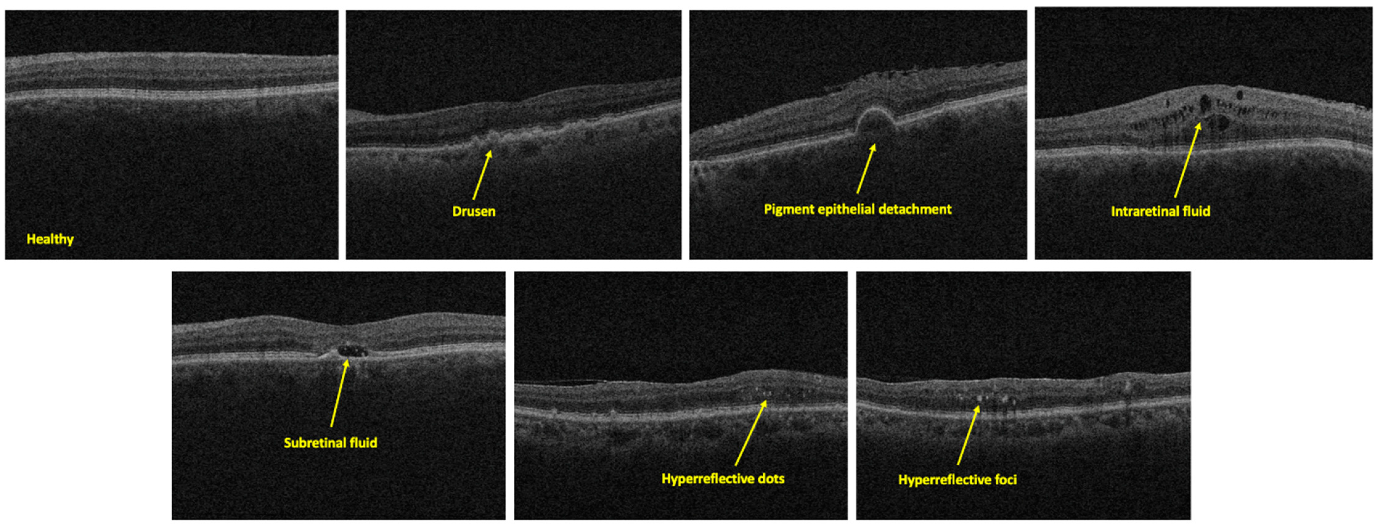
2.2. Feature Description
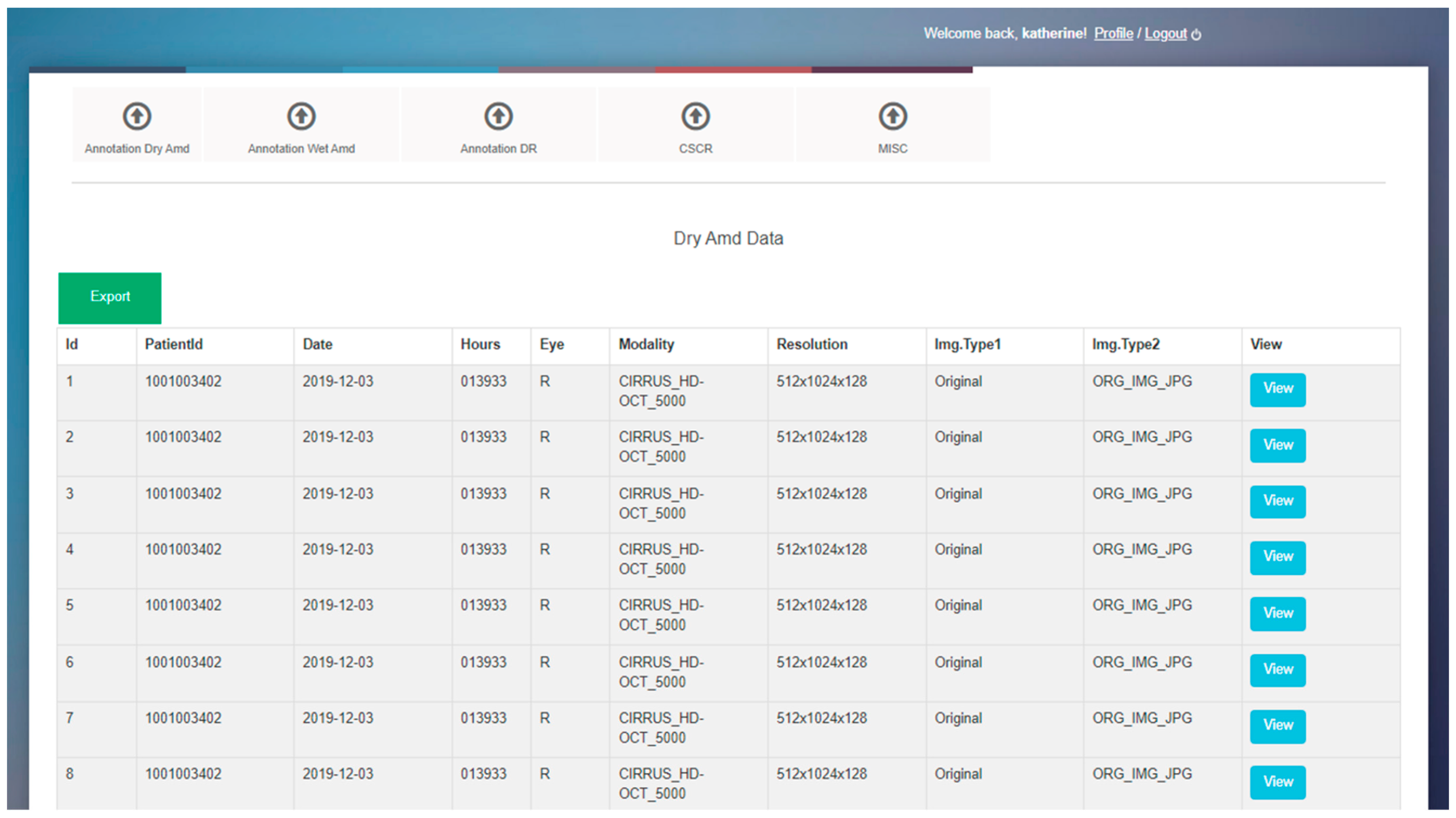
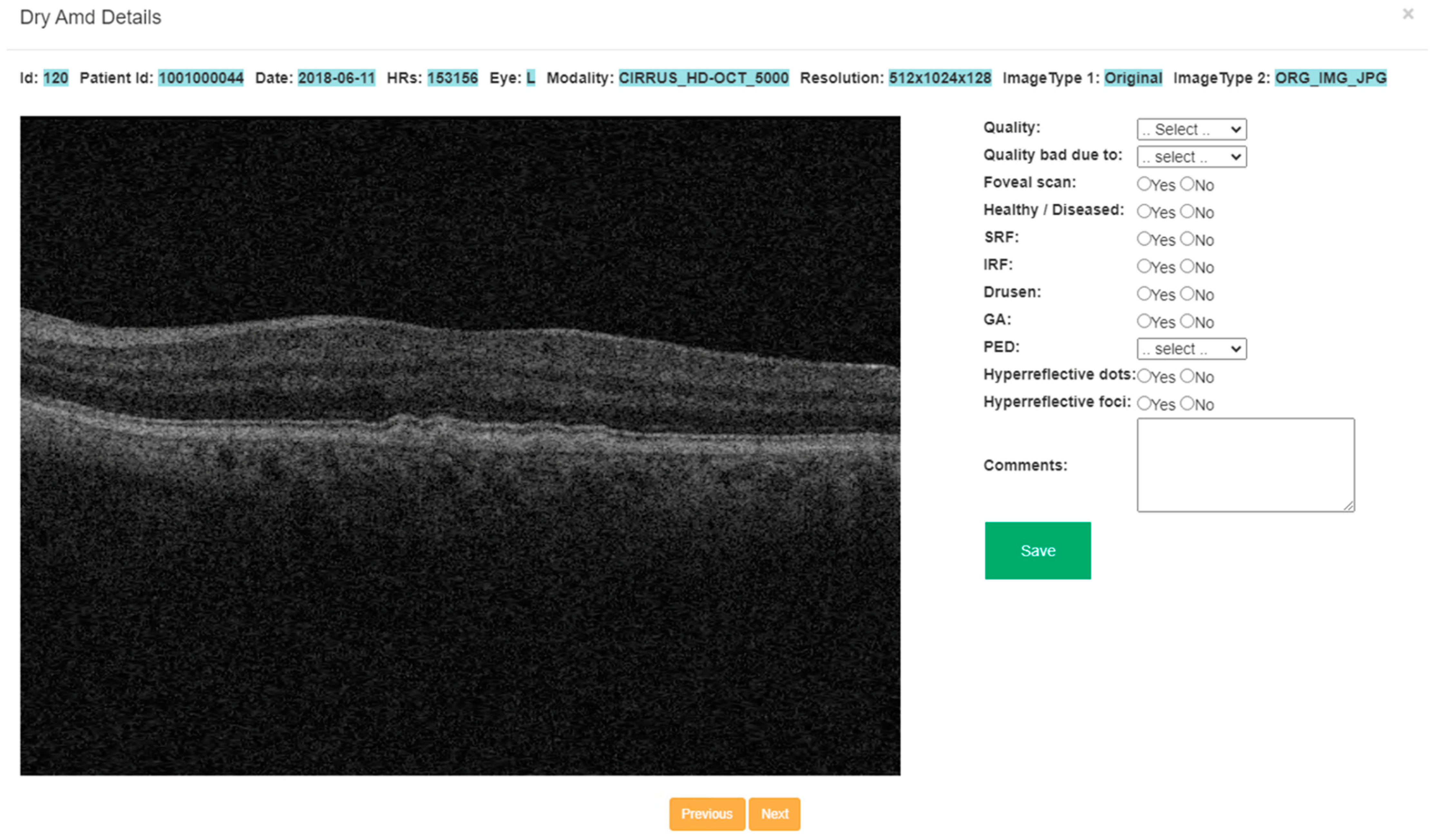
2.3. Annotation Tool
2.4. Annotation Strategy
2.5. Data Preprocessing
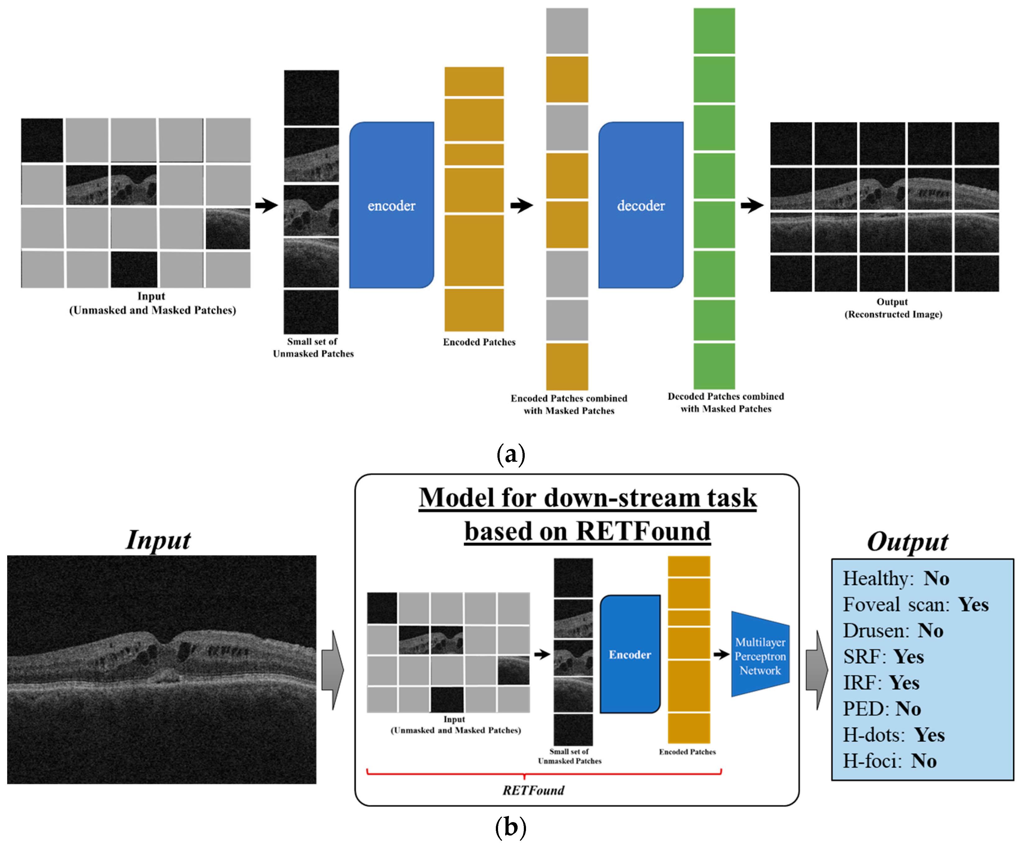
2.6. RETFound Model
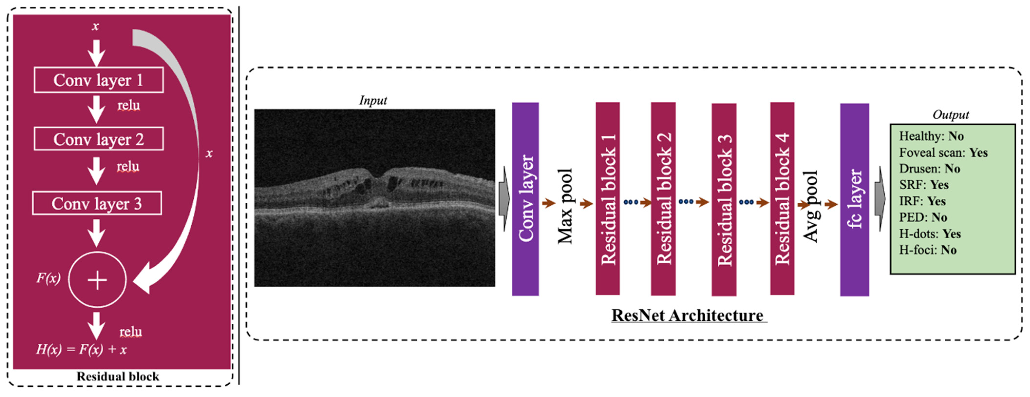
2.7. ResNet-50 Model
3. Results
4. Discussion
Author Contributions
Funding
Institutional Review Board Statement
Informed Consent Statement
Data Availability Statement
Acknowledgments
Conflicts of Interest
References
- Fleckenstein, M.; Keenan, T.D.; Guymer, R.H.; Chakravarthy, U.; Schmitz-Valckenberg, S.; Klaver, C.C.; Wong, W.T.; Chew, E.Y. Age-related macular degeneration. Nat. Rev. Dis. Primers 2021, 7, 31. [Google Scholar] [CrossRef] [PubMed]
- Kollias, A.N.; Ulbig, M.W. Diabetic retinopathy: Early diagnosis and effective treatment. Dtsch. Arztebl. Int. 2010, 107, 75. [Google Scholar] [PubMed]
- Aumann, S.; Donner, S.; Fischer, J.; Müller, F. Optical coherence tomography (OCT): Principle and technical realization. In High Resolution Imaging in Microscopy and Ophthalmology: New Frontiers in Biomedical Optics; Springer: Cham, Switzerland, 2019; pp. 59–85. [Google Scholar]
- Zeppieri, M.; Marsili, S.; Enaholo, E.S.; Shuaibu, A.O.; Uwagboe, N.; Salati, C.; Spadea, L.; Musa, M. Optical coherence tomography (OCT): A brief look at the uses and technological evolution of ophthalmology. Medicina 2023, 59, 2114. [Google Scholar] [CrossRef]
- Dahrouj, M.; Miller, J.B. Artificial intelligence (AI) and retinal optical coherence tomography (OCT). Semin. Ophthalmol. 2021, 36, 341–345. [Google Scholar] [CrossRef] [PubMed]
- Elezaby, S.; Bagherinia, H.; Ren, H.; Sha, P.; Tracewell, L.; Wu, C.; Fard, A.; Durbin, M. A machine learning method for optical coherence tomography scan quality assessment. Investig. Ophthalmol. Vis. Sci. 2020, 61, PB0090. [Google Scholar]
- Kho, A.; Bagherinia, H.; Leahy, C.; Bello, S.A. Automated scan quality assessment in low-cost OCT. Investig. Ophthalmol. Vis. Sci. 2022, 63, 3313-F0122. [Google Scholar]
- Andreu-Perez, J.; Poon, C.C.; Merrifield, R.D.; Wong, S.T.; Yang, G.Z. Big data for health. IEEE J. Biomed. Health Inform. 2015, 19, 1193–1208. [Google Scholar] [CrossRef]
- Mallappallil, M.; Sabu, J.; Gruessner, A.; Salifu, M. A review of big data and medical research. SAGE Open Med. 2020, 8, 2050312120934839. [Google Scholar] [CrossRef]
- Lee, C.H.; Yoon, H.J. Medical big data: Promise and challenges. Kidney Res. Clin. Pract. 2017, 36, 3–11. [Google Scholar] [CrossRef] [PubMed]
- Jee, K.; Kim, G.H. Potentiality of big data in the medical sector: Focus on how to reshape the healthcare system. Healthc. Inform. Res. 2013, 19, 79–85. [Google Scholar] [CrossRef]
- Price, W.N.; Cohen, I.G. Privacy in the age of medical big data. Nat. Med. 2019, 25, 37–43. [Google Scholar] [CrossRef] [PubMed]
- Liao, H.; Tang, M.; Luo, L.; Li, C.; Chiclana, F.; Zeng, X.J. A bibliometric analysis and visualization of medical big data research. Sustainability 2018, 10, 166. [Google Scholar] [CrossRef]
- Merelli, I.; Pérez-Sánchez, H.; Gesing, S.; D’Agostino, D. Managing, analysing, and integrating big data in medical bioinformatics: Open problems and future perspectives. BioMed Res. Int. 2014, 2014, 134023. [Google Scholar] [CrossRef] [PubMed]
- Cheng, C.Y.; Da Soh, Z.; Majithia, S.; Thakur, S.; Rim, T.H.; Tham, Y.C.; Wong, T.Y. Big data in ophthalmology. Asia-Pac. J. Ophthalmol. 2020, 9, 291–298. [Google Scholar] [CrossRef] [PubMed]
- Lee, C.S.; Brandt, J.D.; Lee, A.Y. Big data and artificial intelligence in ophthalmology: Where are we now? Ophthalmol. Sci. 2021, 1, 100036. [Google Scholar] [CrossRef]
- Da Soh, Z.; Cheng, C.Y. Application of big data in ophthalmology. Taiwan J. Ophthalmol. 2023, 13, 123–132. [Google Scholar] [CrossRef]
- Clark, A.; Ng, J.Q.; Morlet, N.; Semmens, J.B. Big data and ophthalmic research. Surv. Ophthalmol. 2016, 61, 443–465. [Google Scholar] [CrossRef]
- Devarakonda, S.T.; Vupparaboina, K.K.; Richhariya, A.; Chhablani, J.; Jana, S. Automated detection of retinal disorders from OCT images using artificial neural network. In Proceedings of the 2016 IEEE Annual India Conference (INDICON), Bengaluru, India, 16 December 2016; pp. 1–6. [Google Scholar]
- Singh, L.K.; Garg, H.; Khanna, M. An artificial intelligence-based smart system for early glaucoma recognition using OCT images. In Research Anthology on Improving Medical Imaging Techniques for Analysis and Intervention; IGI Global: Hershey, PA, USA, 2023; pp. 1424–1454. [Google Scholar]
- Qiu, J.; Sun, Y. Self-supervised iterative refinement learning for macular OCT volumetric data classification. Comput. Biol. 2019, 111, 103327. [Google Scholar] [CrossRef]
- ElTanboly, A.; Shalaby, A.; Mahmoud, A.; Ghazal, M.; Switala, A.; Taher, F.; Suri, J.S.; Keynton, R.; El-Baz, A. Computer Aided Diagnosis System for Early Detection of Diabetic Retinopathy Using OCT Images. In Big Data in Multimodal Medical Imaging; CRC Press: Boca Raton, FL, USA, 2019; pp. 281–299. [Google Scholar]
- DeBuc, D.C. A review of algorithms for segmentation of retinal image data using optical coherence tomography. Image Segmentation 2011, 1, 15–54. [Google Scholar]
- Kumar, Y.; Gupta, S. Deep transfer learning approaches to predict glaucoma, cataract, choroidal neovascularization, diabetic macular edema, drusen and healthy eyes: An experimental review. Arch. Comput. Methods Eng. 2022, 30, 521–541. [Google Scholar] [CrossRef]
- He, K.; Zhang, X.; Ren, S.; Sun, J. Deep residual learning for image recognition. In Proceedings of the IEEE Conference on Computer Vision and Pattern Recognition, Las Vegas, NV, USA, 27–30 June 2016; pp. 770–778. [Google Scholar]
- Hassan, E.; Elmougy, S.; Ibraheem, M.R.; Hossain, M.S.; AlMutib, K.; Ghoneim, A.; AlQahtani, S.A.; Talaat, F.M. Enhanced deep learning model for classification of retinal optical coherence tomography images. Sensors 2023, 23, 5393. [Google Scholar] [CrossRef] [PubMed]
- Szegedy, C.; Vanhoucke, V.; Ioffe, S.; Shlens, J.; Wojna, Z. Rethinking the inception architecture for computer vision. In Proceedings of the IEEE Conference on Computer Vision and Pattern Recognition, Las Vegas, NV, USA, 27–30 June 2016; pp. 2818–2826. [Google Scholar]
- Simonyan, K.; Zisserman, A. Very deep convolutional networks for large-scale image recognition. arXiv 2014, arXiv:1409.1556. [Google Scholar]
- Subasi, M.E.; Patnaik, S.; Subasi, A. Optical coherence tomography image classification for retinal disease detection using artificial intelligence. In Applications of Artificial Intelligence Healthcare and Biomedicine; Academic Press: Cambridge, MA, USA, 2024; pp. 289–323. [Google Scholar]
- Li, F.; Chen, H.; Liu, Z.; Zhang, X.D.; Jiang, M.S.; Wu, Z.Z.; Zhou, K.Q. Deep learning-based automated detection of retinal diseases using optical coherence tomography images. Biomed. Opt. Express 2019, 10, 6204–6226. [Google Scholar] [CrossRef] [PubMed]
- Leandro, I.; Lorenzo, B.; Aleksandar, M.; Dario, M.; Rosa, G.; Agostino, A.; Daniele, T. Oct-based deep-learning models for the identification of retinal key signs. Sci. Rep. 2023, 13, 1–11. [Google Scholar] [CrossRef] [PubMed]
- Bommasani, R.; Hudson, D.A.; Adeli, E.; Altman, R.; Arora, S.; von Arx, S.; Bernstein, M.S.; Bohg, J.; Bosselut, A.; Brunskill, E.; et al. On the opportunities and risks of foundation models. arXiv 2021, arXiv:2108.07258. [Google Scholar]
- Moor, M.; Banerjee, O.; Abad, Z.S.; Krumholz, H.M.; Leskovec, J.; Topol, E.J.; Rajpurkar, P. Foundation models for generalist medical artificial intelligence. Nature 2023, 616, 259–265. [Google Scholar] [CrossRef]
- Zhou, C.; Li, Q.; Li, C.; Yu, J.; Liu, Y.; Wang, G.; Zhang, K.; Ji, C.; Yan, Q.; He, L.; et al. A comprehensive survey on pretrained foundation models: A history from bert to chatgpt. arXiv 2023, arXiv:2302.09419. [Google Scholar] [CrossRef]
- Zhong, W.; Cui, R.; Guo, Y.; Liang, Y.; Lu, S.; Wang, Y.; Saied, A.; Chen, W.; Duan, N. Agieval: A human-centric benchmark for evaluating foundation models. arXiv 2023, arXiv:2304.06364. [Google Scholar]
- Vorontsov, E.; Bozkurt, A.; Casson, A.; Shaikovski, G.; Zelechowski, M.; Severson, K.; Zimmermann, E.; Hall, J.; Tenenholtz, N.; Fusi, N.; et al. A foundation model for clinical-grade computational pathology and rare cancers detection. Nat. Med. 2024, 30, 2924–2935. [Google Scholar] [CrossRef]
- Hong, D.; Zhang, B.; Li, X.; Li, Y.; Li, C.; Yao, J.; Yokoya, N.; Li, H.; Ghamisi, P.; Jia, X.; et al. SpectralGPT: Spectral remote sensing foundation model. IEEE Trans. Pattern Anal. Mach. Intell. 2024, 46, 5227–5244. [Google Scholar] [CrossRef]
- Liang, Y.; Wen, H.; Nie, Y.; Jiang, Y.; Jin, M.; Song, D.; Pan, S.; Wen, Q. Foundation models for time series analysis: A tutorial and survey. In Proceedings of the 30th ACM SIGKDD Conference on Knowledge Discovery and Data Mining, Barcelona, Spain, 25–29 August 2024; pp. 6555–6565. [Google Scholar]
- Wu, C.; Yin, S.; Qi, W.; Wang, X.; Tang, Z.; Duan, N. Visual chatgpt: Talking, drawing and editing with visual foundation models. arXiv 2023, arXiv:2303.04671. [Google Scholar]
- Nguyen, T.; Brandstetter, J.; Kapoor, A.; Gupta, J.K.; Grover, A. ClimaX: A foundation model for weather and climate. arXiv 2023, arXiv:2301.10343. [Google Scholar]
- Wang, J.; Chen, D.; Wu, Z.; Luo, C.; Zhou, L.; Zhao, Y.; Xie, Y.; Liu, C.; Jiang, Y.G.; Yuan, L. Omnivl: One foundation model for image-language and video-language tasks. Adv. Neural Inf. Process. Syst. 2022, 35, 5696–5710. [Google Scholar]
- Zhou, Y.; Chia, M.A.; Wagner, S.K.; Ayhan, M.S.; Williamson, D.J.; Struyven, R.R.; Liu, T.; Xu, M.; Lozano, M.G.; Woodward-Court, P.; et al. A foundation model for generalizable disease detection from retinal images. Nature 2023, 622, 156–163. [Google Scholar] [CrossRef] [PubMed]
- Du, K.; Shah, S.; Gadari, A.; Bollepalli, S.C.; Chhablani, J.; Vupparaboina, K.K. Inter-observer variance in labeling quality and pathology of retinal optical coherence tomography scans [abstract]. In Proceedings of the 2024 Data Science and AI Symposium, Harvard Ophthalmology and Mass Eye & Ear, Boston, MA, USA, 26–27 September 2024; p. 14. [Google Scholar]
- He, K.; Chen, X.; Xie, S.; Li, Y.; Dollár, P.; Girshick, R. Masked autoencoders are scalable vision learners. In Proceedings of the IEEE/CVF Conference on Computer Vision and Pattern Recognition, New Orleans, LA, USA, 18–24 June 2022; pp. 16000–16009. [Google Scholar]
- Deng, J.; Dong, W.; Socher, R.; Li, L.J.; Li, K.; Fei-Fei, L. Imagenet: A large-scale hierarchical image database. In Proceedings of the 2009 IEEE Conference on Computer Vision and Pattern Recognition, Miami, FL, USA, 20–25 June 2009; IEEE: Piscataway, NJ, USA, 2009; pp. 248–255. [Google Scholar]
- Kermany, D.S.; Goldbaum, M.; Cai, W.; Valentim, C.C.; Liang, H.; Baxter, S.L.; McKeown, A.; Yang, G.; Wu, X.; Yan, F.; et al. Identifying medical diagnoses and treatable diseases by image-based deep learning. Cell 2018, 172, 1122–1131. [Google Scholar] [CrossRef]
- Kugelman, J.; Alonso-Caneiro, D.; Read, S.A.; Vincent, S.J.; Chen, F.K.; Collins, M.J. Effect of altered OCT image quality on deep learning boundary segmentation. IEEE Access 2020, 8, 43537–43553. [Google Scholar] [CrossRef]
- Wang, J.; Deng, G.; Li, W.; Chen, Y.; Gao, F.; Liu, H.; He, Y.; Shi, G. Deep learning for quality assessment of retinal OCT images. Biomed. Opt. Express 2019, 10, 6057–6072. [Google Scholar] [CrossRef]
- Lauermann, J.L.; Treder, M.; Alnawaiseh, M.; Clemens, C.R.; Eter, N.; Alten, F. Automated OCT angiography image quality assessment using a deep learning algorithm. Graefes Arch. Clin. Exp. Ophthalmol. 2019, 257, 1641–1648. [Google Scholar] [CrossRef]

| Training Set (1360 Scans) | Testing Set (410 Scans) | |
|---|---|---|
| Healthy scan | 420 | 165 |
| Diseased scan | 940 | 245 |
| Foveal scan | 135 | 33 |
| Subretinal fluid | 89 | 2 |
| Intraretinal fluid | 69 | 6 |
| Drusen | 227 | 91 |
| Pigment epithelial detachment | 220 | 27 |
| Hyperreflective dots | 840 | 257 |
| Hyperreflective foci | 40 | 32 |
| Evaluation Metrics | ||||||||
|---|---|---|---|---|---|---|---|---|
| Accuracy | Sensitivity | Specificity | AUC-ROC | |||||
| Single Task | RN50 | RF | RN50 | RF | RN50 | RF | RN50 | RF |
| H/D | 0.77 | 0.76 | 0.71 | 0.62 | 0.81 | 0.86 | 0.83 | 0.80 |
| Foveal scan | 0.94 | 0.94 | 0.88 | 0.94 | 0.95 | 0.93 | 0.94 | 0.95 |
| Drusen | 0.69 * | 0.76 * | 0.65 * | 0.78 * | 0.70 | 0.75 | 0.75 | 0.83 |
| PED | 0.84 * | 0.79* | 0.44 * | 0.63 * | 0.86 | 0.81 | 0.74 | 0.76 |
| H-Dots | 0.62 | 0.62 | 0.82 * | 0.67 * | 0.28 * | 0.54 * | 0.64 | 0.66 |
| Average | 0.77 | 0.77 | 0.70 | 0.73 | 0.72 | 0.78 | 0.78 | 0.80 |
| Multitask | RN50 | RF | RN50 | RF | RN50 | RF | RN50 | RF |
| H/D | 0.78 * | 0.74 * | 0.66 | 0.72 | 0.87 * | 0.76 * | 0.80 | 0.81 |
| Foveal scan | 0.93 | 0.94 | 0.94 * | 0.85 * | 0.93 | 0.94 | 0.96 | 0.91 |
| Drusen | 0.70 * | 0.60 * | 0.66 * | 0.44 * | 0.72 | 0.65 | 0.74 * | 0.60 * |
| PED | 0.82 | 0.81 | 0.37 | 0.48 | 0.84 | 0.84 | 0.69 | 0.74 |
| H-Dots | 0.62 | 0.64 | 0.80 | 0.72 | 0.30 * | 0.50 * | 0.65 | 0.67 |
| Average | 0.77 | 0.75 | 0.69 | 0.64 | 0.73 | 0.74 | 0.76 | 0.75 |
| Evaluation Metrics | ||||||||
|---|---|---|---|---|---|---|---|---|
| Accuracy | Sensitivity | Specificity | AUC-ROC | |||||
| Single Task | RN50 | RF | RN50 | RF | RN50 | RF | RN50 | RF |
| H/D | 0.94 | 0.94 | 0.90 * | 0.92 * | 0.95 | 0.95 | 0.98 | 0.99 |
| Drusen | 0.94 | 0.94 | 0.91 * | 0.92 * | 0.95 | 0.95 | 0.98 | 0.98 |
| Evaluation Metrics | ||||||||
|---|---|---|---|---|---|---|---|---|
| Accuracy | Sensitivity | Specificity | AUC-ROC | |||||
| Internal Dataset | ||||||||
| Single task | RN50 | RF | RN50 | RF | RN50 | RF | RN50 | RF |
| H/D | 0.69 | 0.57 | 0.61 | 0.58 | 0.74 * | 0.56 * | 0.75 * | 0.59 * |
| Foveal scan | 0.74 | 0.70 | 0.82 * | 0.97 * | 0.73 | 0.68 | 0.79 * | 0.91 * |
| Drusen | 0.59 | 0.69 | 0.57 | 0.56 | 0.60 * | 0.72 * | 0.56 | 0.66 |
| PED | 0.81 * | 0.50 * | 0.26 * | 0.78 * | 0.85 * | 0.48 * | 0.64 | 0.63 |
| H-Dots | 0.51 * | 0.68 * | 0.36 * | 0.71 * | 0.75 * | 0.62 * | 0.54 * | 0.70 * |
| Average | 0.52 | 0.61 | 0.63 | 0.66 | 0.67 | 0.70 | 0.72 | 0.73 |
| Multitask | RN50 | RF | RN50 | RF | RN50 | RF | RN50 | RF |
| H/D | 0.64 | 0.55 | 0.16 * | 0.67 * | 0.96 * | 0.51 * | 0.70 | 0.74 |
| Foveal scan | 0.53 * | 0.95 * | 0.91 | 0.94 | 0.50 * | 0.95 * | 0.75 * | 0.96 * |
| Drusen | 0.52 | 0.55 | 0.32 * | 0.67 * | 0.96 * | 0.51 * | 0.70 | 0.64 |
| PED | 0.70 | 0.72 | 0.44 * | 0.67 * | 0.72 | 0.73 | 0.58 | 0.71 |
| H-Dots | 0.54 | 0.63 | 0.49 * | 0.67 * | 0.63 | 0.56 | 0.60 | 0.69 |
| Average | 0.61 | 0.68 | 0.46 * | 0.73 * | 0.75 * | 0.64 * | 0.67 | 0.75 |
| External (Kermany) Dataset | ||||||||
| Single task | RN50 | RF | RN50 | RF | RN50 | RF | RN50 | RF |
| H/D | 0.84 | 0.89 | 0.82 | 0.91 | 0.81 | 0.88 | 0.92 | 0.95 |
| Drusen | 0.50 * | 0.93 * | 0.38 * | 0.92 * | 0.53 * | 0.94 * | 0.44 * | 0.84 * |
Disclaimer/Publisher’s Note: The statements, opinions and data contained in all publications are solely those of the individual author(s) and contributor(s) and not of MDPI and/or the editor(s). MDPI and/or the editor(s) disclaim responsibility for any injury to people or property resulting from any ideas, methods, instructions or products referred to in the content. |
© 2024 by the authors. Licensee MDPI, Basel, Switzerland. This article is an open access article distributed under the terms and conditions of the Creative Commons Attribution (CC BY) license (https://creativecommons.org/licenses/by/4.0/).
Share and Cite
Du, K.; Nair, A.R.; Shah, S.; Gadari, A.; Vupparaboina, S.C.; Bollepalli, S.C.; Sutharahan, S.; Sahel, J.-A.; Jana, S.; Chhablani, J.; et al. Detection of Disease Features on Retinal OCT Scans Using RETFound. Bioengineering 2024, 11, 1186. https://doi.org/10.3390/bioengineering11121186
Du K, Nair AR, Shah S, Gadari A, Vupparaboina SC, Bollepalli SC, Sutharahan S, Sahel J-A, Jana S, Chhablani J, et al. Detection of Disease Features on Retinal OCT Scans Using RETFound. Bioengineering. 2024; 11(12):1186. https://doi.org/10.3390/bioengineering11121186
Chicago/Turabian StyleDu, Katherine, Atharv Ramesh Nair, Stavan Shah, Adarsh Gadari, Sharat Chandra Vupparaboina, Sandeep Chandra Bollepalli, Shan Sutharahan, José-Alain Sahel, Soumya Jana, Jay Chhablani, and et al. 2024. "Detection of Disease Features on Retinal OCT Scans Using RETFound" Bioengineering 11, no. 12: 1186. https://doi.org/10.3390/bioengineering11121186
APA StyleDu, K., Nair, A. R., Shah, S., Gadari, A., Vupparaboina, S. C., Bollepalli, S. C., Sutharahan, S., Sahel, J.-A., Jana, S., Chhablani, J., & Vupparaboina, K. K. (2024). Detection of Disease Features on Retinal OCT Scans Using RETFound. Bioengineering, 11(12), 1186. https://doi.org/10.3390/bioengineering11121186